Damage to simple DNA components induced
by secondary electrons
YiZheng
Département de médecine nucléaire et de radiobiologie
Thesis presented to the Faculty of Medicine
To obtain a diploma of Philosophy Doctorate (Ph.D.)
Sherbrooke, Québec, Canada
May 2005
E
4~ r-nRE-1:r::
LA 1y
PAc..t A UJ0( 3lesi
tr
P-,1"-el+I
Archives Canada Archives Canada Published HeritageBranch Direction du Patrimoine de l'édition
395 Wellington Street Ottawa ON K1A ON4 Canada
395, rue Wellington Ottawa ON K1A ON4 Canada
NOTICE:
The author has granted a non-exclusive license allowing Library and Archives Canada to reproduce, publish, archive, preserve, conserve, communicate to the public by
telecommunication or on the Internet, loan, distribute and sell theses
worldwide, for commercial or non-commercial purposes, in microform, paper, electronic and/or any other formats.
The author retains copyright ownership and moral rights in this thesis. Neither the thesis nor substantial extracts from it may be printed or otherwise reproduced without the author's permission.
ln compliance with the Canadian Privacy Act some supporting forms may have been removed from this thesis.
While these forms may be included in the document page count,
their removal does not represent any loss of content from the thesis.
•
••
AVIS:
Your file Votre référence ISBN: 0-494-14879-9 Our file Notre référence ISBN: 0-494-14879-9
L'auteur a accordé une licence non exclusive permettant
à
la Bibliothèque et Archives Canada de reproduire, publier, archiver,sauvegarder, conserver, transmettre au public par télécommunication ou par l'Internet, prêter, distribuer et vendre des thèses partout dans le monde,
à
des fins commerciales ou autres, sur support microforme, papier, électronique et/ou autres formats.L'auteur conserve la propriété du droit d'auteur et des droits moraux qui protège cette thèse. Ni la thèse ni des extraits substantiels de celle-ci ne doivent être imprimés ou autrement reproduits sans son autorisation.
Conformément
à
la loi canadienne sur la protection de la vie privée, quelques formulaires secondaires ont été enlevés de cette thèse. Bien que ces formulaires aient inclus dans la pagination, il n'y aura aucun contenu manquant.A major objective of our research group is to understand the mechanism of DNA damage induced by secondary electrons and its relationship to radiosensitization. My project focuses on simple systems, in which small DNA components, nucleosides (dThd), nucleotides (dTp), oligonucleotides (GCAT and CGTA) and modified oligonucleotides, are exposed to low energy electrons, and the subsequent reactions are studied by chemical analysis of the products.
A new low-energy electron irradiation system was constructed in which a relatively large area of target compounds can be irradiated. Thus, this system provides sufficient amount of damage products for further chemical analysis by HPLC, GC/MS and LC/MS. Our systematic studies revealed two main types of LEE-induced fragmentation reactions in DNA: 1) cleavage of the N-glycosidic and 2) cleavage of the phosphodiester bond. The results show that phosphodiester bond cleavage by 4-15 eV electrons involves cleavage of the C-0 bond rather than the P-0 bond. Below 14 eV, the yield of LEE-induced damage products in DNA is dominated by the formation of transient anions located around 6 and 10 eV. Beyond 14 eV, direct LEE impact is believed to contribute substantially to damage. Our studies suggest that electron transfer occurs from the base moiety to the sugar-phosphate backbone in DNA, but the inverse does not occur, in agreement with theoretical studies. The present study provides a chemical basis for the formation of strand breaks by the reaction of LEE with DNA. The capture ofnon-thermalized electrons with 4-10 eV of energy by DNA bases may be an important factor in DNA damage in living cells.
L'un des objectifs majeurs de notre groupe de recherche est la compréhension des mécanismes de dommages à l' ADN induits par les électrons secondaires et de leur lien avec la radiosensibilisation. Mon projet porte sur des systèmes simples où de petits composants del' ADN, soit des nucléosides (dThd), des nucléotides (dTp), des oligonucléotides (GCAT et CGTA) et des oligonucléotides modifiés, sont exposés à des électrons de faible énergie. Les réactions induites sont par la suite étudiées par analyse chimique des produits.
Un nouveau système d'irradiation par des électrons de faible énergie a été construit à l'intérieur duquel une zone relativement grande de produits-cibles peut être irradiée. De cette façon, cela permet de créer suffisamment de produits de dommage pour poursuivre l'analyse par HPLC, GC/MS et LC/MS. Les études systématiques ont révélé deux types principaux de réactions de fragmentation de l' ADN induits par les LEE : 1) le clivage du lien N-glycosidique et 2) du lien phosphodiester. Cela montre que le bris du lien phosphodiester par les électrons de 4-15 eV implique le bris du lien C-0 plutôt que celui du lien P-0. En dessous de 14 eV, le rendement des produits créés par les LEE dans l' ADN est dominé par la formation d'anions transitoires localisés entre 6 et 10 eV. Au-delà de 14 eV, nous croyons qu'un impact direct des LEE contribue aux dommages de façon substantielle. Nos études ont démontré que les LEE transfèrent de la base au squelette de l' ADN, tel que suggéré par des études théoriques. La réaction inverse ne se produit pas. Un tel mécanisme de dommage suggère que la capture d'électrons non thermalisés de 4-15 eV par les bases de l' ADN pourrait être un agent important du dommage à l' ADN cellulaire. Cette étude fournit une base chimique pour la formation des bris à la suite de la réaction des LEE avec l'ADN.
Abstract
Résumé
List of tables and illustrations . . . .. .. . . .. . . .. . . .. . . ... . . ..
mList of abbreviations . . .
ivChapter 1 - Introduction . . . .. . . .. 1
1.1 Ionizing radiation and radiobiology . . . 1
I.1.1 Direct and indirect effect of radiation . . . 1
I.1.2 Process time scale of ionizing radiation . . . 2
1.2 Events induced by the interaction of fast charged particles .. .. .. . . .. . . .. . . .. 3
1.3 Electron-molecule interactions in condensed phase . . . .. 5
I. 3 .1 Nonresonant scattering . . . 5
I. 3.2 Resonant scattering . . . .. . . .. . . .. . .. . . .. . . .. . .. . ... .. . .. . .. . .. 6
I.3.2.1 Major types ofresonances.. ... ... . .. ... . .. . ... .. . .. . .. . .. . .. . ... .. .... .... 7
I.3.2.2 Lifetime of the resonance ~t . . . .. . . . .. . .. . . .. . . .. . .. . .. . ... . . . .. . .. . .. . .. . .. . .. . . 7
I.3.3 LEE interaction with a molecule AB . . . 8
1.4 DNA damage induced by ionizing radiation ... 10
I.4.1 Structure of DNA . . . 10
I.4.2 Hydroxyl radicals ... 11
I.4.3 Solvated electrons ... 12
I.4.4 Base ionization ... 13
I.4.5 Typical methods of detection (PAGE, HPLC, MS) ... 13
1.5 DNA damage induced by LEE ... 14
I.5.1 reaction of LEE with DNA components ... 14
I.5.1.1 DNA bases and uracil ... 14
I.5.1.2 Deoxyribose and phosphate backbone ... 16
I.5.2 Measurement of LEE interactions with DNA ... 29
I.5 .2.1 Measurement of ions and neutral species by mass spectrometry ... 29
I.5.2.2 Measurement by high resolution electron energy loss spectroscopy ... 30
I.5.2.3 Measurement of fragmentation products by X-ray photoelectron spectroscopy (XPS) ... 31
1.6 Description of the research project ... 33
Chapter Il - First article . . . 36
Irradiator to Study damage induced to large nonvolatile molecules by Low-energy Electrons Yi Zheng, Pierre Cloutier, J. Richard Wagner, Léon Sanche Review of Scientific Instruments, 2004, 75, 4534. Chapter III - Second article . . . 44
Glycosidic bond cleavage of thymidine induced by low energy electrons Yi Zheng, Pierre Cloutier, Darel J. Hunting, J. Richard Wagner, Léon Sanche Journal of American Chemical Society, 2004, 126, 1002. Chapter IV -Third article ... 47
Chemical basis ofDNA sugar-phosphate cleavage induced by low-energy electron Yi Zheng, Pierre Cloutier, Darel J. Hunting, Léon Sanche, J. Richard Wagner Journal of American Chemical Society: Accepted. Chapter V - Fourth article . . . .. . . .. . . .. . . .. . . 75
Phosphodiester and N-glycosidic bond cleavage in DNA induced by 4-15 eV electrons Yi Zheng, Pierre Cloutier, Darel J. Hunting, J. Richard Wagner, Léon Sanche Journal of Physical Chemistry: in preparation. Chapter VI - Discussion ... 107
Chapter VII- Conclusions ... .120
Acknowledgement ... 123
Figure 1. The characteristic events observed after the process of ionizing radiation . . . 2 Figure 2. Initial events induced by a fast charged particle that penetrates film of molecules RH . . . 3 Figure 3. Energy distribution of Al Ka X-ray induced SE emission from tantalum ... 5 Figure 4. Energy transfer and unimolecular fragmentation pathways that follow LEE interaction with a molecule AB . . . 9 Scheme 1. Schematic drawing of DNA strands ... 10 Scheme 2. Structure of deoxyribose analogues ... 17 Scheme 3. Hypothetical reaction pathways for thymine ring cleavage, leading to formation/desorption of CN and OCN neutral fragments ... 21 Figure 5. Measured quantum yields for the induction of SSB, DSB, and multiple DSB in DNA films by 4-100 eV electron impact ... 24 Table 1. Quantitative analysis of products obtained from LEE irradiation of dTp using HPLC/lJV and GC/MS ... l 09 Scheme 4. Proposed pathways for phosphate ester bond cleavage ofDNA ... l 12 Scheme 5. Comparison of the distribution of damage by sites of cleavage at three different electron energies . . . .. . . l 16 Scheme 6. Structure of GCXT with arrow and number pointing to the possible direction of electron transfer and sites of phosphodiester bond cleavage ... .l 18
Ade Adenine BrU Bromouracil BrUdR Deoxybromouridine Cyt Cytosine dCyd 2' -deoxycytidine dR 2-Deoxyribose DD Dipolar dissociation
DEA Dissociative electron attachment DFT Density functional theory DNA Deoxyribonucleic acid DSB Double strand break(s) dThd Thymidine
dTp Thymidine monophosphate EEL Electron energy loss
ESD Electron stimulated desorption ESI Electrospray ionization
eV Electron volts
FWHM Full width at half maximum Gua Guanine
GC/MS Gas chromatography/ mass spectrometry HREEL High resolution electron energy loss
keV Kilo electron volts LC Liquid chromatography
LEE Low energy electron(s) (0-30 eV)
LEEEF Low energy electron enhancement factor LUMO Lowest unfilled molecular orbital
MFP Mean free path(s) ML Mono layer( s) MS Mass Spectrometry Oligos 0 ligonucleotides RNA Ribonucleic acid
SAM Self assembled monolayer(s) SE Secondary electron(s)
SSB Single strand break(s) Thy Thymine
THF Tetrahydrofuran Ura Uracil
UHV Ultra high vacuum UV Ultraviolet
Cbapter 1 - Introduction
1.1 lonizing radiation and radiobiology
In 1895 the German physicist Wilhelm Conrad Roentgen discovered "a new kind of ray", which he called X ray ----the X representing the unknown. This unknown ray started the new century of radiation research, which has become a diverse field that covers many areas of knowledge. Ionizing radiation is a type of radiation that produces a separation of charge in matter by transfer of sufficient energy to overcome the electron-binding energy in atoms or molecules. It has been found that ionizing radiation induces a variety of destructive, mutagenic, and potentially carcinogenic modifications, principally in DNA (von Sonntag, 1987). Thus the investigation of ionizing radiation in living organisms is the basis of radiobiology.
Radiation therapy is a clinical treatment modality where ionizing radiation is used to treat patients with malignant neoplasms. The goal is to deliver a measured dose of radiation to a defined volume with minimal damage to surrounding normal tissue, resulting in eradication of the tumor. The application of radiotherapy relies essentially on the understanding ofhow cells respond to stress produced by ionizing radiation.
1.1.1 Direct and indirect effect of radiation
When any form of radiation ---- X or y rays, charged or uncharged particles is absorbed in biologie material, one possibility is that it will interact directly with the critical targets in cells. The target atoms itself may be ionized or excited; thus initiating a chain of events that leads to a biologie change. This is called direct effect of radiation.
Altematively, the indirect effect involves the interaction of cellular water rather than with macromolecules within the cell. The indirect action initiated with the hydrolysis of
water which produces Ho·, eaq, H·, H2, etc. These radicals may diffuse a short distance and further react with critical targets, such as DNA. It is known that 80% of a cell is composed of water, which absorbs about 66% of the energy. Indirect action is estimated to compose nearly 50% of the ionizing radiation. The indirect effect of ionizing radiation on DNA is briefly described in section 1.4. In this thesis all the results are obtained in the condensed phase, therefore, major efforts are focused on the direct effect of radiation.
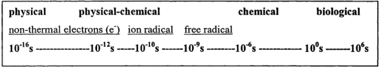
1.1.2 Process time scale of ionizing radiation
The time scale of events triggered by ionizing radiation is illustrated in the fig. 1. There are vast differences in the time scale involved in these various events. First the physical process e.g. the initial ionization, takes place in 1ff15s. Second the primary radicals
are produced by physico-chemical processes having a lifetime of about 10·10 to 10·9 s. The
reaction of those radicals with target molecules leads to chemical changes from bond breakage. Finally, expression of the biological effect may take longer time depending on the consequences involved. For example, if radiation damage is oncogenic, its expression as a cancer may be delayed several years. However, all those biological effects depend on the chemical modifications that have already occurred. Therefore, the chemical analysis of ionizing radiation damages plays an important role in understanding radiobiological pathways.
physical physical-chemical chemical biological non-thermal electrons (e") ion radical free radical
10"16s ---10·12s ---10-10s ---10-9s ---10-6s ---106s -106s
1. 2 Events induced by interaction of fast charged particles
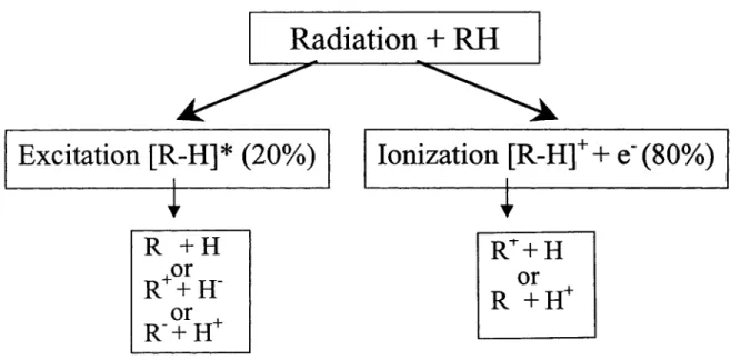
Taking a simple example of the initial interaction of a fast charged particle with a molecular solid composed of organic molecules R-H, the pre-chemical stage of radiation damage is shown in fig. 2 (Sanche, 2002). As the fast charged particle passes near the molecule R-H, the molecule is perturbed by the rapid change of electric field induced by the moving charge. Because this perturbation leaves the energy and momentum of the fast particle practically unchanged, the energy transfer can be described as an absorption of electromagnetic radiation by the molecules of the medium. The most probable energy loss of fast primary charged particles is 22 eV. This absorption can lead to the formation of electronically excited species [R-H]*, and ionization (Fig. 2). Most of the energy of high-energy particles is deposited within irradiated systems by the emission of such succession oflow-energy quanta (22 eV).
Radiation
+
RH
Excitation [R-H]* (20%)
R +H
or
R++H-or
R-+H+
Ionization [R-H]+
+
e-(80%)
1RT+H
or
R +H+
Figure 2. Initial events induced by a fast charged particle that penetrates an organic or biomolecular solid composed of molecules RH (H = hydrogen, R = rest of molecule).
One can estimate that
ca.
20% of the energy deposited by fast charge particles in organic matter leads to [R-H]* production, whereas the rest leads to ionization. The ionization energy is shared as the kinetic energy of secondary electrons and the potential energy of the cation, with the largest portion of the energy going to the secondary electrons. The sequence of events of ionization and electronic excitation may lead to hydrogen-atom abstraction, as an example of possible fragmentation produced by ionizing radiation (Fig.2).
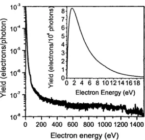
Many species are produced along the ionization track, which consist of excited atoms and molecules, radicals, ions, and secondary electrons. Among these species, secondary electrons are generated abundantly ( 4 x 10⁴ per Me V of deposited energy) by the absorption of energy from primary high energy photons such as X, y rays, and fast charge
particles (Cobut et al., 1998). Secondary electrons have low energies with a distribution that lies essentially below 70 eV and a most probable energy below 10 eV. An example of SE distribution and emission coefficient induced by X-ray irradiation is shown in figure 3 (Cai et al., 2005). The spectrum was recorded with a current of 0.14 ± 0.02 nA of SE emitted from a 1.4 x 1.4 cm2 tantalum substrate. The energy-integrated electron yield was 0.039 ± 0.003 electrons per photon. It indicates that most of SE are LEE with kinetic energy below 10 eV.
Within the thermalization distances of the order of 1-1 Onm, the highly excited atomic, molecular and radical species, ions and low-energy electrons (LEE) can induce non-thermal reactions. A majority of these reactive species, which can initiate chemical reactions, are created by the secondary LEE. Thus, in order to understand the ionizing radiation process in living cell, the mechanism of action of LEE must be understood.
10-3 en 8 c ... c .8 0 7 0 10-4 .c: 6 ë Q_ .c a. b ..- 5
--
Cl) Ù'i 4 10-5 c ce
3 0 ,_ t5 2 0 (!) ~ 10-6 ~ ~ -0 "O Qï Q) >=>=
1 ff7 10-8 -+-...-..--.--.--.-....-T""""'lr--r--r--.-""T"'""..,..:.-~ 0 200 400 600 800 1000 1200 1400Electron energy (eV)
Figure 3. Energy distribution of Al Ka X-ray induced secondary electron emission from tantalum.
1. 3 Electron-molecule interactions in condensed phase
Electrons are small negatively charged particles that can be accelerated to high energy and to a speed close to that of light by means of an electrical device, such as a betatron_ In order to interpret the processes induced by electrons, especially LEE on biomolecular films, it is necessary to discuss the basic interaction between an electron and a molecule.
In general, the interaction of an electron with an atom or a molecule can be described in terms of forces derived from the potential that acts between them. Electron-molecule collisions can be divided into two main types: direct and resonant interactions.
1. 3.1 Nonresonant scattering
Direct scattering has short interaction times characterized by the usual duration of the electron transit through the dimensions of a molecule. Because the potential interaction
is always present, direct scattering occurs at all energies above the energy threshold for observed phenomenon. It produces a smooth, usually rising signal that does not exhibit any particular features.
Depending on the amount of energy transferred from the electron to the target, scattering can be elastic or inelastic. In elastic collisions, according to momentum conservation, the energy transfer, i.e. ôE/E, to the atom or molecule is on the order of m/M (m and M are the mass of the electron and target, respectively) of the electron's initial energy. Because of the very small electron mass (m) compared to even the lightest atom H ( 1: 1836), the loss of electron energy through momentum transfer to the target is negligible.
In contrast, inelastic collisions may create electronically excited states and subsequent reactions which are described as following:
1. 3.2 Resonant scattering
e-+M ~ M*
+e-M* ~ R + X ( dissociative excitation)
M* ~ R+ +
x-
(ion pair dissociation) M* ~ M+ + e- (autoionization)M* ~ M2+ + 2e- (multiple ionizations)
Electron resonances are well described in many reviews (Palmer & Rous, 1992; Sanche, 1991; Sanche, 1995; Sanche, 2000). Resonant electron scattering occurs when the incoming electron is bound into discrete energy level of the target for a much longer time than the usual scattering time in the neighborhood of a target. From a molecular orbital perspective, the resonant state may be considered as a transient negative ion (TNI) formed by an electron which temporarily occupies a previously unfilled orbital of the molecule.
Thus, resonance scattering occurs at specific energies that correspond to the formation of transient anions. At the resonance energy, product yield is usually enhanced, whereas a strong peak is observed in the yield function.
1.3.2.1 Major types of resonances
There are two major types of resonances: shape and core-excited resonance. If the additional electron occupies a previously unfilled orbital of the target in its ground state, the transitory state is referred to as a single-particle or "shape" resonance. Shape resonance applies specifically when temporary trapping of the electron is due to the shape of the electron-molecule potential. When the transitory anion if formed by two electrons occupying previously unfilled orbitais, the resonance is referred as a two-particle, one-hole or core-excited state. The first step in the latter resonance includes one core electron to be excited from the ground state, leaving a hole and an electron in the LUMO. Then a second electron is captured by the positive electron affinity of the electronically excited state or exciton, forming a core-excited anion (Sanche, 1991).
1.3.2.2 Lif etime of the resonance At
When the electron is temporarily captured by the target, it has an increased interaction time. This causes additional distortion of the target whose magnitude depends on the lifetime of the resonance, At. Long-lived resonances with lifetimes larger than 10·14 s cause a significant displacement of the nuclei of a molecule when the additional electron occupies a strongly bonding or antibonding orbital. When the electron leaves the molecule, nuclear motion is initiated toward the initial intemuclear distance, causing excitation of man y vibrational overtones of the molecule, due to the strong overlap between the nuclear wave function of the resonant state and that ofmany vibrational states of the ground state of
the molecule. On the other hand, when the ôt is much smaller than a typical vibrational period (ôt << 10-14 s), the nuclei are not significantly displaced. Thus, for short resonance
times only the lower vibrational levels become excited with considerable amplitude.
According to the uncertainty principle (i.e.,
r •
ôt ~ h/2n), the transient state has awidth in energy
r
which serves to characterize and identify the process in the energy dependence of the scattering cross-sections or excitation functions. Resonance lifetime ôt could be reflected by the energy width. Long-lived resonances (ôt ;:::: 10-14 s) in atomsproduce sharp peaks in elastic and electronic excitation and ionization cross-sections. In molecules such resonances may lead to more decay channels due to the additional degrees of freedom introduced by nuclear motion. When the resonance are short-lived (ôt << 1ff14
s), they produce broad peaks in their decay channels.
1.3.3 LEE interaction with a molecule AB
The possible decay channels of a diatomic molecule AB induced by LEE are illustrated in figure 4 (Bass & Sanche, 2004). The direct electron interaction may produce an excited neutral state of the molecule AB* via pathway a. The departing electron may leave the molecule in a rotationally, vibrationally, or electronically excited state (pathway a). Then, AB* may dissipate its excess energy via photon emission and/or energy transfer to the surrounding medium (al). If the resulting electronically excited neutral state is dissociative, ground state or excited fragments can be produced (a2). Above a certain energy threshold ( ~ 14-16 eV), dipolar dissociation (DD) becomes possible to yield an
anion and a cation (a3).
In the case of resonant scattering, the incident electron temporarily attaches to the molecule via pathway b. The resulting transient anion may autoionize via b3. The other
important process is called dissociative electron attachment (DEA), i.e., the anion may dissociate into a stable anion and a neutral fragment in an excited or the ground state (b 1 ). DEA occurs when the resonance meets several conditions: a) the lifetime of the resonance is, at least, of the order of a vibrational period; b) the Aff state is dissociative in the Franck-Condon (FC) region, c) at least one of the possible fragment has a positive electron affinity. If during its lifetime, the transient anion transfers energy (~E) to another system (e.g., by
collisional interaction with another molecule, or phonon creation in a surrounding medium), it may stabilize as long as the parent molecule has a positive electron affinity (process b2). Finally, the incoming electron can directly ionize the molecule via path c. If the resulting cation is dissociative, it may fragment as shown in c 1.
cl •
A+
B+(*)Figure 4. Energy transfer and unimolecular fragmentation pathways that follow LEE interaction with a molecule AB.
1. 4 DNA damage induced by ionizing radiation 1.4.1 Structure of DNA
Deoxyribonucleic acid (DNA) DNA is a polymer (Adams et al., 1981). The monomer units of DNA are nucleotides, which consists of a 5-carbon sugar ( deoxyribose ), a nitrogen containing base attached to the sugar, and a phosphate group. There are four different types of nitrogenous bases in DNA: adenine (A), guanine (G), cytosine (C) and thymine (T). A DNA molecule consists oftwo polynucleotide antiparallel stands having the form of a right-handed helix. The two strands achieve contact through hydrogen bonds between A-T and G-C and also by base to base 1t stacking between adjacent bases on the
same strand (Scheme 1 ).
'f
1
o--P=O
Scheme 1. Schematic drawing of DNA strands
With such unique chemical structure, DNA achieves a supreme coding effectiveness and serves as the carrier of genetic information in both prokaryotes and eukaryotes. An undifferentiated mammalian fetal cell contains only a few picograms (10-12g) of DNA, which determine the synthesis of as many as 30,000 distinct proteins (Devlin, 1997). In summary, DNA is the macromolecule that ultimately controls cellular functions, primarily through protein synthesis. Also, DNA plays an exclusive role in heredity, by transfer of biological information from one generation to the next (McGowan, 2003).
The double helix exists in various geometries depending on the base composition and physical conditions, i.e., A, B and Z form. B-DNA is the most common form in cells. It should be noted that H20 molecules, which easily fit in the grooves of the helix, are an
integral part of the DNA structure. Even under dry conditions, DNA may still contain on average 2.5 water molecules per base pair (Swarts et al., 1992). In the dried state, B-DNA may convert to A configuration. Therefore, even in the condensed phase the radiation-induced hydrolysis of water should also be considered. ln dilute solution, DNA damage induced by ionizing radiation is mainly caused by the reactive species formed from water, including hydroxyl radicals, solvated electrons and H atoms.
1.4.2 Hydroxyl radicals
The action of hydroxyl radicals on DNA constituents in aqueous solution (indirect effect of ionizing radiation), which is also called "oxidative damage to DNA", has been extensively studied (von Sonntag, 1987; Cadet, 1997; Dizdaroglu et al., 2002; Wagner et al., 1999). The relevant information on the transient
·oH
radical adducts to pyrimidine and purine nucleic acid components has been obtained from electron spin resonanceinvestigations (Davies et al.1995). Most of the ·oH-mediated resulting products have been characterised and reviewed (Cadet, 1997; Cadet 2003).
OH radical can abstract hydrogen atoms within the sugar moiety with a preference at C4' position. ln this respect, each sugar in DNA attacked by an OH radical forms a strand break in alkali, with a G value (the number of specified chemical events produced in an irradiated substance per 1 OO eV of energy absorbed from ionizing radiation) of at least 0.6(lG~10-7 mol/J). It is estimated that about 2.7 OH radicals are formed per 100 eV of
radiation energy absorbed. There is no evidence for reactions of •oH with phosphate groups. Therefore, at least 0.6 (>20%) ·oH react with sugars and less than 2.1 (< 80%) with bases (Hutchinson, 1985). However, the actual distribution favours the sugar because OH radicals are more accessible to the sugar moiety.
1.4.3 Solvated electrons e·aq
Solvated electrons are one of the species formed by ionization process of ionizing radiation after the electron becomes solvated in liquid water (Eq. 1). ln N2 saturated water the G value of e-aq is about 2.65, whereas in N20 it could be scanvagered and converted into OH radicals (von Sonntag, 1987).
(1) Solvated electrons form electron adducts in the reaction with pyrimidines and purine bases and lead to further products by protonation. From the studies of model system poly(U) it was found that e-aq hardly makes any strand break or base release compared to OH and H radicals (von Sonntag, 1987).
1.4.4 Base ionization
From the process of base ionization, the primary species are radical cations and radical anions, which can be observed by ESR. The radical anions have been observed with the nucleotides at liquid He temperature (Box et al. 1975). However, in DNA, due to the difference in electron affinity of bases only the cytosine and thymine anions, guanine and adenine cations were detected (Sevilla et al., 1991). It was also found that menadione-mediated photosensitization to UV A radiation leads to photo-ionization of pyrimidine bases, whereas photoexcited riboflavin is only able to ionize purine bases (Cadet 2003). 1.4.5 Typical methods of detection (PAGE, HPLC, MS)
Owing to its negative charge, DNA migrates on polyacrylamide gels upon electrophoresis (PAGE). This method can clearly distinguish between two pieces of DNA that differ in length by one or more nucleotide unit. The resulting electropherograms are analyzed by autoradiography using labeled DNA and improved by computer-assisted spectrophotometry. For example, supercoiled, circular and linear forms of plasmid DNA induced by LEE were separated by this method (Boudaiffa et al., 2000).
Chemical analysis of products includes first the separation by high-performance liquid chromatography (HPLC), and then characterization using suitable spectroscopie measurements, e.g. mass spectroscopy (MS). Recently HPLC coupled to the electrospray ionization tandem mass spectrometry (HPLC/ESI-MS/MS) represents one of the most powerful analytical tools, providing high sensitivity and specificity to the measurement. It has been used to search for the unidentified radiation-induced DNA lesions (Regulus et al. 2004).
1. 5 DNA damage induced by LEE
According to the fundamental interactions that lead to subsequent events in a target, the biological effects of radiation are not produced by the mere impact of primary quanta, but mostly by the secondary species that are generated along the radiation track. Because LEE are the most abundant of the secondary species produced by the primary interactions, it is crucial to determine their action within cells, particularly in DNA, where LEE lead to mutagenic and potentially lethal DNA damages (Boudaiffa et al., 2000). Since the pioneer study of Sanche's group, much works focuses on the LEE induced processes in DNA and its constituents.
1.5.1 Reaction of LEE with DNA components 1.5.1.1 DNA bases and uracil
Systematic gas-phase investigations of stable anion production by LEE impact on uracil (U) (Denifl et al, 2004a; Feil et al., 2004; Hanel et al., 2003) and other DNA bases (Denifl et al, 2004b; Sukhoviya et al., 2001; Abouaf et al., 2003; Denifl et al., 2003; Gohlke et al., 2003; Abdoul-Carime et al, 2004; Ptasinska et al., 2005) provided considerable insight into the mechanisms of damage to DNA induced by SE of low energies. Electron attachment to these biomolecules leads to dissociation into various fragments without a measurable amount of stable parent anions. The fragment anions with highest abundance from uracil, adenine and the pyrimidines were (U-H)-, (A-H)-, (C-H)- and (T-H)-, respectively. ln addition, five other fragment anions were formed by DEA to cytosine and eight additional product anions were detected in the case of thymine. Yield fonctions were measured for all fragment anions in the electron energy range from about 0 to 14 eV.
Twelve fragments were produced by DEA to uracil but with lower cross sections than for (U-H)-. Energy thresholds for dissociation into cation fragments by LEE impact on U were also determined (Huels et al., 1995).
Subsequent gas-phase studies showed that the high hydrogen loss induced by LEE impact on the DNA bases was site-specific (Abdoul-Carime et al., 2004). In principle, dehydrogenation of nucleobases can arise from either C-H or N-H bond cleavage. To clarify this point, these authors carried out experiments on partly deuterated thymine at non-exchangeable carbon positions, T0 . Below 3 eV, both the energy dependence and the
absolute intensity of the yield function of (T 0-H)- (129 amu) were virtually identical to
those obtained from thymine (T-H)- (125 amu). By switching the mass spectrometer to mass 128 amu [corresponding to T0-D)-], the ion signal completely disappeared. This
observation provided direct evidence that DEA generates the N-dehydrogenated anion (T 0
-H)N - as confirmed by DFT calculations. The structures in the (T 0-H)N - ion yield curve
suggested that different electronic states of the precursor ion are involved. Any of these states, however, decay by hydrogen cleavage from the N sites, but not from the carbon positions (Abdoul-Carime et al, 2004; Ptasinska et al., 2005).
The four DNA bases were also investigated in thin multilayer films, but fewer anions of different masses were measured than in the gas phase. The difference is principal/y due to the inability of the heavier anions to overcome the po/arization potential that they induce in the film causing them to remain trapped in the target (Huels et al.,
1995). In fact, only the light anions H-, 0-, OH-, CN-, OCN-, and CH2- were found to desorb by the impact of 5-35 eV electrons on the physisorbed bases via either single or
H-yield fonctions exhibited resonance structures at around 9 and 20 eV, typical of DEA to the molecules. A monotonie increase in the anion yield fonctions was interpreted to arise from non-resonant stable-anion production via dipolar dissociation (DD). The resonance features were attributed to electron capture by the positive electron affinity of excited states that involves the excitation of the lone-pair n ~ n*, 7t ~ n*, and/or cr ~ cr* (i.e.,
formation of a two-electron one-hole transitory anion). 1.5.1.2 Deoxyribose and phosphate backbone
The backbone of DNA consists of a long chain of repeated deoxyribose-phosphate units. Investigation of LEE-interaction with this unit and its two basic constituents are of special interest in relation to DNA damage. In DNA, a SSB occurs when one of the two backbones is broken. If breaks occur on two chains within a short distance (- 10 base pairs or 30 À), then the damage is referred to as a DSB. The latter damage is difficult to repair by the cell and without reparation the cell can mutate or die. To understand how such breaks can occur via LEE impact, specific sub-units of the backbone were investigated in both the gas and condensed phases (Ptasinska et al., 2004; Antic et al., 1999; Antic et al., 2000; Huels et al., 2004; Lepage et al., 1998; Breton et al., 2004; Pan & Sanche, 2005).
The formation of anions and cations by LEE on the gaseous 2-deoxy-D-ribose (C5H1004) (Ptasinska et al., 2004) as well as solid films of the sugar-like analogs, THF (1),
3-hydroxytetrahydroforan (Il), and a-tetrahydroforyl alcohol (Ill) (Antic et al., 1999; Antic et al., 2000) were investigated by mass spectrometry (Scheme 2). In these experiments, the yield fonctions for H- ion desorbed by the impact of 1-20 eV electrons on 10-ML films were characterized by an onset at 6.0, 5.8, and 6.0 eV, and a maximum centered at 10.4, 10.2, and 10.0 eV for 1, II, and III, respectively. No other anions were observed to desorb.
AU features below 15 eV in H- ESD yield functions were characteristic of DEA to 1, II, and III. A steep rise in the H- signal with an energetic threshold near 15 eV was characteristic of nonresonant DD. Owing to the strong similarity of the H- desorption profiles for 1, II, and III, the authors concluded that the majority of the anion yield for all three systems arises from at least one transient anion associated with electron attachment to the furan ring and located near 10 eV (Antic et al., 1999; Antic et al., 2000). Considering the large Rydberg character of the excited states in 1 near the energy range of the observed resonance, they further suggested that this resonant state is of the core-excited type,
Ô
Do
HO~
HO
I
II
III
possibly with dissociative valence cr* configurational mixing.
Scheme 2. Structure of deoxyribose analogues: tetrahydrofuran (1), 3-hydroxytetrahydrofuran (II), and R-tetrahydrofurfuryl alcohol (Ill).
The only structural difference between compound III and 2-deoxyribose ( dR) is the addition of two OH radicals at the 1 and the 3 positions. However, in the case of gaseous deoxyribose, anions of much larger masses were observed to be formed by LEE impact compared to similar experiments with condensed molecule III. As explained previously, this difference arises from the polarization field present at the surface of dielectrics. Heavy anions such as (D-H)-, C5H10; and C5H6
ü;
in the gas-phase experiments were observed(Ptasinska et al., 2004). The highest measured cross section (l.2x 10-15 cm2) for the
The mechanism leading to this anion could be interpreted as s-wave electron attachment followed by the removal of two water molecules. For C5H60; only this resonance near O
eV was observed. The other two fragments, C5H7
ü;
and (D-Hr reveal, besides a strong resonance at 0 eV, a second resonance at 1.2 and 1.5 eV, respectively, which is about 30 times lower in intensity. ln contrast to the results of DNA bases, dehydrogenation is not the predominant reaction channel for deoxyribose, but the relative amount of fragment ions compared to that of the parent cation is about an order of magnitude larger than in the case of nucleobases. This result indicates the weakness of sugar moieties in the backbone to attack by LEE.In a molecule as complex as DNA, the products of fragmentation are expected not only to involve single-step processes such as DEA, but also the reaction of the primary radicals and ions with other surrounding constituents within the molecule. The possibility of reactive scattering within the backbone of DNA has been demonstrated in experiments with condensed films containing 02 and THF (Huels et al., 2004). Their 0-20 eV electron
impact measurements show that all of the OH- and some of the H- desorption yields are the result of reactive scattering of the 1-5 eV 0- fragments produced initially by DEA to 02. These 0- reactions involve hydrogen abstraction and atom exchange with THF, and
result in the formation of THF-yl radicals such as alkoxyl radicals, as well as THF oxidation products, such as lactones. 0- was found to scatter over nanometer distances, comparable to DNA dimensions, and its reaction involves formation of a transient (OC4H80)* - collision complex.
HREEL spectra of resonance-enhanced vibrational excitations of gaseous and solid THF were recorded (Lepage et al., 1998; Breton et al., 2004). The production of aldehydes
fragments, which remained trapped within the bulk of the THF film, were detected in situ via the 3'1(n n*) and 3(n n*) electronic transitions and vibrational excitation modes.
The synthesis of aldehyde were discussed in terms of the formation of transient anion states, which may lead to the fragmentation of the molecule. The strong rise in the energy dependence data observed from about 6 eV was correlated to the electronic excitation threshold of THF which was suggested, in the solid phase, to involve electron transition to an unoccupied molecular orbital of cr*co character. Core-excited resonances, previously identified from vibrational excitation fonctions in multilayer films of THF around 9 and 10 eV (Lepage et al., 1998), were also suggested to contribute to the strong rise from 6 to 10 eV via the formation of neutral dissociative states. The small feature found around 3 eV was proposed to result from a cr* shape resonance also previously measured in the anion yields of multilayer films ofTHF (Antic et al., 1999), and involving the temporary trapping of an electron in either one of the two lowest unoccupied molecular orbitais (LUMO), both possessing a cr*co character. The features seen in the energy dependence above 11 eV were explained by considering more specific core-excited resonances involving a hole in the 7b or 6b, 7a or Sb, and 4b orbitais, and two electrons in the cr*co orbitais. The DEA process, mentioned previously (Antic et al., 1999; Antic et al., 2000), known to lead to fragmentation of THF via the formation of a core-excited resonance around 10 eV, was also proposed as a possible cause of damage. Non-resonant fragmentation of THF via the formation of several cations was finally suggested to increasingly contribute to the cross section from about 11 eV.
So far, the phosphate unit of DNA has only been investigated in the condensed phase (Pan and Sanche, 2005). They reported ESD of OH- anions from a solid film of Na
P02(0H)z. Their OH- yield function exhibits a single broad peak with a maximum around
8 eV indicating the existence of an intermediate anion state leading to OH- production, possibly via temporary electron localization in antibonding cr* orbitais of the molecule. In trimethylphosphate (Folkard et al., 1993), a surrogate for the DNA phosphate group, and in recent DFT studies of electron attachment to a sugar-phosphate-sugar unit of DNA (Li et al., 2003), electron localization into the lowest antibonding cr* orbitais was found to lead to DEA. These results suggest the existence of a resonance at the phosphate unit resulting from a core-excited state formed by a positive ion core binding two electrons in cr* orbitais. 1.5.1.3 Short single-strand DNA and oligonucleotides
Short DNA strands may easily be prepared as SAM chemisorbed on gold. Such samples have the advantage of being more uniform in coverage, better oriented and more pure than those made from bacterial DNA. Using such short single and double strands of DNA having well defined base sequences, further insight into the mechanisms of LEE-induced DNA damage was obtained (Dugal et al., 1999; Dugal et al., 2000; Abdoul-Carime et al., 2000; Carime & Sanche, 2001; Carime & Sanche, 2002; Abdoul-Carime et al., 2001). Much of the ESD data from short single strand DNA (i.e., oligonucleotides) have been performed by measuring the yields of neutral fragments induced by 1-30 eV electrons impinging on SAM oligonucleotides that consisted of 6 to 12 base units (Dugal et al., 1999; Dugal et al., 2000 and Carime et al.2000; Abdoul-Carime & Sanche, 2001; Abdoul-Carime & Sanche, 2002). Their results were obtained from mass spectrometric measurements of the residual atmosphere near the target during its bombardment in UHV by a 10-8 A electron beam. They showed that LEE-impact
species, as the most intense observable yields. No sugar moiety phosphorus-containing fragments or entire bases were detected. Comparison with anion yield functions, two possible pathways were suggested for the formation of CN and OCN by electron impact (Scheme 3): a DEA route (Eq. 1-3) and attack by OH radical on adjacent bases (Abdoul-Carime et al., 2001). From various results, it was also indicated that the sequence context played an important role in LEE-induced damage to the bases within oligonucleotides
0
•
H,C'{
J NH eH,CL
J NH NAO+ NAO + 1 1 R RH,C'ë
J NHl
0 NAO N + llNH 1 1 R R 0 ~ CN + OH llNH ---OCN + H H3cJ...----OH•
H3c'(o OHll ..
/NH J NH NAO NA0---1 1 R R(Abdoul-Carime & Sanche, 2002).
0 (1) (2) (3) 0 ll
+
Radical NHScheme 3. Hypothetical reaction pathways for thymine ring cleavage, leading to the formation/desorption of CN and OCN neutral fragments via: (a) DEA, (b) secondary reactions induced by an OH radical created via DEA to an adjacent base.
Electron conduction (Nogues et al., 2004) and temporary trapping (Ray et al., 2005) of LEE by DNA were investigated. In the experiments, photoelectrons are ejected by an excimer laser operating at 193 nm (6.4 eV) from a gold substrate on which the molecules are chemisorbed (Ray et al., 2005; Naaman et al., 1998). The LEE (< 2 eV) transmitted
through SAM of short DNA oligomers ML into vacuum are energy analyzed by time of flight. Electrons that are not transmitted are captured by DNA and transferred back to the grounded metal substrate. Because of the short lifetime of the captured electrons and the low-laser intensity and repetition rate, the ML is not charged by electrons between laser pulses. Thus, the instantaneous transmitted current reflects the capturing efficiency of the layer during the duration of the laser pulse (20 µsec). With such transmission experiments, the dependence of the capture probability on the base sequence and the state of the temporarily captured electrons was determined (Ray et al., 2005). It was found that the capture probability scales with the number of guanine bases in the single-strand oligomers and depends on their clustering level.
In the two-photon electron ejection from DNA SAM experiment, electrons are excited in the metal substrate with photon energy below the work function of the substrate (Ray et al., 2005). Sorne of these electrons are transferred to the LUMO of the adsorbed layer. A second photon is used to eject these electrons from the LUMO to the vacuum, where their kinetic energy is measured. The kinetic energy of ejected electrons is related to their binding energy. It was concluded that, (1) once captured, the electron is not localized on one of the bases, but instead lies either on the sugar phosphate backbone or between the molecules in the ML, in a nonlocalized state; (2) the state of the captured electrons is insensitive to the sequence of the oligomer and (3) double-strand DNA does not capture electrons as efficiently as single-strand DNA, but, once captured, the electrons are more strongly bound in the double than in the single strand configuration.
1.5.1.4 Plasmid DNA
Plasmid DNA was first bombarded with electrons of energies lower than 100 eV with threshold energies for SSB and DSB at 25 and 50 eV, respectively (Folkard et al., 1993). Later, dry samples of plasmid DNA films were bombarded with 5 eV to 1.5 keV electrons in a supercoiled configuration (Boudaiffa et al., 2000a, 2000b, 2000c; Huels et al., 2003). Their samples were analyzed by electrophoresis to measure the percentage of circular and linear forms of DNA produced corresponding to SSB and DSB, respectively. By measuring the relative quantities of these forms in their 5-ML sample as a function of exposure to electrons, these authors measured the total effective cross-section(~ 4 x 10-15 cm2) and effective range(~ 13 nm) for the destruction of supercoiled DNA, at 10, 30, and 50 eV (Boudaiffa et al., 2002).
Figure 5 shows the measured yields for the induction of SSB, DSB, and multiple DSB in plasmid DNA induced by 5-100 eV electrons. The apparent SSB yield threshold near 4-5 eV is due to the eut-off of the electron beam at low energies, whereas the DSB yield begins near 6 eV. Both yield functions possess a strongly structured signature below 15 eV and have a peak around 10 eV, a pronounced minimum near 14-15 eV, a rapid increase between 15 and 30 eV, and above 30 eV roughly constant yields up to 100 eV. ln stark contrast, the multiple DSB yield has an apparent threshold near 18-20 eV and a very weak peak at 25 eV, above which it increases monotonically by about 1 order of magnitude up to 100 eV. Both peaks in the SSB and DSB yields around 10 eV incident electron energy are similar in magnitude to the respective yields above 30 eV. The relatively high yield below 15 eV may be due to electron diffraction within DNA, which amplifies the captured cross section at specific DNA sites (Pendry, 1974).
10 SSB c
e
~ 5w
c Q) -0 ·5 c L... Q) a. 2 (a) DSB --··· (b) 0 10 20 30 40 50 60 70 80 90 100Incident Electron Energy (eV)
Figure 5. Open and solid symbols are the measured quantum yields ( events per incident electron) for the induction of SSB (a), DSB (b), and multiple DSB (c) in DNA films by 4-1 OO eV electron impact. At each electron energy, the error bars correspond to the standard deviation of the average reported value, while the experimental uncertainty is about ± 10% (Reprinted from Huels et al., 2003).
The incident-electron energy dependence of the damage to elementary constituents of DNA, probed in the form of desorbed anions and neutral species, exhibits strong variations due to electron resonances. From comparison of the maxima in the anion and neutral production yield functions of these DNA constituents to the DNA results, it becomes qui te obvious that the strong energy dependence of the DNA strand breaks below 15 eV in Fig. 5 can be attributed to the initial formation of transient anions, decaying into the DEA and/or dissociative electronic excitation channels. However, because the basic DNA components (i.e., the sugar, phosphate and base units and structural H20) can all be
fragmented via DEA between 5 and 13 eV, it is not possible a priori to unambiguously attribute SSBs and DSBs to the initial dissociation of a specific component.
A more deterministic interpretation of DNA damage below 15 eV came from the experiments that ESD of anions from plasmid and 40 base-pair synthetic DNA was directly measured within the 3-20 eV range (Pan et al., 2003). Resonant structures were observed with maxima at 9.4 ± 0.3, 9.2 ± 0.3, and 9.2 ± 0.3 eV, respectively, in the yield functions of H-, 0-, and OH-. The yield function for H- desorption, from linear and plasmid double strand DNA exhibit a similar behavior as the yield functions for 0- and OH- desorption. The prominent 9-eV feature observed in all anion yield functions is a typical signature of the DEA process. The maxima in the H-, 0- and OH- yield function from DNA can be correlated with the maximum spreading from 8 eV to 10 eV in the SSB yield and the one occurring at 10 eV in the DSB yield induced by LEE impact on films of supercoiled DNA (Fig. 5) (Boudaiffa et al., 2000a; Huels et al., 2003). Comparing the H- yield functions for desorption from films of thymine (the results obtained for the three other bases are similar to that shown for thymine) (Abdoul-Carime et al., 2001), amorphous ice (Pan et al., 2005), and a-tetrahydrofuryl alcohol (Antic et al., 1999), it was found that the H- peak energy from amorphous water is definitively too low to be associated with DEA to the structural water of DNA, unless the strong hydrogen bonding in DNA shifts considerably the H2
0-resonance energy. In contrast, it indicates that the bases are an important source of desorbed H- desorption with an intensity about 3 times the one arising from the sugar ring. A similar conclusion can be reached from comparison with gas-phase H-/D- abstraction from the carbon position in thymine (Ptasinska et al., 2005). Thus, comparison of line shapes and magnitude of the yield functions in both phases suggests that LEE-induced
H-desorption [rom DNA below 15 eV occurs main/y via DEA to the bases with a substantial contribution from the deoxyribose ring. Similar comparisons with anion yield functions
from basic constituents with those of 0- and OH- from DNA films (Martin et al., 2004) indicate that 0- production arises from temporary electron localization on the phosphate group. The OH- desorption yield function resembled that of the 0- yield, but has a considerably lower intensity. This result suggests a two step process: formation of 0-via DEA to the phosphate group followed by reactive scattering of the 0-ion with the nearby deoxyribose unit (i.e. reactive scattering as described in section 1.5.1.2).
In these ESD experiments, the counter-ion on the phosphate group was Na+ due to the method of preparation. Later, SAM of linear single and double strand DNA chemisorbed on a gold substrate with different orientations with respect to the substrate was studied (Pan & Sanche, 2005), but this time in their samples, the Na+ counter ion was replaced by H+. In this case, electron impact on DNA below 19 eV with OH in the phosphate unit produced OH- essentially via DEA to the phosphate group in the backbone. Between 2 and 5 eV, this process occurred exclusively via direct DEA. Above 5 eV, direct DEA to the phosphate unit was still the dominant mechanism, but they could not completely rule out a possible contribution to the OH- yield, arising from reactive scattering of 0-. Their results showed that the phosphate-counterion part of DNA plays a significant role in LEE induced DNA damage.
It was only after the development of more sensitive techniques to detect SSB and DSB in DNA that the electron energy range below 4 eV was investigated (Martin et al., 2004). Two peaks, with maxima of (1.0 ± 0.1) x 10-2 and (7.5 ± 1.5) x 10-3 SSB per
energies of 0.8 eV and 2.2 eV. The peaked structure provides unequivocal evidence for the role of low lying temporary anion states in the bond breaking process. The curve could be reproduced in magnitude and line shape by a model that simulates the electron capture cross section as it might appear in DNA owing to the n* anion states of the bases (Atlatooni et al., 1998). The lowest peak in the modeled capture cross section, which occurs at 0.39 eV in the gas phase, was shifted by 0.41 eV to match that in the SSB yield and its magnitude normalized. Usually, in moving from the gas phase to the condensed phase, polarization effects shift the n* resonances observed in the gas-phase to lower energies. However, in DNA the electric dipole fields created by the negatively charged phosphate groups and positive counter ions play a major role. The 0.4 eV positive shift could be explained by the phosphate charge which is closer to the bases, thus produces a net destabilization that slightly exceeds that of the polarization induced by the transient anion (Martin et al., 2004).
This interpretation of electron capture by the bases below 5 eV is corroborated by direct comparison with the DEA results (Denifl et al., 2004a, 2004b). The narrow peak near 1 eV has been interpreted as due to a dipole-bound anion (Scheer et al., 2004), which may not exist in DNA due to a eut-off of the long-range potential. There are two other features exhibiting a good energy correspondence with the maxima of 0.8 eV and 2.2 eV in DNA. Thus, both comparisons offer support for the charge transfer mechanism, an anionic potential surface exists that connects the initial n* anion state of the base to a cr* anion state of the phosphate group (Barrios et al., 2002). The cr* state leads to rupture of the C-0 bond connecting the phosphate group to the sugar. Transport of an electron from the base to the C-0 cr* antibonding orbitais of the sugar-phosphate takes place through three saturated
bonds. This is not surprising since there is ample precedence for such transfers leading to bond breaking in gas-phase DEA studies (Barrios et al., 2002; Cai et al., 2005). Furthermore, electron transfer from the bases is consistent with electron localization on the sugar-phosphate backbone leading to DNA damage (Boudaiffa et al., 2000a).
It is difficult to compare directly the yields obtained by LEE impact under UHV conditions, with those obtained from experiments in which DNA or other biomolecules are irradiated by high energy particles, mainly because of different experimental conditions, including the composition and conformation of the DNA. In addition, the dosimetry for LEE beam experiments is not available due to problems related to the energy imparted both to the DNA film and the metal substrate (Boudaiffa et al., 2000a, 2000b). By using an X-ray SE emission source, DNA damage induced by high energy photons (Alka.X-X-rays of 1.5 keV) and LEEs (average energy of 5.8 eV) under identical experimental conditions were directly compared (Cai et al., 2005). The exposure curves for the formation of SSB, DSB, and interduplex cross-links were obtained for both ML and thick films of DNA, respectively. The lower limits of G values for SSB and DSB induced by SE were derived to be 86 ± 2 and 8 ± 2 nmol
r
1, respectively; the average G values were about 2.9 and 3.0times larger than th ose obtained with 1.5 ke V photons.
Taking only the dose imparted by the slow SEs emitted from the tantalum substrate, a LEE enhancement factor (LEEEF) is defined as the ratio of yield of products in ML DNA induced by the LEE (slow SE, E ~ 10 eV) emitted from the metal substrate vs. that induced
by the photons in a particular experiment. The LEEEF for 1.5 keV photons was derived to be at least 0.2 for both SSB and DSB, by taking average G values for SE (Henke et al., 1981). The extrapolated LEEEF for X-rays from 1.5 keV to 150 keV (i.e. to energies of
medical diagnostic X-rays) indicates that SE electrons with the distribution and emission
coefficient of Fig. 3 are 20-30 times more efficient to damage DNA than the X-ray photons of 40-130 keV that create them. It can be seen that when electrons with the distribution of Fig. 3 strike a single DNA molecule, they have on average a probability about J
a5
larger to damage DNA than 40-130 keV photons. Hence, this first comparison study of DNA damage induced by X-rays and SE under identical experimental conditions shows LEE (E <10 eV) to be much more efficient in causing SSB and DSB than X-rays . . 1.5.2 Measurement of LEE interactions with DNA
1.5.2.1 Measurement of ions and neutral species by mass spectrometry
Sorne of the damage induced by LEE impact on biomolecules can be assessed by monitoring the ions and neutral species that desorb in vacuum during bombardment. Such measurements can be performed by placing the effusive beam or sample film near a mass spectrometer (Hervé du Penhoat et al., 2001). Generally, a LEE beam, emanating from an electron monochromator or a focusing electron gun, impinges onto the target. The full-width at half-maximum (FWHM) of the electron energy distribution varies from 0.03 to about 0.5 eV, depending on the type of electron source, for typical beam currents of 2-400 nA. In the gas phase experiments, the electron energy scale and the cross section for ion production are usually calibrated by measuring the anion signal from a gas which exhibits a resonance at a well defined energy, and whose DEA cross section is accurately known. Although this method provides an accurate energy calibration, it may overestimate the magnitude of the cross sections for non volatile biomolecules, since the latter may condense on chamber walls and thus reduce their background pressure relative to the calibrating gas.
Neutral spec1es that desorb from the films and reach the ionizer of the mass spectrometer, can also be ionized and focused onto the quadrupole rods (Hervé du Penhoat et al., 2001). Charged particles are kept from reaching the ionizer by placing suitable potentials on grids or lenses located between the target and the ionizer. To increase the detection efficiency of desorbing neutral species, the latter can be ionized close to the target surface by a laser (Kimmel & Orlando, 1995). With a standard electron ionization source, the background signal can be discriminated by beam modulation Iock-in techniques (Rakhovskaia et al., 1995).
Ions that emerge from an effusive beam or a film can be focused by ion lenses located in front of the mass spectrometer. In certain systems, the ion energies can be determined by a retarding potential or a deflector. Relative ion yields can be obtained from three different operating modes (Sanche, 1995): (1) the ion-yield mode, in which ions of a selected mass are detected as a fonction of incident electron energy, (2) the ion-energy mode, in which the ion current at a selected mass is measured for a fixed electron energy as a fonction of the retarding potential, and (3) the standard mass mode, in which the intensity of mass peaks is measured for a fixed electron energy.
1.5.2.2 Measurement by high resolution electron energy Joss spectroscopy
High resolution electron energy loss (HREEL) spectroscopy, has recently been applied to probe vibrational and electronic excitations in biomolecules. With this technique (Sanche & Michaud, 1984; lbach & Mills, 1982), electrons leaving a monochromator are focused on a gas jet or a metal substrate on which molecules are condensed. Electrons re-emitted from the target within a narrow cone at another angle are energy analyzed by a second electron deflector (i.e. the analyzer). Depending on the
apparatus, it may be possible to vary the angle of incidence or the analyzing angle or both. HREEL spectra are recorded by sweeping the energy of either the monochromator or the analyzer. The energy dependence of the magnitude of a given energy loss event (i.e. the excitation fonction) is obtained by sweeping the energy ofboth the monochromator and the analyzer with a potential difference between them corresponding to the probed energy loss. HREEL spectra are usually recorded at overall resolutions ranging from 6 to 80 meV FWHM with corresponding incident currents in the 10·10 - 10·8 A range.
HREEL spectroscopy has also been applied to the measurement of total absolute cross sections for fragment production by LEE impact on biomolecular films (Breton et al., 2004). In this case, the fragments, which remain trapped within the bulk of the film, are detected and quantified in situ by recording their electronic and/or vibrational HREEL spectra (Sanche & Michaud, 1984, Lepage et al., 1998).
1.5.2.3 Measurement of fragmentation products by X-ray photoelectron spectroscopy (XPS)
As with HREEL spectroscopy, XPS can also be utilized to probe the products formed by LEE impact on thin biomolecular films. In this case, changes in the binding energy of electrons in atomic shells serve to identify fragmentation products remaining in the film. The apparatus is similar to the one used to analyzed neutral and ion species (Hervé du Penhoat et al., 2001), but the mass spectrometer is replaced by an electron analyzer to measure the energy of photoelectrons; a source is added to irradiate the film with X-rays. Two different types of electron sources can be used to fragment the biomolecules: an electron gun or the photoelectrons emitted by the metal or semi-conductor substrate. As an example of a photoelectron electron source, Figure 3 shows the energy
spectrum of Alka X-ray induced SE emission from tantalum; it has a peak at 1.4 eV and an average energy of 5.8 eV (Cai et al., 2005). When the biomolecular film is sufficiently thin (< 5 nm), the number ofX-ray photons absorbed by the biomolecules are negligible and the induced damage may be considered to result from electrons with the energies of the distribution shown in Fig. 3.
The cross sections for electron-induced reactions are determined by assuming that the analytical XPS elemental signais from the original compound and radiation products are described by exponential functions (Klyachko et al., 1999):
L:
= I0 • exp(-oNt); Ir=Io•
[1 - exp(-crNt)] (2)where
L:
and Ir represent the measured signais (XPS intensities) from a particular element in the original compound and the same element in the radiation products, respectively; cr is the cross section for the corresponding process, N is the flux density of incident X rays or electrons; t is the time of irradiation, and I0 is the maximum signal from the same elementin the original compound as well as in the radiation products (i.e. measured at t = 0 and t T, respectively). Generally, measurements fit the above equations to within less than 10-15 % standard deviation (Klyachko et al., 1999). The validity of the above equations requires that the zone of analysis coïncide with that of damage. This condition can be met for the experiments with SE from X-rays, but not with certainty for those with the electron-beam irradiation. Thus the absolute values for the cross sections of electron beam-induced damage may be lower than their true values. In principle, photons of energy lower than that of X-rays (UV and visible photons) could also be used to probe LEE induced processes within condensed biomolecules. So far, only electron trapping and transmission through short DNA oligomers have been measured with such techniques (Ray et al., 2005).
1.6 Description of the research project
The existing LEE techniques are only capable of analysing degradation products trapped in or desorbed from a condensed film, i.e. positive ion, negative ion, and neutral species can be measured. However, most of the non-volatile radiation products, which remain on the surface, have not been identified. There is a missing link of initial fast radiation process to the final damage to elucidate the chemistry involved in the electron-initiated reactions. Therefore, it is necessary by coupling chemical analysis with LEE irradiation to obtain the information of the structure of final products. The objective of this research project is to study chemical modifications of DNA induced by LEE, starting with basic DNA components.
A novel sample spin coating and LEE irradiator system has been set up that can prepare a uniform film of biomolecules and bombard the inside surface of a cylinder target. With this configuration, we are able to irradiate a surface a hundred times larger than with a conventional electron gun, and therefore, provide a sufficient amount of damaged sample for chemical analysis outside vacuum. After recovered from the substrate, the sample is first separated by HPLC, then collected and further analyzed by GC-MS or ESIIMS/MS. The detailed description can be found in chapter Il.
By using this new apparatus, films of biological molecules can be investigated. The initial study is a relatively simple DNA nucleoside, thymidine and constitutes the second article ( chapter Ill). The results show that LEE irradiation induces glycosidic bond cleavage in thymidine and gives thymine as a major product, via a non-ionizing resonant process. ln