To cite this version :
Sandra GUERARD, Mathieu MANASSERO, Véronique VIATEAU, Véronique MIGONNEY, Wafa SKALLI, David MITTON - Biomechanical evaluation of a bioactive artificial anterior cruciate ligament - Biomechanical evaluation of a bioactive artificial anterior cruciate ligament - Vol. 1, n°4, p.239-252 - 2014
Any correspondence concerning this service should be sent to the repository Administrator : archiveouverte@ensam.eu
Copyright © 2015 Techno-Press, Ltd.
http://www.techno-press.org/?journal=aba&subpage=7 ISSN: 2287-2094 (Print), 2287-2272 (Online)
Biomechanical evaluation of a bioactive artificial anterior
cruciate ligament
Sandra Guérard
1, Mathieu Manassero
2a, Véronique Viateau
2b, Véronique
Migonney
3c, Wafa Skalli
1dand David Mitton
1e1Institut de Biomecanique Humaine George Charpak, Arts et Metiers ParisTech, 151 bd de l’hôpital 75013
Paris, France
2Ecole Nationale Vétérinaire d’Alfort, Service de Chirurgie, 7 av du Général de Gaulle,
94700 Maison-Alfort, France
3Université Paris 13, Laboratoire de Biomatériaux et Polymères de Spécialité/CSPBAT,
UMR CNRS 7244, 99 av JB Clément, 93430 Villetaneuse, France (Received March 12, 2014, Revised January 22, 2015, Accepted January 23, 2015)
Abstract. This study aimed to assess the biomechanical performance of a new generation of artificial ligament, which can be considered “bioactive” and “biointegrated,” implanted in sheep. Thirty sheep were implanted: 15 sheep received the artificial ligament grafted with a bioactive polymer (grafted) and 15 received the artificial ligament without a bioactive polymer (non-grafted). The animals were sacrificed 3 or 12 months after implantation. The knee kinematics, namely flexion-extension, anterior drawer, and varus-valgus tests, were evaluated using a fully characterized custom-made device. Afterward, the specimens were tested under uniaxial tension until failure.
The flexion-extension showed significant differences between (grafted or non-grafted) artificial and native ligaments 3 months after implantation. This difference became non-significant 12 months postoperatively.
The anterior tibial drawer was significantly increased 3 months after implantation and remained significantly different only for non-grafted ligament 12 months after implantation.
Twelve months after implantation, the differences between grafted and non-grafted ligament biomechanical properties were significant in terms of stiffness. In terms of load to failure, grafted ligaments seem to have had slightly better performance than non-grafted ligaments 12 months postoperatively. Overall these results suggest that grafted artificial ligaments have slightly better biomechanical characteristics than non-grafted artificial ligaments 12 months after implantation in sheep.
Keywords: ACL reconstruction; biomechanics; in vivo integration; in vitro experiments
1. Introduction
Corresponding author, Ph.D., E-mail: sandra.guerard@ensam.eu
aPh.D., E-mail: mathieumanassero@wanadoo.fr bPh.D., E-mail: veronique.viateau@wanadoo.fr
cProfessor, E-mail: veronique.migonney@univ-paris13.fr d
Professor, E-mail: wafa.skalli@ensam.eu
The anterior cruciate ligament (ACL), which plays a key role in knee stabilization, is commonly injured during sports activities. Although ligament replacement is mostly realized using a tendon autograft or allograft, major drawbacks such as long postoperative recovery or high occurrence of flexion contracture are encountered (Burks et al. 2005, Mascarenhas et al. 2008). Reconstruction with an artificial ligament presents potential advantages, compared to autograft, such as decreased morbidity, a faster recovery, and no viral transmission. The first use of synthetic material for ligament replacement was proposed in the 1980s but yielded poor results and high rates of failure because of the materials used, which did not achieve the expectations for this type of implant (Legnani et al. 2010).
Recent developments in artificial ligaments (ALs) have led to a renewed interest in this type of reconstruction (Gao et al. 2010). The Ligament Advancement Reinforcement System (LARS AC™) is composed of two different parts in polyethylene terephthalate (PET). The first, intra-osseous, part is composed of 25-µm longitudinal fibers bound together by a transverse knitted structure (Nau et al. 2002). The second, intra-articular, segment is made of longitudinal parallel fibers that are twisted at 90° to imitate the natural cruciate ligament. Recently, the biocompatibility of the LARS AC™ ligament was improved by grafting the prosthesis with a polymer, allowing control of the host response and leading to a new generation of synthetic bioactive ligament (Viateau et al. 2011, Vaquette et al. 2013, WO/2004/067051 2004).
The LARS AC™ artificial ligament is chemically grafted by a “bioactive” and “biomimetic” polymer, which has been shown in vitro to mask the synthetic origin of the material to control collagen secretion, fibroblasts proliferation, and inflammatory response (WO/2004/067051 2004, Pavon-Djavid et al. 2007). Bioactive polymer is a polymer able to modulate and/or control the biological response in vitro and in vivo. Radical graft polymerization of sodium salt of styrene sulfonate (NaSS) on the PET surface was performed using the “grafting from” technique. The grafting ratio was about 3 to 5 10-6 mol/g and was found to be perfectly reproducible (Zhou et al. 2007, Ciobanu et al. 2006). The aim was to induce a better rehabilitation of the intra-articular part of the ligament fibers by fibroblast cells able to proliferate and secrete proteins of the extra cellular matrix. Before it can be commonly used in clinical routine, its biocompatibility (in terms of nature and pattern of tissues in-growth within LARS ACTM) as well as its biomechanical performance for the short and long term must be studied in animals.
Goat or sheep models of ACL replacement are the most frequently adopted. Goats and sheep are large animals; their ACL is in vivo loaded and composed of two bundles as human ACL. Their knee joint morphological characteristics are close to those of humans, allowing for use of reconstructed ligament and surgical materials generally employed for human ACL reconstruction. Finally, the mechanical characteristics of ACL are well known for these species (Milano et al. 2005, Zantop et al. 2008, Weiler et al. 2002). All these criteria defined sheep as the most-adapted animal model for the biomechanical evaluation of a bioactive artificial ligament.
The present study has focused on biomechanical performance. Several studies have proposed experimental protocols to assess the biomechanical performance of ACL reconstructions: They have mostly focused on the type of fixation that must be used for tibial fixation (Kleweno et al. 2009, Ferretti et al. 2005) or femoral fixation (Speirs et al. 2010). Other studies have compared reconstruction methods in terms of laxity or three-dimensional kinematics (Hagemeister et al. 2002). In the later studies, experiments were carried out on the “tibia-reconstructed ACL-femur” complex either in a quasi-static pull-out test (Milano et al. 2005) or in a cyclic test to simulate the fatigue behavior of the reconstructed ligament (Scheffler et al. 2002). For all tested loading conditions (quasi-static or cyclic), the orientation between the graft and the bone is not identical:
When the tibia-reconstructed ACL-femur complex is tested, the flexion angle varies from 30° (Scheffler et al. 2002, Shen et al. 2010) to 45° (Monaco et al. 2010). When only the tibia or the femur is tested with the reconstructed ligament, the worst-case scenario (corresponding to the tunnels aligned along the axis of force application in terms of tensile loading) is used (Kleweno et al. 2009, Speirs et al. 2010). For those cadaveric studies, porcine (Speirs et al. 2010, Shen et al. 2010) and sheep (Milano et al. 2006) bones are most commonly used. Testing conditions are then different, which makes comparison difficult. No study has yet assessed the biomechanical performance of artificial ligaments grafted with a bioactive polymer. It is hypothesized that the artificial ligaments grafted with a bioactive polymer may have an improved biomechanical performance over non-grafted ligaments.
Thus, the aims of this study were (1) to describe the methodology for assessing the biomechanical performance of artificial ligaments, including uncertainties quantifications, and (2) to test the hypothesis that the biomechanical performance of the bioactive polymer-grafted artificial ligaments (LARS) (hereafter “grafted”) was improved in comparison to non-grafted with a bioactive polymer ligaments (hereafter “non-grafted”), using an animal model (sheep) at two time points (3 and 12 months).
2. Materials and method
2.1 Surgery and specimen preparation
Two-year-old, female Pré-Alpes sheep (each weighing approximately 65±6.6 kg (mean value±1 standard deviation)) free of degenerative joint disease as evidenced by preoperative radiographs were obtained from a licensed vendor and reared in keeping with the guidelines published by the European Committee for Care and Use of Laboratory Animals (Directive du Conseil 24.11.1986. 86/609/CEE).
Thirty sheep underwent excision of the proximal third of the left ACL and subsequent intra-articular joint stabilization with a 44 strands PET artificial ligament (LARS AC44TM, ultimate tensile strength=2000 N) based on the procedure described for humans (Lavoie et al. 2000). Briefly, the artificial ligament was placed intra-articularly through two 5-mm-wide femoral and tibial bone tunnels. The ligament was inserted so that its knitted portions remained in the bone tunnels and the smooth fibers lay intra-articularly. The ligament was fixed in the bone tunnels with two 6-mm titanium interference screws. Fifteen sheep received the artificial ligament grafted with a bioactive polymer and 15 received the artificial ligament without a bioactive polymer. Fourteen sheep were sacrificed 3 months after implantation and the remaining 16 were sacrificed 12 months after surgery using a barbiturate overdose.
The 60 fresh-frozen cadaveric (operated and unoperated controlateral knees) specimens were used for experimentation: The excised joints (including the synovial capsule, the femur, the tibia, and the distal fourth of the quadricipital tendon) were kept frozen at -20°C and were thawed at room temperature 24 hours before experimentation. The biomechanical analysis of the knees consisted of three successive in vitro experiments. The first two kinematics tests were performed the same day: a flexion-extension analysis followed with a laxity test. The knees were kept overnight at +4°C and, finally, a pull-out destructive test was performed the following day. During all preparation and testing, the samples were kept moisturized using a saline solution.
2.2 Kinematics analysis: flexion-extension and laxity tests
Tripods with passive infrared markers were attached to the bony structures, namely, the femur and tibia, allowing for the assessment of continuous bones kinematics using an optoelectronic tracking device (Polaris, NDI, Waterloo, Ontario, Canada). Before testing, to achieve an accurate anatomic coordinate system (CS), biplanar X-Rays were performed on each specimen using the EOS system (EOS Imaging, Paris, France). From the biplanar X-Rays and using a three-dimensional generic model of sheep’s femur and tibia that is geometrically deformed to fit the real geometry of each sample, three-dimensional (3D) reconstruction (Chaibi et al. 2012) of each specimen was carried out in the EOS global CS. Tripods were also 3D reconstructed in the same global EOS CS, which allowed for identification of the transformation matrix between tripods and anatomic CS. Homogeneous matrices of all rigid bodies (tripods and anatomic CS) expressed in the femoral CS were constructed as written in Eq. (1), giving access to the real-time 3D relative position of bony structures.
(1)
with ‘Femur’ standing for the femoral CS where all the movements are interpreted, ‘Tibia’ the CS associated with the tibia, ‘EOS’ the EOS CS, ‘TFemur’ and ‘TTibia’ the CS associated to the tripod
link to the femur and the tibia, and ‘POL’ the CS associated with the optoelectronic tracking device. The calculation of the bones coordinate system is described in (Azmy et al. 2010).
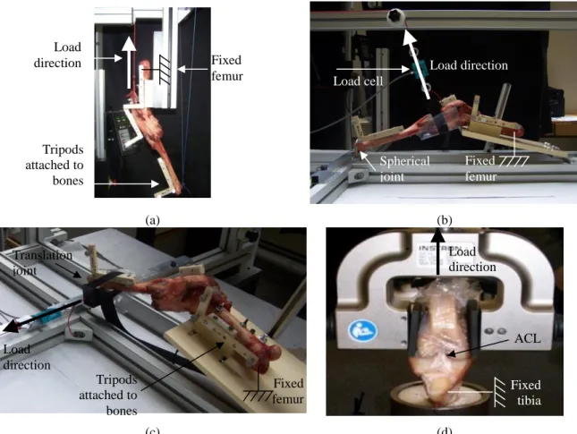
In the experimental kinematics setup (flexion-extension), the femur was fixed to the test rig using bi-cortical screws and the experimental apparatus was mounted in an INSTRON 5500 testing machine (Instron Ltd., Buckinghamshire, England). A pre-load tension was applied to the quadriceps tendon parallel to the femoral shaft using a 30 N deadweight applied on the tibia distal extremity toward the center of the femoral head. The test was conducted for 6 cycles at a speed of 50 mm/min (see Fig. 1(a)) (Azmy et al. 2010, Jenny et al. 2002). The continuous kinematics analysis was performed expressing all the tibial movements (rotations and translations) with respect to the femur.
For the laxity test, a specific experimental device was designed. Two configurations were used in which the femur was fixed and the stifle positioned with a 40° flexion angle: this angle was chosen so that all the specimens could be positioned in the device. For the anterior drawer test, a spherical joint between the distal tibia and the experimental device was used (see Fig. 1(b)) and for the varus-valgus test, the distal tibia could slide on a horizontal plane (see Fig. 1(c)). For both tests, 6 cycles of loading were carried out to reach a tensile force of 100 N on the third part of proximal tibia (the distal third of tibia for varus-valgus test), perpendicular to the tibial shaft in the anterior direction for the drawer test and perpendicular to the transverse plane for the varus-valgus test.
For flexion-extension tests, six degrees of freedom of tibia were studied (three translations, three rotations). For the laxity tests, the anterior translation or of the varus-valgus rotation (for anterior drawer and varus-valgus test, respectively) was considered for a specific load or torque. 2.3 Pull-out destructive tests
(a) (b)
(c) (d)
Fig. 1 Experimental setups: (a) flexion-extension, (b) anterior drawer, (c) varus-valgus, and (d) pull-out test
For the pull-out tests, the distal femur and proximal tibia were cut 10 cm to the joint space. Joint capsule, collateral, and posterior cruciate ligaments as well as the quadricipital tendon were removed and the free bony ends were embedded in steel cylinders using a low temperature melt alloy (MCP70, Mining & Chemical Product, Wellingborough, UK). The sheep’s knees were fixed on an INSTRON 5566 testing machine (Instron Ltd., Buckingham-shire, England) instrumented with a 5 kN load cell (accuracy 0.5%) and tested at room temperature (see Fig. 1(d)). The flexion-angle was set to align the axis of the applied load with the native ACL direction and both interference screws in the case of an artificial ligament. This position was chosen to simulate a worst-case scenario. Knees were conditioned using 10 cycles between 5 and 50 N (5 mm/min) followed by 120 s relaxation at 100 N. Finally, a tension load until total failure was applied to the specimen (5 mm/min). The experimental load-elongation curve was recorded. Stiffness was determined as the most linear region of the load elongation curve and failure mode and ultimate failure load were also documented.
2.4 Evaluation of the technique uncertainties
The uncertainties errors evaluation relies on the assessment of measurement reproducibility.
Tripods attached to bones Load cell Load direction Load direction Load direction Tripods attached to bones Load direction Fixed femur Fixed femur Fixed femur ACL Spherical joint Translation joint Fixed tibia
The latter was carried out using the same method described in (Azmy et al. 2010) for flexion-extension tests and extended to the anterior tibial drawer and the varus-valgus tests. It relied on the Monte Carlo approach to assess uncertainties presented in the “Guide to the Expression of Uncertainty in Measurement” (JCGM 101:2008). Briefly, the occurring uncertain quantities are based on the technical knowledge about an instrumental error source or manual operations such as the tripod adjustment or the calculation of the anatomic coordinate system. All sources of uncertainties are identified and evaluated. Afterward, a set of random results is generated for each random input quantity for which a corresponding random noise has been added. The procedure is repeated 300 times (i.e., the number of Monte Carlo trials=300) and 2 standard deviations are used to evaluate the global uncertainties of the technique. In the case of flexion-extension and anterior drawer and varus-valgus tests, the computed quantities correspond to the six degrees of freedom of the tibia.
2.5 Data analysis
Statistical analyses were performed using Xlstat software (addinsoft SARL, PARIS, France) to evaluate (1) the effect of implantation time (3 months and 12 months) on biomechanical properties considering the controlateral intact knee as control and (2) the difference between grafted and non-grafted artificial ACL for the two implantation durations. Numerical data were expressed as mean±standard deviation. They were analyzed using a non-parametric Mann-Whitney test. The significance level was set at p≤0.05.
3. Results
3.1 Clinical results
All implanted animals recovered successfully from the surgical procedures and remained in good health for the durations of the study. All sheep used their operated limb from the day after surgery until the day of sacrifice and orthopaedic examinations were performed at monthly intervals until the animals were killed.
3.2 Evaluation of measurement uncertainties
Table 1 presents the calculated uncertainties for the different degrees of freedom and for the three different kinematics tests. All the results are presented for 2 standard deviations corresponding to the 95% confidence interval. In terms of rotation uncertainties, their values remain under 4.1° for flexion-extension, under 1.7° for anterior drawer and under 1.6° for varus-valgus. In terms of translation uncertainties, they are less than 3.5 mm for the three kinematic tests.
3.3 Flexion-extension results
Of the 60 initial specimens, some samples could not be analysed due to various experimental problems (2 grafted and 3 non-grafted ligament-reconstructed knees from the 3-month groups and 2 grafted and 1 non-grafted ligament-reconstructed knees from the 12-month groups because either
Table 1 Evaluation of measurement uncertainties (2 standard deviations)
Rotations around axis (°) Translations along axis (mm) Varus/Valgus rotation x Int./Ext.* rotation y Flex./Ext.* rotation z Antero-posterior translation x Cranio-caudal translation y Medio-lateral translation z flexion-extension 4.1 3.5 0.5 2.4 2.4 3.5 anterior drawer 1.7 1.1 0.3 1.2 1.6 3.4 varus-valgus 1.6 1.1 0.9 1.2 1.6 3.4
*Int./Ext.: Internal/External; Flex./Ext.: Flexion/Extension
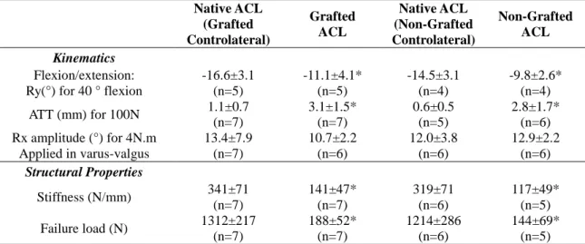
Table 2 Mobilities and structural properties of the tested knees three months after implantation (data expressed as mean1 SD) Native ACL (Grafted Controlateral) Grafted ACL Native ACL (Non-Grafted Controlateral) Non-Grafted ACL Kinematics Flexion/extension: Ry(°) for 40 ° flexion
-16.6±3.1 (n=5) -11.1±4.1* (n=5) -14.5±3.1 (n=4) -9.8±2.6* (n=4) ATT (mm) for 100N 1.1±0.7 (n=7) 3.1±1.5* (n=7) 0.6±0.5 (n=5) 2.8±1.7* (n=6) Rx amplitude (°) for 4N.m Applied in varus-valgus 13.4±7.9 (n=7) 10.7±2.2 (n=6) 12.0±3.8 (n=6) 12.9±2.2 (n=6) Structural Properties Stiffness (N/mm) 341±71 (n=7) 141±47* (n=7) 319±71 (n=6) 117±49* (n=5) Failure load (N) 1312±217 (n=7) 188±52* (n=7) 1214±286 (n=6) 144±69* (n=5) ATT: anterior tibial translation, n: number of specimen.
ACL: Anterior Cruciate Ligament
Grafted: bioactive polymer-grafted artificial ligaments (LARS) Non-grafted: non-grafted with a bioactive polymer ligaments
*Significantly different from the native ACL (p≤.05, Mann-Whitney test)
of failure of the knees during the kinematics tests, a visual sliding of the ACL with regards to the bone during specimen manipulation before the biomechanical experiments or measurement defects).
The flexion-extension knee kinematics was analysed using the internal rotation value for a 40° flexion angle, corresponding to a flexion angle reached by all the specimens. At three months after implantation, the differences between the controlateral native ligaments and the artificial ligaments, both grafted and non-grafted, were significant: the values of flexion-extension rotation for artificial ligaments was 33% lower than the controlateral values (grafted: p=0.010, non-grafted: p=0.011). After 12 months of implantation, no significant difference was observed between
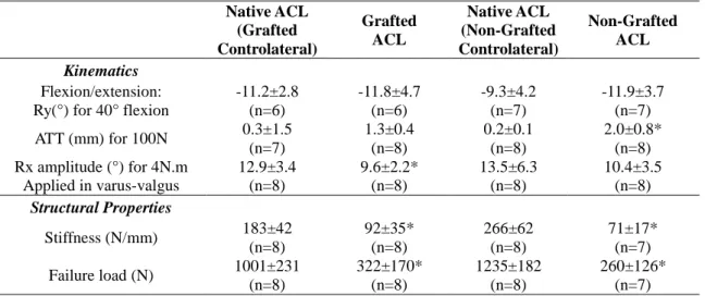
Table 3 Mobilities and structural properties of the tested knees 12 months after implantation (data expressed as mean1 SD) Native ACL (Grafted Controlateral) Grafted ACL Native ACL (Non-Grafted Controlateral) Non-Grafted ACL Kinematics Flexion/extension: Ry(°) for 40° flexion
-11.2±2.8 (n=6) -11.8±4.7 (n=6) -9.3±4.2 (n=7) -11.9±3.7 (n=7) ATT (mm) for 100N 0.3±1.5 (n=7) 1.3±0.4 (n=8) 0.2±0.1 (n=8) 2.0±0.8* (n=8) Rx amplitude (°) for 4N.m Applied in varus-valgus 12.9±3.4 (n=8) 9.6±2.2* (n=8) 13.5±6.3 (n=8) 10.4±3.5 (n=8) Structural Properties Stiffness (N/mm) 183±42 (n=8) 92±35* (n=8) 266±62 (n=8) 71±17* (n=7) Failure load (N) 1001±231 (n=8) 322±170* (n=8) 1235±182 (n=8) 260±126* (n=7) ATT: anterior tibial translation, n: number of specimen.
ACL: Anterior Cruciate Ligament
Grafted: bioactive polymer-grafted artificial ligaments (LARS) Non-grafted: non-grafted with a bioactive polymer ligaments
*Significantly different from the native ACL (p≤.05, Mann-Whitney test)
(grafted or grafted) artificial and native ligaments (p=0.624 and p=0.257 for grafted and non-grafted, respectively). The mean values and associated standard deviation are reported in Tables 2 and 3 for both implantation durations.
3.4 Laxity results
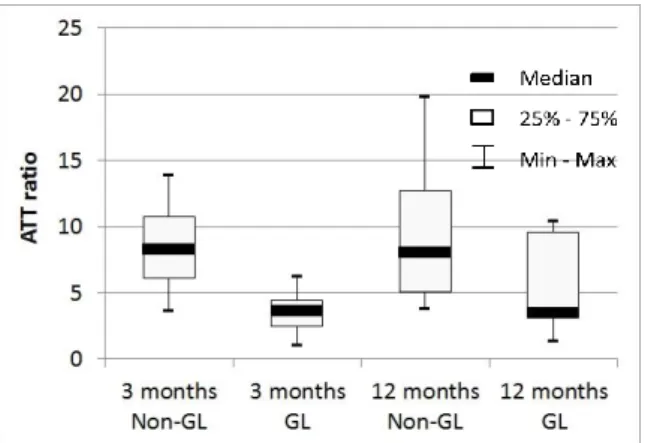
All the results of average anteroposterior tibia translation (ATT) are summarized in Table 2 for specimens explanted 3 months after implantation and in Table 3 for specimens explanted 12 months after implantation. The mean ATT of (grafted or non-grafted) ligament-reconstructed knees drastically increased compared with that of intact controlateral knees for both implantation durations. The ATT at 3 months (and at 12 months) was significantly higher than the ATT of native ACL for both artificial ligaments (p=0.008 and p=0.022 for grafted and non-grafted ligament-reconstructed knees, respectively, for the 3-month group and p<0.0001 for both grafted and non-grafted ligament-reconstructed knees, respectively, for the 12-month group). There were no significant differences between grafted and non-grafted ACL-reconstructed knees (Mann-Whitney test). Fig. 2 presents a boxplot of the ratio between the implanted and the native controlateral knees in terms of anterior tibial translation for the two implantation durations. For the grafted artificial ligaments group, the ATT was 3.5±1.7 times larger three months after implantation in the operated stifle compared to the controlateral group and 5.5±3.7 times larger twelve months postoperatively. For the non-grafted ACL group, the ratio between operated and controlateral stifle was 8.5±4.3 three months after implantation and 9.6±5.8 twelve months postoperatively.
Fig. 2 Ratio between anterior tibial translation (ATT) of artificial and corresponding controlateral native ligament for the two implantation durations (Non-GL: non-grafted artificial ligament; GL: grafted artificial ligament)
Tables 2 and 3 also report the varus-valgus amplitude rotation (in degrees) corresponding to a 4 N.m moment. The values showed no significant difference between either (grafted or non-grafted) artificial and native ligaments or grafted and non-grafted ligaments for three-month implantation (p=0.280 for grafted ligaments and 0.648 for non-grafted ligaments). Twelve months after implantation, the differences remained non-significant for the non-grafted group (p=0.234), but became significant for the grafted group (p=0.035).
3.5 Structural properties
For the three-month group, four specimens implanted with a grafted ACL and two specimens implanted with a non-grafted ACL failed by artificial ligament pull-out from both the tibial and femoral tunnel. Three specimens implanted with a grafted ACL, three specimens implanted with a non-grafted ACL, and three specimens with unoperated intact native ligament failed at mid-substance. Failure mode of seven native ligaments was osteocartilaginous avulsion at the tibial or femoral insertion (Table 4). Three native ligament specimens had to be excluded because of experimental problems.
For the 12-month group, three specimens implanted with a grafted AL and two specimens implanted with a non-grafted ACL failed by graft pull-out from either the tibial and femoral tunnel. Five specimens implanted with a grafted ACL, 5 specimens implanted with a non-grafted ACL, and 11 specimens with intact native ligament failed at mid-substance. Failure mode of five native ligaments was osteocartilaginous avulsion at the tibial or femoral insertion (Table 4).
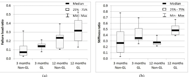
In terms of structural properties of the knees, results drastically decreased between native and grafted or non-grafted ACL for both implantation durations (p<0.0001 for all groups). Data between grafted and non-grafted ligaments were not significantly different (Tables 2 and 3). Fig. 3 presents a boxplot of the ratio between the implanted and the native controlateral knees in terms of stiffness and failure load for the two implantation durations. The ratio, calculated for each sheep, corresponds to the structural value (failure load or stiffness) for the operated limb divided by the value of the controlateral limb.
Table 4 Failure mode for the pull-out tests
3 Months After Implantation 12 Months After Implantation
Native ACL (Graft. Contro. *) Graft. * ACL Native ACL (Non-Graft. Contro. *) Non- Graft. * ACL Native ACL (Graft. Contro. *) Graft. * ACL Native ACL (Non-Graft. Control. *) Non- Graft. * ACL Femoral slippage 0/7 3/7 0/6 1/5 0/8 1/8 0/8 0/7 Tibial slippage 0/7 1/7 0/6 1/5 0/8 2/8 0/8 2/7 Ligamentfailure 1/7 3/7 2/6 3/5 6/8 5/8 5/8 5/7 Osteocartilagino us avulsion 5/7 0/7 2/6 0/5 2/8 0/8 3/8 0/7 Other 1/7 0/7 2/6 0/5 0/8 0/8 0/8 0/7
ACL: Anterior Cruciate Ligament
*Graft.: Grafted: bioactive polymer-grafted artificial ligaments (LARS), Non-graft.: non-grafted with a bioactive polymer ligaments, Contro.: Controlateral
(a) (b)
Fig. 3 Ratio between artificial and corresponding controlateral native ligament for (a) failure load and (b) stiffness for the two implantation durations (Non-GL: non-grafted artificial ligament; GL: grafted artificial ligament)
4. Discussion
evaluate the biomechanical performance of artificial ligaments. The nature and pattern of tissue in-growth within LARS ACTM ligaments as well as the link between biological and biomechanical results using the same sheep are discussed in another publication of our group (Viateau et al. 2013). The aim of the current study was to compare the changes in the biomechanical properties of sheep knees implanted with a grafted or non-grafted ACL for a 3- or 12-month duration. Four different tests were used; they can be classified into kinematics tests and structural tests. For the kinematics tests, the reliability of the method was evaluated. Indeed, for the flexion-extension tests, the 95% confidence interval in rotation along the y axis was equal to 3.5° whereas the measured values were all higher than 9° in absolute value. For the varus-valgus test, the 95% confidence interval in rotation along the x axis was equal to 1.6° whereas the measured values were all higher than 9° in absolute value. The measurements can thus be considered reliable. For the ATT (along the x axis), the measured values for the implanted knees were higher than the 95% confidence interval (equal to 1.2 mm) whereas the values measured for the intact knees were under the 95% confidence interval. However, the differences between implanted and native knees remained higher than the confidence interval limit, thus providing credibility to the results.
In the present study, three months after implantation, no significant difference was observed in the biomechanical results of grafted and non-grafted ligaments. Twelve months after implantation, the differences between grafted and non-grafted ligament biomechanical properties were still not significant in terms of failure load value=0.19), but were significant in terms of stiffness (p-value=0.0012). However, a tendency toward a slightly better performance for the grafted ligaments compared to the non-grafted ligaments in terms of load to failure could be observed 12 months postoperatively (see Fig. 3(a)): The p-value between 12-month grafted AL and non-grafted AL failure load ratio was equal to 0.183.
In terms of flexion/extension, three months after implantation, differences between native and implanted knees were significant, and remained significant twelve months after implantation only for the non-grafted ligaments. The varus/valgus test showed no significant differences three months after implantation for both ligament groups, but twelve months after implantation the differences became significant for the grafted ligaments.
The differences between artificial and native ligaments three months after implantation were mostly due to insufficient fixation of the artificial ligament in bone tunnels. At that time point, failure occurred in most cases by slippage of the ligament from the bone tunnels whereas it mostly occurred by ligament failure 12 months after implantation. Although loading was performed in line with the bone tunnels (which is the least stable position and which does not correspond to clinical situations), these results suggest that ligament fixation (and not the artificial ligament per se) is a weak link in both grafted and non-grafted ligament reconstructed knees in the early postoperative period. This can be explained by the lower biomechanical properties of bone tissue surrounding the implant. Vayron et al. (2012) showed in rabbits that the values of the indentation moduli of new bone tissue surrounding a titanium implant (4, 7, and 13 weeks) were significantly lower than those of mature bone.
The failure load of native ACL has been compared with an autologous Achilles tendon split graft implanted in sheep (Weiler et al. 2002). Three months after implantations, a decrease in failure load was observed in all operated knees compared to unoperated knees (1531.3±180.3 N and 237.8±59.8 N, respectively). Although direct comparisons between different studies is difficult because of variations in methodology (i.e., the flexion angle in Weiler et al. (2002) was set to 60° whereas it was adapted to align ACL and tibial and femoral tunnels in our study, which led to a mean flexion angle of 83°), these results were consistent with the conclusions of our study. Hunt et
al. (2005), using a sheep model to compare different ACL reconstruction methods, concluded that, one year after implantation, reconstructed ACL showed significantly lower load-to-failure and stiffness values compared to the intact ACL. Abramowitch et al. (2003) studied, in goats, the effect of initial graft tension on biomechanical properties. Six weeks after implantation, they evaluated the structural properties of the knee with the ACL orientation aligned along the tension axis. They noted a significant decrease in failure load and stiffness in implanted knees but no difference between the low- and high-tension groups. Even if the implantation duration differs from one study to another, using similar primary fixations, the structural properties of the knees are significantly affected by the ACL replacement.
In terms of the anterior tibial drawer, our results compare favorably with those published in the literature. Zantop et al. (2008) studied the influence of tunnel length on the ATT in goats three months after surgery. The flexion angle, in which the ATT was tested, varied from 30° to 90°. The ATT under 67 N anterior tibial force was 6.3 mm on average. Abramowitch et al. (2003) conducted in vitro an anterior drawer test six weeks after implantation. The differences in ATT at 30° of knee flexion under 67 N anterior tibial force between the implanted and control groups were statistically significant for every initial tension: A mean ATT equal to 13.4 mm was indeed achieved for the reconstructed knees (15 mm and 11.8 mm depending on the initial tension) and a mean ATT equal to 5.1 mm for the intact knees was achieved. These values are higher than the value presented in the current study for a 40° flexion angle and 100 N anterior tibial force (from 1.3 to 3.1 mm for implanted knees during 3 or 12 months in sheep).
In conclusion, structural parameters suffer from an insufficient bone fixation in the early post-operative period, prompting the need for improved primary fixation. It can be expected that a longer in vivo implantation will improve the mechanical response. Indeed, there is a trend toward an increase in both the stiffness and the failure load after 12 months in vivo. The trend in favor of the grafted artificial ligament observed in the present study will have to be confirmed using a larger population.
Acknowledgments
We gratefully acknowledge the financial support provided by the Agence Nationale pour la Recherche (ANR), France [Grant LIGART 06-TECSAN-006-01]. We would also like to thank Bernard Brulez and the LARS society for providing all the artificial ligaments and the orthopaedic surgeons who followed the project: Pr. P. Thoreux and Dr. J. Cournapeau.
References
Abramowitch, S., Papageorgiou, C., Withrow, J., Gilbert, T. and Woo, S. (2003), “The effect of initial graft tension on the biomechanical properties of a healing ACL replacement graft: a study in goats”, J. Orthop.
Res., 21(4), 708-715.
Azmy, C., Guérard, S., Bonnet, X., Gabrielli, F. and Skalli, W. (2010), “EOS® orthopaedic imaging system to study patellofemoral kinematics: assessment of uncertainty”, Orthop. Traumatol. Surg. Res., 96(1), 28-36.
Burks, R.T., Crim J., Fink, B.P., Boylan, D.N. and Greis, P.E. (2005), “The effects of semitendinosus and gracilis harvest in anterior cruciate ligament reconstruction”, Arthroscopy, 21(10), 1177-1185.
(2012), “Fast 3D reconstruction of the lower limb using a parametric model and statistical inferences and clinical measurements calculation from biplanar X-rays”, Comput. Meth. Biomech. Biomed. Eng., 15(5), 457-466.
Ciobanu, M., Siove, A., Gueguen, V., Gamble, L.J., Castner, D. and Migonney, V. (2006), “Radical graft polymerization of styrene sulfonate on poly(ethylene terephthalate) films for ACL applications: “grafting from” and chemical characterization”, Biomacromolecules, 7(3), 755-760.
Ferretti, A., Conteduca, F., Labianca, L., Monaco, E. and De Carli, A. (2005), “Evolgate fixation of doubled flexor graft in anterior cruciate ligament reconstruction: biomechanical evaluation with cyclic loading”,
Am. J. Sports Med., 33(4), 574-582.
Gao, K., Chen, S., Wang, L., Zhang, W., Kang, Y., Dong, Q., Zhou, H. and Li, L. (2010), “Anterior cruciate ligament reconstruction with LARS artificial ligament: a multicenter study with 3- to 5-year follow-up”,
Arthroscopy, 26(4), 515-523.
Hagemeister, N., Duval, N., Yahia, L’H., Krudwig, W., Witzel, U. and De Guise, J-A. (2002), “Comparison of two methods for reconstruction of the posterior cruciate ligament using a computer based method: quantitative evaluation of laxity, three-dimensional kinematics and ligament deformation measurement in cadaver knees”, Knee, 9(4), 291-299.
Hunt, P., Scheffler, S., Unterhauser, F. and Weiler, A. (2005), “A model of soft-tissue graft cruciate ligament reconstruction in sheep”, Arch. Orthop. Trauma. Surg., 125(4), 238-248.
JCGM 101:2008 (2008), “Evaluation of measurement data - Supplement 1 to the “guide to the expression of uncertainty in measurement” - propagation of distributions using a Monte Carlo method”, Joint
Committee for Guides in Metrology.
Jenny, J-Y., Lefebvre, Y., Vernizeau, M., Lavaste, F. and Skalli, W. (2002), “Validation d’un protocole expérimental d’étude optoélectronique de la cinématique active continue de l’articulation du genou in
vitro”, Rev. Chir. Orthop. Reparatrice Appar. Mot., 88(8), 790-796.
Kleweno, C.P., Jacir, A.M., Gardner, T.R., Ahmad, C.S. and Levine, W.N. (2009), “Biomechanical evaluation of anterior cruciate ligament femoral fixation techniques”, Am. J. Sports Med., 37(2), 339-345. Lavoie, P., Fletcher, J. and Duval, N. (2000), “Patient satisfaction needs as related to knee stability and
objective findings after ACL reconstruction using the LARS artificial ligament”, Knee, 7(3), 157-163. Mascarenhas, R. and MacDonald, P.B. (2008), “Anterior cruciate ligament reconstruction: a look at
prosthetics - past, present and possible future”, Mc Gill J. Med., 11(1), 29-37.
Milano, G., Mulas, P.D., Sanna-Passino, E., Careddu, G-M., Ziranu, F. and Fabbriciani, C. (2005), “Evaluation of bone plug and soft tissue anterior cruciate ligament graft fixation over time using transverse femoral fixation in a sheep model”, Arthroscopy, 21(5), 532-539.
Milano, G., Mulas, P.D., Ziranu, F., Piras, S., Manunta, A. and Fabbriciani, C. (2006), “Comparison between different tibial fixation devices for ACL reconstruction with doubled hamstring tendon graft: a biomechanical analysis”, Arthroscopy, 22(6), 660-668.
Monaco, E., Labianca, L., Speranza, A., Agrò, A.M., Camillieri, G., D'Arrigo, C. and Ferretti, A. (2010), “Biomechanical evaluation of different anterior cruciate ligament fixation techniques for hamstring graft”,
J. Orthop. Sci., 15(1), 125-131.
Nau, T., Lavoie, P. and Duval, N. (2002), “A new generation of artificial ligaments in reconstruction of the anterior cruciate ligament: Two-year follow-up of randomised trial”, J. Bone Joint Surg. [Br], 84(3), 356-360.
Pavon-Djavid, G., Gamble, L.J., Ciobanu, M., Gueguen, V., Castner, D. and Migonney, V. (2007), “Bioactive PET fibers and fabrics: grafting, chemical characterization and biological assessment”,
Biomacromolecules, 8(11), 3317-3325.
Scheffler, S.U., Südkamp, N.P., Göckenjan, A., Hoffmann, R.F. and Weiler, A. (2002), “Biomechanical comparison of hamstring and patellar tendon graft anterior cruciate ligament reconstruction techniques: the impact of fixation level and fixation method under cyclic loading”, Arthroscopy, 18(3), 304-315. Shen, H.C., Chang, J.H., Lee, C.H., Shen, P.H., Yeh, T.T., Wu, C.C. and Kuo, C.L. (2010), “Biomechanical
com-parison of cross-pin and endobutton-CL femoral fixation of a flexor tendon graft for anterior cruciate ligament reconstruction- a porcine femur-graft-tibia complex study”, J. Surg. Res., 161(2), 282-287.
Speirs, A., Simon, D. and Lapner, P. (2010), “Evaluation of a new femoral fixation device in a simulated anterior cruciate ligament reconstruction”, Arthroscopy, 26(3), 351-357.
Vaquette, C., Viateau, V., Guérard, S., Anagnostou, F., Manassero, M., Castner, D. and Migonney, V. (2013), “The effect of polystyrene sodium sulfonate grafting on polyethylene terephthalate artificial ligaments on in vitro mineralisation and in vivo bone tissue integration”, Biomat., 34(29), 7048-7063. Vayron, R., Barthel, E., Mathieu, V., Soffer, E., Anagnostou, F. and Haiat, G. (2012), “Nanoindentation
measurements of biomechanical properties in mature and newly formed bone tissue surrounding an implant”, J. Biomech. Eng., 134(2), 021007.
Legnani, C., Ventura, A., Terzaghi, C., Borgo, E. and Albisetti, W. (2010), “Anterior cruciate ligament reconstruction with synthetic grafts. A review of literature”, Int. Orthop., 34, 465-471.
Viateau, V., Zhou, J., Guérard, S., Manassero, M., Thourot, M., Anagnostou, F., Mitton, D., Brulez, B. and Migonney, V. (2011), “LIGART: Synthetic “bioactive” and “biointegrable” ligament allowing a rapid recovery of patients: chemical grafting, in vitro and in vivo biological evaluation, animal experiments, preclinical study”, IRBM, 32(2) 118-122.
Viateau, V., Manassero, M., Anagnostou, F., Guérard, S., Mitton, D. and Migonney, V. (2013), “Evaluation of the Ligament Advanced Reinforcement System (LARS™) in a sheep model of anterior cruciate ligament (ACL) replacement. A 3- and 12-month study”, Arthroscopy, 29(6), 1079-1088.
Weiler, A., Peine, R., Pashmineh-Azar, A., Abel, C., Südkamp, N. and Hoffmann, R. (2002), “Tendon healing in a bone tunnel. Part I: Biomechanical results after biodegradable interference fit fixation in a model of anterior cruciate ligament reconstruction in sheep”, Arthroscopy, 18(2), 113-123.
WO/2004/067051 (2004), “Biomimetic prosthetic ligament and production method thereof - Ligament prothétique biomimétique et procédé d'obtention”, European Patent.
Zantop, T., Ferretti, M., Belle, K., Brucker, P., Gilbertson, L. and Fu, F. (2008), “Effect of tunnel-graft length on the biomechanics of anterior cruciate ligament - reconstructed knees intra-articular study in a goat model”, Am. J. Sports Med., 36(11), 2158-2166.
Zhou, J., Ciobanu, M., Pavon-Djavid, G., Gueguen, V. and Migonney, V. (2007), “Morphology and adhesion of human fibroblast cells cultured on bioactive polymer grafted ligament prosthesis”, 29th
Annual International Conference of the IEEE Engineering in Medicine and Biology Society, Lyon,
France, August.