Université de Montréal
Differences in brain structure between males and females diagnosed with schizophrenia
par
Adham Mancini Marïe
Département de Psychiatrie, Université de Montréal Faculté de Médecine
Thèse présentée à la Faculté de Médecine en vue de l’obtention du grade de Doctorat
Option Science Biomédicale
August 2016
Université de Montréal
Faculté des études supérieures et postdoctorales Ce mémoire intitulé
Differences in brain structure between males and females diagnosed with schizophrenia
Présentée par : Adham Mancini Marïe
a été évaluée par un jury composé des personnes suivantes : Dr. Lan Xiong, représentant de la doyenne
Dr. Tania Lecomte, président-rapporteur Dr. Adrianna Mendrek, directeur de recherche
Dr. David Luck, membre du jury Dr. Donna Lang, membre du jury externe
Résumé
Les progrès dans le domaine de la neuroimagerie cérébrale ont permis une certaine compréhension des maladies mentales comme la schizophrénie. Cependant, peu de résultats sont cohérents et ils sont souvent contradictoires, ce qui rend difficile de tirer des conclusions concrètes par rapport à la maladie.
Plusieurs facteurs jouent un rôle dans les résultats divergents et convergents : Les différentes techniques d'imagerie et les analyses, le nombre de patients inclus dans les études, l'âge des patients, l'âge de l’'apparition de la maladie, les critères de diagnostic, les effets du traitement antipsychotique, le statut social, ainsi que les comorbidités, font partie de ces facteurs. Bien que les différences cérébrales entre femmes et hommes « normaux » sont bien établies, ce n’est que ces dernières années que des études en neuroimagerie de la schizophrénie ont abordé les différences homme-femme comme une explication potentielle des résultats discordants de l’imagerie cérébrale.
L'objectif de cette thèse est de comprendre le rôle du sexe (genre féminin et masculin) dans les anomalies anatomiques observées dans la schizophrénie; ceci, en réalisant des études qui contrôlent, autant que possible, l'effet de différentes variables confondantes et en utilisant des analyses d’IRM automatisées chez des patients et des sujets sains de même âge et du même sexe. Une brève revue globale des résultats actuels dans le domaine de la schizophrénie ainsi que des résultats liés aux différences entre les sexes dans la schizophrénie vont être présentés.
La première étude visait à étudier l'influence des différences de sexe sur des mesures de la gyrification corticale de la schizophrénie. Étant donné que la schizophrénie est une maladie dont les «symptômes cliniques » ont un impact négatif sur la qualité de vie des patients qui en souffrent, nous avons exploré la relation entre la gyrification corticale et les différents symptômes de la schizophrénie chez les hommes et les femmes atteints de ce trouble psychiatrique. Le rôle du sexe sur la gyrification corticale et son association aux symptômes a été à peine étudié chez les patients atteints de schizophrénie ; c’est pour cette raison que, nous croyons que cette étude est d’une importante valeur.
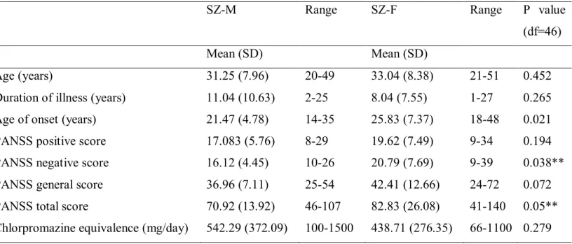
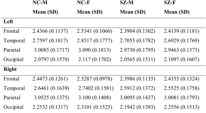
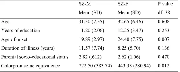
Dans cette première étude, des images 3T T1 ont été acquises auprès de 48 patients atteints de schizophrénie (24 hommes [SZ-M] et 24 femmes [SZ-F]) et 48 volontaires sains (24 hommes [NC-M] et 24 femmes [NC-F]), appariés en fonction de l'âge et du sexe. Des mesures d’indice de
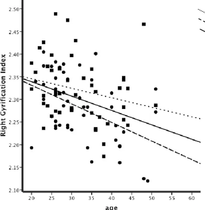
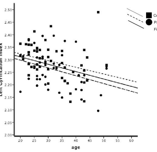
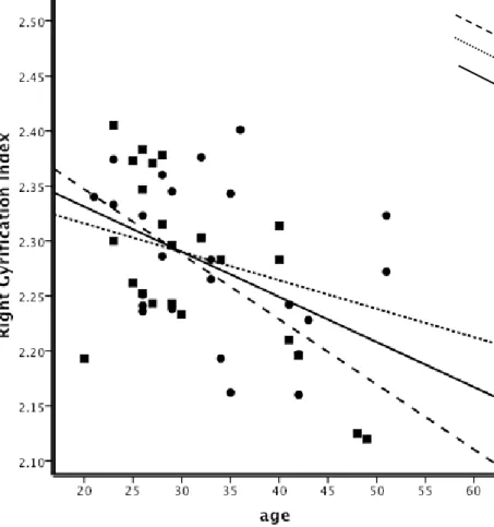
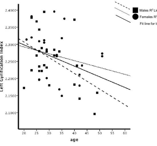
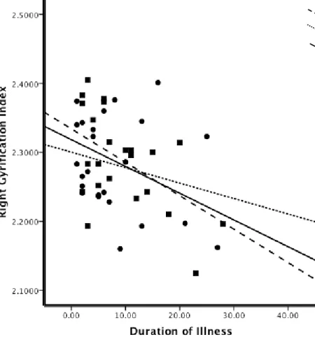
gyrification (IG) pour chaque hémisphère et les quatre lobes cérébraux (frontaux, temporal, pariétal, et occipital) ont été effectuées en utilisant le pipeline de CIVET, lequel est entièrement automatisé. Plusieurs résultats intéressants ont émergé: les patients avaient des valeurs inférieures importantes de l’IG global par rapport aux témoins; SZ-M avaient des valeurs d'IG hémisphériques significativement inférieurs par rapport à NC-M, cela n'a pas été observé dans les groupes de femmes. Aucune différence entre les sexes dans les valeurs de diminution de l’IG avec l'âge n’a été observés chez les témoins sains par contre, une diminution de la valeur de l’IG avec l’âge chez les patients était plus importante chez les patients homme que les patients femmes. Une détérioration plus progressive dans l'hémisphère droit dans les deux groupes de patients a été observée, tout comme des réductions significatives des valeurs d’IG en relation avec la durée de la maladie chez SZ-M, mais pas chez SZ-F.
Dans les groupes de patients, on observe des diminutions des valeurs d’IG dans les lobes frontaux bilatéraux et, le lobe occipital droit; le groupe SZ-M a montré une valeur d’IG significativement plus élevée par rapport à NC-M dans le lobe temporal droit; SZ-F a montré des valeurs d’IG significativement plus faibles dans les lobes bilatéraux frontaux, temporaux, pariétaux et le lobe occipital droit, par rapport à NC-F. Aucune corrélation significative n'a été trouvée entre les valeurs de l'IG et le profil de la symptomatologique dans les deux groupes de patients.
Etant donné que l’IG reflète, en partie, des altérations dans le développement et la connectivité cérébrale, la diminution de l’IG observée chez les patients est en accord avec le modèle de développement neurobiologique de disconnectivité dans la schizophrénie. De plus, nous soulignons l'importance de l'âge ainsi que la durée de la maladie lorsque nous comparons les hommes et les femmes atteints de schizophrénie. Cependant, nous n'avons pas observé de corrélation significative n'a été trouvée entre les valeurs de l'IG et les symptômes, ce qui est d'un intérêt particulier et inattendu compte tenu des résultats de la neuroimagerie montrant par exemple certaines corrélations entre les symptômes positifs et certaines anomalies du lobe temporal dans la schizophrénie.
Considérant ces résultats, nous avons décidé d'investiguer, dans notre deuxième étude, l'association entre les symptômes et les densités de matière grise (DMG) et de matière blanche (DMB) à la place des mesures de gyrification corticale. Nous avons utilisé la morphométrie basée sur le voxel "Voxel Based Morphometry (VBM8.0 with Diffeomorphic Anatomical Registration (Through Exponentiated Lie Algebra [DARTEL])" et la modélisation linéaire automatique
(SPSS21.0 ALM) sur les images 3T T1 MPRAGE acquises auprès de 40 patients atteints de schizophrénie (SZ) et 41 témoins sains (NC).
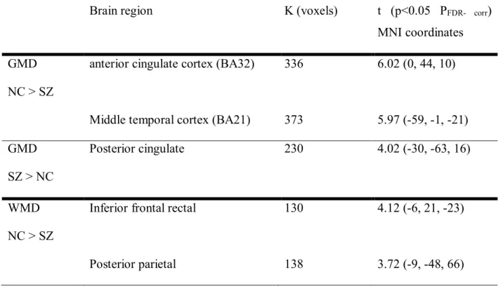
Nous avons trouvé que les patients atteints de schizophrénie avaient une DMG réduite dans le cortex cingulaire antérieur, le cortex temporal médian gauche et une DMG plus élevée dans le cortex cingulaire postérieur gauche par rapport aux sujets sains. Une diminution significative de DMB dans la région fronto-rectal inférieure gauche et la région pariétale postérieure gauche a été observée chez les patients comparés aux sujets sains.
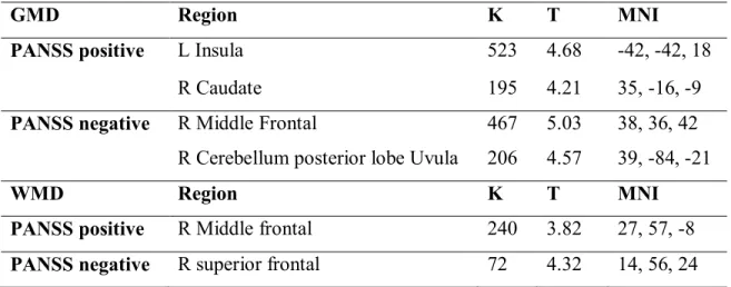
Nous avons trouvé des corrélations positives entre les symptômes positifs et la DMG dans l'insula gauche et le noyau caudé droit; et entre les symptômes négatifs et la DMG dans le cortex frontal médian droite et le lobe postérieur de cervelet droit. Nous avons aussi trouvé des corrélations négatives de DMG dans la région pariétale droite (précuneus), le lobe postérieur du cervelet gauche et les symptômes positifs; ainsi qu'entre la DMG du lobe antérieur du cervelet gauche et les symptômes négatifs. En outre, des corrélations positives ont été trouvées entre la DMB dans le cortex frontal médian droit et les symptômes positifs et entre le DMB dans la région frontale supérieure droite et les symptômes négatifs. Des corrélations négatives ont été trouvées entre les symptômes positifs et la DMB dans la région occipitale inférieure droite et le cunéus occipital droit, tandis que des corrélations négatives ont été trouvées entre la DMB et la région frontale supérieure gauche.
Il est intéressant de noter que lorsque les symptômes ont été analysés par regroupement, nous avons trouvé que le symptôme de la désorganisation conceptuelle corrélait positivement avec la DMG totale et la DMB totale. L’augmentation de DMG a été associée à une diminution de la gravité des hallucinations et du manque de spontanéité; tandis que l'augmentation de DMB totale a été associée à la diminution de la sévérité de l'hostilité et des idées de grandeur. Une comparaison entre les groupes d'hommes a montré une diminution de la DMG chez les patients schizophrènes, tandis qu’aucune différences n’a été observée dans les groupes de femmes. Nous n’avons trouvé aucune corrélation entre la DMG, la DMB, le liquide cérébro-spinal, le volume total du cerveau, les symptômes individuels et la schizophrénie chez les sujets féminins. Chez les hommes atteints de schizophrénie, on observe des corrélations négatives importantes entre les idées de grandeur et la DMB; des corrélations positives entre la désorientation et la DMB. De plus on observe des corrélations entre et les déficits d'attention et de DMG et DMB. Nos résultats montrent que ces associations sont différentes chez les hommes et les femmes atteints de la schizophrénie.
La symptomatologie de schizophrénie est un mélange de déficits cognitifs et socio-affectifs. Dans ce contexte, le but de notre troisième étude est d'étudier chez les patients atteints de la schizophrénie des DMG et DMB et leur relation avec l’acuité mnésique avec des contenus émotionnelles (négatives, positives et neutres) ainsi que étudier l'effet des différences de sexe sur nos résultats. Quarante et un patients droitiers, traités par antipsychotique, souffrant de schizophrénie (SZ) et 40 témoins sains (NC), tous droitiers, ont participé à l’étude. Nous avons utilisé des images de l'International Affective Picture System (IAPS), une banque d'images émotionnelles, et de l’IRM. On observe chez les témoins sains des corrélations entre les valeurs élevées de DMG du cortex pariétal postérieur, du lentiform, du putamen, noyau caudé, le cortex orbitofrontal inférieur gauche et la reconnaissance des images négatives. On observe des corrélations entre la DMG dans la région temporale gauche, fusiforme et la reconnaissance des images positives ; et également dans le cervelet antérieur gauche et l’acuité des images neutres. Chez les patients on observe des valeurs élevées des DMG dans le cortex occipital inférieur gauche et la reconnaissance des images négatives, mais aucune corrélation entre la capacité de reconnaissance des images positives ou neutres.
Nous avons observé chez les témoins sains: des relations significatives entre la DMB dans le cortex pariétal postcentral gauche et la capacité de reconnaître des images négatives; dans le cortex temporal inferieur gauche, le cortex pariétal gauche (précuneus), le cortex frontal gauche et la capacité de reconnaissance des images positives; des valeurs de DMB du cortex temporel médian et l’acuité des images neutres.
Les patients atteints de schizophrénie ont montré des relations significatives entre de DMB dans le cortex occipito-lingual gauche et la reconnaissance des images négatives ; dans le cortex pariétal angulaire gauche et la reconnaissance des images positives ; et dans le cortex temporal supérieur droit et les images neutres. Les différences de sexe dans la schizophrénie ont été observées : chez les patients de sexe masculin, des corrélations négatives ont été trouvées entre les DMB et la capacité de reconnaître des images négatives et positives. Chez les hommes sains, nous avons trouvé des corrélations positives entre des valeurs totales de DMG et la capacité de reconnaître des images négatives. Nous n’avons pas observé de corrélations dans les groupes de femmes. Ces résultats soutiennent l'hypothèse de l'atrophie fronto-temporale régionale chez les patients schizophrènes. Toutefois, nous notons qu’ils ont des augmentations relatives des valeurs de DMB dans le cortex occipito-pariétal.
Nous avançons l'hypothèse que les déficits mnésiques chez les patients sont liés à des perturbations dans la coordination des réseaux cérébraux, ce qui peut être affecté par des déficits structuraux plus évidents chez les patients masculins. Par conséquent, nous préconisons que les futures études devraient utiliser le connectome ou l’approche « réseaux cérébraux » pour étudier l’impact du sexe (genre masculin-féminin) sur les déficits cognitifs et symptomatologiques dans la schizophrénie. Nos résultats globaux soulignent l'importance de la différence entre homme et femme dans la modulation de manifestations cliniques et fonctionnelles de la schizophrénie. Ainsi, nous croyons que le contrôle des covariables comme l'âge, la durée de la maladie et le statut social est insuffisant et que les études futures sur la schizophrénie devraient systématiquement séparer les hommes des femmes, afin de mieux comprendre cette maladie mentale complexe et dévastatrice.
Mots-clés: Schizophrénie, les différences de sexe, l'imagerie par résonance magnétique, les
symptômes, les déficits cognitivo-affectifs, indice de gyrification, substance grise, la substance blanche.
Abstract
Advances in cerebral neuroimaging techniques have helped our understanding of mental illnesses, such as schizophrenia. Few findings remain consistent and are often contradictory, making it difficult to draw informative conclusions about the disease. Several factors play a role in both diverging and converging results. Imaging technique and analyses, number of patients involved, age of patients, age at onset of the disease, diagnostic criteria, antipsychotic treatment effects, social status, comorbidities, are among some of the reasons. Despite well established cerebral sex differences in healthy population, it is only in recent years that neuroimaging studies in schizophrenia have addressed sex differences as a major possible explanation for discrepant neuroimaging finding.
The aim of this thesis is to help understand the role of sex on brain structures in schizophrenia, by conducting studies that control as much as possible for other variables and by using MRI automated analyses for patients and controls matched for age and sex. This work will briefly present findings in schizophrenia in general, and then an extensive review of the literature on sex differences in schizophrenia will be presented. From it, we are able to conclude that sex differences have been reported with rare exception in almost all aspects involved in the life of patients with schizophrenia.
Chapters
1. The first study investigated sex differences in cortical gyrification in schizophrenia patients (SZ). In addition, considering that schizophrenia is a disease of “clinical symptoms” that determine the quality of life of patients afflicted by it, we explored the relation between cortical gyrification and symptoms in males and females with schizophrenia. The role of sex on cortical gyrification and its association with symptoms has been scarcely investigated in patients with schizophrenia. In this study, 3T T1 images were acquired from 48 schizophrenia patients (24 males [SZ-M] and 24 females [SZ-F]) and 48 normal controls [NC] (24 males [NC-M] and 24 females [NC-F]) matched for age, sex, and handedness. Gyrification Index (GI) analyses for each hemisphere and four cerebral regions (frontal, temporal, parietal, and occipital) were performed using the fully automated CIVET pipeline. Patients had significant lower values of the overall GI relative to normal controls and SZ-M had significant lower right hemispheric GI values compared to NC-M. This was not observed in either NC-F or in SZ. No gender difference in GI values decreases with
age were observed in NC. In patients, GI decreases with age were greater in SZ-M than SZ-F, with a more progressive deterioration in the right hemisphere in both patient groups. Significant GI value reductions in association with duration of illness were observed in SZ-M but not in SZ-F. Patient groups had lower GI in bilateral frontal, temporal, and parietal lobes than controls. SZ-F had significant lower GI values in left frontal, bilateral temporal and left parietal lobe compared to NC-F. No significant correlations were found between GI values and symptom scores in either group of patients. Since GI reflects, in part, alterations in cerebral development and connectivity, the decrease in GI observed in patients is in agreement with the neurodevelopmental model of disconnectivity in schizophrenia, and may explain the worse prognosis and social outcome observed in male patients. Furthermore, we emphasize the importance of age and duration of illness when comparing males and females with schizophrenia. Observed differences between male and female patients may reflect a more diffuse and generalized cortical loss in males. Female patients had cortical loss in specific regions, while preserving cortical gyrification in compensatory regions. Our latter finding -no significant correlation between GI values and symptom scores- was of particular interest and was unexpected in view of neuroimaging findings of correlations between positive symptoms and temporal lobe abnormalities.
2. In the second study, we examined the association between symptoms and brain structure using gray (GMD) and white matter (WMD) densities. Voxel-based morphometry (VBM8.0 with Diffeomorphic Anatomical Registration Through Exponentiated Lie Algebra [DARTEL]) and Automatic Linear Modeling (SPSS21.0 ALM) were used on 3T T1 MPRAGE images acquired from 40 schizophrenia patients (SZ) and 41 normal controls (NC). We found that SZ had lower GMD in the anterior cingulate cortex and left middle temporal gyrus, and higher GMD in the left posterior cingulate in comparison to NC. SZ had significantly lower WMD in the left inferior fronto-rectal and the left posterior parietal regions in comparison to NC. Significant positive correlations were found between positive symptoms and GMD in the left insula and right caudate, and between negative symptoms and GMD in the right middle frontal and the posterior lobe of the right cerebellum (uvula). Inverse relationships between GMD in the right parietal (precuneus), the left posterior lobe of the cerebellum (uvula) and positive symptoms, and between GMD in the left anterior lobe of the cerebellum and negative symptoms were observed in SZ. In addition, positive correlations were found between WMD in the right middle frontal lobe, and between positive symptoms and WMD in the right superior frontal region with negative symptoms. Negative
correlations were found between positive symptoms and WMD in the right inferior occipital and the right occipital cuneus, while negative symptoms correlated negatively with the WMD of the left superior frontal.
When symptom clusters were analyzed, conceptual disorganization symptom positively correlated with both total GMD and WMD. While increases in GMD were associated with decreased severity of lack of spontaneity and hallucinations symptom, increases in total WMD were associated with decreased severity of hostility and grandiosity symptoms. Comparison between male subjects revealed decreased GMD in male schizophrenia patients, while no differences were observed between females across groups. No correlations were found in female groups between GMD, WMD, CSF, or total brain volume and individual symptoms. In males with schizophrenia, significant negative correlation between ideas of grandiosity and WMD, a positive correlation between disorientation and WMD, and attention deficits and GMD and WMD were found. The current data suggest region-specific GMD and WMD association with negative and positive symptoms. In addition, it reveals that such associations are different in male and female schizophrenia patients.
3. The third study investigated the relationships of GMD and WMD with memory accuracy for emotionally negative, positive, and neutral pictures in schizophrenia patients relative to normal controls. Schizophrenia is characterized by an amalgam of cognitivo-socio-emotional deficits. The relationship between emotion processing on cognition and neurobiological underpinnings merit more attention than it has received so far. Memory deficits are among the most common deficits in schizophrenia and have a widespread impact on cognition in general. Additionally, consistently with the major theme of the present thesis, we investigated the effect of gender on the observed effect. Forty one, right-handed medicated patients with schizophrenia (SZ) and 40 right-handed normal controls (NC) matched by age and sex were assessed for memory accuracy using negative, positive and neutral pictures taken from the International Affective Picture System (IAPS). Imaging methods and analyses were similar to our second study. Fifteen minutes after presentation of selected IAPS images (incidental encoding), subjects were asked to recognize the previously seen images among other images. We found higher GMD in NC in the right posterior parietal cortex, lentiform, putamen, and caudate, as well as the left inferior orbitofrontal cortex, in relation with the negative images accuracy. NC had higher GMD in the left temporal and fusiform regions in relation with the positive images accuracy, and higher GMD in the left anterior cerebellum in
relation with neutral images. Schizophrenia subjects had higher GMD in the left inferior occipital cortex in relation with the negative images accuracy, but GMD was not correlated with positive or neutral images accuracy in this group. WMDs correlations were higher in NC in the left postcentral parietal region for negative images; in the left inferior temporal, left precuneus parietal, and left frontal regions for positive images; and in the left middle temporal region for neutral images. Schizophrenia patients had higher WMD in the left lingual occipital for negative images; in the left angular parietal for positive images; and in the right superior temporal region for neutral images. While examining the two sexes separately, we observed inverse correlations between WMD and both negative and positive pictures in male patients. In addition, only in male controls, GMD positively correlated with negative pictures and this correlation was absent in female SZ subjects and NC females. These findings support the hypothesis of fronto-temporal regional atrophy in schizophrenia. Schizophrenia patients have relatively increased occipito-parietal WMD, advancing the hypothesis that the core pathophysiological problem underlying recall memory in SZ may be related to disruptive alterations in the coordination of large-scale brain networks, and this may be affected by structural deficits that are more evident in male patients. It is recommended that future studies should use the connectomes or the brain networks approach to investigate the effect of sex on memory deficits in schizophrenia.
Our overall findings point out to the importance of sex in modulating the clinical and functional manifestations of schizophrenia. We believe that controlling for covariates as age, duration of illness, social status, etc. is insufficient and that future studies in schizophrenia should systematically separate male and female findings, if we wish to understand this complex and devastating mental illness.
Keywords: Schizophrenia, sex differences, magnetic resonance imaging, positive and negative symptoms, cognitivo-emotion deficits, gyrification index, voxel based morphometry, gray matter densities, white matter densities.
Table of contents
Title ……... i
Identification of jury …………...i
Résumé ...iii
Abstract ...ix
Table of contents ...xiii
List of tables ...xvi
List of figures...xvii
Abbreviations ...xviii
Thank you ...xxi
Table of contents 1 Introduction………..1 1.1 History of schizophrenia………...1 1.2 Overview………...1 1.3 Epidemiology of schizophrenia………...4 1.4 Etiology of schizophrenia…….……….4 1.5 Cognition in schizophrenia………5 1.6 Emotion in schizophrenia………..6
1.7 Pathogenesis and Pathophysiology………...8
1.8 Treatment of schizophrenia….………..9
1.9 Neuroimaging findings………...11
2 Sex differences in schizophrenia………13
2.1 Epidemiology………13 2.2 Age of onset………...14 2.3 Premorbid history………..14 2.4 Birth complication……….16 2.5 Psychosocial factors………..17 2.6 Clinical expression………19 2.7 Course of illness………21 2.8 Cognition………...22 2.9 Treatment………..24
3 Summary……...………...….…...33
4 Hypotheses and rationale..………..….…...35
5 Article 1………..37
6 Article 2………..73
7 Article 3……….…116
8 Discussion………..155
8.1 Summary of main findings………156
8.2 Limitation………..173
9 Conclusion……….175
10 Clinical and research implications………..……….….176
11 References……….177
List of tables Article 1. Table 1………64 Table 2………65 Article 2. Table 1………..100 Table 2………..101 Table 3………..102 Table 4………..103 Table 5………..104 Table 6………..106 Article 3. Table 1………..144 Table 2………..145 Table 3………..146 Table 4………..147 Appendix Table X………..237 Table Y………..252
List of figures Article 1 Figure 1a……….…66 Figure 1b……….67 Figure 2a……….……68 Figure 2b………...…..69 Figure 3………...70 Figure 4..………...71 Figure 5………...72 Article 2. Figure 1..………...107 Figure 2..………...108 Figure 3..………...109 Figure 4..………...110 Figure 5..………...111 Figure 6..………...112 Figure 7..………...113 Figure 8..………...114 Article 3. Figure 1..………...148 Figure 2..………...149 Figure 3..………...…150 Figure 4..………...151 Figure 5..………...152 Figure 6..………...153
Abbreviations
2D 2 dimensional
3D 3 dimensional
3T 3 Tesla
AICC Akaike’s Information Criterion Corrected ALM Automatic Linear Modeling
ANCOVA Analysis of covariance ANOVA Analysis-of-variance BA32 Broadman Area 32
BDNF Brain-derived neurotrophic factor
CATIE Clinical Antipsychotic Trials of Intervention Effectiveness CCTCC Cortico-cerebellar-thalamic-cortical circuit
CIHR Canadian Institutes of Health Research
CIVET Brain image-processing pipeline for fully-automated corticometric, morphometric and volumetric analyses of magnetic resonance
CLASP Constrained Laplacian Anatomic Segmentation using Proximity CSF Cerebral spinal fluid
CT scans Computerized tomography
DARTEL Diffeomorphic Anatomical Registration Through Exponentiated Lie Algebra DMB Densité de la matière blanche
DMG Densité de la matière grise
DRSC Developmentally reduced synaptic connectivity
DSM-IV The Diagnostic and Statistical Manual of Mental Disorders, fourth edition DSM-V The Diagnostic and Statistical Manual of Mental Disorders, fifth edition ERP Event Related brain Potentials
FDR False discovery rate
fMRI Functional magnetic resonance imaging FOV Field of view
FRSQ Fond de recherche en santé du Québec GABA Gamma-aminobutyric acid
GLM General Linear Model
GM Gray matter
GMD Gray matter density GMV Gray matter volume
IAPS International Affective Picture System
ICD-10 International Statistical Classification of Diseases, 10th revision IG Indice de gyrification
IGH The Institute of Gender and Health IRM L'imagerie par résonance magnétique IQ Intelligence quotient
L Left
MATLAB Matrix Laboratory
MNI Montreal Neurological Institute
MPRAGE Magnetization Prepared Rapid Gradient Echo Imaging MRI Magnetic resonance imaging
MSUC Meiotic suppression of unpaired chromosomes NC Normal controls
NC-M Normal control males NC-F Normal control females NFG Nerve growth factor
NICE The National Institute for Health and Care Excellence NMA Negative memory accuracy
NOC National Occupational Classification NOC OAR Orbitofrontal cortex and amygdala ratio OC Obstetrical complications
PANSS Positive and negative syndrome scale PFWE- corr Familywise error rate
PMA Positive memory accuracy
PORT Patient Outcomes Research Team
QT QT; the time between the start of the Q wave and the end of the T wave in the cardio electric cycle
R Right
SANS Scale for the Assessment of Negative Symptoms SZ Schizophrenia patients
SCID Clinical Interview for DSM-IV SD Standard deviation
SNR Signal to noise ratio
SPM8 Statistical Parametric Mapping version 8 SPM-t Statistical parametric map of the t-statistic
SPSS 17 Statistical Package for the Social Sciences, version 17.0 SPSS 21 Statistical Package for the Social Sciences, version 21.0 SZ-M Males with schizophrenia
SZ-F Females with schizophrenia
TE Echo Time
TR Repetition Time
VBM Voxel based morphometry VBR Ventricular-brain ratios WHO World Health Organization
WM White matter
WMD White matter density ZMA Neutral memory accuracy
Thank you
I dedicate this work to my parents, my wife, and my children.
Working as a clinician and at the same time on my Ph.D., has been a great challenge. There were moments when I thought it was impossible, however I feel blessed to be surrounded by people who inspire me, encourage me, and most of all believe in me. Here I would like to take the time to thank them for the force they have ignited in me, especially at the times when the road was rough and the purpose of what I wanted to achieve was distorted. I have to start where all things start…with my parents, you taught me the importance of perseverance and that it is “never too late”, you taught me the magical effect of “…it’s ok…you’re almost there!”. You inspired me by being yourselves a model to follow and by obtaining your degrees decades before. I want to thank you for that, for your unconditional love and for doing all what you could to provide me with the bases that would make me the independent and the “not afraid of challenges” person I am now.
To my beloved wife Cherine, whom without your support I would not have been able to finish this work. With all your daily tasks, and being a great mother for our three children, you have been a beacon guiding me through to achieve this doctoral project. To our children Zein, Gawad and Chiraz who always made me smile with their contemplations….why I study “ALL the time…” and expressed their concerns for me (and probably also for themselves) “…but we thought when we are grown-ups we don’t have to go to school anymore!” ….to you I dedicate this work.
To Dr. Adrianna Mendrek: Without hesitation, you put your confidence in me to take on this project. Through the years, your leadership, spontaneous scientific generosity, and humanity have profoundly touched me and were my propeller to finish this work. Thank you for making work fun and rewarding, for your insightful advice, and constructive comments throughout this doctoral project.
To Dr. Stephane Potvin: My dear friend and colleague, you do not cease to surprise me! Your dynamic energy and baggage of great ideas as a researcher….your grand tranquility and open mindedness as a person have been inspiring to me and to this work. Thank you…and do not change! To Nadia Lakis and José Jiminez: I would like to thank you for being my friends and colleagues and for your excellent collaboration on this research project. Even when the M.Scs.
Ph.Ds., and postdocs are over and each of us moved on in life, the team spirit and friendship seem unstoppable! Thank you for the encouragement on this challenging project and most of all thank you for your great sense of humor in stressful moments…. when for seconds it felt like the end of the world - as losing a week’s work of pipeline imaging analyses because of technical problems!
I would like to thank Dr. Emmanuel Stip, for his scientific enthusiasm, his valuable advice and for making it possible to conduct this project using the imaging lab facilities at the centre de recherche Fernand Seguin (now the IUSMM).
I would like to thank my friend and colleague Dr. Umberto Giardini for being an inspiration during my clinical years in psychiatry, helping me bridge the worlds of clinical practice and research. Being the precious reminder that ….yes it is very important to understand the disease, but even more important is to understand the person suffering from it.
I would like to thank Dr. Tania Pampoulova for her efforts in recruitment of patients and clinical evaluations in this project. Josiane Bourque, thank you for the time you dedicated to this project and your competence in completing the collection of data.
It would not have been possible to accomplish this work without the help of the participants, especially patients, who willingly dedicated their time and efforts to shed some light on this complex mental illness “schizophrenia”.
1 Introduction
1.1 History of schizophrenia
The term schizophrenia, which literally means “split mind or mind torn apart” was first used by Euegen Bleuler (1911). Bleuler defined schizophrenia as essentially a splitting of thoughts (cognition) from feelings (emotion). Bleuler considered symptoms such as loose associations, blunted affect, ambivalence, and autism as the core psychopathologies of the illness. Bleuler defined a set of basic symptoms considered to be unique to the disorder and present in all patients with the disorder. The pathogenesis and prognosis were considered to be variable where patients may present a stable, deteriorating, or improving clinical tableau after the first onset (1). Kraepelin (1919) used the term dementia praecox to describe schizophrenia; he believed it was a disease with a single etiology and a distinct pathogenesis (2). More specifically, Kraepelin described the disease as neurodegenerative with a dementia-like characteristics that started in adolescence or early adulthood. Symptoms described were catatonia, hebephrenia, and paranoid dementia. Positive symptomatology in schizophrenia was conceptualized by Schneider (1959) who described primary symptoms of schizophrenia like hallucinations as disease specific (3). Since then several authors have reported these symptoms in other somatic/physical (4, 5), neurological (6), genetic (7-9) and psychiatric pathologies (10).
1.2 Overview
Schizophrenia is a severe, chronic and complex psychiatric disorder characterized by a significant functional decline impacting cognitive, affective and social domains (11). Its complexity is reflected in the heterogeneous clinical presentation with symptoms ranging from hallucinations and delusions, disorganized speech and behavior, to flat affect, lack of motivation, and cognitive deficits linked to areas of attention, memory and executive functions (12, 13). In addition, schizophrenia heterogeneity is further complicated by the presence of important sex differences with respect to age at disease onset, symptomatology, medication responsiveness, prognosis, social outcomes, comorbidities, premorbid functioning, and structural brain
abnormalities (14-21). Schizophrenia typically manifests during late adolescence or early adulthood between the ages of 16 and 30 years. It usually has a progressive and insidious onset over several years. Approximately 10-15% of schizophrenia patients commit suicide (22, 23) and almost half of the patients will have attempted suicide during their lifetime (24-26). Mortality in schizophrenia population for all causes, has been shown to be two to threefold higher than in the normal population (27).
Schizophrenia is characterized by a large array of symptoms that can be seen in other mental diseases. Symptoms are classified under three broad types: I- Positive symptoms (i.e., conceptual disorganization, delusional ideation, altered perceptual experiences as acoustic- verbal, tactile, and gustatory hallucinations, hostility, grandiosity, and paranoid ideation). II- Negative symptoms (i.e. blunted affect, emotional withdrawal, passive/apathetic social withdrawal, anhedonia, avolition/apathy, and poverty of speech). III- Cognitive symptoms (i.e. poor attention, lack of judgment and insight, unusual thought content, difficulties in abstract thinking, disorientation in time and space, alteration in the perception of self and one’s situation, and concentration and memory deficits.
Criterion-based systems have been developed to decrease the complexity and improve the reliability of diagnosis. These systems include the International Classification of Diseases, tenth edition (ICD-10) (1994), used mostly in European countries, the Diagnostic and Statistical Manual of Mental Disorders, fourth edition (DSM-IV) (1994), and more recently the Diagnostic and Statistical Manual of Mental Disorders, fifth edition (DSM-V) (2013) which describe characteristic symptoms of schizophrenia (28). The two systems have some fundamental differences (for details please see table Y) (29). The release of the DSM-V introduced two changes to DSM-IV Criterion A for schizophrenia: (1) bizarre delusions and special auditory hallucinations (Schneiderian first-rank symptoms) are not considered as schizophrenia specific and (2) that the individual must have at least one of the core “positive symptoms”: delusions, hallucinations, and disorganized speech for a reliable diagnosis of schizophrenia. Both DSM-IV and V criteria require the presence of social and occupational dysfunction (30). Subtypes of schizophrenia were removed because of lack of diagnostic and course stability. Since the release of DSM-V several clinicians and researchers questioned, criticized, and debated the coherence of its criteria for schizophrenia (29, 31-34). Overall, diagnostic criteria improve reliability and improve clinical communication.
The emphasis on psychotic symptoms (delusions and hallucinations) in the diagnosis of schizophrenia has overshadowed the importance of other symptoms that are less acute, particularly those symptoms that have a great negative impact on social and interpersonal relations. Symptoms of social and cognitive deterioration are usually present before and persist after remission of the acute phase of positive symptoms. Interestingly, a recent meta-analysis demonstrated that contrary to the current belief, negative symptoms were found to decrease in almost all schizophrenia outpatient samples (35). These findings suggest that negative symptoms may improve over time to a greater extent than what has previously been assumed (36-38). Blanchard & Cohen (2006) examined the structure of negative symptoms in schizophrenia and whether they represent an independent group of symptoms. They also investigated if negative symptoms are themselves subtyped. They consistently found across studies that negative symptoms seemed to be independent from positive, affective, depression, anxiety, or disorganization symptoms. Two separate clusters of negative symptoms were consistently identified in the literature: diminished expression and anhedonia-asociality (39).
The relation between negative symptoms (e.g. apathy, avolition, blunted affect) and loss of social and interpersonal abilities are now well documented in the literature (40, 41). Patients suffering from schizophrenia are typically unable to continue their education or maintain their employment (42). Tsang et al, (2010) found that cognitive functioning, negative symptoms, social, community, and mental health support were significant predictors of employment outcome Positive symptoms, substance abuse, gender and hospitalization history were found to be non-significant predictors (43). Negative symptoms may be the best symptomatic predictor of functioning in individuals with early psychosis is an important treatment target to improve remission (44, 45)
A cornerstone theory to explain schizophrenia symptoms is the ‘dopamine hypothesis’ (46) It was posited that positive symptoms were related to dopamine neuron overactivity in the mesolimbic pathway, and that the negative symptoms were related to underactivity of dopamine neurons in the prefrontal cortex (47-49).
1.3 Epidemiology of schizophrenia
Schizophrenia is a severe mental disease with a combined economic and social burden (50) ranking among the world’s top causes of disability in life-years (51, 52). Lifetime prevalence is between 0.7 and 1% worldwide (53-55). More recent studies suggest that the rate of 1% could be an overestimate and that arguably, the prevalence of the disorder is typically higher in developed than in developing countries, and higher in migrant groups than in native-born populations (56, 57). Additionally, a higher prevalence of schizophrenia among lower socio-economic classes within communities has been steadily reported over the past century (58-60). Incidence, which represents the annual number of newly diagnosed patients in the population varies significantly in the literature (61, 62). McGrath and colleagues (2008) have reported that the median incidence of schizophrenia was 15.2/100,000 persons and that the central 80% of estimates varied significantly between 7.7 and 43/100,000 persons. In addition, the authors found urbanity, migration, and male gender to be associated with higher developing of the disorder. The changeability in incidence rates might be attributed to changes in diagnostic approaches (27).
1.4 Etiology of schizophrenia
Factors that may play a role in the development of schizophrenia include , perinatal/prenatal viral infections (63, 64), obstetric complications, hypoxia at birth, Rh incompatibility, famine and malnutrition, severe environmental stresses, urbanity, winter births, and heritability as demonstrated by twin and adoption studies. To date the etiology of schizophrenia remains unclear. Several theories on the origin of schizophrenia have been proposed. One central hypothesis is the neurodevelopmental theory, which posits that neurodevelopmental changes in utero through young adulthood play a role (65-70). Neurodevelopmental factors are influenced by the combined impact of genetics and prenatal events triggering pathological processes during adolescence (71-73). The mechanism by which these factors lead to schizophrenia remains unknown. Neurodevelopmental anomalies during uterine development in the 1st and 2nd trimesters have been proposed to result in pathological neural circuitry during critical development in adolescence (74) especially if the adolescent is exposed to stress or abused drugs (65, 71, 75-80).
Recent advances in neuroimaging, genetics, and neuropharmacology have indicted the pivotal role played by abnormalities in key neurotransmitters (i.e. dopamine, serotonin, glutamate, GABA, acetylcholine, and noradrenaline) in development of schizophrenia (81-91).
The risk of developing schizophrenia is considerably higher in individuals with an affected family member. This risk increases with the degree of genetic proximity: it is higher in persons with first-degree relative with the disorder (92, 93). Twin and adoption studies of schizophrenia consistently show higher concordance rates in monozygotic of approximately 50 to 80% in comparison with approximately 17% in dizygotic twins(94-99). Approximately 20 to 50% of monozygotic twins are discordant for schizophrenia, suggesting a role for the environmental factors (100, 101). There is little evidence available to isolate one specific or group of genes in schizophrenia (60), although it is likely to be a polygenic/multifactorial disease (60, 102, 103). Additionally, both pre- and perinatal complications (e.g. maternal influenza, fetal hypoxia), comorbidities as obsessive-compulsive disorder and other environmental elements (e.g. season of birth, cannabis use) have been linked to a substantial increase in risk for schizophrenia (60, 104-108). Lastly, subjects with a family history of schizophrenia were found to have a lower average age at onset (109)
1.5 Cognition in schizophrenia
Schizophrenia was a term created by Bleuler in 1911 to characterize the fragmentation and disintegration of the mind and behavior of this disorder (1). Since then, research has intensively investigated the splitting of thoughts, feelings, and actions in schizophrenia (110-114). Specifically, studies have addressed how and why these functions are dissociated (115-117). Despite the robust evidence for the influence of emotion on cognition and vice versa, the nature of these interactions and their role in psychotic symptoms are not yet clearly understood (118-120).
A meta-analysis by Reichenberg (2010) revealed that schizophrenia patients manifest a wide range of cognitive deficits, including reduced capacity in executive function, declarative memory, and processing speed. Other cognitive deficits encompass working memory, motor speed, language and perception deficits (121). Chronic schizophrenia patients have disturbances in both selective attention and maintaining attention (122-124). Neuroimaging studies show that these deficits are associated with a diminished activation in superior temporal and frontal gyri, cingulate,
thalamus, and basal ganglia. Abnormal activation correlated with positive and negative symptoms (125). Increased activation in other prefrontal areas in patients is thought to be compensatory (126). A meta-analyses by Fioravanti et al., (2012) reported that executive functions are severely affected in patients with schizophrenia (127). Functional neuroimaging studies on executive function show that schizophrenia patients activate a similar neural network when compared with healthy subjects; however, patients had altered activity with deficits in the dorsolateral prefrontal cortex, anterior cingulate and mediodorsal nucleus of the thalamus.
Memory deficits are among the most common deficits in schizophrenia with widespread impacts on other cognitive problems, ranging from attentional and visuo-spatial deficits to problems in higher cognitive functions such as reasoning, planning and problem solving (124, 128-137). Deficits in working memory have been associated with hypoactivation in the medial temporal lobe (137). A meta-analysis of episodic memory in patients with schizophrenia showed a robust pattern of decreased cerebral activations in the prefrontal cortex, cerebellum and temporal regions (138). Temporal lobe neuroanatomical abnormalities, contributing to memory deficits, observed in structural neuroimaging studies remain among the most robust findings (139-142).
Overall, memory impairments in schizophrenia are considered to be core endophenotypes because they are common, are relatively stable across the course of the illness, are present regardless of psychotic symptoms, and are found to a lesser degree in unaffected relatives (143).
1.6 Emotion in schizophrenia
Affective deficits are characteristics of schizophrenia and can be summarized in three general domains: perception, expression, and experiencing of emotions (144-154). Habel et al., (2000) assessed discrimination and experience of emotion (mood induction) in three different ethnic groups of patients with schizophrenia (American, German, and Indian) using happy, sad, and neutral facial expressions of Caucasian actors. Face discrimination performance was impaired across the three patient groups in comparison with healthy controls. This finding is consistent with the literature on schizophrenia features being stable across cultures (155, 156). A recent meta-analysis by Barkl and colleagues (2014), found that deficits in facial emotion identification were already present in patients at the onset of psychosis, and that emotion identification impairment
represents a trait susceptibility marker, rather than a consequence of the disease (144). Impairments in perception of facial emotions vary from moderate to severe in schizophrenia (146), and appear to be influenced by clinical and demographic factors (151). Collectively, these studies provide substantial support for a socio-emotional deficit in schizophrenia (147, 149, 157-162). Disturbances in socio-emotional cognition observed in patients with schizophrenia may represent an abnormal interaction between frontal lobe and its functionally connected cortical and subcortical areas (163). Significant variations in deficits are not explained by age, education, or gender. Severity of clinical symptoms and duration of illness were associated with greater deficits in emotion processing (164).
Inconsistent findings of emotional valence and arousal deficits are reported in schizophrenia patients. Patients with anhedonia show deficits in valence but not in arousal compared to controls. Reduced valence experience was associated with increased degree of anhedonia in patients and controls (165). Schizophrenia patients with flat affect seemed to rate emotional valence similarly to healthy controls however they have reduced neural activity in the anterior cingulate, right parahippocampal gyrus and multiple visual areas (166). Foucher et al, (2011) showed that coupling arousal with cognitive tasks improve cerebral activity in hypoactive regions in patients with schizophrenia (167). In healthy population, a weak but consistent V-shaped relation between arousal and valence was reported. This relation showed a large variation at the individual level depending on person or circumstances (Kuppens et al., 2013).
Many studies have identified structural and functional abnormalities in the medial prefrontal cortex, orbitofrontal and anterior cingulate gyrus regions pivotal to the regulation of affective states and emotional behavior in schizophrenia (168). Additionally, abnormalities of the amygdala have been reported during processing of emotions in patients with schizophrenia (153). Gur and colleagues (2004) (169) found that schizophrenia patients had no activation of the limbic regions during emotional valence discrimination tasks. Conversely, a study by Kosaka et al., (2002) found that schizophrenia patients activated their amygdala bilaterally during negative face discrimination tasks in comparison to healthy controls who activated the right amygdala alone (170). Interestingly, studies comparing subgroups of schizophrenia patients show varying degrees of emotional impairments (108, 171-184). Impairment in emotional functioning is closely associated with to poor clinical and social outcomes including unemployment, social dysfunction,
and worse prognosis (42, 185-188).Cognitive and affective deficits in schizophrenia have been associated with altered multisensory integration also termed perceptual incoherence (189). Postmes et al., (2014) theorize that perceptual incoherence in schizophrenia may evoke incoherent self-experiences including depersonalization, ambivalence, diminished sense of agency, and 'loosening of associations' between thoughts, feelings and actions. Thus patients with schizophrenia are unable to apprehend the world in holistic manner or to experience the unity of his/her self, thoughts and feelings (190)
1.7 Pathogenesis of schizophrenia
The combined effects of gene and environment have been associated with brain morphometric abnormalities in schizophrenia. These abnormalities are suggestive of alterations in synaptic, dendritic, and axonal organization, resulting in abnormalities in connectivity between cerebral neural circuitry (141, 191-198). Environmental and developmental processes further impact these abnormalities (22, 77, 199).
It has been postulated that schizophrenia is a result of disturbances in the later phases of cerebral cortical development especially in the last phases of neuronal migration and pruning of cortical connections (e.g. Jones, EG, 1995, 1997)(200, 201). A disturbance of migration or preprogrammed pruning in the white matter immediately below layer VI of the cortex causing a failure to establish normal patterns of connections in the overlying cortex was proposed as an explanation. This vulnerable circuitry is associated with altered gene expression for neurotransmitter and receptor-related molecules, which in turn could lead to schizophrenia (200, 201). Based on studies showing altered neuronal density, Selemon and Goldman-Rakic (1999) proposed that reduction in prefrontal cortex interneuronal neuropil compromised cell structure, and impoverished neuronal connectivity caused deficits in functional communication between neurons. They reported that the increased neuronal density seen on histological examination as reduced neuropil without neuronal loss, indicated a subtle loss of connections between neurons with devastating consequences on cortical function in patients with schizophrenia (202). Andreasen et al., (1998) proposed a connectivity model in schizophrenia that leads to what the authors termed cognitive dysmetria. This model implicates dysfunction of the cortico-cerebellar-thalamic-cortical circuit (CCTCC) connectivity in schizophrenia (203).
McGlashan and Hoffman (2000) proposed that schizophrenia resulted from developmentally reduced synaptic connectivity (DRSC). The model suggests that schizophrenia arises from critically reduced synaptic connectedness because of developmental disturbances of synaptogenesis during gestation and early childhood and/or synaptic pruning during adolescence. The DRSC model describes reduced synaptic density in prefrontal and other areas of association cortex as the “final common pathway” to the symptoms and course of schizophrenia (198). Buckley et al., (2007) found that disturbances in brain-derived neurotrophic factor (BDNF) and nerve growth factor (NFG) might contribute to the pathogenesis of schizophrenia. These disturbances were found in first-episode patients. The preceding findings point to the presence of altered connectivity patterns in schizophrenia. These alterations may manifest as micro (i.e. the synaptic/neural level) or macro (i.e. white matter wiring/cabling level) circuit anomalies (204).
1.8 Treatment of schizophrenia
Schizophrenia remains an incurable mental illness partly due to the heterogeneity of symptoms and the limited efficacy of current treatments (205-209). Existing treatments aim at managing the symptoms in their acute phase and attaining a clinical stability over time using multidisciplinary approaches such as medication, cognitive remediation, individual and group psychotherapy, social enforcement and integration, appropriate housing, adapted employment and financial aid, and supportive family education (210). These approaches are recommended guidelines in North America by The Schizophrenia Patient Outcomes Research Team (PORT) (2009) and The National Institute for Health and Care Excellence (NICE) in UK (2009).
In general, typical antipsychotics, also termed first generation antipsychotic drugs have been shown to be effective in improving positive symptoms and in decreasing relapses of schizophrenia, while having little therapeutic effects on negative symptoms, mood symptoms and cognitive deficits (11, 211-215). Atypical antipsychotics are characterized by fewer extrapyramidal side-effects (i.e. pseudo Parkinson’s-like symptoms), better efficacy in treatment of negative symptoms, fewer prolactin disturbances, and additional benefits on cognitive deficits (216). It should be noted however, that recent data regarding efficacy of atypical versus typical antipsychotics does not support the greater benefit of atypicals for management of negative symptoms or for cognitive amelioration (217-222). Clozapine, an atypical antipsychotic, has also
shown more efficacy in some non-respondent patients (223-227). A multicenter randomized double blind controlled study demonstrated that olanzapine had the lowest rates of discontinuation and was most strongly associated with metabolic syndrome and weight gain. The efficacy of the first generation antipsychotic perphenazine appears similar to that of the second generation antipsychotics quetiapine, risperidone, and ziprasidone (219, 228-231). Observational and naturalistic studies report that: patients treated with olanzapine and clozapine had somewhat better outcomes than patients treated with other atypical or typical antipsychotics in terms of response, relapse, remission and treatment discontinuation. Additionally, atypical antipsychotics had also a lower frequency of extrapyramidal symptoms and anticholinergic use compared to typical antipsychotics (e.g. haloperidol, flupenthixol, fluphenazine, perazin and zuclopenthixol). Weight gain was associated with all antipsychotics, however it was greater with olanzapine and clozapine (229).
Other treatment interventions are gaining momentum in the management of schizophrenia, including cognitive training, psychoeducation, social skills training, cognitive behavior therapy and cognitive remediation therapy (232-248) The lack of consensus on assessment methods of efficacy of alternative therapies, such as cognitive behavioral and remediation therapy remains problematic (249). Cognitive remediation therapy show a neurobiological enhancing effect in patients with schizophrenia by increasing activations in several cerebral regions, specifically the frontal. Improved working memory and executive tasks in these patients was suggested to be in relation with post therapeutic neuroplasticity (250, 251). FMRI and PET studies comparing pre to post cognitive remediation therapy found increased brain activation in the frontal and parietal lobes, including the left medial frontal gyrus, the left inferior frontal gyrus, the right middle frontal gyrus, the right postcentral gyrus, and the inferior parietal lobule in patients with schizophrenia after therapy. Increased brain activity in medial and middle frontal gyrus in patients after therapy was related to the improvement in working memory performance (251). Cognitive remediation therapy has a significant association with improvement of psychosis symptoms and these effects remain at follow-up (252). Combined approaches using both computerized cognitive training and group therapy in schizophrenia patients show significant improvements in verbal memory and in symptom intensity (253). Cognitive training programs have shown little or non-significant effects on positive and negative symptoms in schizophrenia (254). Psychoeducation has been reported to reduce relapse and length of hospital stay. Additionally, it encourages medication compliance
(232). Cognitive behavior therapy has beneficial effect on positive symptoms (255). Additionally, it may attenuate brain responses to threatening stimuli and thus mediate symptom reduction in patients with schizophrenia (256). More evidence is needed to support other approaches such as social skills training (257).
Recently, consensus about the combination of pharmacological and psychosocial/cognitive therapies having more benefits for schizophrenia patients than one or the other individually has been reached (258, 259).
1.9 Neuroimaging findings
One of the most robust neuroanatomical findings in schizophrenia is enlargement of ventricles and related decreases in total brain volume and in cortical and subcortical tissue, which appear to be particularly pronounced in the prefrontal cortex, superior temporal cortex and hippocampal formation (260-264).
Schizophrenia is thought to be underpinned by neurodevelopmental abnormalities of brain gray matter and white matter function, structure and connectivity (191, 192, 265-268). Numerous brain regions have been implicated in the pathophysiology of this genetically and behaviorally complex disorder, suggesting that schizophrenia is not caused by any focal brain abnormality, but results from disturbed interactions between brain regions. In this regard, Friston and Frith (1995) have advanced that the core pathology of schizophrenia is an impaired neuromodulation of synaptic plasticity, leading to abnormal functional integration of neural systems (269).
Research evidence repeatedly demonstrated functional and structural alterations in cerebral gray matter (GM) and white matter (WM) in schizophrenia, particularly in the prefrontal cortex (including the anterior cingulate), the superior temporal gyrus, the limbic system (medial temporal lobe, hippocampus, entorhinal cortex, amygdala) as well as in the striatum, the insula, the thalamus and the cerebellum (50, 270-280).
Imaging studies demonstrate a large spectrum of inconsistent alterations associated with schizophrenia symptoms. GM losses in the orbitofrontal, medial prefrontal, lateral prefrontal and temporal cortices, thalamus, as well as limbic and subcortical structures were associated with negative symptoms. Conversely, positive symptoms were linked to the perisylvian regions and extended thalamic, superior temporal cortex GM losses. GM reductions in the temporal, insular
and medial prefrontal cortices, medial temporal and cerebellar regions was correlated with disorganized symptoms
2 Sex differences in schizophrenia 2.1 Epidemiology
Sex differences in schizophrenia have been reported in almost all facets of the disease: onset and course of the illness, clinical manifestation, treatment response, side effects of medication, cognitive and emotion deficits, social outcome, prognosis, comorbidities, genetics, in addition to cerebro-functional and morphological differences. Lewine’s hypothesis stating the presence of sex differences in schizophrenia has been tested (281-285). A recent review of the literature by Van der werf and colleagues (2014) reported that overall males had a 1.15-fold greater risk of schizophrenia than females. Specifically, males had higher risk up until the age of 39, while females had higher risks between the ages of 50-70 (286). In addition, females had two incidence peaks of age, the first between 20 and 29 years, the second between 30 and 39 years; compared to males who had one incidence peak between the ages of 20-29 years. These finding suggest that sex differences are present in both incidence and risk of schizophrenia (27, 287). Okkels et al., (2013) found similar findings in a recent study on early onset schizophrenia; however, they report that higher male incidence than females in the periods 1971-1993 and 1971-2010. In the period 1994-2010 incidence rates were higher for females than males. They propose that changes in the diagnostic tools in recent years account for this finding (288). Anderson (2013) argued that, these time-periods coincided with a change in availability of data, with data from outpatient services only available in the latter time-period. These additional data may have partially contributed to the observed increase in incidence, and may have led to the narrowing of the sex differential, as males are more likely to be treated in an in-patient setting than females (289). Vanasse et al., (2012) conducted a population-based cohort study in Quebec using a population health services database between January 1996 and December 2006. They found that males were diagnosed at early ages while diagnosis in females was distributed in a uniform manner over age (290). This difference in prevalence was not reported in other studies (27, 291-293). In contrast, few studies have found higher incidences in females or no significant gender differences (294, 295) and no sex difference in terms of rates of mortality (26, 27).
2.2 Age of onset
Earlier age of onset in males compared to females is one of the most consistent finding in schizophrenia and later onset schizophrenia is reported in females (283, 286, 296-307). Meesters et al., (2012) performed a study on 100 males and 100 females unequivocally meeting DSM-III criteria for schizophrenia. The mean age at onset was about five years younger in males than in females. Approximately 9/10 males were diagnosed with schizophrenia before the age of 30 compared to only 2/3 of females. Late onset after the age of 35 years occurred in 17% of females and in only 2% of males (308). Several studies show that males usually develop the illness at age 18–25, while in females, the mean age of onset is 25–35 (301, 303). Similar to finding by Van der werf and colleagues (2014), females were found to have two periods during their life in which they may manifest a first onset: after menarche and after 40 years old (286, 291, 299, 300, 309, 310). These findings have also been replicated in first episode psychosis patients (311-313). Hafner et al., (1993), found that females at first onset were 3.2 to 4.1 years older than males (310). This sex difference disappears in the presence of family history (109, 299, 314).
Reduced estrogens after menopause may explain the increased prevalence in females over 40 (310, 315). In contrast, studies showing no sex difference in the age of onset were explained by the use of diagnostic criteria of DSM-III limiting the age to 45 and/or positive family history influencing the age of onset in both males and females equally (301, 305, 309, 310, 316-321). Several reports have found that even though age of onset is significantly earlier in males, no sex differences in the time from onset of symptoms to first hospitalization were found, suggesting that age of onset is related to biological rather than psychosocial factors (299, 322-324).
2.3 Premorbid history
Retrospective studies of patients and follow-up data of females with schizophrenia and their children show great premorbid deficits and more pronounced decline in males than females on several aspects of the illness. (303, 313, 325-335).
Males are more frequently diagnosed with substance abuse and antisocial behavior than females, while females have superior family and occupational function and superior clinical
responses (326). Males with schizophrenia have poorer premorbid functioning and more rapid deterioration than females especially when closer to the disease onset. Additionally male have significantly longer duration of untreated psychosis (DUP). Poor premorbid function was related to a more insidious onset and presence of negative symptoms, especially among males. Positive symptoms do not necessarily precede the onset (313). Males appear to have more severe negative symptoms and lower functioning than females and these differences are stable overtime (336). Poorer premorbid social adjustment is more common in male patients with primary persisting negative symptoms.(337). Males exhibit more severe premorbid impairment than females, however no sex differences were found in the decline of academic adjustment. Social dysfunctioning is spared in females until late adolescence (332, 338, 339). Several authors have suggested that these differences are due to the neuroprotective effects of estrogen (339-341) which might explain other sex differences, such as older age of onset, milder negative symptoms, and better response to antipsychotics (325, 342, 343).
Subgroups of patients with a good prognosis were characterized by: female sex, absence of premorbid deviant behavior and a high education level at first admission (344). These findings are consistent with findings that females with schizophrenia are more likely to be married and to have started a family at the time of first hospitalization compared to males schizophrenia (345). Males were reported to be more socially isolated, withdrawn, more likely to be single, have lower levels of education and work capacities (328, 346). It is suggested that enhanced premorbid functioning in females is due to later onset allowing them to complete their education, form personal relationships, marry and work before the manifestation of the illness (42, 301, 347). For example, Skokou and Gourzis (2014) found that earlier onset patients were characterized by significantly more single status, more avoidant premorbid personality disorder traits, and less passive-aggressive premorbid personality disorder traits, than late onset patients. Additionally, earlier age of onset seems to be associated with increased social inhibition and worse psychosocial adaptation in the premorbid period of paranoid schizophrenia (347). Males presented more schizoid-schizotypal traits before the first onset (348).
In summary, most studies show that females have better premorbid functioning than males (301, 313, 329, 344, 349-351). Few studies have reported no sex differences in premorbid function specifically at early stages of schizophrenia (352, 353). Worse premorbid functioning depended on age of onset and the presence of negative symptoms (352).