Université de Montréal
Nucleotide Complementarity Features in the Design
of Effective Artificial miRNAs
par Yifei Yan
Département de biochimie et médecine moléculaire
Faculté médecine
Thèse présentée
en vue de l’obtention du grade de doctorat en biochimie
option biologie structurale
avril, 2018
ii
iv
Résumé
L'importance du miARN dans la régulation des gènes a bien été établie. Cependant, le mécanisme précis du processus de reconnaissance des cibles n'est toujours pas complètement compris. Parmi les facteurs connus, la complémentarité en nucléotides, l'accessibilité des sites cibles, la concentration en espèces d'ARN et la coopérativité des sites ont été jugées importantes. En utilisant ces règles connues, nous avons précédemment conçu des miARN artificiels qui inhibent la croissance des cellules cancéreuses en réprimant l'expression de plusieurs gènes. De telles séquences guides ont été délivrées dans les cellules sous forme de shARN.
Le VIH étant un virus à ARN, nous avons conçu et testé des ARN guides qui inhibent sa réplication en ciblant directement le génome viral et les facteurs cellulaires nécessaires au virus dans le cadre de mon premier projet. En utilisant une version mise à jour du programme de conception, miRBooking, nous devenons capables de prédire l'effet de concentration des espèces à ARN avec plus de précision. Les séquences guides conçues fournissaient aux cellules une résistance efficace à l'infection virale, égale ou meilleure que celles ciblant directement le génome viral par une complémentarité quasi-parfaite. Cependant, les niveaux de répression des facteurs viraux et cellulaires ne pouvaient pas être prédits avec précision. Afin de mieux comprendre les règles de reconnaissance des cibles miARN, les règles de couplage des bases au-delà du « seed » ont été approfondies dans mon deuxième projet. En concevant des séquences guides correspondant partiellement à la cible et en analysant le schéma de répression, nous avons établi un modèle unificateur de reconnaissance de cible par miARN via la protéine Ago2. Il montre qu'une fois que le « seed » est appariée avec l'ARN cible, la formation d'un duplex d'ARN est interrompue au niveau de la partie centrale du brin guide mais reprend plus loin en aval de la partie centrale en suivant un ordre distinct. L'implémentation des règles découvertes dans un programme informatique, MicroAlign, a permis d'améliorer la conception de miARN artificiels efficaces.
Dans cette étude, nous avons non seulement confirmé la contribution des nucléotides non-germes à l'efficacité des miARN, mais également défini de manière quantitative la manière dont ils fonctionnent. Le point de vue actuellement répandu selon lequel les miARN peuvent cibler efficacement tous les gènes de manière égale, avec uniquement des correspondances de semences, peut nécessiter un réexamen attentif.
Mots-clés: le miARN, la semance, Ago2, le VIH, répression, le miARN artificiel, le
vi
Abstract
The importance of miRNA in gene regulation has been well established; however, the precise mechanism of its target recognition process is still not completely understood. Among the known factors, nucleotide complementarity, accessibility of the target sites, and the concentration of the RNA species, and site cooperativity were deemed important. Using these known rules, we previously designed artificial miRNAs that inhibit cancer cell growth by repressing the expression of multiple genes. Such guide sequences were delivered into the cells in the form of shRNAs.
HIV is an RNA virus. We designed and tested guide RNAs that inhibit its replication by directly targeting the viral genome and cellular factors that the virus requires in my first project. Using an updated version of the design program, miRBooking, we become capable to predict the concentration effect of RNA species more accurately. Designed guide sequences provided cells with effective resistance against viral infection. The protection was equal or better than those that target the viral genome directly via near-perfect complementarity. However, the repression levels of the viral and cellular factors could not be precisely predicted. In order to gain further insights on the rules of miRNA target recognition, the rules of base pairing beyond the seed was further investigated in my second project. By designing guide sequences that partially match the target and analysing the repression pattern, we established a unifying model of miRNA target recognition via Ago2 protein. It shows that once the seed is base-paired with the target RNA, the formation of an RNA duplex is interrupted at the central portion of the guide strand but resumes further downstream of the central portion following a distinct order. The implementation of the discovered rules in a computer program, MicroAlign, enhanced the design of efficient artificial miRNAs.
In this study, we not only confirmed the contribution of non-seed nucleotides to the efficiency of miRNAs, but also quantitatively defined the way through which
they work. The currently popular view that miRNAs can effectively target all genes equally with only seed matches may require careful re-examination.
Keywords: miRNA, seed, Ago2, HIV, repression, artificial miRNA,
viii
Table of Contents
Résumé ... iv
Abstract ... vi
Table of Contents ... viii
List of Tables ... xii
List of Figures ... xiii
List of Abbreviations ... xv
Contribution of Authors ... xviii
Chapter 2: ... xviii
Chapter 3: ... xviii
Original Contribution to Knowledge ... xx
Chapter 2: ... xx
Chapter 3: ... xx
Acknowledgement ... xxii
GENERAL INTRODUCTION ... 1
CHAPTER 1: LITERATURE REVIEWED ... 3
1.1 MicroRNA Biogenesis ... 4
1.1.1 Encoding gene structure ... 4
1.1.2 Biosynthesis/Transcription ... 7
1.1.3 Nuclear Processing ... 7
1.1.4 Transport ... 11
1.1.5 Cytoplasmic processing by Dicer ... 11
1.1.6 Argonaute loading ... 12
1.1.7 Regulation of miRNA biogenesis ... 13
1.1.8 Biogenesis from engineered constructs ... 15
1.1.9 miRNA definition and annotation conventions ... 15
1.1.10 siRNA discovery, definition, and functions ... 16
1.2 Messenger RNA (mRNA) architecture ... 20
1.3 The Argonaute genes ... 22
1.3.2 Structural organization of the Argonaute protein ... 24
1.3.3 Argonaute slicer activity ... 28
1.3.4 Structural studies of Argonaute proteins ... 29
1.4 miRNA functional overview ... 32
1.4.1 Slicer-dependent silencing ... 33
1.4.2 Slicer-independent silencing ... 33
1.5 Factors that influence the efficiency of silencing ... 38
1.5.1 The intrinsic factors for guide RNA-mediated silencing ... 38
1.5.2 Extrinsic factors ... 49
1.6 Computational approaches to study miRNA targets ... 53
1.6.1 Classification of prediction programs ... 53
1.6.2 Limitations of existing prediction algorithms ... 60
1.7 Development of RNAi strategy against HIV ... 62
1.7.1 HIV is an RNA virus ... 62
1.7.2 RNAi technology against HIV ... 62
1.8 Rationale of the thesis: refocusing on non-seed base-pairing to gain mechanistic insights ... 64
CHPATER 2: APPLYING MIRBOOKING AS A DESIGN TOOL ... 65
2.1 Abstract ... 66
2.2 Introduction ... 67
2.3 Results ... 70
2.3.1 Using mirDesign for Smart RNA design and selection ... 70
2.3.2 Optimization of the renilla luciferase construct for dual luciferase assay 74 2.3.3 mirDesign smart RNAs inhibit HIV gene expression ... 77
2.3.4 Protective effect against viral infection in transiently transduced cells .... 82
2.3.5 Protective effects in stably transduced cells ... 86
2.3.6 Assessment of the effects of mismatched nucleotides in the non-seed region ... 89
x
2.5.2 Categorization of seeds mirDesign-predicted seeds ... 98
2.5.3 Cloning of designed smart RNAs ... 99
2.5.4 Plasmid Construction ... 99
2.5.5 Cell culture and transduction of gene expression ... 100
2.5.6 Establishing stable cell lines that express designed smart RNAs ... 101
2.5.7 Pseudoviral particle packaging using pNL4.3-luc ... 101
2.5.8 Dual luciferase assay ... 101
2.5.9 Immunoblot Analysis ... 102
2.5.10 Measuring reporter transcript and mature RNA guide abundance using RT-qPCR ... 103
2.6 Acknowledgements ... 104
CHAPTER 3: A NEW MODEL FOR BASE PAIRING BEYOND THE SEED ... 105
3.1 Abstract ... 106
3.2 Introduction ... 106
3.3 Results ... 110
3.3.1 Mismatched modules cause disturbance in silencing efficiency ... 110
3.3.2 Variation in target concentration is not a dominant factor that perturbs the silencing efficiencies ... 116
3.3.3 Confirmation of the effects of MRE location, accessibility, and repeats116 3.3.4 The pattern of repression levels is not associated with the levels of mature guide RNAs ... 120
3.3.5 Sequence alterations in the non-seed region display a decidable pattern in repression levels ... 124
3.3.6 Establishing a computational model using the pattern ... 129
3.3.7 Correlation with larger mismatched regions ... 132
3.3.8 Correlation with other siRNA studies ... 132
3.3.9 Enrichment in designing effective artificial miRNAs ... 135
3.3.10 Enrichment effect in public data from genome-wide studies ... 138
3.3.11 Structural analysis supports the modular functioning of AGO2 ... 140
3.3.12 A possible model for non-seed nucleotide binding to AGO2 ... 146
3.4.1 Simplicity and consistency of the sequential recognition model ... 150
3.4.2 Limitations of the current model ... 152
3.5 Materials and Methods ... 154
3.5.1 Plasmid Construction ... 154
3.5.2 Cell culture and monitoring shRNA efficiencies ... 155
3.5.3 Measuring reporter transcript and mature RNA guide abundance using qRT-PCR ... 156
3.5.4 Cells and Retroviral-Mediated Gene Transfer ... 158
3.5.5 Growth Curve ... 158
3.5.6 Western blot ... 158
3.5.7 Molecular modeling of AGO protein structures ... 159
3.5.8 Implementation and validation of MicroAlign and the miScore ... 160
3.6 Acknowledgements ... 163
CHAPTER 4: GENERAL DISCUSSION ... 164
4.1 The multiple-target approach in designing anti-HIV shRNAs ... 165
4.2 Essential features of a guide RNA for effective silencing ... 166
4.3 Analysis of the limitation of linear regression-based target prediction algorithms ... 171
4.4 Recent updates of representative target prediction programs ... 173
4.5 Known limitations of the non-seed base pairing model we proposed. ... 175
4.6 Validation issues of MicroAlign ... 176
4.7 Application of the MicroAlign algorithm ... 179
4.8 Evolutionary perspectives ... 180
4.9 RNAi in comparison with other genome editing methods ... 180
CONCLUSION ... 182
REFERENCES ... i THE APPENDICES ... xliii APPENDIX A ... xliv APPENDIX B ... liii
xii
List of Tables
Table I. List of tools used for miRNA target prediction by features that they
include in computation. ... 55
Table II. MiRBooking predicted repression effects on each target gene ... 72
Table III. MirBooking designed guide sequences ... 73
Table IV. Selected guide RNA sequences against HIV to be tested. ... 79
List of Figures
Figure 1. Four types of genomic locations of miRNA genes. ... 5
Figure 2. The overall biogenesis pathway of miRNA. ... 8
Figure 3. The human Ago2 protein with a modeled guide-target RNA duplex bound to it. ... 26
Figure 4. Types of target sites of miRNA. ... 40
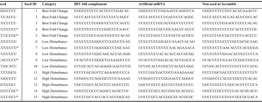
Figure 5. The identity of mismatched nucleotides affects repression efficiency.47
Figure 6. Testing and selection of the transfection controls for dual luciferase assay. ... 75
Figure 7. Reporter assay identifies RNA guides that inhibit HIV gene expression... 80
Figure 8. Protection against infection in transiently transduced cells. ... 84
Figure 9. Cells stably transduced with MiRBooking designed shRNAs showed protection against viral infection ... 87
Figure 10. Non-seed nucleotide complementarity is important for HIV-targeting shRNAs ... 91
Figure 11. Silencing profile of the coding region and 3’UTR sites in the reporter plasmid ... 111
Figure 12. Verification of our assay system being reliable to assess effets of base pairing ... 114
Figure 13. Silencing profile in FR(-)tat and pNL-luc reporters resemble ... 118
Figure 14. Different combinations of guides and target sites to generate mismatches at modules A-D to produce the repression profile ... 122
Figure 15. Combined effects of mismatches reveal the interdependency between the modules. ... 125
Figure 16. Repression profile of miB target sorted by mismatched modules ... 127
Figure 17. Validation of the non-seed model ... 130
xiv
Figure 20. Structural analysis supports the proposed mechanism ... 142
Figure 21. Additional features of the interaction between Ago2 and the guide strand ... 144
Figure 22. Summary of the skipped-propagation and coordinated annealing model ... 148
Figure 23. Simulation of Concentration Effect of miRNA. ... 169
Figure 24. Efficiently repressed miR-20a targets predicted by MicroAlign algorithm. ... xlv
List of Abbreviations
ADAR: adenine deaminase AGO (Ago): Argonaute ARE: AU-rich elements BBR: bicoid binding region BMP: bone morphogenetic protein bp: base pairCAF1: CCR4-associated factor 1 CAT-1: catecholamine transferase 1
CCR4-NOT1: carbon catabolite repression 4-negative on TATA-less ceRNA: competing endogenous RNA
CHX: cyclohexamide CMV: cytomegalovirus CrPV: Cricket paralysis virus cS7: conserved segment 7
DGCR8: DiGeorge Syndrom critical region 8 dsRBDs: double strand RNA binding domain DUF: domain of unknown function
eEF: enkaryote Elongation Factor eIF: eukaryote Initialtion Factor
eIF4E-BP: eukaryotic Initiation Factor 4E binding protein EMCV: encephalomyocarditis virus
ES cells: embryonic stem cells
FACS: Fluorescence activated cell sorting FFluc: firefly luciferase
GDP: guanosine diphosphate GFP: green fluorescence protein GTP: guanosine triphosphate
xvi
HEK 293T: human embryonic kidney 293 transformed
HITS-CLIP: high-throughput sequencing of RNAs isolated by crosslinking immunoprecipitation
miRNA: microRNA
MRE: miRNA response elements MSCV: murine stem cell virus
mTOR: mammalian target of rapamycin ncRNA: non-coding RNA
nt: nucleotide
ORF: open reading frame PABP: poly(A) binding protein PARN: poly(A)-specific ribonuclease PCR: polymerase chain reaction PI3K: Phosphoinositide-3 kinase piRNA: PIWI domain interacting RNA PIWI: P-element induced wimpy testis Pri-miRNA: primary transcript of miRNA PTEN: phosphatase and Tensin homolog puro: puromycin
PVDF: Polyvinylidene fluoride Rap: rapamycin
RIPA: Radio Immunoprecipitation Assay RISC: RNA-induced silencing complex RLC: RISC loading complex
Rluc: renilla luciferase RNAi: RNA interference RRM: RNA recognition motif shRNA: small hairpin RNA siRNA: small interfering RNA TBF-β: transforming growth factor-β TF: transcription factor
TNRC6: trinucleotide repeat containing 6 TRBP: TAR RNA-binding protein tRNA: transfer RNA
TU: transcription unit UTR: untranslated region wt: wild type
xviii
Contribution of Authors
Chapter 2:
I performed all experiments and data analyses except for:
The design of all SM shRNAs using miRBooking was performed by Nicolas Scott (Table I-III). One repeat of the Western blot of Rela, Akt, and tubulin proteins was performed by Roqaya Imane. The predicted alignment between the designed SM shRNA guide strands and the target sites were computed by Albert Feghaly (Table V). Etienne Gagnon provided stable cell generation strategies, technical training in tissue culture, as well as some of the stable cell lines. Gerardo Ferbeyre designed and directed the project, and proofread the manuscript. Francois Major designed and directed the project, defined the categories of the small RNA guides to be tested, supervised the adaptation of miBooking by mirDesign, which was used for the design of guide RNA sequences; in addition, Francois proofread and edited the manuscript.
Chapter 3:
I performed all experiments, wrote the MicroAlgin program, and conducted structural modeling and analyses except for:
Maria Acevedo generated stable cell lines that express the SM1-5 shRNA, performed Western blots on the targeted proteins, and conducted the cell growth assay to validate the efficiency and function of the knockdown of the E2Fs (Fig. 17
and 19). Lian Mignacca performed the RT-qPCR to validate the primers and
quantitate the levels of mature miB-A to D guide RNAs (Fig. 12G). Philippe Desjardins made single nucleotide mutations in the seed nucleotides of miB guide strand and tested their activities using luciferase reporter assay (Fig. 12A). Nicolas Scott utilized external (Robertson and Wee) datasets and verified MicroAlign algorithm using them (Fig. 17E). Julie Robitaille cloned and extracted pPRIME
empty and miB-mod1 to 4 DNA plasmids (Fig. 17C and D). Jordan Quenneville performed cloning of the pPRIME-miB-D plasmid that I used when testing modified target sites (part of Fig. 13H and Fig. 14). Roqaya Imane performed cloning of pPRIME miB, miB A-C, as well as technical repeats of luciferase assay (Fig. 14); she also performed one repeat of the RT-qPCR to quantitate the level of mature miB guide strand (Fig. 12E). Albert Feghaly performed two repeats of the bioinformatics validation of MicroAlign outputs. Etienne Gagnon designed some of the validation strategies, provided technical training in cell culture, and proofread and edited the manuscript as well as the figures. Gerardo Ferbeyre directed the experimental validation of the model, and proofread the manuscript. Francois Major directed the bioinformatics validation of the model, designed computational approaches, and proofread and edited the manuscript.
xx
Original Contribution to Knowledge
Chapter 2:
• High density of coding sequence in the HIV genome is one main reason for the ineffectiveness of siRNA, in addition to hindrance due to RNA structure.
• Puromycin can enhance miRNA-mediated repression of HIV when coding region is targeted.
• Artificial miRNAs targeting both the viral and the cellular genes provide cells with equal or better protection against invading viral particles than the perfectly complementary ones.
Chapter 3:
• The positional mismatches beyond the seed display a silencing pattern • This pattern is only observable for 3’UTR target sites.
• The cooperativity of multiple sites enhances silencing but not as much as base pairing at key positions.
• The enhancement by 3’ mismatches is only observable when no other mismatch occurs.
• Concentration of the mature guide RNA was not the cause of the silencing profile
• Base pairing propagates from the seed, skips the central part, and resumes downstream, then comes back to the central part to form the completely paired guide-target RNA duplex.
xxii
Acknowledgement
I would like to thank my supervisor, Dr. François Major, for his support and guidance during my training. I am especially thankful to François for allowing me to pursue my own scientific interest and ideas, allowing the full development of a rather complex and fundamental science project. His emphasis on the quality of research sets a standard of undoubtedly strong work ethics in science. I feel well prepared for a career in science because of that. I would like to thank my co-supervisor, Dr. Gerardo Ferbeyre, for his guidance and motivation that helped me overcome every formidable challenge. I am especially grateful for the crucial support that he provided, together with the members of his lab, to appropriately and timely address the key issues that the referees raised during the review process of my articles.
I would like to thank Dr. Etienne Gagnon, for his unparalleled scientific intuition, shrewd foresights, and dedicated commitment that provided invaluable guidance for my training. His contribution to both manuscripts is integral and essential.
I would also like to thank the members of the Major and Ferbeyre lab, both past and present, for their contributions to my development in research projects. In addition to the acknowledgements inside the chapters, I would like to thank Julie Pelloux, for great discussions and suggestions that were later proved valuable. I would like to thank Julie Robitaille, for inspiring discussions and technical help.
I need to thank the members of my thesis committee, Dr. Pascal Chartrand and Dr. Eric Lecuyer, in addition to my supervisor and co-supervisor, for their great involvement and advices. I thank the Jury Members, Dr. Phillip Zamore, Dr. Franz Lang, and Dr. Eric Lecuyer for generously offering their time to evaluate this dissertation and my work.
In addition, I would like to express my special thank my Master supervisor, Dr. Gertraud Burger, who initially accepted me for graduate studies and inspired me with her patience and extraordinary visions about the wonders of the RNA world.
I would like to express my deep thanks to Dr. Jerry Pelletier and his lab, where I obtained invaluable training that transformed my way of thinking and prepared me for scientific research. He and his lab provided crucial theoretical and technical support for some of the experiments in this thesis.
And I would also like to thank Dr. Raquel Aloyz, Dr. Lawrence Panasci, and Dr. Jerry Price, who helped me to set my first footsteps into the realm of research. I thank my friends Dr. John Mills and Dr. Abba Malina, whose insights and advices helped me enormously with my project. To John and Isabelle, you guys are great; and to Lillian and Amelia, thank you for supporting “uncle Yifei”.
Lastly, I thank my parents, Dr. Ju Yan and Bingzhen Chen, for their unconditional love and support throughout the years.
1
GENERAL INTRODUCTION
MicroRNAs are genome-encoded small RNA molecules that regulate gene expression post-transcriptionally. Mature microRNAs (miRNA) are single stranded RNA molecules of 21 nucleotides in length. Argonaute (Ago) protein associates with miRNA to form an essential component of the miRISC (miRNA-induced silencing complex), which downregulates target gene expression by cleaving the mRNA at the binding site, removing its poly-A tail, removing the 5’ cap structure, or repressing its translation (Fabian et al., 2010).
First discovered as a gene that does not encode any protein but control the larval development in Caenorhabditis elegans by the Ambros lab (Lee et al., 1993), lin-4 was the first functional microRNA molecule identified. Its sequence is complementary to that of the 3’ untranslated region (UTR) of the lin-14 RNA and its regulatory roles were confirmed by the Ruvkun lab (Wightman et al., 1993). In the past two decades, miRNAs were found to play important roles in cell growth, division and differentiation, as well as metabolism and development.
As each metazoan miRNA is predicted to target hundreds of mRNAs due to the promiscuous base pairing between their seeds and multiple gene sequences, a large proportion of the human transcriptome is suggested to be under the control of miRNAs (Bushati and Cohen, 2007; Carthew and Sontheimer, 2009). Over half of the human genes are predicted to be directly regulated by miRNAs and hence the unique combination of miRNAs in each cell type is likely to be a determinant for the fate of thousands of mRNAs (Friedman et al., 2009; Kim et al., 2009). Supporting this view, genomic approaches such as Ago-CLIP and its derived methods identified 17,000 miRNA-target interactions in human, as well as new types of miRNA target sites (Chi et al., 2009; Grosswendt et al., 2014; Pasquinelli, 2012).
Based on the understanding of the miRNA machinery, RNAi technology has been widely applied as a gene knockdown method. However, off-targeting represent one of the main challenges due to the lack of accuracy in the prediction algorithms. Undesired gene knockdown can cause cell death and prevented its application on a larger scale. Correlations were found with base complementarity, target site location,
AU-content, secondary structure, and sequence conservation.(Agarwal et al., 2015; Grimson et al., 2007); yet precise quantifications are still required to improve the prediction algorithms. We started a project with an in-house algorithm, which implements the known targeting rules, to design RNA guide sequences that target the HIV genomic RNA as a proof of concept for the application of our artificial miRNA design strategy. Some of the guide strand showed significant protective effects against HIV infection; however, others showed activities that do not correspond well with the computer predictions. Upon analysis of the results, we further investigated features that are essential for the design of artificial miRNAs. We demonstrated that base pairs beyond the seed in the guide-target RNA duplex are formed following a particular order. In effect, such ordered base pairing has hierarchical impacts on the efficiency of the guide RNA. We validated this rule both experimentally and computationally and proposed a unifying model that describes the roles of non-seed base pairing in Ago2-mediated silencing.
3
1.1 MicroRNA Biogenesis
1.1.1 Encoding gene structure
The miRBase database (http://www.mirbase.org/), release of June 2013, contains 24,521 microRNA loci from 206 species, processed to produce 30,424 mature microRNA products (Kozomara and Griffiths-Jones, 2014). MiRNAs were also identified in simple multi-cellular organisms, such as poriferans, cnidarians (Grimson et al., 2008), as well as protists (such as Dictyostelium) (Avesson et al., 2012). Except for the placozoan Trichoplax, miRNAs have been identified in every animal species with a sequenced genome (Maxwell et al., 2012). According to current (at the time of writing this thesis) miRBase, there are 1,917 miRNA genes in humans, 1,234 in mouse, 258 in fly, 253 in worm, and 326 in Arabidopsis thaliana. Conserved through evolution, around 55% of C. elegans miRNAs have homologs in human (Kim et al., 2009). Considering the advantages of short hairpins for generating guide RNAs in gene silencing, it is suggested that miRNAs have arisen more than once in eukaryotic evolution (Bartel, 2018).
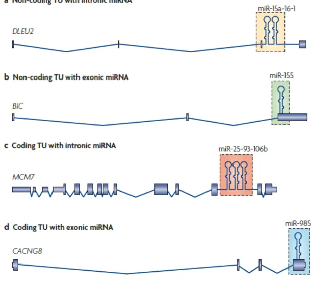
The location of miRNA-encoding sequences relative to the transcription units yielded information about miRNA biogenesis. Bradley and colleagues found that about 70% of mammalian miRNA genes (161 out of 232) are located in defined transcription units, and 117 of them are located in introns. Among the 117 intronic miRNA genes, 90 of them are located in protein-coding genes, and 27 are located in non-coding RNA genes (ncRNAs) (Rodriguez et al., 2004). Later studies revealed that miRNA genes can occur in four types of transcripts: 40% occurs in the introns of non-coding RNA ncRNA transcripts units (TU) (Fig. 1a); 10% occurs in the exons of ncRNAs (Fig. 1b); 40% occurs in the introns of coding RNA (Fig. 1c); some occur in either the introns or the exons (Fig. 1d) depending on the result of alternative splicing (Kim et al., 2009). About 50% of microRNA genes are endogenous genes that occur in clusters as polycistronic genes. They are transcribed as a single TU. Further processing is needed to generate the mature miRNA sequences. In rare cases, individual miRNA genes
5
Figure 1. There are four types of genomic locations of miRNA genes. a. Intronic miRNAs
in non-coding transcripts, exemplified by the miR-15a~16-1 cluster. b. Exonic miRNAs in non-coding transcripts. This is shown by miR-155, which was found in a non-coding RNA gene, BIC198. c. Intronic miRNAs in protein-coding transcripts. An example is the miR-25~93~106b cluster, which is embedded in the intron of the DNA replication licensing factor MCM7 transcript. d. miRNAs located in exons of protein-coding transcripts. The last exon of CACNG8 mRNA contains the miR-985 hairpin. (Kim 2009, Nature Molecular Cell Biology)
7
1.1.2 Biosynthesis/Transcription
Since miRNA mature sequence is only ~21 nt in length, it was originally thought that the RNA polymerase that transcribes it should belong to the RNA pol III family, which transcribes short RNA genes. However, primary transcripts of miRNA (pri-miRNAs) were later identified to be rather long, often containing thousands of nucleotides (Lee et al., 2002b). It was later confirmed that RNA pol II was mainly responsible for its transcription (Lee et al., 2004). This conclusion is supported by the fact that pri-miRNA transcripts are capped and poly-adenylated, which is the signature characteristics of the pol II transcribed genes; in addition, α-amanitin, which specifically inhibits pol II, greatly reduces the pri-miRNA levels (Lee et al., 2004). Mature miRNA can also be generated by polymerase III using transgenic constructs (Zhou et al., 2008a; Zhou et al., 2005).
1.1.3 Nuclear Processing
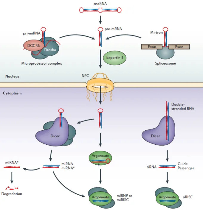
The pri-miRNAs are usually very long (up to several kilo bases), capped and poly-adenylated. Stem-loop structures that contain miRNA sequences need to be recognized and processed to eventually yield a mature ~21nt miRNA. Processing occurs in two steps: the first step occurs in the nucleus where the stem-loop structure of ~75nt will be cut out (pre-miRNA); the second step occurs in the cytoplasm, where the loop is removed and the double-stranded RNA molecule will dissociate and load into the RNA-induced silencing complex (RISC) to function as a guide for target sequences. We will look at these two steps as well as the transport step from the nucleus to the cytoplasm in detail.
9
Figure 2. The overall biogenesis pathway of miRNA. The primary transcripts of
microRNAs are called pri-miRNA that are processed into pre-miRNA hairpins within the nucleus. Processing by the nuclear microprocessor complex, which contains the RNase III enzyme Drosha, releases the hairpin: this part is referred to as the precursor miRNA (pre-miRNA). The primary transcript can also be generated from Mirtrons and some of the snoRNA. Mirtrons are processed by the spliceosome, while the processing machinery of snoRNA is unclear. The generated pre-miRNAs are exported into the cytoplasm by Exportin-5. They are processed there into mature miRNA by Dicer and loaded into the RNA induced silencing complex, where it directly binds to the AGO protein. The other strand is referred to as miRNA* and is normally degraded. Mature miRNAs then guide the RISC to target select mRNA transcripts for translational silencing or degradation. In the case of siRNA processing, the loaded functional strand is referred as the guide; the other, the passenger strand. Figure adapted from a 2013 review article by Meister (Meister, 2013).
As aforementioned, pri-miRNA is usually a long (up to several kilo bases) capped and poly-adenylated molecule that contains stem-loop structures. RNAse III-type protein, Drosha, which recognizes the stem of the hairpin structure, cuts out the ~75nt stem-loop from its primary transcript (Lee et al., 2003). The released stem-loop structure is termed the pre-miRNA (Lee et al., 2002b).
Drosha requires a cofactor called DiGeorge syndrome critical region 8 (DGCR8) in humans, and Pasha, in D. melanogaster and C. elegans (Denli et al., 2004; Gregory et al., 2004; Han et al., 2004; Landthaler et al., 2004). In humans, Drosha and DGCR8 form a large protein complex of ~650 kD, called the Microprocessor complex (Gregory et al., 2004; Han et al., 2004). DGCR8 recognizes two features in pri-miRNA: the single stranded base segments and stem of about 33 bp. With its assistance, Drosha is able to cleave the substrate at ~11 bp away from the ssRNA-dsRNA junction (Han et al., 2006; Zeng and Cullen, 2005). Mouse embryonic stem (ES) cells that fail to produce miRNAs suffer from defects in proliferation and differentiation when they are deficient in the Dgcr8 gene. This phenomenon establishes the necessity of DGCR8 in the miRNA pathway as well as miRNA function in ES cells (Wang et al., 2007).
As mentioned in Section 1.1.1, many miRNA genes are located in the introns of the coding and non-coding RNAs. This suggests the possible coordination between transcription, miRNA processing, and splicing. Indeed, studies revealed that pri-miRNA processing is a co-transcriptional process (Kim and Kim, 2007b). Moreover, by mutating the Drosha recognition sequence in miR-26 or depleting Drosha in cells, Drosha processing is shown to precede intron splicing, and cleavage of the stem-loop structures within the intron does not impair splicing (Kim and Kim, 2007a). The “exon-tethering” model is favoured where the exons of Pol II transcripts are co-transcriptionally assembled into the spliceosome; then, Drosha complex processing takes place before the intron is excised (Dye et al., 2006). Evidence from chromatin precipitation and nuclear run-on assays support this model (Morlando et al., 2008; Pawlicki and Steitz, 2008).
11
1.1.4 Transport
Exportin-5 is the main transporter protein that is responsible for delivering pre-miRNA molecules out of the nucleus (Fig. 2). It mediates the transport of pre-miRNAs across the nuclear membrane by cooperatively binding to a cofactor called Ran. Upon the completion of transport, a molecule of GTP is hydrolyzed to GDP to release the cargo molecule into the cytoplasm (Bohnsack et al., 2004; Kim, 2005).
Exportin-5 was originally discovered as a minor transporter for tRNA molecules when Exportin-t, the main transporter for tRNA, is knocked down or overloaded. Later studies showed that Exportin-5 is indeed the main transporter protein of miRNA (Lund et al., 2004; Yi et al., 2003) because its depletion causes a significant decrease of the pre-miRNA level in cytoplasm.
1.1.5 Cytoplasmic processing by Dicer
Further processing of the ~70 nt stem-loop structure of pre-miRNA is carried out by the RNase III family protein, Dicer (Bernstein et al., 2001; Grishok et al., 2001; Hutvagner et al., 2001; Ketting et al., 2001; Knight and Bass, 2001). It was originally discovered as the processing protein required for siRNA generation (Bernstein et al., 2001; Hammond et al., 2000). Dicer is a highly conserved protein of about 200 kD across all Eukaryotes. It cuts away the loop structure and generates the ~22 nt double-stranded RNA molecule with two nucleotides of 3’ overhang. One of the two strands becomes the mature miRNA. Knocking down Dicer causes the accumulation of pre-miRNA and the diminishment of the 22 nt mature miRNA (Grishok et al., 2001; Hutvagner et al., 2001; Ketting et al., 2001; Knight and Bass, 2001)
Dicer interacts with protein partners to carry out its dicing function. In C. elegans, it interacts with RDE-4 to convert the precursor RNA into a short dsRNA (Tabara et al., 2002). In D. melanogaster, there are two isoforms of Dicer, Dcr-1 and Dcr-2. Dcr-1 interacts with Loquacious (Loqs) and converts pre-miRNA to a miRNA/miRNA* duplex (Forstemann et al., 2005). Dcr-2 interacts with R2D2 to process long dsRNA into short siRNA duplex
(Forstemann et al., 2007; Liu et al., 2003). Human Dicer interacts with TRBP (TAR RNA-binding protein; also known as TRBP2) (Chendrimada et al., 2005; Haase et al., 2005) and PACT (also known as PRKRA) proteins (Lee et al., 2006).
1.1.6 Argonaute loading
The selection of strand to be loaded into Argonaute is not random. The selected strand, called the guide strand or anti-sense strand, will be functional and the other strand, the passenger strand or the miRNA* strand, will be degraded or discarded through an ATP-dependent process (Hammond et al., 2000; Nykanen et al., 2001; Suzuki and Miyazono, 2010, 2011). The mechanism of strand selection is based on thermodynamic stability of the RNA duplex. It was first discovered in D. melanogaster, where R2D2 protein senses the asymmetry in stability of the RNA duplex and binds to the more stable end of the duplex, while it forms a stable heterodimer with Dicer 2 protein and orients Ago2 on the RNA duplex (Liu et al., 2003; Siomi and Siomi, 2010; Tomari et al., 2004). It was shown that the flyAgo1-RLC operates in a similar manner via the interaction between Dcr-1 and Loqs (Chendrimada et al., 2005; Gregory et al., 2005; MacRae et al., 2008; Maniataki and Mourelatos, 2005; Tomari et al., 2004). Together they sense central mismatches in fly miRNA and preferentially load the guide strand into Ago1 (Tomari et al., 2007). Later studies demonstrated that both fly Ago1 and 2 are also able to sense the thermodynamic asymmetry as well as mismatches at key positions in the RNA duplex; strand separation subsequently takes place within the Argonaute protein (Iwasaki et al., 2009).
A similar postulate was originally made about the mammalian Ago-loading process, thinking that AGO isoforms may distinguish miRNA and siRNA. Yet studies revealed that such mechanism is partially lost in mammals. Ago1-4 do not have distinguishable preference for miRNAs; on the other hand, Ago1 and 2 have preference for siRNA duplexes compare to Ago3 and 4 (Su et al., 2009). Evidence suggests that for mammalian siRNAs, after Dicer cleavage, the siRNA duplex is released from Dicer and the more stable end binds to TRBP in the RLC and its less stable end binds to the Argonaute (Aza-Blanc et al., 2003; Khvorova et
13
al., 2003; Preall and Sontheimer, 2005; Schwarz et al., 2003; Tomari et al., 2004). It has also been shown that Dicer, TRBP (and/or PACT) help orient the dsRNA by the asymmetry rule when loading dsRNA into Argonaute (Noland et al., 2011). However, other studies indicate that Dicer and TRBP may not be the only ones responsible for the implementation of the asymmetry rule (Betancur and Tomari, 2012; Murchison et al., 2005). For siRNA, endonucleolytic activity of the AGO2 cleaves the passenger strand and, as the consequence, the guide strand remains in the AGO2 protein while the cleaved passenger strand undergoes degradation (Leuschner et al., 2006; Matranga et al., 2005; Miyoshi et al., 2005). Moreover, overexpression of mammalian Ago1-4 causes increase in mature miRNA levels, indicating that they all contribute to miRNA processing (Diederichs and Haber, 2007). It was later demonstrated that each human Ago isoform alone is sufficient to implement the asymmetry rule in strand selection (Suzuki et al., 2015). However, these pieces of evidence do not dismiss the importance of Dicer-interacting partners in RLC since without them loading will be inefficient (Cenik and Zamore, 2011).
1.1.7 Regulation of miRNA biogenesis
There are three main ways to regulate miRNA biogenesis: transcriptional control, post-transcriptional control, and feedback circuits (Kim et al., 2009).
1.1.7.1 Transcriptional control
Among the known miRNAs, miR-1 and miR-133 are specifically expressed in adult cardiac and skeletal muscle tissues (Horak et al., 2016). They are transcriptionally regulated. Myogenic TFs, such as myogenin and myoblast determination 1 (MYOD1), bind upstream of miR-1 and miR-133 loci and promote the transcription of these two miRNA genes (Chen, 2006; Rao et al., 2006). Some miRNAs can be potentially used to assess cancer progression due to their correlations with transcription levels of factors involved in tumour. For instance, some tumour suppressor TF can also regulate miRNA gene expression. Tumour suppressor p53 activates the transcription of the miR-34 family (He et al., 2007). On the other hand, MYC, an oncogenic protein, activates or represses a number of miRNAs that are involved in
the cell cycle and apoptosis (Chang, 2008; He, 2005). Interestingly, epigenetic control such as DNA methylation contributes in the regulation of miR-203 locus in the T-cell lymphoma but not in normal T-cells (Bueno, 2008).
1.1.7.2 Post-transcriptional control
Drosha processing represents the first post-transcriptional control point of miRNA biogenesis. As an example, in the case of the induction of miR-21, bone morphogenetic protein (BMP)/transforming growth factor-β (TBF-β) activates SMAD protein, which interacts with Drosha and DDX5 (also known as p68) and enhances Drosha processing (Davis et al., 2008). An additional post-transcriptional regulation point is the nuclear transport step. In some human cell types, miR-31, miR-128, and miR-105 precursors are retained in the nucleus while mature miRNAs are not produced, suggesting that they are regulated at the transport step (Lee, 2008).
1.1.7.3 Feedback loop control
Two types of feedback circuits are usually observed: single-negative feedback and double-negative feedback loops. Drosha and Dicer levels are regulated by the former type of feedback (Forman et al., 2008; Tokumaru et al., 2008). Drosha and DGCR8 form a single-negative feedback loop: Drosha downregulates DGCR8 by cleaving DGCR8 mRNA, while DGCR8 upregulates Drosha by stabilizing its Drosha protein (Han, 2009; Yeom et al., 2006). Human Dicer, on the other hand, constitutes a single-negative feedback with its product, let-7 miRNA, which binds to the 3’UTR of Dicer mRNA and represses its expression (Forman et al., 2008; Tokumaru et al., 2008).
Double negative feedback loops are often regarded as an efficient genetic switch of specific miRNAs during differentiation (Kim et al., 2009). They are also referred to as bistable switches in biochemical networks (Tyson and Novak, 2010). The interaction between let-7 and LIN28 falls into this category: let-7 represses LIN28 mRNA expression while LIN28 represses let-7 maturation (Newman et al., 2008; Viswanathan et al., 2008). Another example of this
15
Their repressive action upon each other constitutes an important switch in the epithelial-mesenchymal transition (Bracken, 2008).
1.1.8 Biogenesis from engineered constructs
Custom synthesis of RNA oligos are relatively expensive and not every lab can afford to test them in large quantities. Cloning the guide sequence of interest into an expression construct becomes a cost-effective choice for most researchers. The guide sequences are usually designed to appear on one arm of the stem of a small hairpin which can be recognized and processed by endogenous miRNA processing machinery; such engineered RNA species is termed “small hairpin RNA” (shRNA). Upon transcription, the hairpin structure of RNA is recognized by the microRNA processing machinery and subsequently processed and loaded into Argonaute. Among all constructs tested, the engineered construct based on miR-30 backbone from the Hannon lab has been most widely accepted as an efficient method of producing mature guide sequences (Dickins et al., 2005; Paddison et al., 2004; Stegmeier et al., 2005).
1.1.9 miRNA definition and annotation conventions
Given the description of miRNA gene structure, biogenesis, and function, we now know that miRNAs are almost exclusively endogenous in origin, and possibly goes through more concerted processing steps, in which both Drosha and Dicer are essential. But one may still ask the question: “How are the miRNA genes annotated?” The definition of miRNA determines its annotation in the genome. Researchers who greatly contributed to the study of miRNA across different species reached an agreement on the traits that miRNAs should possess (Ambros et al., 2003); consequently, these agreed criteria shaped the definition of miRNA. The annotation standards set in 2003 is still in use in most of the miRNA databases as well as prediction software such as MirBase and TargetScan.
In brief, five criteria were agreed upon (listed below). The first two are expression criteria that verify the existence of the miRNA, and the other three are called biogenesis criteria. They verify the authenticity of miRNA via their biogenesis characteristics.
Expression criteria:
A. Detection of a distinct ~22-nt RNA transcript by hybridization to a size-fractionated RNA sample, often by Northern blotting.
B. Identification of the ~22-nt sequence in a library of cDNA made from
size-fractionated RNA. Such sequences must precisely match the genomic sequence of the organism from which they were cloned.
Biogenesis criteria:
C. Prediction of a potential fold-back precursor structure that contains the ~22-nt miRNA sequence within one arm of the hairpin.
D. Phylogenic conservation of the ~22-nt miRNA sequence and its predicted fold-back precursor secondary structure.
E. Detection of increased accumulation of organisms with reduced Dicer function. Since it is not always possible to verify all five criteria for a particular candidate miRNA, some relaxation in the criteria is allowed. For example, A+D+E, A+D, A+C, B+D, D+E are all accepted as sufficient to annotate an miRNA.
According to the above definition, the conservation criterion plays an important role in the identification of miRNA, while the function of the miRNA is not mentioned. This leads to the fact that many of the miRNA collected in the existing databases do not have any known function, or any well validated target. Experimental validation of the targets is currently an on-going process for many annotated miRNA genes. Hence the targeting rules that facilitate the prediction of target for a given miRNA became crucial to the study of miRNA functions.
1.1.10 siRNA discovery, definition, and functions
RNA interference (RNAi) is a phenomenon by which double-stranded (ds) RNA induces sequence-specific post-transcriptional gene silencing. The term RNAi came into existence after Fire and Mello confirmed that dsRNA in both sense and anti-sense transcripts were responsible for the silencing in C. elegans (Fire et al., 1998). RNAi was later observed in various organisms including plants, Drosophila, nematodes and protozoa (Hannon, 2002;
17
Mello and Conte, 2004). It was first demonstrated in plants that the long dsRNAs were converted to small ones of ~25 nt RNA molecules in order to function as silencing triggers (Hamilton and Baulcombe, 1999). Supplying long dsRNAs to mammalian cells induces non-specific suppression of gene expression; this is because the host defense system against viral infections is activated when dsRNAs are introduced into the cell (Manche et al., 1992; Minks et al., 1979). Elbashir et al. resolved this problem by utilizing small (21-23 nucleotide) dsRNAs instead of long dsRNAs to avoid the non-specific gene suppression (Elbashir et al., 2001a). They named such small dsRNA as small interfering RNA (siRNA). With this method, they confirmed in animal cell extracts that the small RNAs were derived from the long dsRNAs and functioned as silencing triggers (Elbashir et al., 2001a). Similar siRNAs were later identified in Drosophila S2 (Hammond et al., 2000; Tuschl et al., 1999) as well as human HeLa cells (Martinez et al., 2002; Schwarz et al., 2003), where both sense and anti-sense strands were processed into 21-23 nt segments (Zamore et al., 2000). It was later found that Dicer processes the long dsRNA and generate siRNAs to direct silencing of specific targets (Meister and Tuschl, 2004; Tomari and Zamore, 2005).
The original inducers of RNAi were long, perfectly base-paired, linear dsRNA species and they were exogenously supplied to the cell or taken up from the environment. They are hence referred to as exo-siRNAs. Exo-siRNAs were identified in flies as a defense mechanism against invading viruses that produces long strands of dsRNA during infection (van Rij et al., 2006; Wang et al., 2006). Though the initial discovery is based on exogenous RNA, the origin of siRNA could also be endogenous. Heterochromatin sequence, including centromeres, transposons, and other repetitive sequences were found to give rise to a variety of siRNA (Lippman and Martienssen, 2004). Functional studies in plants also identified trans-acting siRNAs (ta-siRNAs) that are generated from well-defined transcription units and regulate the expression of specific genes (Allen et al., 2005; Vazquez et al., 2004). Deep sequencing revealed that in somatic tissue, cultured cells, and ovaries of D. melanogaster, siRNAs are derived from transposon transcripts, sense-antisense transcript pairs and long stem-loop structures (Babiarz et al., 2008; Chung et al., 2008; Czech et al., 2008; Ghildiyal et al., 2008;
Kawamura et al., 2008; Okamura et al., 2008a; Okamura et al., 2008b). In mouse, numerous types of endo-siRNAs were identified in oocytes (Tam et al., 2008; Watanabe et al., 2008), and, to a lesser extent, in ES cells (Babiarz et al., 2008). In other species such as plants and C. elegans, endo-siRNAs were also discovered (Chapman and Carrington, 2007). In plants, RNA-dependent RNA polymerases (RdRPs) are required to generate functional endo-siRNAs, adding more complexity to its pathway (Tang et al., 2003); on the other hand, fly and mammalian generate endo-siRNAs in an RdRP-independent manner.
So far, only flies are known to differentiate between miRNA- and siRNA-like precursor molecules by the two isoforms of Dicer. For siRNAs, Dicer 2 was the choice of processing nuclease. With the help of R2D2, the resulting guide strand RNA is preferentially loaded in Ago2 (Forstemann et al., 2007; Tomari et al., 2007). On the other hand, endogenous miRNA precursors, especially those that contain mismatched bulges, were preferentially cleaved by Dicer 1, with the help of Loquacious (LOQS; also known as R3D1) (Czech et al., 2008; Kawamura et al., 2008; Okamura et al., 2008b; Tomari et al., 2007). Such origin-dependent preference was not detected in mammals and it was suggested that siRNAs and miRNAs can functionally mimic each other depending on their complementarity with targets (Doench et al., 2003; Hutvágner and Zamore, 2002). Plant miRNA are usually highly complementary to their targets and preferentially mediates silencing through cleavage of the target mRNA. Due to the processing and targeting processes closely resemble those of siRNAs, plant miRNAs are suggested to be able to function as siRNAs (Tang et al., 2003).
In S. pombe, siRNAs were found to induce heterochromatin formation and lead to transcriptional gene silencing (Lippman and Martienssen, 2004). Similar observations were made in plants, animals, and ciliates. Transcriptional gene silencing is mediated via a complex, called the RNA-induced transcriptional silencing (RITS) complex, which contains Ago1 loaded with the siRNA. The interaction between RITS complex and RNA polymerase II facilitates siRNA’s recognition of the nascent transcript (Buhler et al., 2006; Djupedal et al., 2005; Kato et al., 2005). RITS association promotes histone H3 methylation on lysine 9 (H3K9) by histone methyltransferases (MHTs); as a consequence, the
chromodomain-19
containing protein Swi6 is recruited and chromatin compaction takes place (Lippman and Martienssen, 2004).
1.2 Messenger RNA (mRNA) architecture
As mRNA is targeted by miRISC, understanding the functional organization of mRNA is rudimentary to the study of functions of miRNA. Messenger RNA is transcribed in the nucleus by RNA polymerase II (Pol II). It is used as blue print for protein synthesis in the cytoplasm. In some cases, mRNA is targeted to specific subcellular locations for translation or temporary storage (Rodriguez et al., 2008). In Eukaryotes, it usually consists of the following regions: the 5’ cap structure, the 5’untranslated region (5’UTR), the start codon that marks the start of the coding region (CD), the stop codon which signals the end of the coding region and the start of the 3’ untranslated region (3’UTR), and the poly-A tail. The 5’ cap and the 5’ UTR are important for the recruitment of initiation factors that signals the ribosomes to start translation. Coding region is located between the start codon and the termination codon and it contains a series of codons that encode the amino acid sequence. When a ribosome scans over the start codon in a suitable context, methionine initiator tRNAi is brought to the P site of the ribosome
and it initiates protein synthesis. In mammalians, the start codon (AUG) itself encodes the methionine residue (Sonenberg and Hinnebusch, 2009).
Further downstream of the stop codon, the mRNA is not translated. This region is called the 3’UTR. The primary role of the 3’UTR is to regulate mRNA stability. The AU-rich elements (AREs) control mRNA degradation and translation via interactions with specific binding proteins (ARE-BP) (Helfer et al., 2012). The AREs usually contain AUUUA pentamers and U-rich sequences; however, there is little sequence homology among AREs besides those motifs (Chen and Shyu, 1995). For instance, AUF1 protein binds to the ARE and, depending on the mRNA it binds, it could either stabilize or destabilize the mRNA (Loflin et al., 1999; Xu et al., 2001). On the other hand, HuR binds AREs and universally stabilizes mRNA by inhibiting 3’-5’ degradation (Brennan and Steitz, 2001). In Drosophila, the Caudal mRNA contains a bicoid binding region (BBR) in its 3’UTR, which recruits the Bicoid protein and causes the tethering of the 5’ end to the 3’end of the mRNA. As the result, the Caudal mRNA is maintained in a translationally inactive state (Cho et al., 2005).
21
One important way that 3’UTR contribute to the stability of mRNA is via the presence of miRNA response elements (MREs). As elaborated later in this thesis (section 1.5.2.5), the miRNAs prefer certain regions of the 3’UTR as their targets for effective repression of the encoded gene. The reason why miRNAs preferentially target the 3’UTR is thought to avoid the impeding ribosomes (Guo et al., 2010). The MREs present in the coding regions are likely protected by the traveling ribosomes, which can even displace bound miRISC. Recent report has shown that the coding region can also be targeted by miRNA and consequently repressed via slicer-independent mechanisms (Zhang et al., 2018).
When RNA polymerase II completes the transcription of precursor mRNAs, a process called polyadenylation takes place (Albert L. Lehninger, 1993; Colgan and Manley, 1997). It refers to the addition of adenosine to the 3’end of the mRNA (normally 200-300 adenosines) and the added long tract of adenosines is called the poly(A) tail. Nuclear poly(A) binding protein (PABP) binds to the nascent poly(A) tail and enhances the polymerase activity of PAP, facilitating further extension of the poly(A) tail. Cytoplasmic PABP binds to 3’UTR and it inhibits mRNA degradation as well as promotes translation (Bernstein and Ross, 1989; Borman et al., 2000) by facilitating the circularization of mRNA through its binding to the eIF4G scaffolding protein (Sonenberg and Hinnebusch, 2009).
1.3 The Argonaute genes
1.3.1 The Argonaute genes
Many Argonaute orthologs are found in metazoan and plant genomes. The orthologs are usually identified in piRNA (PIWI domain interacting RNA) pathways in germ cells or other classes of small RNAs. Only the original AGO proteins identified in miRNA and siRNA pathways mediate gene silencing (Peters and Meister, 2007). Argonaute proteins are classified into three orthologous groups: Argonaute-like proteins are similar to Arabidopsis thaliana AGO1; Piwi-like proteins are closely related to D. melanogaster PIWI (P-element induced wimpy testis); and the last one contains the C. elegans-specific group 3 Argonautes (Yigit et al., 2006). Argonaute-like and Piwi-like proteins are found in bacteria, archaea, and eukaryotes, implying their ancient origin (Cerutti and Casas-Mollano, 2006). The number of Argonaute genes varies depending on the species. In human, there are 8 Argonaute paralogous genes, including four Argonaute-like and four Piwi-like genes; five were found in D. melanogaster, including two Argonaute-like and three Piwi-like proteins; in A. thaliana, 10 Argonaute-like were identified; only one Argonaute-like protein was identified in Schizosaccharomyces pombe; at least 26 Argonaute genes in C. elegans (5 Argonaute-like, 3 Piwi-like and 18 group 3 Argonautes) (Hutvagner and Simard, 2008).
The human Argonaute has four paralogs, hAgo1-4, which are ubiquitously expressed. Human AGO1, 3, and 4 genes are located next to each other on chromosome 1, while hAGO2 gene is located on chromosome 8 separately (Nakanishi et al., 2013). AGO2 is well known for being the only cleavage-active form of the four. It was later shown that each one of Ago1-4 is essential in mouse embryonic stem (ES) cells as they all prevent cells from undergoing apoptosis (Su et al., 2009).
Ago1-4 were thought to have redundant functions for repression of translation; it was later demonstrated that Ago3 is a more potent translational repressor than other non-cleaving
23
hematopoietic cells results in deficiency of B-cell and red blood cell development (O'Carroll et al., 2007). Germ line Ago2 deficiency is embryonically lethal (Liu, 2004; Meister, 2004). Interestingly, male mouse germ line cells express high levels of Ago4 as well as Ago3 (Gonzalez-Gonzalez et al., 2008). Ago4 is demonstrated to be specifically responsible for the silencing of many sex-linked transcripts in male germ line. Loss of Ago4 results in fertility defects including reduced testis sized and lower sperm counts in male mice (Modzelewski et al., 2012). On the other hand, Ago1 and Ago3 are required for RNAi pathways of cellular defence against influenza A viral infection. Ago1 and Ago3 double-knockout cells are significantly more vulnerable to this RNA virus (Van Stry et al., 2012). Hence recent evidence may suggest that each Ago paralog may take specialized role when acting as the repressor of translation.
As the overexpression of each of the four paralogs is demonstrated to enhance the production of mature miRNAs, it was postulated that they may also have redundant functions in miRNA maturation (Diederichs and Haber, 2007). However, it was shown in mouse ES cells that Ago1 and 2 have preferences for siRNA duplexes during Ago-loading comparing to Ago3 and 4. This indicates that the four paralogs have non-redundant roles in miRNA maturation because they may distinguish RNA duplexes based on their complementarity (Su et al., 2009).
In addition, a recent study has revealed that Ago proteins can be differentially regulated by phosphorylation. Phosphorylation at Ago2 Y529 inhibits it from being loaded with small RNAs (Rudel et al., 2011); on the other hand, EGFR-dependent phosphorylation of Ago2 Y393 hinders the processing of looped precursor RNAs (Shen et al., 2013). Moreover, Akt3 is shown to phosphorylate Ago2 S387 and alters its activity from cleavage toward translational repression (Horman et al., 2013; Zeng et al., 2008). It was recently shown that phosphorylation of Ago2 S387 by Akt3 induces LIMD1 binding, which enables the recruitment of TNRC6 by Ago2. The assembly of Ago2-TNRC6 complex switches the Ago2 activity to favour translational repression. In the absence of LIMD1, Ago2 miRNA-silencing function is lost and translational repression is mainly mediated by Ago3 (Bridge et al., 2017).
1.3.2 Structural organization of the Argonaute protein
Eukaryotic Argonaute proteins usually consist of four domains: the N-terminal, PAZ (PIWI-Argonaute-Zwille), MID and PIWI domains (Fig. 3). Between the N-terminal and PAZ domains, there is a loop called L1; similarly, between PAZ and the MID domain, there is another loop called L2. These two flexible loops render the PAZ domain more flexible relative to the rest of the Argonaute protein.
The N-terminal domain is commonly found in animal Argonautes, unlike the other three domains, which are ubiquitously found in all species. The N-terminal domain was thought to prevent base pairing of the target to the guide beyond nt 16 in the guide sequence (denoted as g16) (Faehnle et al., 2013; Kwak et al., 2012).
PAZ domain is found in both Argonautes and Dicer. It contains an OB-like fold (oligonucleotide/oligosaccharide binding fold). Data from structural as well as biochemical studies shows that it binds to single-stranded (ss) nucleic acids (Lingel et al., 2003, 2004; Ma et al., 2004; Song, 2003; Yan, 2003). The binding appears to be sequence-independent; however, an intriguing feature is that PAZ recognizes the 3’ end overhang of ssRNAs, where two such overhang nucleotides are typically produced after Dicer processing of the pre-miRNA. For human Ago2, the entire protein structure looks like a bird with two wings (MID and N domains) spread out and the head (PAZ) tilted upward, holding the 3’ end of the guide RNA in its bill. The RNA guide strand is threaded through the N-PAZ channel.
MID domain of Argonaute protein binds to the 5’ end of the small RNA guides , with some preference to U or A at position 1 (Boland et al., 2010; Frank et al., 2010; Parker et al., 2005). The nucleotide preference is achieved via specific contacts with amino acid residues in the MID lobe. Nucleotide 2-10 of the RNA guide strand is threaded through the RNA binding groove between PIWI and MID lobes (Elkayam et al., 2012).
PIWI domain contains the catalytic residues that cleave the target RNA. The catalytic center is a RNase-H-like fold (containing the DEDH catalytic tetrad), which was originally found to cleave RNA that are base-paired with DNA(Ma et al., 2005; Parker et al., 2004; Song et al., 2004; Yuan et al., 2005). In human, Ago2 is the only isoform that is capable of cleaving
25
target RNA. Ago1, 3, and 4 mediate repression by slicer-independent pathways. The requirement for divalent cation as well as 5’-phosphate and 3’ OH detected in the products confirmed its RNase H characteristic (Tolia and Joshua-Tor, 2007). Such catalytic tetrad is conserved in hAgo3 but altered to be DEDR in hAgo1and hAgo4, which are compromised in catalytic activity. Another characteristic of the PIWI domain is that it accommodates the GW182 protein by binding to its WG/GW repeats (Till et al., 2007).
Figure 3. The human Ago2 protein with a modeled guide-target RNA duplex bound to it.
27
Figure 3. The human Ago2 protein with a modeled guide-target RNA duplex bound to it.
The domains are labeled in bold letters. The Ago2 structure is adapted from PDB 4F3T from Elkayam et al. (Elkayam et al., 2012). The 3’ end of the guide RNA is bound to the PAZ domain while the nucleotides between the nt15 and the 3’ end could not be resolved.
1.3.3 Argonaute slicer activity
The Argonaute protein is a multi-functional protein. It plays versatile roles in small RNA biogenesis and gene silencing. The most prominent and potent effect is its RNase activity. All AGOs are not slicer-active. In humans, Ago2 is the only isoform that is capable of cleaving target mRNA. It cleaves the mRNA via RNase H-like mechanism, utilizing Mg2+ as a co-factor. Mutational experiments were conducted to understand the protein features that are important for nuclease reaction to occur as well as the reasons why some Argonautes are RNase-inactive. The first structural and functional study on hAgo2 was conducted by Liu et al., who investigated the mammalian slicer Ago2 (Liu, 2004). When a human RISC is assembled with a guide RNA, the RISC becomes an enzyme for the highly complementary MREs (Haley and Zamore, 2004).
Guide-loaded RISC is a multiple-turnover enzyme; after cleaving a perfectly paired target, it leaves with the guide strand intact and becomes ready to bind next target (Haley and Zamore, 2004; Hutvágner and Zamore, 2002; Martinez and Tuschl, 2004). In Zamore’s study, they measured values of Km and kcat, which are measurements of the affinity and the turnover
rate of an enzyme, respectively. In addition, the ratio between kcat and Km, which is the
“specificity constant”, is a classical measure of catalytic efficiency and corresponds to the second order rate constant of the reaction when the substrate concentration is much lower than the Km. In the study of wild-type D. melanogaster RISC loaded with let-7, kcat/Km is ~8.4X10⁻⁴
nM-1S-1 (Haley and Zamore, 2004). The measured values are much slower than the expected rate of collision of RISC with mRNA (>= 10-2 nM-1S-1) (Haley and Zamore, 2004). This indicates that there are factors that rate-limit the reaction, possibly due to the conformational changes required during the target recognition and cleavage (Haley and Zamore, 2004).
This suspicion eventually led to the proposition of the “two-state model” of Ago2’s cleavage mechanism. Cross-linking experiments suggests that the 3’ end of the guide strand binds PAZ domain (Tomari et al., 2004), confirmed by structural studies (Lingel et al., 2003, 2004; Ma et al., 2004; Song, 2003; Yan, 2003). Upon seed binding to the target, the guide