DEVELOPMENT OF NEW APPROACHES FOR THE
SYNTHESIS AND DECODING OF BEAD
ONE-COMPOUND CYCLIC PEPTIDE LIBRARIES
Thèse
Xinxia Liang
Doctorat en sciences pharmaceutiques
Philosophiae doctor (Ph.D.)
Québec, Canada
DEVELOPMENT OF NEW APPROACHES FOR THE
SYNTHESIS AND DECODING OF BEAD
ONE-COMPOUND CYCLIC PEPTIDE LIBRARIES
Thèse
Xinxia Liang
Sous la direction de :
Éric Biron, directeur de recherche
III
RÉSUMÉ
La plupart des processus cellulaires et biologiques reposent, à un certain niveau, sur des interactions protéine-protéine (IPP). Leur manipulation avec des composés chimiques démontre un grand potentiel pour la découverte de nouveaux médicaments. Malgré la demande toujours croissante en molécules capables d'interrompre sélectivement des IPP, le développement d'inhibiteurs d’IPP est fortement limité par la grande taille de la surface d'interaction. En considérant la nature de cette surface, la capacité à mimer des structures secondaires de protéines est très importante pour lier une protéine et inhiber une IPP. Avec leurs grandes capacités peptidomimétiques et leurs propriétés pharmacologiques intéressan-tes, les peptides cycliques sont des prototypes moléculaires de choix pour découvrir des ligands de protéines et développer de nouveaux inhibiteurs d’IPP. Afin d’exploiter pleinement la grande diversité accessible avec les peptides cycliques, l’approche combinatoire «one-bead-one-compound» (OBOC) est l’approche la plus accessible et puissante. Cependant, l'utilisation des peptides cycliques dans les chimiothèques OBOC est limitée par les difficultés à séquencer les composés actifs après le criblage. Sans amine libre en N-terminal, la dégradation d'Edman et la spectrométrie de masse en tandem (MS/MS) ne peuvent pas être utilisées.
À cet égard, nous avons développé de nouvelles approches par ouverture de cycle pour préparer et décoder des chimiothèques OBOC de peptides cycliques. Notre stratégie était d'introduire un résidu sensible dans le macrocycle et comme ancrage pour permettre la linéarisation des peptides et leur largage des billes pour le séquençage par MS/MS. Tout d'abord, des résidus sensibles aux nucléophiles, aux ultraviolets ou au bromure de cyanogène ont été introduits dans un peptide cyclique et leurs rendements de clivage évalués. Ensuite, les résidus les plus prometteurs ont été utilisés dans la conception et le développement d’approches en tandem ouverture de cycle / clivage pour le décodage de chimiothèques OBOC de peptides cycliques. Dans la première approche, une méthionine a été introduite dans le macrocycle comme ancrage pour simultanément permettre l’ouverture du cycle et le clivage des billes par traitement au bromure de cyanogène. Dans la seconde approche, un résidu photosensible a été utilisé dans le macrocycle comme ancrage pour permettre l’ouverture du cycle et le clivage suite à une irradiation aux ultraviolets. Le
IV
peptide linéaire généré par ces approches peut alors être efficacement séquencé par MS/MS. Enfin, une chimiothèque OBOC a été préparée et criblée la protéine HIV-1 Nef pour identifier des ligands sélectifs.
Le développement de ces méthodologies permttra l'utilisation de composés macrocycliques dans les chimiothèques OBOC et constitue une contribution importante en chimie médicinale pour la découverte de ligands de protéines et le développement d'inhibiteurs d’IPP.
V
ABSTRACT
A great number of cellular and biological processes depend, at some level, on protein-protein interactions (PPI).Their manipulation with chemical compounds has provided a great potential for the discovery of new drugs.Despite the increasing demand for molecules able to interrupt specific PPI, the development of small PPI inhibitors is beset by a number of challenges such as the large size of the interaction interface. Based on the interface’s nature, the ability to mimic protein secondary structures is very important to bind a protein and inhibit PPI. With their interesting peptidomimetic abilities and pharmacological properties, cyclic peptides are very promising templates to discover protein ligands and development new PPI inhibitors. To fully exploit the great diversity accessible with cyclic peptides, the one-bead-one-compound (OBOC) combinatorial method is certainly the most accessible and powerful approach. Unfortunately, the use of cyclic peptides in OBOC libraries is limited by difficulties in sequencing hit compounds after the screening. Lacking a free N-terminal amine, Edman degradation cannot be used on cyclic peptides and complicated fragmentation patterns are obtained by tandem mass spectrometry (MS/MS). In this regard we have designed and developed new convenient ring-opening approaches to prepare and decode OBOC cyclic peptide libraries. Our strategy was to introduce a cleavable residue in the macrocycle and as a linker to allow linearization of peptides and their release from the beads for sequencing by MS/MS. First, amino acid residues sensible to nucleophiles, ultraviolet irradiation or cyanogens bromide were introduced in a model cyclic peptide. Afterward, the most promising residues were used to design and develop tandem ring-opening/cleavage approaches to decode OBOC cyclic peptide libraries. In the first approach a methionine residue was introduced in the macrocycle and as a linker to allow a simultaneous ring-opening and cleavage from the beads upon treatment with cyanogens bromide. In the second approach, a photosensitive residue was used in the macrocycle and as a linker for a dual ring-opening/cleavage upon UV irradiation. The linear peptide generated by these approaches can be efficiently sequenced by tandem mass spectrometry. Finally, an OBOC library has been prepared and screened against the HIV-1 Nef protein to identify selective ligands.
The development of these methodologies will prompt the use of macrocyclic compounds in OBOC libraries and be an important contribution in medicinal chemistry for the discovery of protein ligands and the development of PPI inhibitors.
VI
TABLE OF CONTENTS
RÉSUMÉ ... III ABSTRACT ... V TABLE OF CONTENTS ... VI LIST OF TABLES ... IX LIST OF FIGURES ... X ABBREVIATION ... XV ACKNOWLEDGMENT ... XX Chapter 1 Introduction ... 1 1.1 Protein-Protein Interactions ... 21.1.1 Protein-Protein Interactions Interface and Hot Spots ... 3
1.1.2 Peptides as Protein-Protein Interactions Inhibitors ... 6
1.1.3 Cyclic Peptides ... 8
1.2 Combinatorial Chemistry Approaches to Prepare Peptide Libraries ... 9
1.2.1 Biological Libraries Method ... 13
1.2.2 Spatially Addressable Parallel Library Method ... 15
1.2.3 Synthetic Libraries Requiring Deconvolution ... 17
1.2.4 Synthetic Library Method Using Affinity Chromatography Selection ... 19
1.2.5 One-bead One-compound Library Method ... 19
1.3 Screening Strategies for One-Bead-One-Compound Libraries ... 21
1.3.1 Colorimetric Assays... 23
1.3.2 Fluorescent Assays ... 24
1.3.3 Complex Object Parametric Analyzer and Sorter (COPAS) ... 25
1.3.4 Magnetic Beads ... 26
1.4 Strategies to Decode One-Bead-One Compound Cyclic Peptide Libraries ... 27
1.4.1 The Ladder Synthesis Approach ... 27
1.4.2 One-Bead-Two-Compound (OBTC) Method ... 29
1.4.3 Ring-Opening Approach ... 32
Chapter 2 Working Hypothesis and Objectives ... 39
2.1 Working Hypothesis ... 40
2.2 Objectives ... 41
VII
Chapter 3 Evaluation of Different Cleavable Residues and Linkers for Ring-Opening/
Cleavage Approaches ... 43
3.1 Selection of the Ring-Opening Residues and Linkers ... 44
3.2 Model Cyclic Peptides Synthesis and Ring-Opening ... 45
3.3 Evaluation of the Ring-Opening and Cleavage Combinations ... 46
3.4 Conclusion ... 59
3.5 Experimental Section ... 60
Chapter 4 Practical Ring-Opening Strategy for the Sequence Determination of Cyclic Peptides from One-Bead-One Compound Libraries ... 62
Forward ... 63
Résumé ... 64
Practical Ring-Opening Strategy for the Sequence Determination of Cyclic Peptides from One-Bead-One-Compound Libraries ... 65
Supporting Information (SI) ... 78
Chapter 5 A Dual Photochemical Ring-Opening/Cleavage Approach for the Synthesis and Decoding of One-Bead-One-Compound Cyclic Peptide Libraries ... 99
Forward ... 100
Résumé ... 101
A Dual Photochemical Ring-Opening/Cleavage Approach for the Synthesis and Decoding of One-Bead-One-Compound Cyclic Peptide Libraries ... 102
Supporting Information (SI) ... 114
Chapter 6 Synthesis and Screening of One-Bead-One-Compound Combinatorial Peptide Libraries for the Development of HIV-1 Nef Protein Inhibitors. 143 6.1 Introduction... 144
6.2 Results ... 145
6.2.1 Preparation of OBOC library ... 145
6.2.2 OBOC Library Screening and Sequence Determination ... 146
6.2.3 Binding Assay of Selected Peptides by Nanoporous Optical Interferometry ... 149
6.3 Conclusion ... 150
6.4 Experimental Section ... 151
6.4.1 Materials and Equipment ... 151
6.4.2 Peptides Synthesis ... 152
6.4.3 On-bead Library Screening ... 154
VIII
Chapter 7 Discussion and conclusion ... 156
7.1 General Discussion ... 157
7.2 Conclusion ... 161
IX
LIST OF TABLES
Chapter 1
Table 1. Milestones in early combinatorial chemistry55 ... 10
Table 2. Combinatorial peptide library methods ... 12 Chapter 3
Table 1. Ring-opening/cleavage Approaches ... 45 Table 2. Calculated [M+H] + (Da) of model peptides at different stage ... 46
Chapter 6
Table 1. Sequenced peptides from positive beads ... 148 Table 2. Kinetic characterization of selected peptides by NPOI ... 150
X
LIST OF FIGURES
Chapter 1
Figure 1. Regulation of PPI involved in apoptosis signalling pathway.5 ... 2
Figure 2. Surface of interaction between two proteins.18 ... 3
Figure 3. Cellular functions of the pro-apoptotic p53 protein. ... 4
Figure 4. Interaction surface between a MDM2 binding domain and a peptidic fragment from p53. ... 5
Figure 5. Molecular inhibitors of the p53-MDM2 protein-protein interaction. ... 6
Figure 6. Structures of peptide-based marcocycles used as therapeutic agents. ... 7
Figure 7. Strategies to prepare cyclic peptides. ... 8
Figure 8. Synthetic approaches used in combinatorial chemistry: the parallel synthesis method (left) and the split-and-pool synthesis method (right). ... 11
Figure 9. Peptides displayed on M13 phage surface as fusion to the amino terminus of (A) pVIII or (B) pIII; (C) affinity-based selection procedure adapted in phage display technology.75 ... 14
Figure 10. Techniques based on the spatially addressable parallel library strategy. (A) Multi-pin technology, (B) Tea bag method and (C) Spot synthesis technique. ... 15
Figure 11. Descriptive model of (A) iterative process and (B) positional scanning deconvolution approaches. In the case of positional scanning, the sequence is reconstructed at the end of the process, whereas in the iterative process the sequence is obtained step by step.97 ... 18
Figure 12. The “split-and-pool” synthesis method to generate a one-bead-one-compound combinatorial library (A) a number of permutations for random peptide libraries (P, E, and T are building blocks in this case amino acids).(B) the generated diversity.102 ... 20
Figure 13. On-bead screening of OBOC libraries and hit identification by MS/MS. ... 21
Figure 14. Protein-labelling strategies and sorting methods for the screening of OBOC libraries and selection of positive beads. ... 22
Figure 15. Colorimetric sorting with SA-AP... 23
Figure 16. Schematic of COPAS sorting. ... 25
XI
Figure 18. The Ladder Synthesis Approach.136 ... 28
Figure 19. One-bead-two-compound approach on topological segregated bilayer beads. .. 30 Figure 20. Partial Edman degradation approach described by Chait et al. (h = homoserine lactone).150 ... 31
Figure 21. Alkylthioaryl bridge ring-opening strategy described by Lee et al.155 ... 32
Figure 22. The dual ring-opening/cleavage strategy described by Simpson and Kodadek.127
... 33 Figure 23. Mechanism involved in methionine ring-opening strategy. ... 34 Figure 24. The dual ring-opening/cleavage strategy with reverse methionine linker described by Liang et al.156 ... 34
Figure 25. Homocysteine system one-pot ring-opening/cleavage reaction by CNBr mediated.157 ... 35
Figure 26. The cyclic dilactone approach to generate linear peptides.159 ... 36
Figure 27. Dual ring-opening/cleavage of cyclic depsipeptides with ammonia described by Gurevich-Messina et al.161... 37
Chapter 2
Figure 1. Design of a ring-opening approach to prepare and decode OBOC cyclic peptide libraries and its use in screening and ligands identification. ... 41
Chapter 3
Figure 1. Combination of methionine as ring-opening residue and ANP as linker with MALDI-TOF MS and MS/MS spectra. A) Synthesis of the cyclic peptides and opening/cleavage. B) MALDI-TOF MS spectrum of model peptide obtained after ring-opening/cleavage on a single bead and C) MS/MS spectra of the [M+H]+ 895.50 Da
molecular ion. ... 47 Figure 2. Combination of methionine as ring-opening residue and HMBA as linker with MALDI-TOF MS and MS/MS spectra. A) Synthesis of the cyclic peptides and ring-opening/cleavage. B) MALDI-TOF MS spectrum of model peptide obtained after ring- opening/cleavage on a single bead and C) MS/MS spectra of the [M+H+17]+ 912.52 Da
XII
Figure 3. Combination of methionine as ring-opening residue at the N-terminal position and HMBA as linker with MALDI-TOF MS and MS/MS spectra. A) Synthesis of the cyclic peptides and ring-opening/cleavage. B) MALDI-TOF MS spectrum of model peptide obtained after ring-opening/cleavage on a single bead and C) MS/MS spectra of the [M+H]+ 895.50 Da molecular ion. ... 50
Figure 4. Combination of methionine as ring-opening residue at the N-terminal position and spacer sequence added to the HMBA linker with MALDI-TOF MS and MS/MS spectra. A) Synthesis of the cyclic peptides and ring-opening/cleavage. B) MALDI-TOF MS spectrum of model peptide obtained after ring-opening/cleavage on a single bead and C) MS/MS spectra of the aminolyzed specie [M+17+H]+ 1352.85 Da. (Spacer = LBBKG) ... 52
Figure 5. Combination of methionine as ring-opening residue at the N-terminal position and spacer sequence added to the ANP linker with MALDI-TOF MS and MS/MS spectra. A) Synthesis of the cyclic peptides and ring-opening/cleavage. B) MALDI-TOF MS spectrum of model peptide obtained after ring-opening/cleavage on a single bead and C) MS/MS spectra of the molecular ion [M+H]+ 1335.83 Da. (Spacer = LBBKG) ... 53
Figure 6. Combination of ANP as ring-opening residue at the N-terminal position and Met as linker with MALDI-TOF MS spectrum. A) Synthesis of the cyclic peptides and opening/cleavage. B) MALDI-TOF MS spectrum of model peptide obtained after ring-opening/cleavage on a single bead. (Spacer = LBBK) ... 55 Figure 7. Combination of ANP as ring-opening residue at the N-terminal position and spacer sequence added to the HMBA linker with MALDI-TOF MS and MS/MS spectra. A) Synthesis of the cyclic peptides and ring-opening/cleavage. B) MALDI-TOF MS spectrum of model peptide obtained after ring-opening/cleavage on a single bead and C) MS/MS spectra of the molecular ion [M-H2O+H]+ 1408.83 Da. (Spacer = LBBKG) ... 56
Figure 8. Use of ANP residue in the macrocyle and linker for dual ring-opening and cleavage with MALDI-TOF MS and MS/MS spectra. A) Synthesis of the cyclic peptides and ring-opening/cleavage. B) MALDI-TOF MS spectrum of model peptide obtained after ring-opening/cleavage on a single bead. C) MS/MS spectra of the dehydrated molecular ion [M-H2O+H]+ 1408.84 Da and D) MeOH adduct molecular ion [M+14+H]+ 1440.84 Da.
(Spacer = LBBKG) ... 58
Chapter 4
Figure 1. Incorporation of a Met residue in the macrocycle at different positions and the corresponding linear peptide generated after ring reopening and cleavage from the resin. . 69 Figure 2. Design and synthesis of cyclic peptides for the tandem ring-opening/cleavage approach.Reagents and conditions: (a) succinic anhydride, DIPEA, DMF; (b) H-Met-OFm, HATU, DIPEA, DMF; (c) 20% piperidine/DMF; (d) Fmoc-Lys-OAll, HATU, DIPEA, DMF; (e) standard Fmoc solid-phase peptide chemistry with HCTU; (f) Pd(PPh3)4, PhSiH3,
CH2Cl2; (g) PyBOP, HOBt, DIPEA, DMF; (h) TFA/H2O/TIS (95:2.5:2.5); (i) CNBr,
XIII
Figure 3. HPLC profiles (λ = 220 nm) and ESI-MS spectra of crude model peptides. (A) cyclo[AYKPFNMK(M-succinamide)] 5a and (B) H2N-KAYKPFNh* 6a after tandem
ring-opening/cleavage from TG resin. (h* = homoserine lactone) ... 72 Figure 4. MALDI MS and MS/MS spectra of peptides after tandem ring-opening/cleavage on a single bead. (A) H2N-KAYKPFNh* 6a, MS/MS for precursor ion m/z950.59. (B) H2N
-KGYGSKh* released from a bead randomly selected from the OBOC cyclic heptapeptide library, MS/MS for precursor ion m/z722.37. (h* = homoserine lactone)... 74
Chapter 5
Scheme 1. Synthetic Route to Cyclic Peptides with ANP Residue and Ring-Opening/Cleavage
Reaction………118
Figure 1. HPLC and ESI-MS profiles of crude products showing cyclization and ring-opening. (A) cyclo[ANP-LGYGKFE]-NH2 2a; and (B) 3-oxo-3-(2-nitrosophenyl)-propionyl-LGYGKFQ-NH2 3a. (C) Proposed structures for the dehydrated product 3a* and its adduct 3a’ observed during ESI-MS analysis. ... 106 Figure 2. MALDI-TOF MS and MS/MS spectra obtained after dual ring opening/cleavage on a single bead of cyclic peptide 4a. (A) MS of the crude product; (B) MS/MS of the dehydrated specie 3a* (968.4 Da) [3a-18+H]+. ... 108
Figure 3. MS and MS/MS spectra of ANP*-HFSKGQLBBKG-NH2 after dual ring
opening/cleavage on a randomly selected bead from the OBOC cyclic peptide library. (A) MS; (B) MS/MS for precursor ion m/z 1299.5. (B = β-alanine). ... 109
Chapter 6
Figure 1. A) Structure of an HIV virion particle; B) Structure of the HIV-1 Nef protein; C) Sequential steps governing the downmodulation of MHC class I molecules and CD4. (Left) Nef accelerates the endocytosis of MHC I molecules (a). Nef activates PI3K (b). ARF6 becomes activated (c). Together with Nef, ARF6 mediates the internalization of MHC I molecules (d). The latter are retrieved to the TGN, where they remain trapped (e). (Right) The two steps of Nef-induced CD4 downmodulation. Nef connects the cytoplasmic tail of CD4 triggering rapid endocytosis of the CD4 receptor (a). In the early endosome, Nef interacts with the COPI coatomer, which targets CD4 for lysosomal degradation (b).169 . 144
Figure 2. Solid-phase synthesis of the OBOC peptide library by the split & pool method. ... 146 Figure 3. A) First screening round with Dynabeads® M-280 Streptavidin; B) Second
XIV
Figure 4. A) MALDI-TOF MS and B) MS/MS spectrum of selected peptide NO.8 ... 148 Figure 5. A) Artist's rendering of nano-porous biosensor principle; B) Schematic representation of interferogram of a typical nanoporous silicon biosensor experiment.176 149
XV
ABBREVIATION
Chemicals and solvents ACN Acetonitrile AcOH Acetic acid
ANP 3-Amino-3-(2-nitrophenyl)propionic acid BCIP 5-Bromo-4-chloro-3-indolyl phosphate BSA Bovine serum albumin
CNBr Cyanogen bromide
DBU 1,8-Diazabicycloundec-7-ene DCM Dichloromethane
DMF Dimethylformamide EtOAc Ethyl acetate
Fmoc 9-Fluorenylmethyloxycarbonyl
HATU 1-[Bis(dimethylamino)methylene]-1H-1,2,triazolo[4,5-b]pyridinium 3-oxid hexafluorophosphate
HCTU 1-[Bis(dimethylamino)methylene]-5-chlorbenzotriazolium 3-oxid-hexafluorophosphate
HMBA 4-Hydroxymethylbenzoic acid HOAt 1-Hydroxy-7-azabenzotriazole MeOH Methanol
MgSO4 Magnesium sulfate
NMM N-Methylmorpholine PBS Phosphate-buffered saline Pip Piperidine
PIC Phenylisocyanate PITC Phenylisothiocyanate
XVI
SA-AP Streptavidin-Alkaline Phosphatase SDS Sodium dodecyl sulfate
TBST Tris-buffered saline and tween 20 TFA Trifluoroacetic acid
TIPS Triisopropylsilane Units ℃ Degrees Celsius cm Centimeter cm-1 Reciprocal centimeter g Gram h Hour mg Milligram min Minute mL Milliliter µL Microliter mmol Millimole mol Mole m/z Mass-to-charge ratio s Second nm Nanometer
ppm Part per million
µm Micrometer
mm Millimeter
Ǻ Angstrom
equiv Equivalent m/v Mass to volume
XVII
v/v Volume to volume
Analytical methods
1H NMR Proton nuclear magnetic resonance 13C NMR Carbon-13 nuclear magnetic resonance
COPAS Complex object parametric analyzer and sorter ESI Electrospray ionization
LC-MS Liquid chromatography mass spectrometry
MALDI-MS Matrix assisted laser desorption ionization mass spectrometry MS Mass spectrometry
RP-HPLC Reversed-phase high-performance liquid chromatography
UV Ultraviolet
Others
ELISA Enzyme linked immunosorbent assay FP Fluorescence polarization
HTS High-throughput screening HIV Human immunodeficiency virus IC50 Half maximal inhibitory concentration
MDM2 Mouse double minute 2 homolog MW Molecular weight
Nef Negative regulatory factor
NPOI Nanoporous optical interferometry OBOC One bead one compound
OBTC One bead two compound PED Partial Edman degradation PID Protein interaction domain
XVIII
PPI Protein-protein interaction PTM Post translational modification RT Room temperature
SPPS Solid phase peptide synthesis SPR Surface plasmon resonance TGN Trans-Golgi network
XIX
It does not matter how slowly you go, so long as you do not stop. Confucius 551 - 479 BC
XX
ACKNOWLEDGMENT
It would not have been possible to achieve the degree of Doctor of Philosophy (Ph.D.) without unlimited support, assistance, and cooperation from numerous people in the process of my study. I would like to express to my deep and sincere appreciation to all of these people although I know it will have never been enough.
First and foremost, I would like to appreciate my supervisor, Dr. Éric Biron, for providing me the opportunity to pursue my Ph.D. degree in his lab and his constant intellectual guidance, patience, and encouragement throughout these years. It is that he guides me to the field of solid phase synthesis which is a powerful and great potential methodology for life-science research. His impressive knowledge, brilliant ideas, and sincere passion about science direct me to grow as a researcher, in which undoubtedly I will continuously benefit in my further career. With his humour and dynamism, I have been able to work in a pleasant environment.
I would like to express my gratitude to the programme director, Dr. Thérèse Di Paolo, for her understanding of my language barrier and managing my study processes. I wish to thank my committee members for valuable observations and thesis defense. I would also like to appreciate secretary department, Lysanne Tanguay who helped me a lot at beginning of my study, Sylvie Lacasse and Jessica Jean who organized my exams and defence. I will also take this chance to thank all my great colleagues in CHU de Quebec Research Centre. My lab life would not be going well without their kind help and accompanying. Anick Girard taught me a lot and gave me great help for experiments when I joined in the lab. Marie-Pier Thibeault showed her clean and tidy labbook to make me have the good habit. Simon Vézina-Dawod gave suggestions for NMR analysis and we had lots of fun during working with his humour. François Bédard has been brought a lot of convenience with his skills in repairing various lab equipments. Marie-Claude Trottier, technical assistant, is responsible for the manipulation of NMR and LC-MS. I am really happy and lucky to have known and worked with all lab mates.
I would like to thank all my friends who shared happiness and sorrow with me in Quebec City. Without your accompanying, it will be hard and lonely of both my life and study.
XXI
They are Yi Dong, Lei He, Xiaoqiang Wang, Dan Xu, Ruixuan Wang, Xiaoye Sang, Hui Han, Huan Liang, Lucie Carolle Kenmogne, Guy Bertrand Djigoué, and Dossa Richi. China Scholarship Council which is the most appreciated sponsor provided four-year financial support for my study in Université Laval. I also appreciate the Consortium Québécois sur la découverte du medicament (CQDM) supports me to finalize my thesis. Finally, I will present my deepest acknowledgement from the bottom of my heart to my family. My parents, they are continuously and unselfishly inspiring me to pursue my dream with their infinite love. My boyfriend, Cang, is accompanying me and making my life to be wonderful. Thank you for your consistent comprehension and encouragements. Without my family support, I would not be where I am now today.
1
Chapter 1
2
1.1 Protein-Protein Interactions
The cellular function of a vast majority of proteins is performed through physical interactions with other molecules, which are most frequently other proteins. Protein-protein interactions (PPI) occur when two or more proteins interact together to carry out their biological function. Essentially all of the known cellular and biological processes depend, at some level, on PPIs (Figure. 1).1,2 These interactions play important roles in a wide range
of diseases and infections. Therefore, the controlled interference of PPI with chemical compounds has provided tremendous potential for the discovery of novel molecular tools to improve our understanding of biochemical pathways as well as the development of new therapeutic agents.3,4
3
1.1.1 Protein-Protein Interactions Interface and Hot Spots
There is a growing interest for PPI in biotech and pharmaceutical companies and the global market for PPI technology and therapeutics is expected to reach $50 billion in 2015.6,7 As a
result, the demand for molecules able to selectively bind protein interaction domains and interrupt specific PPI is rapidly increasing.4,8,9 However, the development of small PPI
inhibitors is beset by a number of challenges such as the large size of the surface area for specific recognition and the intracellular localization of most PPI (Figure. 2).3,10–13 The
interfaces involved in PPI are large (~ 1500 – 3000 Å2)14,15 compared to those involving
small molecules with proteins (~ 300 – 1000 Å2).16,17 Therefore the size of the interfaces
complicates the structure-activity relationship studies and impedes the discovery and development of effective small molecule inhibitors.
Figure 2. Surface of interaction between two proteins.18
Fortunately, different studies have showed that most interfaces involved in PPI contain compact and centralized regions that contribute to high affinity binding and are crucial for the interaction. These small high affinity regions, which are found on both sides of the PPI interface, are called “hot spots”. It has been demonstrated that it is possible to prevent the interaction between two protein partners by blocking a hot spot with a small molecule.19
4
disrupt the PPI and that small molecules may modulate PPI when binding to these “hot spots’’.3,20
A good example of PPI inhibitors targeting interface hot spots is the development of MDM2-p53 interaction inhibitors. The p53 protein, also known as the p53 tumour suppressor, is a transcription factor composed of 393 amino acids playing a major role in cell cycle, apoptosis and DNA repair via the regulation of key genes expression.21–24 The
MDM2 protein (Mouse Double Minute 2 or HMD2, its equivalent in man) is an E3 ubiquitin ligase involved in p53 downregulation. Composed of 491 amino acids, MDM2 binds to the transcription activation domain of p53 to inhibit its role in healthy cells (Figure 3). Virtually all cancer cells exhibit a malfunction of p53 due to p53 gene mutation or overexpression of the protein MDM2.25–27 Given the crucial role of the MDM2-p53
interaction in cancer cells survival, the development of inhibitors for this PPI was extremely promising for the treatment of many cancers. Therefore, the strategy was to develop a molecule capable of binding to MDM2 and prevent its interaction with the p53 protein.
Figure 3. Cellular functions of the pro-apoptotic p53 protein.
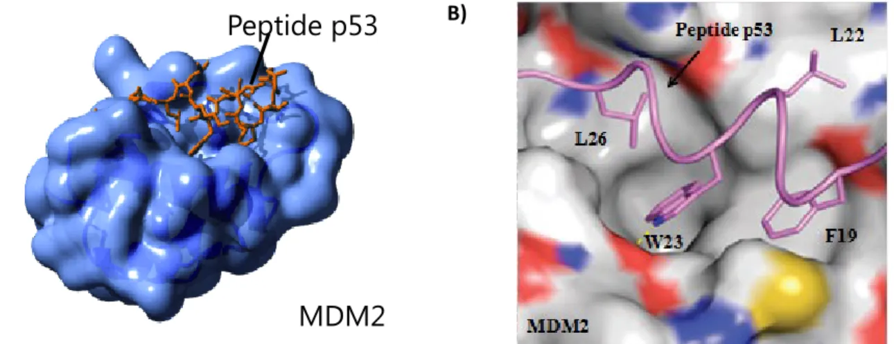
Analysis of the MDM2 protein crystal structure with a short 15-residue peptide fragment from p53 transactivation domain (N-terminus) showed the presence of a small well-defined interaction surface. This p53 fragment was then used as a starting point for the development of several inhibitors for this PPI.28–30 From this structure, researchers observed that side
5
chains from Phe19, Leu22, Trp23 and Leu26, which are contained in an α helix section of p53, interact with a large hydrophobic pocket in MDM2 (Figure 4).
A) B)
Figure 4. Interaction surface between a MDM2 binding domain and a peptidic fragment from p53.
These observations led to the development of several MDM2-p53 interaction inhibitors (Figure 5). In 2001, Moore and his collaborators discovered chlorofusine 1, a naturally occurring cyclic peptide isolated during the screening of bacterial extracts.31 Subsequently,
Robinson’s group has developed the macrocyclic peptide inhibitor 2 showing an IC50 of
140 nM. This macrocycle has a β-hairpin conformation in which the bioactive section is spatially a mimetic of Phe19 and Trp23 from p53 helical section.32 In 2004, Vassilev’s
team have discovered the Nutlin, cis-imidazoline derivatives 3 identified by library screening.33 These molecules were able to efficiently inhibit the formation of the
p53-MDM2 complex with IC50 between 100 and 300 nM. A bromophenyl group is found in the
Trp23 pocket, another bromophenyl group is found in the Leu22 pocket and the ethyl ether chain in the Phe19 pocket. Finally, in 2005 1,4-benzodiazepine-2,5-dione derivatives 4 have been reported as p53-MDM2 interaction inhibitors.34 Compounds showing IC
50
ranging from low µM to high nM have been identified by the combined screening of combinatorial chemical and virtual libraries.
Other examples for the successful rational development of small PPI inhibitors include interactions between XIAP and SMAC, HSP90 and GR, co-activators and estrogen alpha-receptor, Bcl-Xl and Bak, TNF- and TNFRc1.4,9,35–37
MDM2
Peptide p53
6
Figure 5. Molecular inhibitors of the p53-MDM2 protein-protein interaction. 1.1.2 Peptides as Protein-Protein Interaction Inhibitors
Considering the interaction surface nature, protein secondary structures play a major role in the molecular recognition and are essential for specific binding to protein interaction domains (PID). Therefore the ability to mimic protein secondary structures is very important in the design of PPI inhibitors and peptides represent a template of choice to mimic these structures and modulate PPI.20 Peptides are a very interesting class of
therapeutics since they show strong activity, high selectivity, low toxicity and few drug-drug interactions.38,39 However, linear peptides show poor oral bioavailability and are easily
degraded by proteases, limiting their use as therapeutic agents. To overcome these drawbacks, peptide cyclization is increasingly used. Compared to small organic molecules (MW < 500 Da), the size of most macrocycles (MW = 500~2000 Da) is large enough to compete with proteins for flat interaction surfaces and are small enough compared to antibody (MW > 50000 Da) to retain many drug properties. Peptide macrocyles are a rich source of biologically active compounds and are widely produced in nature by plants,
7
bacteria, fungi, marine invertebrates and many other organisms. A wide range of peptide-based macrocycles such as cyclosporine A (immunosuppressant), daptomycin (antibiotic), caspofungin (antifungal), eptifibatide (antiplatelet), octreotide (anticancer), and oxytocin (uterine contractions) are clinically used as therapeutic agents (Figure 6).40
8
1.1.3 Cyclic Peptides
With a wide spectrum of activity and a great therapeutic potential, cyclic peptides have gained a lot of interests in drug discovery. Cyclic peptides show many advantages compared to their linear counterparts. With increased structural rigidity, cyclic peptides show greater stability against endo- and exo-proteases41,42 and the entropic advantages
make them tighter-binding and may confer a higher binding specificity for a given macromolecular receptor.43 Moreover, their conformational analyses in solution by 2D
NMR are more precise and reliable;42,44 and in some cases, they show increased cell
permeability.45
Based on the type of chemical bonds found in the cycle backbone, two different classes of cyclic peptides have been established, namely homodetic and heterodetic cyclopeptides. For homodetic cyclic peptides, the macrocyle backbone is exclusively formed by amide bonds. In contrast, the backbone of heterodetic cyclic peptides may contain also one or many ester, ether, thioester, thioether or disulfide bonds in addition to the amide bonds. Cyclic peptides can be obtained from linear precursors by four different cyclization approaches: head-to-tail (C-terminus to N-terminus), head-to-side chain, side chain-to-tail, and side chain-to-side chain (Figure 7). In most conditions, the ring-closure reaction is performed by lactamization, lactonization or disulfide bridge formation. For the cyclization step, synthetic yields can vary greatly and will depend on various parameters such as reaction conditions, ring size, specific structure, and even amino acid sequence.
9
Cyclic peptides are very convenient molecular probes in chemical biology and very useful tools in drug discovery and development. Most importantly, cyclic peptides are a class of privileged structures since they have been reported to bind to multiple, unrelated classes of receptor with high affinity.46–48 The great degree of molecular complexity and diversity that
can be accessed by simple changes in their linear sequence, makes the use of cyclic peptide-based scaffolds very appealing in drug design and discovery.49 Upon cyclization,
small changes, e.g. in α-carbon stereochemistry, ring size, or constraining residues, can have a dramatic effect on the backbone overall conformation. These conformational effects allow the presentation of side-chains with diverse relative orientations. Their abilities to mimic protein secondary structures such as β- and γ-turns, β-hairpins, β-sheets, -helices and other helix types have been widely studied and demonstrated.50–52
Based on the characteristics described above, cyclic peptides represent a very attractive template to discover protein ligands and develop promising PPI inhibitors. In order to fully exploit the great conformational and functional diversity accessible with cyclic peptides, combinatorial chemistry is certainly the most powerful tool. A major asset of combinatorial cyclic peptide libraries over classic combinatorial libraries, where the scaffold is fixed, is the possibility to generate conformational diversity as well as functional diversity in order to encompass a very large chemical space (diversity).
1.2 Combinatorial Chemistry Approaches to Prepare Peptide Libraries
During the last three decades, the development of combinatorial methodologies has been greatly stimulated by the progress made in biological assays and their increasing testing capacities. To match the growing needs in high-throughput screening (HTS), the preparation of libraries containing a large number of chemical entities was required and combinatorial chemistry represented the most convenient and efficient approach to prepare such libraries. Allowing the simultaneous generation of a large number of synthetic compounds, combinatorial libraries have been used by many research groups to quickly test millions of compounds for their activity on a selected target in a short period of time. This approach is probably the most powerful approach that has occurred in pharmaceutical and life sciences to reduce the time and cost for generating high affinity protein ligands.10
A combinatorial library is usually referred as a collection of 1 × 10⁴ to 1 × 10¹⁰ different
molecules generated by synthetic or biological approaches. The development of combinatorial peptide library technologies can be attributed to Bruce Merrifield who won a Nobel Price for the use of functionalized styrene-divinylbenzene beads as polymer supports in 1963 and his important contribution to the development of solid phase peptide synthesis.53 This approach has faced a renascence in combinatorial chemistry in the middle
1980s with the synthesis of the first limited peptide library using multi-pin technology in 1984,54 and has since become a powerful tool that not only facilitates the drug discovery
process but also provides important information for the fundamental understanding of molecular recognition. These applications led to the development of a wide variety of combinatorial approaches to prepare peptide and chemical compound libraries (Table 1).
Table 1. Milestones in early combinatorial chemistry55
Year Milestone
1984 Limited peptide library with the multi-pin technology 1985 Limited peptide library using tea-bag method
1986 Iterative approach on solid phase peptide library screening using the multi-pin synthesis
1986-90 Development of polynucleotide library methods
1988 Introduction of the split synthesis method on synthesizing a limited library of solution peptides
1990 Light directed parallel peptide synthesis of a library of 1024 peptides on chip 1990 Successful use of the filamentous phage to display a peptide library
1991 Introduction of the one-bead-one-compound method and successful application of this concept to a huge bead-bound peptide library
1991 Successful application of the interactive approach on a huge solution phase peptide library
1992 Synthesis of a limited benzodiazepine-based small molecule library
1992-93 Development of encoding methods for the one-bead-one-compound non-peptide library
11
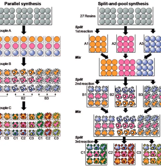
Among the different synthetic approaches to prepare combinatorial libraries, the two most commonly used are the parallel synthesis method and the split-and-pool synthesis method. In parallel synthesis, like in a general organic synthesis, each reaction is performed in separate reaction conditions (Figure 8). The main difference is that many reactions are performed simultaneously in many reactors. To treat many reactions, easy treatment of each reaction is needed, and so solid-phase reaction is the more preferred method. The other option is to use expensive automatic synthesizer. If resins are not used as solid supports, it is possible to synthesize as a chip form by using photolithography of semi-conductor chips.
Figure 8. Synthetic approaches used in combinatorial chemistry: the parallel synthesis
12
The split-and pool synthesis method is only used with solid phase reactions (Figure 8). This method is most commonly used in the one-bead-one-compound combinatorial technology and will be described in details in section 1.2.5. The “split-and-pool synthesis” method was first described by Furka et al.56 also developed by Lam et al.57 and Houghten et al..58 The
beads are split and placed into separate reaction vessels. Different building blocks are coupled on and within the beads. After each coupling step, the beads from each reaction vessel are combined and then randomly split again to the different reaction vessels for next coupling step. A library is prepared after repeating those processes several times. For example, if we try to make all the possible oligomers from monomers A, B and C, the total possible number of compounds is 27 (3 × 3 × 3) and only three coupling steps are required in the case of the split-and pool method. The number of total solid supports should be larger than 27 total compounds to be synthesized so that statistics of split-and-pool process is even. The parallel synthesis approach is more convenient for small size libraries while the split-and-pool approach is more efficient to prepare large size libraries.
Table 2. Combinatorial peptide library methods Combinatorial peptide
library methods Key points of method
Biological libraries Phage-display Plasmid Polysome Bacterial display Yeast-display Spatially addressable parallel
libraries
Multi-pin technology
Tea bags and NanoKan technologies SPOTs-membrane method
Light-directed peptide synthesis on chip
Synthetic libraries requiring deconvolution
Iterative approach Positional scanning Recursive deconvolution Orthogonal partition approach Dual recursive deconvolution Select library by affinity
column Using affinity chromatography selection One-bead one-compound
13
Most combinatorial methods are composed of three main steps: (i) preparation of the library, (ii) screening of the library components, and (iii) determination of the chemical structures of active compounds.59 The key feature of combinatorial chemistry is that a large
range of analogs is synthesized using the same reaction conditions with different building blocks and in the same of different reaction vessels.60 In this way, chemists can synthesize
many hundreds to millions of compounds simultaneously instead of preparing only a few compounds one by one. To date, there are five general methods of preparing and screening combinatorial libraries: (i) the biological peptide library method (e.g., phage-display peptide library),61–63 (ii) the spatially addressable parallel library method,64,65 (iii)
combinatorial library methods requiring deconvolution,58,66 (iv) the affinity selection
method67 and (v) the OBOC combinatorial library method (Table 2).57,59
1.2.1 Biological Libraries Method
Polypeptide libraries can be obtained by molecular biology techniques with different biological entities, such as bacteria, phages, yeast and ribosomes. Among these biological peptide library methodologies, the phage-display technique based on phage exposition with peptides displayed by fusion with a viral coat protein is the most widely used.68–70 Many
filamentous bacteriophages are used for displaying peptides but M13 is the most commonly used because of its high capacity for replication and the ability to receive large DNA inserts into its genome.71,72 Smith firstly developed the “fusion phage” method by inserting foreign
DNA fragments into the encoding gene of the pIII protein to display short peptide library on the surface of filamentous M13-derived bacteriophage for mapping antibody epitopes.73
This led to the expression of L-amino acid containing peptide on the virion surface, which did not affect virus infectivity. Bacteriophages are single-stranded DNA viruses that infect bacteria.74 The major coat protein pVIII which is represented by about 2700 copies is the
most abundant capsid component. At one end of the phage virion, there are 5 copies each of two minor coat proteins pIII and pVI for bacterial cell binding and the termination of phage particle assembly. Three to five copies of minor coat proteins pVII and pIX are at the other end of the phage for initiation and maintenance of phage assembly. The most commonly used coat proteins for peptide libraries displayed on M13 are pIII and pVIII (Figure 9). Higher affinity ligands are usually obtained by protein pIII.
14
Figure 9. Peptides displayed on M13 phage surface as fusion to the amino terminus of (A) pVIII or (B) pIII; (C) affinity-based selection procedure adapted in phage display
technology.75
In general, the extremely powerful biologic library method provides the following advantages. (i) It enables the researcher to routinely generate 108 to 109 different phages; (ii)
a large number of different peptide sequences are easily produced on viral surface for screen. Longer peptides or proteins can be easily constructed without the limitation as in the case of the synthetic peptide library; (iii) it is convenient to generate the library by simply growing the microorganisms; (iv) random oligopeptides can be grafted on such tertiary folds by taking advantage of known protein folds, such as immunoglobulin fold, zinc-finger fold, or conotoxin fold. However, phage display for high-quality, large-diversity library requires specialized resources and skills. And several major disadvantages are present, such as (i) only the natural L-amino acid peptide libraries (20 natural proteinogenic amino acids) can be incorporated into these libraries, unnatural amino acids or other organic building blocks is not feasible; (ii) although simple disulfide cyclization is feasible, complicated bicyclic, compact scaffolding, branched structures, or molecules with special chemistry of cyclization are impossible; and (iii) screening assays of the biological libraries are generally limited to the binding assays (e.g., panning) and some functional assays such as protease substrate determination.
A)
B)
15
1.2.2 Spatially Addressable Parallel Library Method
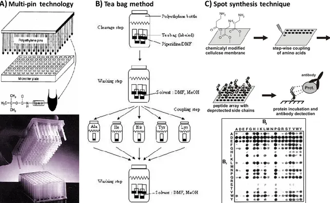
Combinatorial libraries can also be generated by using another effective method, spatially addressable parallel library method. In this method, a collection of compounds is synthesized on a variety of carriers or in solution in a spatially addressable format. Depending on the library method used, screening can be performed either by a solid-phase binding assay or by a solution-phase assay. The chemical structure of positive compound can be inferred by the location, decoding is not needed. The reported techniques based on this strategy include multi-pin technology,54 tea bag method,76,77 SPOT synthesis,78 and
peptide microarray64,79 (Figure 10). The main advantages of this method is structural
determination of the library member is not required because these compounds is spatially addressable. In additional, D-amino acids, other unnatural amino acids or even other chemical moieties can be included in the library’s structure. The major drawback of this method is that only limited number of compounds can be synthesized and therefore the library is very small.
Figure 10. Techniques based on the spatially addressable parallel library strategy. (A) Multi-pin technology, (B) Tea bag method and (C) Spot synthesis technique.
16
Geysen et al.54 initially introduced Multi-pin method in 1984 (Figure 10A). In this method,
amino-functionalized polyacrylic-acid-grafted polyethylene rods which were called “pins” were inserted into an adapter fitting over a 96-well microtiter plate containing the reagent solutions. A unique compound is performed in each individual well of the microtiter plate. Biological screening was performed by an ELISA on the peptide pin. Latterly, peptides were able to release by cleavable linkers for solution assay in this pin method.80–82 The
chemical structure of any component is easily determined by its spatial location. Today, the multi-pin technology has been commercialized by Mimotopes (San Diego, CA, USA) as Pepsets. Peptide loading of each pin has been increased significantly by introducing the so-called “lantern” to fit into the tip of each pin.
A year later, Houghten introduced the “Tea bag” method for simultaneous multi-peptide synthesis (Figure 10B). 83 In this method, batches of resin (~25-100 mg) are sealed inside
labeled, porous polypropylene packets (tea bags). Amino acid coupling reaction is performed in individual bag to generate activated monomers. After completion of each coupling, all tea-bags are collected into a large vessel and all common steps, such as resin washing and amino acid protecting group removing, are treated simultaneously. In 1995, a update method by using radio-frequency microchips (Rf tagging) for encoding
combinatorial chemical libraries and a sample of resin beads for peptide synthesis in mini-baskets (e.g., NanoKan) was reported.76,77 Each of these mini-baskets will be scanned with
an electronic reader prior to or right after each coupling cycle during the “split-and-pool” synthesis.56–58 Therefore, the construction of each compound can be identified by the
recording history. At the end of the synthesis, single compounds are released from the beads which are inside of each mini-basket and placed in a 96-well plate to form a spatially addressable compound library. This approach takes advantage of the power of “split-and-pool” synthesis method. The main advantage of this method is that it offers considerable synthetic flexibility as one may use any resin beads in the synthesis.
Frank and coworkers synthesized peptides on a cellulose membrane or paper as the solid support instead of using polyethylene pins (Figure 10C).84,85 In this SPOT-synthesis
method, a circular spot is created by the volume of amino acids and coupling reagent. The size of the spot determines both the scale of reaction and the number of compounds. Different peptides are synthesized at different locations in a single sheet of cellulose paper.
17
The common steps of synthesis are treated by washing the whole membrane or surface with the respective reagents and solvents. The compounds synthesized can be evaluated using conventional high-throughput screening techniques while still attached to the membrane or in solution following membrane release. Cotton86,87 (another form of cellulose) and
polystyrene-grafted polyethylene film88 segments have also been used as solid supports.
SPOT synthesis is amenable to miniaturization and automation. The advantages of the method are that it is simple, cheap and provides sufficient quantities of peptides for various applications.89
Pioneered by the Affymax group64 peptide microarrays are prepared by immobilizing
many peptide molecules on the surface of a solid support in a small area in an addressable fashion. In 1991, Fodor et al. synthesized minute quantities of 1024 peptides on a single glass slide by using a photolithographic masking process in conjunction with light-directed peptide synthesis.64 Each peptide spot occupies a 50 × 50 µm area. This accomplishment, in
fact, predated DNA microarray. The immobilization can be achieved via in situ synthesis or chemical ligation through a covalent bond. A hydrophilic linker between the solid surface and the peptide usually is added to minimize steric hindrance caused by the solid support. The most commonly used solid support for microarray printing is a standard microscope glass slide but other solid supports include cellulose sheets65 and polymer-based
membranes.90,91 Because of the limited quantity of peptide available, biological assay is
restricted primarily to binding or functional assay on the slide. In addition, this method is not widely available since the requirement of complicated instrumentation.
1.2.3 Synthetic Libraries Requiring Deconvolution
In this method, mixtures of compounds are first synthesized and then screened for specific biological property. On the basis of the results from biologic assays, the structural determination of active compound may be able to be deduced without need of further structure elucidation. The term “deconvolution” has been used to describe the process whereby the active molecules in a library are identified, usually by the iterative testing of mixtures of compounds for a specific biologic property. The general deconvolution approaches includes the iterative approach,94 the positional scanning approach,93 the
18
recursive deconvolution approach.96 There are following advantages in the method. A large
number (106 to 108) of peptide library is able to be synthesized and screened for many
existing biological assays. Decoding library is also not necessary because the structure of the active compounds can be deduced. However, in general, additional synthesis and testing, which are time-consuming, are needed to reach a final solution.
Figure 11. Descriptive model of (A) iterative process and (B) positional scanning deconvolution approaches. In the case of positional scanning, the sequence is reconstructed at the end of the process, whereas in the iterative process the sequence is obtained step by step.97
The two main deconvolution approaches are the iterative process92 and the positional
scanning (Figure 11).93,98 The iterative approach was first reported by Geysen et al. in the
multi-pin system. 92 Each position is chosen by one amino acid using a progressive
selection way. Sublibraries are generated based on the result of the previous one; therefore, the structure does not need to be sequenced. A related approach, positional scanning, was later introduced by Dooley & Houghten.93 In this method, sublibraries are synthesized with
one of 20 amino acid fixed at one specific position while randomized in other positions. Then the sublibraries are tested for biologic activities. At the end of process, the sequence of active peptides can be deduced based on the biologic assay results. This method assumes that the contribution of each amino acid residue to the biologic activity is independent of
A)
19
each other and it works very well for one existing predominant. If there are multiple binding motifs, scramble or uninterpretable results may be obtained.
1.2.4 Synthetic Library Method Using Affinity Chromatography Selection
In this method, the peptides, which are usually synthesized by “split-and-pool” method, are cleaved off the resins to form a solution phase peptide library.56–58 The generated solution
of peptide mixture is then loaded onto an affinity column with immobilized receptor to select the active molecules. After thorough washing, the bound peptides are eluted and structure are determined.99 This method has been applied successfully for combinatorial
oligodeoxynucleotide library in which the bound oligodeoxynucleotides are eluted and amplified. However, there are only few applications for peptide library because peptides cannot be amplified or cloned. Zuckermann et al. were able to apply the affinity selection method to retrieve three peptides from a very small peptide library (19 compounds) in solution.100 Cantley et al. reported the successful application of the affinity selection
method to identify peptide motifs for SH2 domains and kinase domains of protein tyrosine kinases.67,101 The following points should be considered (i) nonspecific binding, if a huge
peptide library is used or the affinity binding is not very high; (ii) uninterpretable sequencing data, if there is more than one predominant motif in the peptides mixture; (iii) To have enough material for structure determination may be another problem since the peptides cannot be amplified. Generally this method can be applied only to a relatively small peptide library (e.g. <10 000 peptides).
1.2.5 One-Bead One-Compound Library Method
The «one-bead one-compound approach» (OBOC) is a powerful approach that has been widely used to generate large peptide libraries. The concept was initially reported by Lam et al. where a library including 1013 compounds was prepared by “split-and-pool
synthesis”.57 The described library contained a large number of single beads displaying
many copies of a single compound and was named one-bead-one-compound library. In the OBOC method, the beads are split and placed into separate reaction vessels. Different building blocks are coupled on and within the beads. After each coupling step, the beads from each reaction vessel are combined and then randomly split again to the different reaction vessels for next coupling step. A library is prepared after repeating those process
20
several times. A simple example is illustrated in Figure 12 with the preparation of a tripeptide library by the “split-and-pool” methodology with proline (P), glutamic acid (E) and threonine (T) as building blocks by standard solid-phase peptide synthesis.56,57 After
each coupling step, beads from each reaction vessel are combined then split into the three vessels again for the next coupling step. After three such steps the 27 possible peptide sequences are all represented on separate beads (Figure 12). By using standard amino acid coupling procedure, an octapeptide library with 20 amino acids (including 208 possible
copies) can be generated in 2-3 days. In this efficient method to identify ligands, thousands to millions of compounds are rapidly generated using the “split-and-pool” synthesis approach on solid support, in such a manner that each bead displays only a single compound entity.
Figure 12. The “split-and-pool” synthesis method to generate a one-bead-one-compound combinatorial library (A) a number of permutations for random peptide libraries (P, E, and
T are building blocks in this case amino acids).(B) the generated diversity.102
After its construction, high-throughput on-bead screening can be performed on the OBOC library to identify ligands, inhibitors or modulators for the molecular target of interest (Figure 13). OBOC libraries have been successfully used to discover linear peptidic ligands and modulators for various receptors,103,104 enzymes,53,105–108 transcription factors,109 and
other protein targets.43,110–114 Because of its accessibility, flexibility and huge potential , the
OBOC technology is particularly interesting to discover ligands for a protein of interest and develop new PPI inhibitors.
A) Building Blocks Reaction cycles Library entities 3 2 9 20 2 400 20 3 8 000 20 4 160 000 20 5 3 200 000 20 6 64 000 000 Generated diversity B)
21
1.3 Screening Strategies for One-Bead-One-Compound Libraries
In general, the screening of OBOC libraries is performed on-bead. Most on-bead screening methods allow a large number of beads to be screened simultaneously against a target of interest and identify the positive beads containing the bioactive peptides. Therefore, the nature and behavior of the solid support become very important. The polymer beads used for the preparation and screening should show good swelling properties and be compatible with organic solvents for library synthesis and aqueous solution for biological assays. Among the different amphiphilic solid supports, PEG-based resins like ChemMatrix or PEG-grafted resins such as TentaGel® are the most commonly used. Compared to
ChemMatrix which is composed of cross-linked PEG, TentaGel® have a polystyrene core
grafted with poly(ethylene glycol) chains.115,116 For TentaGel® resin, the loading capacity is
typically around ~0.3 mmol/g and a wide variety of bead size can be used. The most commonly used are the 90 µm and the 130 µm which carry ~0.1 and 0.35 nmol compound per bead, respectively.117 This amount is more than enough to identify the sequence of the
peptide on the beads by tandem mass spectrometry (MS/MS). Moreover, the uniform size of TentaGel® beads makes the load capacity of beads consistent.
22
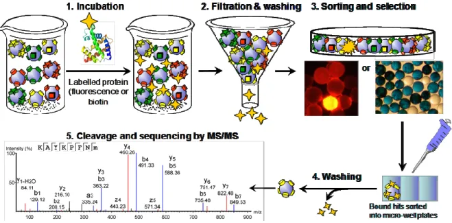
Currently, the reported screening procedures for OBOC libraries generally involve the following steps: (1) incubation of the library with the target; (2) filtration and washing to remove unbound material; (3) sorting of the library and identification/selection of positive beads; (4) washing to remove bound material; (5) cleavage and sequence determination. Steps 1 to 4 can be repeated a few times to eliminate false positive. In the first step, tens of thousands to millions of compounds/beads are incubated with a target of interest that has been labelled. The choice of the label is extremely important in the screening process and will be based on the detection method used for the sorting in step 3. After incubation with the labelled target, beads interacting with the target are identified and isolated for compound structure determination.
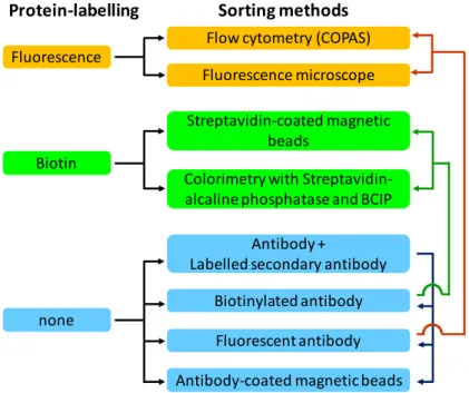
Two main strategies have been used to label proteins of interest, direct protein-labelling and the use of antibodies (Figure 14). In the first strategy, the protein is directly labelled with a fluorescent derivative or with biotin. In the other strategy, a primary or secondary labelled antibody can be used. Whether direct protein labeling or antibody is used, the choice of on-bead detection technique is the same. The three most commonly used detection and sorting techniques for screening OBOC libraries are the colorimetric assays, fluorescence assays and the magnetic sorting.
Figure 14. Protein-labelling strategies and sorting methods for the screening of OBOC libraries and selection of positive beads.
Fluorescence
Biotin
Protein-labelling Sorting methods
Flow cytometry (COPAS) Fluorescence microscope Streptavidin-coated magnetic
beads
Colorimetry with Streptavidin-alcaline phosphatase and BCIP
none
Antibody +
Labelled secondary antibody
Antibody-coated magnetic beads Biotinylated antibody Fluorescent antibody
23 1.3.1 Colorimetric Assays
In colorimetric assay, an enzyme conjugate is used to transform a soluble colorless molecule into a colored precipitate. During the transformation the beads become colored and can be easily isolated when the library is visualized under a microscope. The target protein is usually labelled with a biotin moiety which is a well-known molecule that binds very tightly to streptavidin, a tetrameric protein with four biotin binding sites.118 The
binding of biotin to streptavidin, with a dissociation constant KD on the order of 10−15 M, is
one of the strongest known protein-ligand interactions. The small size of biotin means that biological activity of the protein will most likely be unaffected. The coupling of a biotin moiety to a protein or other molecule has been used in a wide variety of biotechnological applications and is called biotinylation.
Figure 15. Colorimetric sorting with SA-AP.
To detect beads interacting with the target biotinylated protein, a streptavidin-alkaline phosphatase (SA-AP) conjugate is first added to the library that has been incubated with the target and washed (Figure 15). After some time to allow the binding of streptavidin to
24
biotin, 5-bromo-4-chloro-3-indolyl phosphate (BCIP) is added to the mixture to color the positive beads. The alkaline phosphatase conjugated to the streptavidin molecule hydrolyses the phosphate group of BCIP to generate the indole derivative, which is oxidized by oxygen to form an insoluble blue dye. After washing, the beads can be observed under a microscope and the positive colored ones picked up individually with a micropipette. When antibodies are used, the assay is called an on-bead ELISA (Enzyme-Linked Immunosorbent Assay). After the incubation step with the target protein and washings, the assay is performed with a primary antibody against the target protein followed by a secondary antibody conjugated to an alkaline phosphatase. The coloration process is the same as described above.
1.3.2 Fluorescent Assays
In the fluorescence assay, a fluorochrome is directly linked to the target protein or to a secondary antibody. Fluorescence is a very sensitive detection technique that involves the absorption of light or other electromagnetic radiation and emission of light at longer wavelength. The main advantage of this technique is that, in contrast to the colorimetric assay, fluorescence does not require post-incubation chemical reaction. After incubation and washing, the positive beads can be directly identify and isolated under a fluorescence microscope. Amongst the great number of fluorochrome commercially available, Fluorescein and Texas red are the most commonly used for OBOC library screening. They differ in optical properties, such as the intensity and spectral range of their excitation and emission wavelengths. The choice of the fluorochrome depends on the sorting methodology that will be used and the available filters. Special caution should be taken to avoid auto-fluorescence from the solid support. To reduce interference of beads auto-auto-fluorescence, the protein can be labeled by dyes with emission maximum in the red that emitted in a region where the TentaGel beads fluorescence is less intense.112
However, after screening by colorimetry or fluorescence assays individual positive beads are picked manually using a pipette by naked eye under a microscope, which is a time-consuming and labour-intensive process. Therefore it was necessary to introduce more high-throughput sorting techniques for efficient selecting positive beads from a library containing millions of compound beads.