Antimicrobial and molecular analysis of Salmonella serovar Livingstone
strains isolated from humans in Tunisia and Belgium
Intissar Guedda1,2, Bernard Taminiau1, Asma Ferjani2, Jalel Boukadida2, Sophie Bertrand3, Georges
Daube1
1 Department of Food Sciences, Microbiology, Faculty of Veterinary Medicine, University of Liege, Liege, Belgium 2 Microbiology and Immunology Departement UR02SP13, CHU Farhat Hached, Sousse, Tunisia
3 National Reference Center for Salmonella and Shigella, Bacteriology Division, Scientific Institute of Public Health, Brussels, Belgium
Abstract
Introduction: Salmonella Livingstone is one of the most common serotypes responsible for nosocomial outbreaks in Tunisia. In this study, 42 isolates of Salmonella Livingstone were analyzed. Most of these were isolated from humans (31 strains from Tunisia and 9 strains from Belgium) and 2 isolates came from food products (beef and pork).
Methodology: All strains were characterized by antibiogram, multilocus sequence typing (MLST), and virulotyping. This last technique was carried out by simple PCR of five chromosomal genes (agfA, hin/H2, iroB, phoP/Q, and slyA) and two plasmid genes (spvA and spvC). Results: All Tunisian strains were resistant to amoxicillin, amoxicillin-clavulanic acid, ticarcillin, cefalotin, gentamicin, and kanamycin. They were also resistant to third-generation cephalosporin antibiotics (cefotaxim and ceftazidim). Belgian isolates were susceptible to all antibiotics tested. Further to MLST analyses, Tunisian strains belonged to the same sequence type, ST543. For Belgian isolates, eight strains had a ST543 profile, two strains had a ST638 profile, and one strain had a ST457 profile. Analyses of the virulence gene contents showed that strains isolated in different years and from different origins had the same virulence profile. These carried all five chromosomal genes and lacked plasmid-located virulence genes spvA and spvC.
Conclusions: A combination of different typing methods showed that the majority of Belgian strains and all Tunisian strains were closely related; they belonged to the same sequence type (ST543) and had the same virulence profile, but different antibiotic resistance profiles depended on the country of origin.
Key words:Salmonella Livingstone; MLST; virulence genes; antimicrobial resistance. J Infect Dev Ctries 2014; 8(8):973-980. doi:10.3855/jidc.3989
(Received 16 July 2013 – Accepted 28 January 2014)
Copyright © 2014 Guedda et al. This is an open-access article distributed under the Creative Commons Attribution License, which permits unrestricted use, distribution, and reproduction in any medium, provided the original work is properly cited.
Introduction
Salmonella enterica is among the most important
causes of foodborne gastroenteritis worldwide [1-2]. It is also responsible for nosocomial outbreaks, particularly in developing countries. Salmonella
enterica serovar Livingstone is one of the more than
2,500 serovars of S. enterica known [3]. It was first isolated from human feces in 1951 [4]; since then, it has been rarely isolated from human beings and animals, particularly poultry and feed products [5-6].
In Tunisia, there was a dramatic increase in the number of isolations of Salmonella Livingstone from humans and animals between 1999 and 2003 [7]. This particular serotype was one of the most common
Salmonella isolates responsible for nosocomial
In Europe, the number of reported sporadic human cases of Salmonella Livingstone infections was low, and outbreaks were rare [10]. The larger outbreaks were recorded in Tayside, Scotland, during 1989–1991 [5], and in several European countries in 1996 related to travel to Tunisia [11]. In Belgium, Salmonella Enteritidis and S. Typhimurium were the most common serovars causing salmonellosis in humans;
Salmonella Livingstone was rarely isolated [12].
Numerous phenotypic and genotypic methods, among them biotyping, antimicrobial susceptibility profiling, plasmid profiling, ribotyping, random amplification of polymorphic DNA (RAPD-PCR), enterobacterial repetitive intergenic consensus (ERIC-PCR) and pulsed field gel electrophoresis (PFGE)
this serotype [7,9,13]. Currently, alternative molecular typing methods such as multilocus sequence typing (MLST) and virulotyping [1,14-15] are used to characterize many Salmonella enterica serovars. Multilocus sequence typing [16] has been increasingly recognized as a method of choice for genotyping a number of bacterial pathogens [17-18], including Salmonella [1,19-21], and has been used successfully in epidemiological studies and outbreak investigations [17,22]. This technique is based on determination of the DNA sequence of a series of selected housekeeping, ribosomal, and/or virulence-associated genes [23-24].
Virulence genes have been screened using simple [25] or multiplex PCR [26] to determine the distribution of these genes among Salmonella enterica strains [25,27] and to genotype many Salmonella serovars [15,28-29].
The objective of this study was to investigate the phylogenetic relationship between clinical Salmonella Livingstone strains isolated from Tunisia and Belgium between 2002 and 2010 and to analyze the differences in antimicrobial susceptibility profiles and the genomic content of some well-known determinants of virulence in these isolates.
Methodology Bacterial strains
Forty-two strains of Salmonella Livingstone were included in this study, of which 31 were collected from human stools between 2002 and 2009 in Farhat Hached University Hospital located in the town of Sousse (central-east of Tunisia).
The majority of these strains (n = 27) were recovered from stool specimens of babies up to two months of age, during a nosocomial outbreak that occurred in the neonatology ward of the maternity department in 2005. Nine unrelated strains isolated in 2010 from humans were provided by Belgium’s National Reference Centre for Salmonella and
Shigella. Two Belgian meat isolates (pork and beef)
were used as comparison strains (Table 1).
All isolates were identified with the API 20E system (bio-Mérieux, Marcy l’Etoile, France) and were serotyped by agglutination tests with antisera (Bio-Rad, Hercules, USA) as specified by the White-Kauffmann-Le Minor scheme [30].
Antimicrobial susceptility testing
Antibiotic susceptibility was determined by the disk diffusion method on Mueller-Hinton agar (Bio-Rad) according to the guidelines of the Antibiogram Committee of the French Society for Microbiology (CA-SFM).
The following antimicrobial agents (Bio-Rad) were tested: amoxicillin (AMX, 25 μg), amoxicillin-clavulanic acid (AMC, 20/10 μg), ticarcillin (TIC, 75 μg), ticarcillin-clavulanic acid (TCC, 75/10 μg), piperacillin (PIP, 75 μg), imipenem (IMP, 10 μg), cefalotin (CF, 30 μg), cefoxitin (FOX, 30 μg), cefotaxim (CTX, 30 μg), ceftazidime (CAZ, 30 μg), kanamycin (K, 30 UI), tobramycin (TB, 10 μg), gentamycin (GM, 15 μg), amikacin (AN, 30 μg), nalidixic acid (NA, 30 μg), ofloxacin (OFX, 5 μg), ciprofloxacin (CIP, 5 μg), colistin (CS, 50 μg), trimethoprim (TM, 5 μg), chloramphenicol (C, 30 μg), fosfomycin (FOS, 50 μg), tetracyclin (TE, 30 UI), and trimethoprim sulfathomexazole (SXT, 1.25/23.75 μg).
Multilocus sequence typing (MLST)
Multilocus sequence type analysis was carried out using the MLST protocols described on the MLST website (MLST.net).
Briefly, all isolates were grown overnight in LB medium at 37°C and total DNA was extracted by thermal extraction. For each strain, two to three colonies were dissociated in 150 µL of sterile distilled water; this suspension was heated to 95°C for five minutes, then cooled immediately in ice. After two minutes of centrifugation at 13,000 rpm, the supernatant was conserved at -20°C [31].
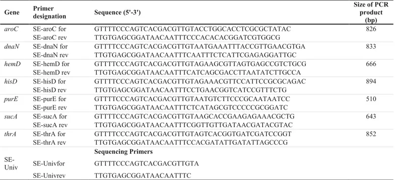
PCR amplification for the seven genes of the MLST panel was carried out as follows: primer pairs (Table 2) were used to amplify the DNA for the presence of the following genes: aroC, dnaN, hemD,
hisD, purE, sucA, and thrA. PCR reactions were
carried out in 20 µL volumes containing 2 µL of DNA template, 5 U/µL of Taq DNA polymerase (Eurogentec, Seraing, Belgium), 1X PCR buffer, 0.5 µM of forward and reverse primers (Eurogentec, Seraing, Belgium), and 200 µM of DNTPs (Promega Fitchburg, USA).
PCR cycling conditions were 94°C for 5 minutes followed by 34 cycles of 94°C for 1 minute, 55°C for 1 minute and 72°C for 1 minute, with a final extension of 72°C for 5 minutes followed by hold at 4°C.
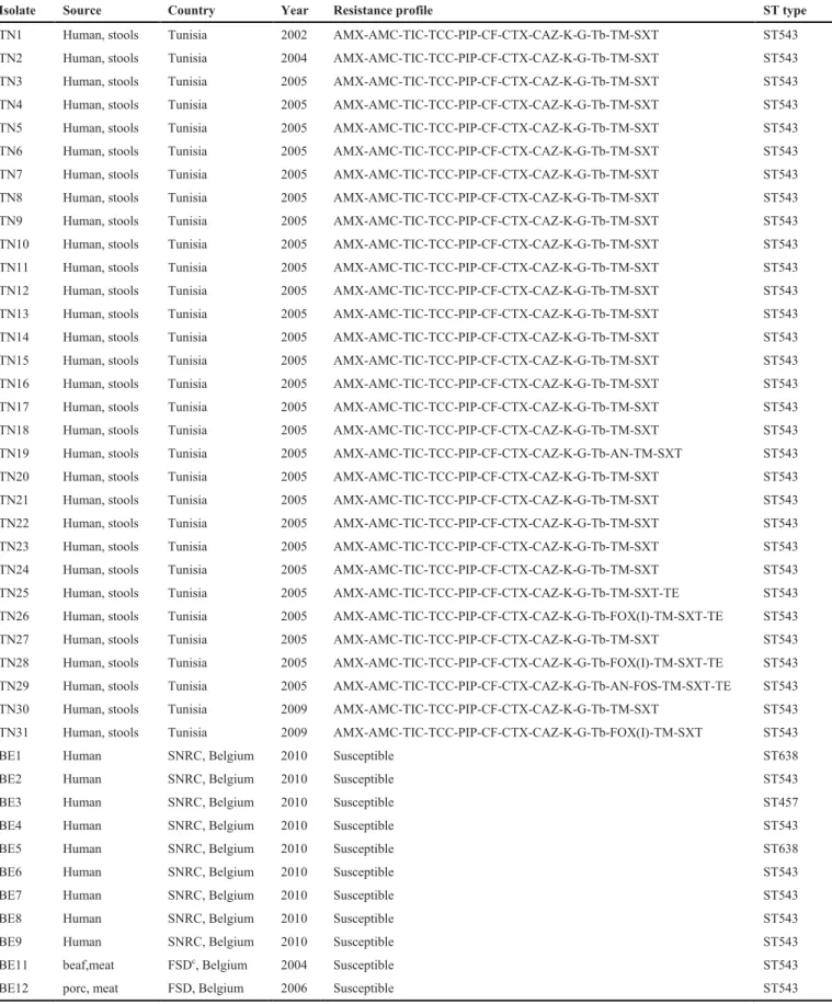
Table 1. Origin and characterization of Salmonella Livingstone isolates used in this study
Isolate Source Country Year Resistance profile ST type
TN1 Human, stools Tunisia 2002 AMX-AMC-TIC-TCC-PIP-CF-CTX-CAZ-K-G-Tb-TM-SXT ST543 TN2 Human, stools Tunisia 2004 AMX-AMC-TIC-TCC-PIP-CF-CTX-CAZ-K-G-Tb-TM-SXT ST543 TN3 Human, stools Tunisia 2005 AMX-AMC-TIC-TCC-PIP-CF-CTX-CAZ-K-G-Tb-TM-SXT ST543 TN4 Human, stools Tunisia 2005 AMX-AMC-TIC-TCC-PIP-CF-CTX-CAZ-K-G-Tb-TM-SXT ST543 TN5 Human, stools Tunisia 2005 AMX-AMC-TIC-TCC-PIP-CF-CTX-CAZ-K-G-Tb-TM-SXT ST543 TN6 Human, stools Tunisia 2005 AMX-AMC-TIC-TCC-PIP-CF-CTX-CAZ-K-G-Tb-TM-SXT ST543 TN7 Human, stools Tunisia 2005 AMX-AMC-TIC-TCC-PIP-CF-CTX-CAZ-K-G-Tb-TM-SXT ST543 TN8 Human, stools Tunisia 2005 AMX-AMC-TIC-TCC-PIP-CF-CTX-CAZ-K-G-Tb-TM-SXT ST543 TN9 Human, stools Tunisia 2005 AMX-AMC-TIC-TCC-PIP-CF-CTX-CAZ-K-G-Tb-TM-SXT ST543 TN10 Human, stools Tunisia 2005 AMX-AMC-TIC-TCC-PIP-CF-CTX-CAZ-K-G-Tb-TM-SXT ST543 TN11 Human, stools Tunisia 2005 AMX-AMC-TIC-TCC-PIP-CF-CTX-CAZ-K-G-Tb-TM-SXT ST543 TN12 Human, stools Tunisia 2005 AMX-AMC-TIC-TCC-PIP-CF-CTX-CAZ-K-G-Tb-TM-SXT ST543 TN13 Human, stools Tunisia 2005 AMX-AMC-TIC-TCC-PIP-CF-CTX-CAZ-K-G-Tb-TM-SXT ST543 TN14 Human, stools Tunisia 2005 AMX-AMC-TIC-TCC-PIP-CF-CTX-CAZ-K-G-Tb-TM-SXT ST543 TN15 Human, stools Tunisia 2005 AMX-AMC-TIC-TCC-PIP-CF-CTX-CAZ-K-G-Tb-TM-SXT ST543 TN16 Human, stools Tunisia 2005 AMX-AMC-TIC-TCC-PIP-CF-CTX-CAZ-K-G-Tb-TM-SXT ST543 TN17 Human, stools Tunisia 2005 AMX-AMC-TIC-TCC-PIP-CF-CTX-CAZ-K-G-Tb-TM-SXT ST543 TN18 Human, stools Tunisia 2005 AMX-AMC-TIC-TCC-PIP-CF-CTX-CAZ-K-G-Tb-TM-SXT ST543 TN19 Human, stools Tunisia 2005 AMX-AMC-TIC-TCC-PIP-CF-CTX-CAZ-K-G-Tb-AN-TM-SXT ST543 TN20 Human, stools Tunisia 2005 AMX-AMC-TIC-TCC-PIP-CF-CTX-CAZ-K-G-Tb-TM-SXT ST543 TN21 Human, stools Tunisia 2005 AMX-AMC-TIC-TCC-PIP-CF-CTX-CAZ-K-G-Tb-TM-SXT ST543 TN22 Human, stools Tunisia 2005 AMX-AMC-TIC-TCC-PIP-CF-CTX-CAZ-K-G-Tb-TM-SXT ST543 TN23 Human, stools Tunisia 2005 AMX-AMC-TIC-TCC-PIP-CF-CTX-CAZ-K-G-Tb-TM-SXT ST543 TN24 Human, stools Tunisia 2005 AMX-AMC-TIC-TCC-PIP-CF-CTX-CAZ-K-G-Tb-TM-SXT ST543 TN25 Human, stools Tunisia 2005 AMX-AMC-TIC-TCC-PIP-CF-CTX-CAZ-K-G-Tb-TM-SXT-TE ST543 TN26 Human, stools Tunisia 2005 AMX-AMC-TIC-TCC-PIP-CF-CTX-CAZ-K-G-Tb-FOX(I)-TM-SXT-TE ST543 TN27 Human, stools Tunisia 2005 AMX-AMC-TIC-TCC-PIP-CF-CTX-CAZ-K-G-Tb-TM-SXT ST543 TN28 Human, stools Tunisia 2005 AMX-AMC-TIC-TCC-PIP-CF-CTX-CAZ-K-G-Tb-FOX(I)-TM-SXT-TE ST543 TN29 Human, stools Tunisia 2005 AMX-AMC-TIC-TCC-PIP-CF-CTX-CAZ-K-G-Tb-AN-FOS-TM-SXT-TE ST543 TN30 Human, stools Tunisia 2009 AMX-AMC-TIC-TCC-PIP-CF-CTX-CAZ-K-G-Tb-TM-SXT ST543 TN31 Human, stools Tunisia 2009 AMX-AMC-TIC-TCC-PIP-CF-CTX-CAZ-K-G-Tb-FOX(I)-TM-SXT ST543
BE1 Human SNRC, Belgium 2010 Susceptible ST638
BE2 Human SNRC, Belgium 2010 Susceptible ST543
BE3 Human SNRC, Belgium 2010 Susceptible ST457
BE4 Human SNRC, Belgium 2010 Susceptible ST543
BE5 Human SNRC, Belgium 2010 Susceptible ST638
BE6 Human SNRC, Belgium 2010 Susceptible ST543
BE7 Human SNRC, Belgium 2010 Susceptible ST543
BE8 Human SNRC, Belgium 2010 Susceptible ST543
BE9 Human SNRC, Belgium 2010 Susceptible ST543
BE11 beaf,meat FSDc, Belgium 2004 Susceptible ST543
PCR products were purified with a PCR purification kit from Promega, and two independent amplified fragments were sequenced using the primers SE-Univfor and SE-Univrev (Table 2).
Sequencing was done with the Sanger sequencing (Big Dye) method and an ABI 3730 capillary sequencer
(http://www.giga.ulg.ac.be/jcms/c_5015/fr/accueil). The sequences were submitted to the MLST database website (http://mlst.ucc.ie/mlst/dbs/Senterica) and assigned existing or novel allele type numbers and sequence type numbers defined by the database. This database defined a novel allele type if it contained one
or more nucleotide changes from existing allele sequences. Composite sequence types (STs) were assigned based on the set of allele types derived from each of the seven loci.
Virulotype PCR screening
The PCR-based assays were described by Del Cerro et al. [32]. Seven Salmonella virulence genes were screened in all the isolates, including five chromosomally located genes (agfA, hin/H2, iroB,
phoP/Q and slyA) and two plasmid-encoded virulence
factors (spvA and spvC) (Table 3). These virulence determinants represented regions known to be either Table 2. PCR and sequencing primers used for MLST
Gene Primer designation Sequence (5'-3')
Size of PCR product
(bp)
aroC SE-aroC for GTTTTCCCAGTCACGACGTTGTACCTGGCACCTCGCGCTATAC 826
SE-aroC rev TTGTGAGCGGATAACAATTTCCCACACACGGATCGTGGCG
dnaN SE-dnaN for GTTTTCCCAGTCACGACGTTGTAATGAAATTTACCGTTGAACGTGA 833
SE-dnaN rev TTGTGAGCGGATAACAATTTCAATTTCTCATTCGAGAGGATTGC
hemD SE-hemD for GTTTTCCCAGTCACGACGTTGTAGAAGCGTTAGTGAGCCGTCTGCG 666
SE-hemD rev TTGTGAGCGGATAACAATTTCATCAGCGACCTTAATATCTTGCCA
hisD SE-hisD for GTTTTCCCAGTCACGACGTTGTAGAAACGTTCCATTCCGCGCAGAC 894
SE-hisD rev TTGTGAGCGGATAACAATTTCCTGAACGGTCATCCGTTTCTG
purE SE-purE for GTTTTCCCAGTCACGACGTTGTAATGTCTTCCCGCAATAATCC 510
SE-purE rev TTGTGAGCGGATAACAATTTCTCATAGCGTCCCCCGCGGATC
sucA SE-sucA for GTTTTCCCAGTCACGACGTTGTAAGCACCGAAGAGAAACGCTG 643
SE-sucA rev TTGTGAGCGGATAACAATTTCGGTTGTTGATAACGATACGTAC
thrA SE-thrA for GTTTTCCCAGTCACGACGTTGTAGTCACGGTGATCGATCCGGT 852
SE-thrA rev TTGTGAGCGGATAACAATTTCCACGATATTGATATTAGCCCG Sequencing Primers
SE-Univ SE-Univfor GTTTTCCCAGTCACGACGTTGTA SE-Univrev TTGTGAGCGGATAACAATTTC aSNRC: Salmonella National Reference Center; b FSD: Food Sciences Department
Table 3. Target genes for virulo-polymerase chain reaction screening
Target Oligonucleotide primers
Sequence (5’–3’)
iroB gene Fw TGCGTATTCTGTTTGTCGGTCC
Rev TACGTTCCCACCATTCTTCCC
agfA gene Fw TCCGGCCCGGACTCAACG
Rev CAGCGCGGCGTTATACCG
hin/H2 gene Fw CTAGTGAAATTGTGACCGCA
Rev CCCATCGCGCTACTGGTATC
phoP/Q gene Fw ATGCAAAGCCCGACCATGACG
Rev GTATCGACCACCACGATGGTT
slyA gene Fw GCCAAAACTGAAGCTACAGGTG
Rev CGGCAGGTCAGCGTGTCGTGC
spvA gene Fw GTCAGACCCGTAAACAGT
Rev GCACGCAGAGTACCCGCA
spvC gene Fw ACTCCTTGCACAACCAAATGCGGA
highly conserved (SPIs) or variable (plasmid) within the Salmonella genome.
The program consisted of a hot start cycle 94°C for 5 minutes, followed by 30 cycles of 94°C for 30 seconds, 60°C for 30 seconds and 72°C for 1 minute, and a final extension step of 72°C for 5 minutes. In all experiments, a negative control without template DNA was introduced. Aliquots of 10 µL of amplification products were analyzed by electrophoresis on 1.5% agarose gels and run in TAE buffer.
Results
Antibiotic susceptibility
Susceptibility test by the disk diffusion method demonstrated that all Tunisian isolates (n = 31) were resistant to amoxicillin, amoxicillin-clavulanic acid, ticarcillin, ticarcillin-clavulanic acid, piperacillin, cefalotin, ceftazidim, cefotaxim, trimathoprim, and trimathoprim sulfathomexazol. These isolates were also resistant to aminoglygosides (kanamycin, gentamicin, and tobramycin). Only two isolates were resistant to amikacin. None of the Tunisian isolates were resistant to nalidixic acid, ofloxacin, chloromphenicol, colistin, ciprofloxacin, or imipinem. Four isolates were resistant to tetracycline, and three strains had intermediate resistance to cefoxitin. Belgian isolates were susceptible to all antibiotics tested (Table 1).
MLST
MLST consists of the sequencing of seven housekeeping genes dispersed over the entire genome. A Salmonella Typhi isolate was used as an outgroup. Overall, in the seven genes, MLST defined three sequence types: ST543 (n = 39), ST638 (n = 2), and ST457 (n = 1). The 31 Salmonella Livingstone isolated in the neonatology ward (Hospital of Sousse, Tunisia) between 2002 and 2009 were related. They possessed identical alleles at seven loci and were all assigned to ST543. The nine clinical isolates recovered from humans in Belgium belonged to three different sequence types: ST543 (n = 6), ST638 (n = 2), and ST457 (n = 1). The strains isolated from pork and beef meat in 2004 and 2008 had the same allelic profile and belonged to the ST543 sequence type. ST543 was the most commonly detected sequence type among Tunisian and Belgian isolates.
Virulotyping
The presence or absence of seven selected
strains isolated from humans and two strains from food products, was investigated by PCR.
All Salmonella isolates tested had the same virulence profile. They carried all tested chromosomal virulence genes: agfA (thin aggregative fimbriae),
hin/H2 (flagellar phase variation), iroB (regulation by
iron), phoP/Q (intra-macrophage survival and enhanced bile resistance), and slyA (salmolysin), and lacked spvA and spvC genes, which were plasmid-located virulence genes.
Discussion
The purpose of this study was to investigate phenotypic and genotypic diversity among clinical strains of Salmonella Livingstone isolates from Tunisia and Belgium between 2002 and 2010. These isolates were subtyped by three well-established typing methods that had been shown to provide adequate discriminatory power for other serotypes.
MLST is becoming an evolutionary method for typing many bacterial pathogens [33-34]. Many MLST sequence typing schemes have been described for a variety of different Salmonella serotypes [1,14,35-36], but only limited information is available on the subtype by MLST of Salmonella Livingstone [21,37]. In the present report, we characterized a set a 42
Salmonella Livingstone isolates from Tunisia and
Belgium by MLST based on sequencing of seven housekeeping genes (genes required for basic cellular functions).
Previous studies showed that MLST of several housekeeping genes may be a good molecular epidemiological option for discriminating among isolates that were shown to be indistinguishable by PFGE [38-39]. In our study, we found that the majority of Salmonella Livingstone tested by MLST (39/42) had the same allelic profile without any nucleotide differences in the seven genes and belonged to the ST543 sequence type. This finding was in agreement with earlier studies, which showed that isolates with the same serotype often had a similar sequence type, and that in general, STs were restricted to the same serovar [40-41].
Recently, many authors proposed new MLST schemes based on sequencing of other genes with higher discriminatory ability than housekeeping genes. They deduced that MLST using the combination of housekeeping genes and virulence or flagellin genes was more discriminatory than MLST using only housekeeping genes [21,38,42-43].
data that ST543 lineage had possibly acquired a niche in the neonatology ward since the first nosocomial outbreak occurred in 2002. In previous studies [8,13], genetic analysis by PFGE of serotype Livingstone isolates collected from different hospitals in Tunisia showed that a single clone of Salmonella enterica serotype Livingstone was responsible for an inter-hospital outbreak in the pediatric ward of Sfax inter-hospital (South of Tunisia) and also the outbreaks in Monastir (central-east) and Tunis (north) hospitals. It seems that the ST543 clone identified in our study may be the same clone that emerged in different hospitals in Tunisia. MLST analysis of Salmonella enterica serotype Livingstone strains isolated from humans (n = 9) and food products (n = 2) in Belgium by MLST showed a genetic diversity among these isolates. In fact, three different sequence types were identified: ST543 (n = 8), ST638 (n = 2), and ST457 (n = 1). Strains isolated from pork and beef had the same allelic profile as the majority of clinical strains isolated in Tunisia and Belgium and belonged to the ST543 sequence type. The same sequence type has also been detected in food samples in Thailand [44]. More isolates from different parts of the world and from different origins need to be tested to confirm that the ST543 sequence type was a major worldwide clone based on MLST analysis. Several PCR-based procedures for the detection of known DNA sequences, including virulence genes, have been successfully applied for many Salmonella serovars [15,25,28-29,45-46]. The main advantages of these approaches were the simplicity, rapidity, and cost effectiveness, which means that many strains could be easily virulotyped [15,32] . In this study, we used the same primers of virulence genes developed previously by Del Cerro et al. [32] to characterize Salmonella strains isolated from humans and from animals. We found that all Salmonella Livingstone analyzed had the same virulence gene profile (virulotype). They carried all the chromosomal genes tested (phoP/Q,
iroB, slyA, hin/H2, and agfA) and lacked the plasmid
virulence genes spvA and spvC. The same profile was observed for S. Panama and S. Wien, whereas S. Enteritidis and S. Derby lacked the hin/H2 gene.
Salmonella Hadar and Salmonella Typhimirium
usually lacked the agfA gene [32]. Heun et al. [15] suggested that the virulotype did not vary with the host source or geographical location. Our results confirmed this data. In fact, the strains of Salmonella Livingstone isolated from food products and from humans in two different countries had the same virulence profile. Another study conducted by Soto et al. [28] showed
that 86 Salmonella Panama strains isolated from different origins (humans, octopi, beef, eggs, sea water, and sewage) had the same virulence profile. On the other hand, the spv operon is present in large plasmids found in few serotypes of Salmonella subspecies I [47-48]. This locus harbors five genes designated spvRABCD. Expression of the spv genes might play an important role in systemic infection and survival of Salmonella in the reticulo-endothelial system [48]. The absence of plasmid virulence genes for all Salmonella Livingstone strains tested in this study was an indicator of low virulence of these strains. This result confirmed the hypothesis of Reilly
et al. [6], suggesting that this Salmonella serotype
might have low virulence for humans. The absence of
spv genes may be explained by the origin of the
isolates tested. In fact, all clinical Salmonella Livingstone strains were isolated from patients suffering from gastroenteritis. It has been proven that the spv locus is strongly associated with strains that cause non-typhoid bacteremia [49].
Heitchoff et al. [27] reported that Salmonella Typhimurium derived from gastroenteritis patients did not possess the Salmonella virulence plasmid, in contrast to all animal and human bacteremia isolates tested.
In Tunisia, there was a significant increase in the number of the non-typhoid Salmonella isolates expressing resistance to extended spectrum cephalosporins (ESC). It has been reported that these isolates were the major cause of nosocomial outbreaks in pediatric wards in Tunisia, and involved various serotypes, including Wien, Mabandaka, and Livingstone [9]. Salmonella Livingstone producing CTX-M-27 and ACC1 has been reported in Tunisia [7-8]. In this report, Salmonella Livingstone strains collected during the outbreak occurred in the neonatology ward in 2005 and strains isolated in individual cases in 2002, 2004, and 2009 were all resistant to the majority of antibiotics tested and had a particular resistance to third-generation cephalosporins (cefotaxim and ceftazidim). To confirm that these strains were producers of β-lactamases, it is important to conduct further phenotypic tests (diffusion test, CMI) followed by genetic analysis (amplification, sequencing). On the other hand, all isolates collected in Belgium from humans and food products were susceptible to all antibiotics tested.
Conclusions
The combination of results of the three typing methods showed no correlation between multilocus
sequence typing, virulotyping, and antibiotic profiling. In fact, we found that strains that belonged to the same virulotype can have different antibiotic profiles and belong to different genotypes.
References
1. Litrup E, Torpdahl M, Malorny B, Huehn S, Christensen H, Nielsen EM (2010) Association between phylogeny, virulence potential and serovars of Salmonella enterica. Infect Genet Evol 10: 1132-1139.
2. Foley SL, Lynne AM, Nayak R (2008) Salmonella challenges: prevalence in swine and poultry and potential pathogenicity of such isolates. J Anim Sci 86: 149-162. 3. Popoff MY (2001) Antigenic formulas of the Salmonella
serovars, 8th revision. WHO Collaborating Centre for Reference and Research on Salmonella. Paris, France: Institut Pasteur.
4. Picton WH, Stirrup W, Price A, Taylor J (1953) A new
Salmonella type, Salm. Livingstone. J Pathol Bacteriol 66:
310-312.
5. Old DC, Porter-Boveri M, Munro DS (1994) Human infection in Tayside, Scotland due to Salmonella serotype Livingstone. J Med Microbiol 40: 134-140.
6. Reilly WJ, Forbes GI, Sharp JC, Oboegbulem SI, Collier PW, Paterson GM (1988) Poultry-borne salmonellosis in Scotland. Epidemiol Infect 101: 115-122.
7. Bouallègue-Godet O, Ben Salem Y, Fabre L, Demartin M, Grimont P, Mzoughi R, Weill FX (2005) Nosocomial outbreak caused by Salmonella enterica serotype Livingstone producing CTX-M-27 extended-spectrum beta-lactamase in a neonatal unit in Sousse, Tunisia. J Clin Microbiol 43: 1037-1044.
8. Ktari S, Arlet G, Verdet C, Jaoua S, Kachrid A, Ben Redjeb S, Mahjoubi-Rhimi F, Hammami A (2009) Molecular epidemiology and genetic environment of acquired bla ACC-1 in Salmonella enterica serotype Livingstone causing a large nosocomial outbreak in Tunisia. Microb Drug Resist 15: 279-286.
9. Ktari S, Mahjoubi F, Jaoua S, Karray A, Marty N, Ben Redjeb S, Hammami A (2006) Application de marqueurs génotypiques dans l’investigation de deux épidémies d’infections nosocomiales à Salmonella Livingstone dans le CHU de Sfax, Tunisie. Pathol Biol 54: 331-336. 10. Guerin PJ, De Jong B, Heir E, Hasseltvedt V, Kapperud G,
Styrmo K, Gondrosen B, Lassen J, Andersson Y (2004) Outbreak of Salmonella Livingstone infection in Norway and Sweden due to contaminated processed fish products. Epidemiol Infect 132: 889-895.
11. Fisher I (1997) An international outbreak of Salmonella Livingstone recognised by Enter salm-net. Euro Surveill 4. 12. Centre National de Référence des Salmonella et Shigella
(2009) Données de surveillance du Centre National de Référence des Salmonella et Shigella, Belgique.
13. Ktari S, Arlet G, Mnif B, Gautier V, Mahjoubi F, Ben Jmeaa M, Bouaziz M, Hammami A (2006) Emergence of multidrug-resistant Klebsiella pneumoniae isolates producing VIM-4 metallo-lactamase, CTX-M-15 extended-spectrum beta-lactamase, and CMY-4 AmpC beta-lactamase in a Tunisian university hospital. Antimicrob Agents Chemother 50:
4198-14. Bell RL, González-Escalona N, Stones R, Brown EW (2011) Phylogenetic evaluation of the “Typhimurium” complex of
Salmonella strains using a seven-gene multi-locus sequence
analysis. Infect Genet Evol 11: 83-91.
15. Huehn S, La Ragione R, Anjum M, Saunders M, Woodward MJ, Bunge C, Helmuth R, Hauser E, Guerra B, Beutlich J, Brisabois A, Peters T, Svensson L, Madajczak G, Litrup E, Imre A, Herrera-Leon S, Mevius D, Newell DG, Malorny B (2010) Virulotyping and antimicrobial resistance typing of
Salmonella enterica serovars relevant to human health in
Europe. Foodborne Pathog Dis 7: 523-535.
16. Maiden MC, Bygraves JA, Feil E, Morelli G, Russell JE, Urwin R, Zhang Q, Zhou J, Zurth K, Caugant DA, Feavers IM, Achtman M, Spratt BG (1998) Multilocus sequence typing: a portable approach to the identification of clones within populations of pathogenic microorganisms. Proc Natl Acad Sci 95: 3140-3145.
17. Revazishvili T, Mamuka Kotetishvili M, Stine OC, Kreger AS, Morris JG, Sulakvelidze A (2004) Comparative analysis of multilocus sequence typing and pulsed-field gel electrophoresis for characterizing Listeria monocytogenes strains isolated from environmental and clinical sources. J Clin Microbiol 42: 276-285.
18. Lévesque S, Frost E, Arbeit RD, Michaud S (2008) Multilocus sequence typing of Campylobacter jejuni isolates from humans, chickens, raw milk, and environmental water in Quebec, Canada. J Clin Microbiol 46: 3404-3411.
19. Liu B, Zhang L, Zhu X, Shi C, Chen J, Liu W, He X, Shi X (2011) PCR identification of Salmonella serogroups based on specific targets obtained by comparative genomics. Int J Food Microbiol 144: 511-518.
20. Ross IL, Heuzenroeder MW (2005) Discrimination within phenotypically closely related definitive types of Salmonella
enterica serovar Typhimurium by the multiple amplification
of phage locus typing technique. J Clin Microbiol 43: 1604-1611.
21. Tankouo-Sandjong B, Sessitsch A, Liebana E, Kornschober C, Allerberger F, Hächler H, Bodrossy L (2007) MLST-v, multilocus sequence typing based on virulence genes, for molecular typing of Salmonella enterica subsp. enterica serovars. J Microbiol Methods 69: 23-36.
22. Nicolas P, Raphenon G, Guibourdenche M, Decousset L, Stor R, Gaye AB (2000) The 1998 Senegal epidemic of meningitis was due to the clonal expansion of A:4:P1.9, clone III-1, sequence type 5 Neisseria meningitidis strains. J Clin Microbiol 38: 198-200.
23. Enright MC, Spratt BG (1999) Multilocus sequence typing. Trends Microbiol 7: 482-487.
24. Urwin R, Maiden MCJ (2003) Multi-locus sequence typing: a tool for global epidemiology. Trends Microbiol 11: 479-487. 25. Sánchez-Jiménez MM, Cardona-Castro NM, Canu N, Uzzau
S, Rubino S (2010) Distribution of pathogenicity islands among Colombian isolates of Salmonella. J Infect Dev Ctries 4: 555-559. doi:10.3855/jidc.670.
26. Skyberg JA, Logue CM, Nolan LK (2006) Virulence genotyping of Salmonella spp. with multiplex PCR. Avian Dis 50: 77-81.
27. Heithoff DM, ShimpWR, Lau PW, Badie G, Enioutina EY, Daynes RA, Byrne BA, House JK, Mahan MJ (2008) Human
Salmonella clinical isolates distinct from those of animal
Salmonella serovar Panama studied by DNA fingerprinting
and antimicrobial resistance. Int J Food Microbiol 71: 35-43. 29. Smith KP, George J, Cadle KM, Kumar S, Aragon SJ,
Hernandez RL, Jones SE, Floyd JL, Varela MF (2010) Elucidation of antimicrobial susceptibility profiles and genotyping of Salmonella enterica isolates from clinical cases of Salmonellosis in New Mexico in 2008. World J Microbiol Biotechnol 26: 1025-1031.
30. Grimont PAD, Weill FX (2007) Antigenic formulae of the
Salmonella serovars. WHO Collaborating Centre for
Reference. Paris, France: Institut Pasteur.
31. Honoré S, Lascols C, Malin D, Targaouchi, Cattoir V, Legrand P, Soussy CJ, Cambau E (2006) Émergence et diffusion chez les entérobactéries du nouveau mécanisme de résistance plasmidique aux quinolones Qnr (résultats hôpital Henri-Mondor 2002–2005). Pathol Biol 54: 270-279. 32. Del Cerro A, Soto SM, Mendoza MC (2003) Virulence and
antimicrobial-resistance gene profiles determined by PCR-based procedures for Salmonella isolated from samples of animal origin. Food Microbiol 20: 431-438.
33. Climent Y, Yero D, Martinez I, Martín A, Jolley KA, Sotolongo F, Maiden MCJ, Urwin R, Pajón R (2010) Clonal Distribution of Disease-Associated and Healthy Carrier Isolates of Neisseria meningitidis between 1983 and 2005 in Cuba. J Clin Microbiol 48: 802-810.
34. Ozawa M, Baba K, Asai T (2010) Molecular typing of avian pathogenic Escherichia coli O78 strains in Japan by using multilocus sequence typing and pulsed-field gel electrophoresis. J Vet Med Sci 72: 1517-1520.
35. Kidgell C, Reichard U, Wain J, Linz B, Torpdahl M, Dougan D, Achtman M (2002) Salmonella typhi, the causative agent of typhoid fever, is approximately 50,000 years old. Infect Genet Evol 2: 39-45.
36. Sangal V, Harbottle H, Mazzoni CJ, Helmuth R, Guerra B, Didelot X, Paglietti B, Rabsch W, Brisse S, Weill FX, Roumagnac P, Achtman M (2010) Evolution and population structure of Salmonella enterica serovar Newport. J Bacteriol 192: 6465-6476.
37. Liu F, Barrangou R, Gerner-Smidt P, Ribot E, Knabel S, Dudley E (2011) Novel Virulence Gene and Clustered Regularly Interspaced Short Palindromic Repeat (CRISPR) Multilocus Sequence Typing Scheme for Subtyping of the Major Serovars of Salmonella enterica subsp. enterica. Appl Environ Microbiol 77: 1946-1956.
38. Foley SL,White D, McDermott PF, Walker RD, Fedorka-Cray PJ, Simjee S, Zhao S (2006) Comparison of Subtyping Methods for Differentiating Salmonella enterica Serovar Typhimurium Isolates Obtained from Food Animal Sources. J Clin Microbiol 44: 3569-3577.
39. Kotetishvili M, Stine OC, Kreger A, Morris JG, Sulakvelidze A (2002) Multilocus sequence typing for characterization of clinical and environmental Salmonella strains. J Clin Microbiol 40: 1626-1635.
40. Ikumapayi UN, Antonio M, Sonne-Hansen J, Biney E, Enwere E, Okoko B, Oluwalana C, Vaughan A, Zaman SM, Greenwood BM, Oluwalana C, Vaughan A, Zaman SM, Greenwood BM, Cutts F, Adegbola R (2007) Molecular epidemiology of community-acquired invasive non-typhoidal
Salmonella among children aged 2-29 months in rural
Gambia and discovery of a new serovar, Salmonella enterica Dingiri. J Med Microbiol 56: 1479-1484.
41. Torpdahl M, Skov MN, Dorthe Sandvang D, Baggesen DL (2005) Genotypic characterization of Salmonella by multilocus sequence typing, pulsed-field gel electrophoresis and amplified fragment length polymorphism. J Microbiol Methods 63: 173-184.
42. Sukhnanand S, Alcaine S, Warnick LD, Su WL, Hof J, Craver MP, MCDonough P, Boor KJ, Wiedmann (2005) DNA sequence-based subtyping and evolutionary analysis of selected Salmonella enterica serotypes. J Clin Microbiol 43: 3688-3698.
43. Fakhr MK, Nolan LK, Logue CM (2005) Multilocus sequence typing lacks the discriminatory ability of pulsed-field gel electrophoresis for typing Salmonella enterica serovar Typhimurium. J Clin Microbiol 43: 2215-2219. 44. Liu WB, Liu B, Zhu X, Yu SJ, Shi XM (2011) Diversity of
Salmonella isolates using serotyping and multilocus sequence
typing. Food Microbiol 28: 1182-1189.
45. Wannaprasat W, Padungtod P, Chaunchuen R (2011) Class 1 integrons and virulence genes in Salmonella enterica isolates from pork and humans. Int J Antimicrob Agents 37: 457-461. 46. Kumar R, Surendran PK, Thampuran N (2009) Detection and
characterization of virulence factors in lactose positive and lactose negative Salmonella serovars isolated from sea food. Food Control 20: 376-380.
47. Marcus SL, Brumell JH, Pfeifer CG, Finlay BB (2000)
Salmonella pathogenicity islands: big virulence in small
packages. Microbes Infect Inst Pasteur 2: 145-156.
48. Van Asten AJ, Van Dijk JE (2005) Distribution of “classic” virulence factors among Salmonella spp. Fems Immunol Med Microbiol 44: 251-259.
49. Guiney DG, Fierer J (2011) The Role of the spv Genes in
Salmonella Pathogenesis. Front Microbiol 2: 129.
Corresponding author
Intissar Guedda
Department of Food Sciences, Microbiology Faculty of Veterinary Medicine
University of Liege, B-400, Liege, Belgium Phone: +21624901327
Email: intissar_guedda@yahoo.fr