Accepted Manuscript
Towards a better understanding of increased sleep duration in the chronic phase of moderate to severe traumatic brain injury: an actigraphy study
Héjar El-Khatib, Caroline Arbour, Erlan Sanchez, Marie Dumont, Catherine Duclos, Hélène Blais, Julie Carrier, Jean Paquet, Nadia Gosselin
PII: S1389-9457(18)30416-7
DOI: https://doi.org/10.1016/j.sleep.2018.11.012 Reference: SLEEP 3905
To appear in: Sleep Medicine
Received Date: 19 July 2018 Revised Date: 15 November 2018 Accepted Date: 20 November 2018
Please cite this article as: El-Khatib H, Arbour C, Sanchez E, Dumont M, Duclos C, Blais H, Carrier J, Paquet J, Gosselin N, Towards a better understanding of increased sleep duration in the chronic phase of moderate to severe traumatic brain injury: an actigraphy study, Sleep Medicine, https:// doi.org/10.1016/j.sleep.2018.11.012.
This is a PDF file of an unedited manuscript that has been accepted for publication. As a service to our customers we are providing this early version of the manuscript. The manuscript will undergo copyediting, typesetting, and review of the resulting proof before it is published in its final form. Please note that during the production process errors may be discovered which could affect the content, and all legal disclaimers that apply to the journal pertain.
M
AN
US
CR
IP
T
AC
CE
PT
ED
1 TitleTowards a better understanding of increased sleep duration in the chronic phase of moderate to severe traumatic brain injury: an actigraphy study
Authors
Héjar El-Khatib1,2, Caroline Arbour1,3, Erlan Sanchez1,4, Marie Dumont1,5, Catherine Duclos1,5, Hélène Blais1, Julie Carrier1,2, Jean Paquet1, Nadia Gosselin1,2
Affiliations
1. Center for Advanced Research in Sleep Medicine, Hôpital du Sacré-Coeur de Montréal, Montreal, Canada
2. Department of Psychology, Université de Montréal, Montreal, Canada 3. Faculty of Nursing, Université de Montréal, Montreal, Canada
4. Department of Neurosciences, Université de Montréal, Montreal, Canada 5. Department of Psychiatry, Université de Montréal, Montreal, Canada Submitted to: Sleep medicine
Date: 19 July 2018 Corresponding Author: Nadia Gosselin, PhD
Center for Advanced Research in Sleep Medicine Hôpital du Sacré-Cœur de Montréal
5400 boul. Gouin Ouest, local E-0330 Montréal, Québec Canada H4J 1C5 Tel: 514-338-2222 ext. 7717
Fax: 514-338-3893
M
AN
US
CR
IP
T
AC
CE
PT
ED
2 AbstractIntroduction: Most adults with moderate to severe traumatic brain injury (TBI) report
persistent sleep-wake disturbances. Whether these complaints are either associated with abnormal sleep-wake patterns or can be explained by TBI-related characteristics is unclear. The present study aimed at characterising the subjective and objective sleep-wake patterns in TBI adults by taking into consideration the influence of TBI severity, common comorbidities and psychoactive medication.
Methods: Thirty-four adults with moderate-severe TBI (one to four years post-injury) were
compared to 34 controls. Sleepiness, fatigue, sleep quality, mood, and pain were assessed with questionnaires. A seven day sleep diary and actigraphy were used to document sleep and wake patterns.
Results: Compared to controls, TBI participants reported more sleepiness and fatigue, as well
as poorer sleep quality. On actigraphy, they had earlier bedtime and longer time spent in bed, but equivalent sleep efficiency during the nighttime episode compared to controls. TBI participants also took more naps and accumulated more time asleep over the 24h period than controls. These group differences were accentuated when only TBI adults using psychoactive medication were included. More comorbidities, more severe injuries and longer hospital stay were positively correlated with fatigue, sleepiness and sleep duration.
Conclusions: Our results showed that despite complaints regarding sleep and diurnal
functioning, TBI survivors have very marginal changes in their objective sleep-wake schedules. Prolonged time spent in bed may reflect an attempt to increase their sleep duration in response to fatigue and sleepiness. TBI adults who use psychoactive medication are those with more evident changes in their sleep-wake schedules.
M
AN
US
CR
IP
T
AC
CE
PT
ED
3 1 IntroductionSleep-wake disturbances (SWD) are reported by up to 70% of patients in the months following traumatic brain injury (TBI) [1-3]. An excessive need for sleep and the inability to maintain long periods of wakefulness during the daytime are an important part of the reported SWD [4,5]. As documented with sleep diaries and questionnaires [1,3,6], these difficulties may translate into earlier bedtimes, longer nighttime sleep duration, more daytime naps and a greater level of sleepiness and fatigue.These complaints are not specific to the early recovery period following the TBI, as they last several years after the injury in 20-30% of adults with TBI [6-8], leading to significant disabilities and impeding a return to normal life [6,9,10].
Despite the high frequency of complaints of increased sleep need and difficulties sustaining consolidated periods of wakefulness following TBI, objective evidence of altered sleep and wakefulness patterns are relatively scarce. Studies using overnight polysomnography found both increased [11] or decreased [12] duration of nighttime sleep in the chronic stages of mild to severe TBI compared to controls. Although being the gold standard to assess sleep architecture, polysomnography does not provide an overview of sleep-wake behavior over the 24 h period. Three prospective and controlled studies have investigated sleep-wake patterns during the chronic phase of TBI (time post-TBI at testing > 1 year) using actigraphy in adults with mild to severe TBI [8,11,13]. The first study found that patients who report a chronic increase in their sleep duration post-TBI have 2 h more sleep per 24 h cycle compared to controls, for up to four years following the TBI [8]. This same study also found a higher percentage of TBI patients with excessive daytime sleepiness as measured with multiple sleep latency tests, and more daytime naps as documented with actigraphy, when compared to controls[8]. The second study observed a longer total sleep time (1 h more per 24 h on average) compared to controls, in a sample of adults with mild to severe TBI recruited on a prospective basis without considering the presence or the absence of sleep
M
AN
US
CR
IP
T
AC
CE
PT
ED
4disorders [11]. Conversely, increased sleep duration over the 24 h period was not present among another sample of mild to severe TBI patients tested in average at three years post-TBI, despite the fact that they had significant self-reported sleepiness, fatigue or sleep complaints on questionnaires [13]. Actigraphy results in this study may have been influenced by the particularly high variability in terms of time since TBI ranging from four months to 13 years.
Among possible contributing factors, characteristics related to the TBI itself (eg, TBI severity and the presence of intracranial haemorrhage) or to its consequences, and comorbidities (eg, pain, depression and anxiety) have all been associated with objective increases in total sleep time [7,8,14,15]. Yet, in previous prospective studies using actigraphy, all TBI severities (mild to severe) were included in the studied samples [8,11,13] and among TBI comorbidities, only symptoms of depression were evaluated by one study [13]. Furthermore, post-TBI symptoms necessitate long-term pharmacological management in 38-77% of patients, particularly in individuals with moderate to severe TBI [1,16,17]. Yet, the use of psychoactive medication is rarely accounted for in TBI research on sleep [6,7,18-20]. Among previous prospective studies using actigraphy, medication status at the time of testing was not reported [11] or partially reported (only stimulant use was specified) [8] or limited (exclusion of patients using sleep medication, but inclusion of those using other psychoactive medications) [13], and the potential impact of medication use on SWD measures was not controlled. Patients taking psychoactive medications may represent those with a more complex recovery and be at higher risk of presenting SWD; their exclusion may therefore lead to an unrepresentative sample of TBI participants. In addition to physiological factors, psychosocial characteristics may also contribute to SWD. TBI patients, who are often young and unemployed, may sleep more than non-TBI individuals just because they have fewer obligations. Family obligations and occupational burdens have not been consistently
M
AN
US
CR
IP
T
AC
CE
PT
ED
5considered in previous TBI studies using actigraphy during the chronic stage of TBI [8,11,13].
The present study aimed (1) to evaluate sleep, daytime sleepiness and fatigue complaints in adults in the chronic stage of moderate to severe TBI; (2) to determine how these complaints are associated with sleep-wake patterns over the 24h period, measured with actigraphy; (3) to examine the association of subjective and actigraphic sleep-wake measures with potential contributing factors, such as injury severity and TBI-related symptoms (ie, depression, anxiety and chronic pain), as well as the presence of obligations curtailing morning sleep; and (4) to explore whether SWD are more severe in TBI patients using psychoactive medication. We hypothesised that TBI patients with persistent comorbidities requiring the use of psychoactive medication would have greater injury severity than non-medicated TBI survivors, report greater level of sleepiness/fatigue, and show longer sleep duration compared to both non-medicated TBI and controls groups.
2 Materials and methods 2.1 Participants
In this cross-sectional study, we reviewed charts of all patients admitted to our tertiary trauma center hospital with a diagnosis of moderate to severe TBI from 2010 to 2015. TBI severity was confirmed by a neurosurgeon using standard criteria [21]. Briefly, TBI was defined as an alteration in brain function or other evidence of brain pathology caused by an external force. Moderate TBI was diagnosed in the case of an initial Glasgow Coma Scale (GCS) between 9 and 12, a loss of consciousness of 30 min to 24 h, and a post-traumatic amnesia of 1 to 14 days. Severe TBI was diagnosed when the initial GCS was between 3 and 8, a loss of consciousness of more than 24 h was observed, and a post-traumatic amnesia persisted for several days or weeks. Inclusion criteria were: (1) being between 18 and 60 years old at the time of the TBI; and (2) having sustained a moderate to severe TBI in the last one to
M
AN
US
CR
IP
T
AC
CE
PT
ED
6four years. Based on the medical chart review, patients were excluded if they presented with: (1) diagnosed psychiatric, neurological or sleep disorders prior to or after the TBI; (2) a history of multiple TBI; or (3) chronic substance abuse.
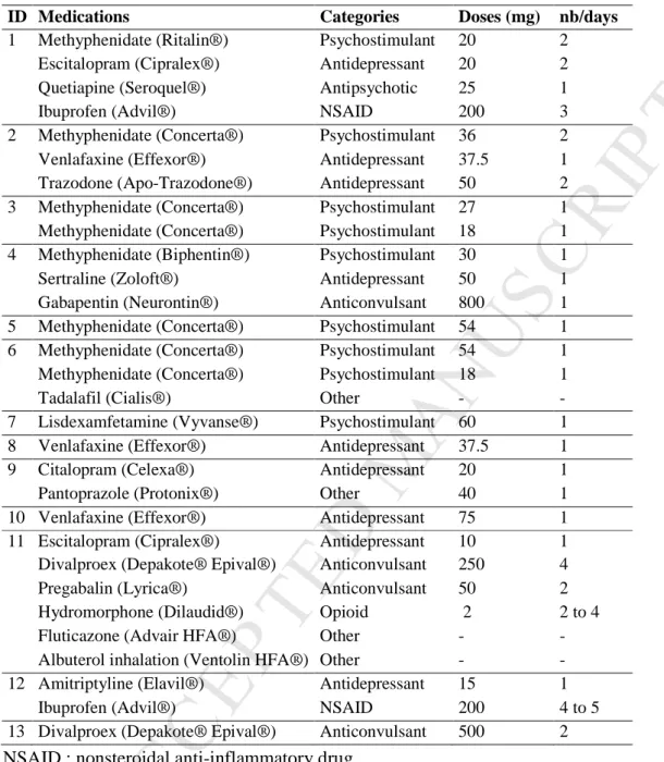
Based on our inclusion and exclusion criteria, 120 patients were contacted by phone (see Figure 1). Based on the telephone interview, participants were excluded if they presented: (1) daily use of hypnotic medication; (2) a self-reported body mass index > 40; 3) pregnancy; 4) jet lag due to trans-meridian travel in the month preceding the study or night work or shift work, leading to an atypical sleep schedule; or 5) inability to communicate in French or English. Inability to reach participants and eligibility assessment performed in the phone interview excluded 70 subjects. Among the 50 remaining subjects, 40 agreed to participate in our study. Thirteen of these patients were using psychoactive medication (see Table 4) at the time of the study.
For comparison, a group of 38 healthy controls having a similar age range and sex distribution that TBI group was recruited through local advertisements and newspaper ads. We excluded subjects with a self-history of diagnosed TBI (for the control group only), psychiatric, neurologic or sleep disorders. The same criteria were used for TBI subjects; although, in TBI subjects, we also had access to their medical records (allowing confirmation of self-reported medical history). Control and TBI subjects with clinically significant scores on questionnaires were not excluded, as we wanted to apply the same criteria in both groups. This study was approved by the Research Ethics Board of the institution and written informed consent was obtained from each participant.
2.2 Overview of the protocol
Recruitment of participants and data collection were performed by the research team. Participants were first asked to wear a wrist actigraphy and to fill out a sleep diary for seven consecutive days at home. All subjects were then admitted to the sleep laboratory for a
one-M
AN
US
CR
IP
T
AC
CE
PT
ED
7night polysomnographic recording to assess sleep disorders. We then conducted a structured interview and a series of questionnaires to assess subjective SWD, TBI outcome, and current symptomatology.
2.3 Subjective measures of sleep-wake disturbances
The Epworth Sleepiness Scale (ESS) [22], the Fatigue Severity Scale (FSS) [23] and the Pittsburgh Sleep Quality Index (PSQI) [24], were used to assess symptoms of daytime sleepiness, fatigue, and sleep quality, respectively. Higher scores on any of these scales or index indicate higher levels of symptomatology.
A seven day sleep diary provided information on the sleep schedules (bedtime and rise time) for nighttime sleep and daytime naps, on daily medications, and on the presence of personal obligations in the morning (eg, childcare, work, school, medical appointments).
2.4 Objective measures of sleep-wake disturbances
Participants wore an actigraph device (Actiwatch Spectrum or Actiwatch-L, Philips-Respironics, Andover, MA) on their non-dominant wrist for seven consecutive days to measure their sleep-wake patterns. The actigraph measures physical motion in all direction in 1-min epochs. The device concurrently records light exposure. Participants were instructed to press a button on the actigraph to indicate when they laid down at any time during the day for sleep, and when they rose from bed. For each day of recording, we manually determined intervals of sleep first using the sleep diary, but when sleep information was deemed invalid (eg, the participant forgot to enter information), sleep episodes were determined according to button presses, level of motor activity and changes in light intensity [25].
For each identified nighttime and daytime sleep episode, each minute of activity data was scored as “wake” or “sleep” by the dedicated software (Actiware, version 6.0.8, Philips-Respironics), using a medium wake threshold (40 activity counts). We used the “wake threshold 40 counts” because of its sensitivity (capacity to detect sleep), specificity (capacity
M
AN
US
CR
IP
T
AC
CE
PT
ED
8to detect wakefulness) and accuracy to measure the rest-activity cycle, as showed among healthy individuals and clinical TBI population [26,27]. The duration of an identified sleep episode corresponds to the total time spent in bed, and total sleep time corresponds to the number of minutes scored “sleep” during the sleep episode. For the main nocturnal sleep episode, sleep efficiency ([percentage of time asleep] / [duration of time spent in bed]) was also computed as an objective estimate of sleep quality. For naps, time spent in bed and total sleep time was also computed, and the percentage of days with naps was calculated manually.
2.5 TBI severity and outcomes
TBI severity was assessed with data from the medical charts (ie, GSC upon admission, duration of post-traumatic amnesia, duration of hospital stay). To assess overall trauma severity of patients with multiple injuries, we computed the Injury Severity Score (ISS) [28], an established medical scale that scores each injury in different body regions (head, face, chest, abdomen, extremities, and external). The Glasgow Outcome Scale Extended (GOSE) [29] administered during the structured interview was used to evaluate functional outcome (3 = lower severe disabilities, 4 = upper severe disabilities, 5 = lower moderate disabilities, 6 = upper moderate disabilities, 7 = lower good recovery, 8 = upper good recovery). The Beck Anxiety Inventory (BAI) [30] and the Beck Depression Inventory-second revision (BDI-II) [31] were used to assess symptoms of anxiety and depression, respectively. These questionnaires have also been previously validated in individuals with moderate to severe TBI [32,33]. Finally, the mean pain intensity experienced over the last week was measured using a pain Numeric Scale (NS) ranging from 0 (no pain) to 10 (worst pain ever felt).
2.6 Statistical analyses
To investigate subjective and objective SWD during chronic TBI, we assessed group differences between TBI participants and controls using Student t-tests when normality assumption was respect and Mann-Whitney tests when normality assumption was not
M
AN
US
CR
IP
T
AC
CE
PT
ED
9appropriate for quantitative data. Chi-square (χ2) tests were used to assed group differences for nominal data. Pearson correlations were performed in the TBI group to test the association between subjective and objective estimates of sleep. We also used Pearson correlations to assess the association of subjective and objective sleep-wake measures with clinical variables of injury severity and chronic comorbidities after TBI.
To test whether the use of psychoactive medication was associated with daytime sleepiness, fatigue, time spent in bed and total sleep time, TBI participants were divided into medicated and non-medicated groups. We assessed group differences between medicated TBI participants, non-medicated TBI participants and controls using analyses of variance (ANOVA) for quantitative data, and χ2 tests for nominal data. To determine between-group differences in the case of significant ANOVA, Tukey HSD post-hoc comparisons tests were used. Analyses were performed using SPSS (SPSS Inc., Chicago, IL) and statistical significance was set at p < 0.05.
3 Results
3.1 Patient characteristics
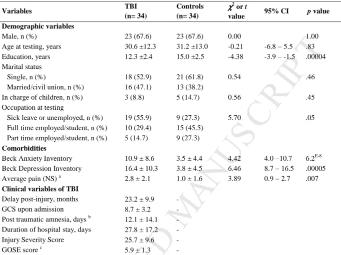
Six TBI participants and four controls were excluded from the initial sample due to artefacts in actigraphic recordings or failure to consistently wear the actigraph, leaving 34 participants in each group for analyses. Table 1 shows participants’ characteristics and statistics. TBI and control groups were comparable on age, sex, and marital status. However, compared to healthy controls, TBI participants had less years of education. TBI participants were tested on average 23.2 ± 9.9 months following the injury. Most TBI participants had recovered sufficiently to carry out activities of daily living without supervision (eg, toileting and self-care, preparing food, driving), and to reintegrate social activities as reflected by GOSE scores (range 4 - 8). However, 55.9% of TBI participants were on sick leave or unemployed. The TBI group reported greater levels of anxiety and depression than controls.
M
AN
US
CR
IP
T
AC
CE
PT
ED
10Clinically significant symptoms of anxiety (Beck Anxiety Inventory above 9) was reported by 52% (18/34) of TBI patients vs. 11% (4/34) of controls, and significant symptoms of depression (Beck Depression Inventory above 13) was reported by 67% (23/34) of TBI participants versus 2% (1/34) of controls. Self-reported pain level was also greater in TBI group compared to controls. Two of the medicated participants (ID 9 and 11 in Table 4 and Figure 2) had an apnea-hypopnea index of 20.8/h and 17.2/h. Sleep diary was missing or incomplete for 14.7% of the controls and 23.5% of the TBI participants.
3.2 Group differences on subjective measures of sleep-wake disturbances
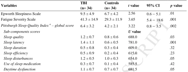
Results and statistics for sleep-wake questionnaires are presented in Table 2. The TBI group reported greater levels of daytime sleepiness and fatigue than controls. As shown by higher scores on the PSQI, global quality of sleep was significantly poorer in TBI participants compared to controls. More specifically, scores on sub-components of the index indicate that the TBI group complained of delayed sleep onset and lower sleep quality, and experienced sleep disturbances and daytime dysfunction more frequently than controls.
3.3 Group differences on objective measures of sleep-wake disturbances
Results and statistics for actigraphy are presented in Table 3. The percentage of days without morning obligations was almost identical in the two groups: 56.9 ± 32.3% in TBI patients compared to 54.4 ± 33.2% in controls (t53 = 2.81; 95% CI = -15.3 − 20.2; p = 0.7).
Therefore, all analyses were performed by combining days with and without occupational obligations.
We found that TBI participants had earlier bedtime, longer time spent in bed during the nighttime episode and more frequent daytime naps. Compared to controls, TBI patients also spent more time in bed and accumulated more time asleep when all sleep episodes over the 24 h period were considered. No group differences were found for sleep efficiency.
M
AN
US
CR
IP
T
AC
CE
PT
ED
113.4 Associations between subjective and objective sleep-wake measures in the TBI group
We then investigated whether sleep, fatigue and sleepiness complaints measured with questionnaires were associated with objective sleep-wake patterns measured with actigraphy in the TBI group. The percentage of days with naps was associated with the ESS and the FSS, such that a greater percentage of days with naps was associated with higher levels of sleepiness (r = 0.36, p = 0.03) and fatigue (r = 0.31, p = 0.03). We also found that TBI participants who spent more time asleep per 24 h tended to report more fatigue (r = 0.31, p = 0.06), as per the FSS. No other significant associations were found between subjective and objective sleep-wake measures.
3.5 Associations between sleep-wake measures and severity of traumatic injuries Correlations were performed between sleep-wake measures and TBI characteristics in order to identify clinical variables that could be associated with higher risk of SWD. Two clinical variables measured in the acute stage of injury were associated with SWD in the chronic stage: the Injury Severity Score and the hospital stay duration. Higher Injury Severity Scores were associated with more fatigue (r = 0.44; p = 0.009), more sleepiness (r = 0.34, p = 0.04), a higher proportion of days with a nap (r = 0.38, p = 0.02) and more total sleep time per 24 h (r = 0.41, p = 0.01). Similarly, longer hospital stay duration was associated with more fatigue (r = 0.59; p = 0.007) and higher proportion of days with a nap (r = 0.48, p = 0.004).
3.6 Associations between sleep-wake measures and comorbidities in the TBI group Overall, subjective measures of fatigue were significantly correlated with psychological symptoms (eg, anxiety and depression), while pain was associated with a poorer subjective sleep quality. Significant correlations were found between fatigue and mood questionnaires, such that increased levels of fatigue were associated with more severe symptoms of anxiety (r = 0.41; p = 0.01) and depression (r = 0.47; p = 0.005). We also observed an association
M
AN
US
CR
IP
T
AC
CE
PT
ED
12between PSQI (global score) and pain intensity (r = 0.67; p = 0.00001), such that TBI patients with greater pain complained of worse sleep quality.
No associations were found between objective measures of sleep and wakefulness measured with actigraphy (ie, nocturnal, daytime and the 24 h period sleep analyses) and comorbidity variables measured with questionnaires (ie, anxiety, depression, and pain).
3.7 Sleep-wake disturbances in medicated versus non-medicated TBI participants Of the 34 TBI participants, 13 were using psychoactive medication on a regular basis at time of testing. All started their treatment after occurrence of the TBI. Details of drug categories for each participant are presented in Table 4.
More TBI participants who used medication were unemployed (11/13) compared to non-medicated TBI participants (8/21), but they did not differ significantly from TBI participants without medication and from controls for the percentage of days without morning obligations (71.9 ± 29.4% in medicated TBI, 49.0 ± 32.5% in non-medicated TBI, and 54.4 ± 33.2% in controls, F2,52 = 1.52; p = 0.22). Compared to TBI participants without medication, TBI
participants with medication had a longer hospital stay in the acute stage (t30= 2.74; 95% CI =
3.9 – 29.3; p = 0.01), a higher Injury Severity Score (t32 = 2.21; 95% CI = 0.6 – 13.6; p =
0.03) and lower GOSE scores (t30= -2.56; 95% CI = -1.9 − -0.2; p = 0.01). Regarding
co-morbidities, there was no significant difference between medicated and non-medicated TBI participants for the measures of depression, anxiety or pain level.
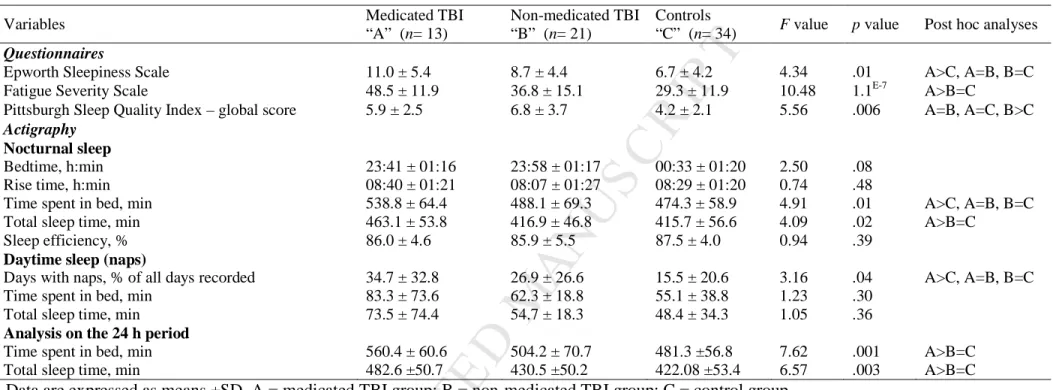
Table 5 presents the comparisons between medicated TBI participants, non-medicated TBI participants and controls for subjective and objective measures of SWD. For subjective measures, reported fatigue level was higher in medicated TBI participants compared to non-medicated TBI participants (post-hoc test, p = 0.03) and compared to controls (post-hoc test, p = 0.00007). However, reported level of fatigue in TBI participants without medication did not differ from that of controls.
M
AN
US
CR
IP
T
AC
CE
PT
ED
13For objective measures, total sleep time during nighttime period was higher in medicated TBI participants when compared to both non-medicated TBI participants (post-hoc test, p = 0.02) and controls (post-hoc test, p = 0.04). Similarly, a significant group effect was found for total time asleep per 24 h and total time spent in bed per 24 h; post-hoc analyses showed that medicated TBI participants had longer total sleep time and time spent in bed per 24 h than both the non-medicated TBI participants (p = 0.01 and p = 0.03 respectively) and controls (p = 0.002 and p = 0.001 respectively) (Figure 2a and 2b respectively). We also observed a
group effect on the percentage of days with naps, which showed that medicated TBI participants tended to take more naps than controls (p = 0.05). Again, TBI participants without medication did not differ from controls.
4 Discussion
The first objective of our study was to evaluate SWD using subjective and objective measures in adults during the chronic phase of a moderate to severe TBI. Our results confirmed previous findings that showed more complaints of fatigue, sleepiness, and poorer sleep quality in the TBI population compared to non-TBI adults [1-3,34]. Despite a significantly lower subjective sleep quality, TBI participants had a total sleep time and sleep-efficiency during nighttime nearly identical to that of control participants, which is in line with Sinclair et al., findings [13]. These results are in contradiction with those reported in two previous studies where they found increased total sleep time, but no significant complaints of fatigue and sleepiness in TBI subjects compared to controls [8,11]. Differences between studies regarding subjective fatigue/sleepiness complaints and objective sleep measures could be due to the heterogeneity of the TBI population in terms of injury severity, recovery, psychoactive medication used and comorbidities. Although it is noteworthy that our group subjects were comparable to those included in previous studies in terms of self-reported sleep-wake and mood values [1,8,11,13,17,19].
M
AN
US
CR
IP
T
AC
CE
PT
ED
14The most striking difference between TBI participants and controls in our study was that TBI participants spent more time in bed over a 24 h period, due to an earlier bedtime and a higher frequency of daytime naps. The significant association between frequency of naps and scores of fatigue and sleepiness suggests that our TBI survivors attempted to relieve fatigue and sleepiness by increasing their sleep time. This strategy was partially successful for increasing time asleep: by spending an additional 45 min in bed, TBI participants were able to sleep on average 28 min more that controls on the 24 h period. However, reporting daytime impairment, spending more time in bed and adopting an early bedtime may delay sleep onset at night, which are all consistent with insomnia symptoms [35]. The longer sleep latency reported by TBI participants on questionnaires, coupled with a lack of feeling refreshed after sleeping, may explain their dissatisfaction with sleep quality. Our findings suggest that monitoring time spend in bed over the 24 h period could be of clinical interest to quantify sleep effort and insomnia-related symptoms following TBI. However time spent in bed on the 24 h period was not reported in previous prospective studies using actigraphy [8,11,13].
If daytime fatigue and sleepiness are not related to sleep patterns, they could result from perturbations in wake regulation. Potential pathophysiological mechanisms include a reduction in the secretion of the wake-promoting neurotransmitter hypocretin, which may increase sleepiness and total sleep time during the acute stage of TBI, and contribute to maintaining this sleep-wake pattern during the chronic stage of TBI [15,36]. Furthermore, the study of post-mortem brain tissue of TBI patients has revealed an important loss of wake-maintaining histamine neurons located in the posterior hypothalamus [37,38]. Therefore, higher daytime sleepiness may reflect a physiological difficulty maintaining wakefulness rather than an increased need for sleep.
The second objective of this study was to investigate whether comorbidities and injury severity were associated with increased SWD. Our findings indicate that subjective
M
AN
US
CR
IP
T
AC
CE
PT
ED
15complaints of fatigue, sleepiness and low sleep quality are strongly associated with a high degree of distress (anxiety, depression, pain), which is in line with previous studies [1,2,39]. Conversely, the level of distress was not associated with actigraphic measures of sleep. We cannot, however, rule out the possibility that mood disturbances or pain were associated with more subtle measures of sleep fragmentation, as previously observed in TBI studies using polysomnography [18,19]. It is well established that SWD have a strong two-way relationship with mood disorders [40,41] and with medical conditions producing pain [42-44], each worsening the symptoms of the other. Therefore, interventions aiming at improving sleep hygiene (eg, spending less time in bed when not sleeping), and acquisition of better coping strategies to reduce fatigue and sleepiness could not only improve sleep, vigilance and energy levels, but also mood and pain symptoms in the TBI population. Cognitive-behavioral therapy [45,46], melatonin administration [47] and blue light therapy [48] all showed promising results in improving subjective sleep quality and reducing fatigue and sleepiness.
Factors other than common TBI comorbidities may also explain SWD. In the present study, both the overall severity score of injuries and longer hospital stays were associated with increased subjective fatigue and sleepiness, as well as with more napping and increased total time asleep per 24 h. However, correlation analyses failed to demonstrate significant associations between the severity of the TBI itself (duration of PTA, GCS score) and sleep-wake complaints or measures. This finding is in agreement with previous studies, in which no relationship was found between TBI severity and sleep duration more than one-year post-TBI [8,11,13]. The exploratory analyses of SWD in the 13 TBI participants who were taking psychoactive medications showed that this sub-group sustained greater overall injury severity, and accounted for the majority of differences between TBI adults and controls for fatigue, sleepiness, total sleep time and time spent in bed. Indeed, participants taking psychoactive medication slept an average of 60.5 min more than controls over the 24 h period. This
M
AN
US
CR
IP
T
AC
CE
PT
ED
16increased time asleep was not observed in TBI participants who were not taking psychoactive medication. Moreover, despite that medicated TBI participants tended to have fewer morning obligations than controls, the same results were found when only days without obligations were analysed (data not shown). When comparing our results with those reported in previous studies, we found interesting similarities. For example, medicated TBI subjects had a total time asleep of 8.0 ± 0.8 h per 24 h, while previous studies reported 8.1 ± 0.5 h in TBI subjects (whose medication status at time of testing was not reported) [7,11]. However, in the present study, total sleep time for TBI participants who did not use psychoactive medication was similar to what was observed in controls of our study and one previous study (Imbach et al., [11]: 7.1 ± 0.7 h vs. our study: 7.0 ± 0.8 h). In the study by Sinclair et al., (where psychoactive medication was taken by a part of their TBI participants), no significant increase in total sleep time per 24 h was found in their TBI participants compared to their controls [13].
Based on our findings, it is plausible that studies using more strict exclusion criteria (ie, excluding TBI participants taking psychoactive medications) may fail to observe an increased sleep duration following TBI, while those using more liberal criteria would be more likely to observe this sleep pattern. Our study raises questions regarding the inclusion of those participants in studies investigating the consequences of TBI, as these persons represent a significant proportion of all moderate to severe TBI, but may differ from non-medicated TBI adults in terms of functional outcome. Studies excluding TBI survivors taking medications can miss out on an important reality experienced by TBI adults and observed by clinicians. Yet, mixing participants using and not using medications can blur the interpretation of results. We therefore suggest that future studies clearly describe medications taken by participants and perform separate analyses for medication users and non-users.
M
AN
US
CR
IP
T
AC
CE
PT
ED
17The present study had certain limitations. Although our findings indicated normal sleep duration and sleep efficiency among TBI participants, we cannot exclude the possibility of more subtle nighttime sleep disturbances. Indeed, certain characteristics associated with the recuperative aspects of sleep (eg, slow waves, microarousals, percentage of specific sleep stages, etc.) can only be assessed using polysomnography. We also cannot exclude the possibility that the medication itself may have negative impacts on the sleep quality and, consequently, affect daytime fatigue and vigilance. In addition, while we agree that it is essential to control the possible effects of psychoactive medication on outcome measures, the small sample of medicated patients (n=13) allowed us to establish only preliminary associations between pharmacological treatment and change in sleep pattern following TBI. Exclusion of patients with daily use of hypnotics may have reduced the differences observed between TBI participants and controls on sleep-wake measures. Moreover, having excluded patients with comorbidities (eg, diagnosed mood disorders, substance abuse) may reduce the generalizability of our results. Finally, missing data may not be at random, and may have impacted our statistics.
5 Conclusion
No objective trace of SWD could be found in our sample of moderate to severe TBI adults, despite the fact that they report high levels of daytime sleepiness and fatigue as well as low sleep quality. Nevertheless, our results show that individuals with TBI spend more time in bed and sleep slightly more, most probably in an effort to decrease daytime sleepiness and fatigue. It is important to highlight that daytime complaints and increased sleep duration were mainly found in TBI survivors using psychoactive medications. Therefore, our results demonstrate that taking into account the use of psychoactive medications during both recruitment and data analysis may be of prime importance in future studies that aim to understand sleep and daytime drowsiness complaints during the chronic stage of TBI.
M
AN
US
CR
IP
T
AC
CE
PT
ED
18 FundingThis study was supported by research grants to N.G. from the Canadian Institutes of Health Research (#FDN154291 and #MOP115172) and from the Consortium pour le développement de la recherche en traumatologie du Québec (#34851). N.G. and J.C. receive salary awards from the Fonds de la recherche du Québec – Santé. E.S. receives a studentship from the Canadian Institutes of Health Research.
Conflicts of interest
The authors declare that they have no conflicts of interest in relation to this work. Figure legends
Figure 1. Flow chart presenting patient recruitment and inclusion in the study
Figure 2. Total sleep time (A) and time spent in bed (B) on the 24 h period for TBI participants and controls (med+: with medication, med-:without medication). Medicated TBI are identified by their number ID (cf. Table 4). No group difference was found between TBI med- and Controls.
M
AN
US
CR
IP
T
AC
CE
PT
ED
19 References1. Ponsford, J.L., D.L. Parcell, K.L. Sinclair, et al., Changes in sleep patterns following traumatic brain injury: a controlled study. Neurorehabilitation and neural repair, 2013. 27(7): p. 613-21.
2. Parcell, D.L., J.L. Ponsford, S.M. Rajaratnam, et al., Self-reported changes to nighttime sleep after traumatic brain injury. Arch Phys Med Rehabil, 2006. 87(2): p. 278-85.
3. Cantor, J.B., T. Ashman, W. Gordon, et al., Fatigue after traumatic brain injury and its impact on participation and quality of life. J Head Trauma Rehabil, 2008. 23(1): p. 41-51.
4. Duclos, C., M. Dumont, C. Wiseman-Hakes, et al., Sleep and wake disturbances following traumatic brain injury. Pathol Biol (Paris), 2014. 62(5): p. 252-61. 5. Ouellet, M.C. and C.M. Morin, Cognitive behavioral therapy for insomnia associated
with traumatic brain injury: a single-case study. Arch Phys Med Rehabil, 2004. 85(8): p. 1298-302.
6. Kempf, J., E. Werth, P.R. Kaiser, et al., Sleep-wake disturbances 3 years after traumatic brain injury. J Neurol Neurosurg Psychiatry, 2010. 81(12): p. 1402-5. 7. Imbach, L.L., P.O. Valko, T. Li, et al., Increased sleep need and daytime sleepiness 6
months after traumatic brain injury: a prospective controlled clinical trial. Brain, 2015. 138(Pt 3): p. 726-35.
8. Sommerauer, M., P.O. Valko, E. Werth, et al., Excessive sleep need following traumatic brain injury: a case-control study of 36 patients. J Sleep Res, 2013. 22(6): p. 634-9.
9. Cantor, J.B., T. Bushnik, K. Cicerone, et al., Insomnia, fatigue, and sleepiness in the first 2 years after traumatic brain injury: an NIDRR TBI model system module study. The Journal of head trauma rehabilitation, 2012. 27(6): p. E1-14.
10. Theadom, A., V. Rowland, W. Levack, et al., Exploring the experience of sleep and fatigue in male and female adults over the 2 years following traumatic brain injury: a qualitative descriptive study. BMJ Open, 2016. 6(4): p. e010453.
11. Imbach, L.L., F. Buchele, P.O. Valko, et al., Sleep-wake disorders persist 18 months after traumatic brain injury but remain underrecognized. Neurology, 2016. 86(21): p. 1945-9.
12. Grima, N., J. Ponsford, S.M. Rajaratnam, et al., Sleep Disturbances in Traumatic Brain Injury: A Meta-Analysis. J Clin Sleep Med, 2016. 12(3): p. 419-28.
13. Sinclair, K.L., J. Ponsford, and S.M. Rajaratnam, Actigraphic assessment of sleep disturbances following traumatic brain injury. Behavioral sleep medicine, 2014. 12(1): p. 13-27.
14. Suzuki, Y., S. Khoury, H. El-Khatib, et al., Individuals with pain need more sleep in the early stage of mild traumatic brain injury. Sleep Medicine. 33: p. 36-42.
15. Baumann, C.R., E. Werth, R. Stocker, et al., Sleep-wake disturbances 6 months after traumatic brain injury: a prospective study. Brain : a journal of neurology, 2007. 130(Pt 7): p. 1873-83.
16. Wiseman-Hakes C, M.B., Mollayeva T, Gargaro J, Colantonio A, A Profile of Sleep Architecture and Sleep Disorders in Adults with Chronic Traumatic Brain Injury. J Sleep Disord Ther, 2016. 5(1).
17. Beaulieu-Bonneau, S. and C.M. Morin, Sleepiness and fatigue following traumatic brain injury. Sleep Med, 2012. 13(6): p. 598-605.
M
AN
US
CR
IP
T
AC
CE
PT
ED
2018. Parcell, D.L., J.L. Ponsford, J.R. Redman, et al., Poor sleep quality and changes in objectively recorded sleep after traumatic brain injury: a preliminary study. Arch Phys Med Rehabil, 2008. 89(5): p. 843-50.
19. Shekleton, J.A., D.L. Parcell, J.R. Redman, et al., Sleep disturbance and melatonin levels following traumatic brain injury. Neurology, 2010. 74(21): p. 1732-8.
20. Verma, A., V. Anand, and N.P. Verma, Sleep disorders in chronic traumatic brain injury. J Clin Sleep Med, 2007. 3(4): p. 357-62.
21. Menon, D.K., K. Schwab, D.W. Wright, et al., Position statement: definition of traumatic brain injury. Arch Phys Med Rehabil, 2010. 91(11): p. 1637-40.
22. Johns, M.W., A new method for measuring daytime sleepiness: the Epworth sleepiness scale. Sleep, 1991. 14(6): p. 540-5.
23. Krupp, L.B., N.G. LaRocca, J. Muir-Nash, et al., The fatigue severity scale. Application to patients with multiple sclerosis and systemic lupus erythematosus. Arch Neurol, 1989. 46(10): p. 1121-3.
24. Buysse, D.J., C.F. Reynolds, 3rd, T.H. Monk, et al., The Pittsburgh Sleep Quality Index: a new instrument for psychiatric practice and research. Psychiatry Res, 1989. 28(2): p. 193-213.
25. Boyne, K., D.D. Sherry, P.R. Gallagher, et al., Accuracy of computer algorithms and the human eye in scoring actigraphy. Sleep Breath, 2013. 17(1): p. 411-7.
26. Kamper, J.E., J. Garofano, D.J. Schwartz, et al., Concordance of Actigraphy With Polysomnography in Traumatic Brain Injury Neurorehabilitation Admissions. J Head Trauma Rehabil, 2016. 31(2): p. 117-25.
27. Martin, J.L. and A.D. Hakim, Wrist actigraphy. Chest, 2011. 139(6): p. 1514-27. 28. Baker, S.P., B. O'Neill, W. Haddon, Jr., et al., The injury severity score: a method for
describing patients with multiple injuries and evaluating emergency care. J Trauma, 1974. 14(3): p. 187-96.
29. Wilson, J.T., L.E. Pettigrew, and G.M. Teasdale, Structured interviews for the Glasgow Outcome Scale and the extended Glasgow Outcome Scale: guidelines for their use. J Neurotrauma, 1998. 15(8): p. 573-85.
30. Beck, A.T., R.A. Steer, R. Ball, et al., Comparison of Beck Depression Inventories -IA and -II in psychiatric outpatients. J Pers Assess, 1996. 67(3): p. 588-97.
31. Beck, A.T., Sterr R.A., Brown, G.K., Beck depression inventory: Manual. 2nd ed. 1996, Boston, MA: Harcourt Brace.
32. Mollayeva, T., T. Kendzerska, and A. Colantonio, Self-report instruments for assessing sleep dysfunction in an adult traumatic brain injury population: A systematic review. Sleep Medicine Reviews, 2013. 17(6): p. 411-423.
33. Ziino, C. and J. Ponsford, Measurement and prediction of subjective fatigue following traumatic brain injury. J Int Neuropsychol Soc, 2005. 11(4): p. 416-25. 34. Ponsford, J.L., C. Ziino, D.L. Parcell, et al., Fatigue and sleep disturbance following
traumatic brain injury--their nature, causes, and potential treatments. J Head Trauma Rehabil, 2012. 27(3): p. 224-33.
35. American Academy of Sleep Medicine Board of, D., ed. International Classification of Sleep Disorders. Third ed. Darien, IL 2014.
36. Baumann, C.R., C.L. Bassetti, P.O. Valko, et al., Loss of hypocretin (orexin) neurons with traumatic brain injury. Ann Neurol, 2009. 66(4): p. 555-9.
37. Valko, P.O., Y.V. Gavrilov, M. Yamamoto, et al., Damage to histaminergic tuberomammillary neurons and other hypothalamic neurons with traumatic brain injury. Ann Neurol, 2015. 77(1): p. 177-82.
M
AN
US
CR
IP
T
AC
CE
PT
ED
2138. Valko, P.O., Y.V. Gavrilov, M. Yamamoto, et al., Damage to Arousal-Promoting Brainstem Neurons with Traumatic Brain Injury. Sleep, 2016. 39(6): p. 1249-52. 39. Ouellet, M.C., S. Beaulieu-Bonneau, and C.M. Morin, Insomnia in patients with
traumatic brain injury: frequency, characteristics, and risk factors. J Head Trauma Rehabil, 2006. 21(3): p. 199-212.
40. Kahn, M., G. Sheppes, and A. Sadeh, Sleep and emotions: bidirectional links and underlying mechanisms. International Journal of Psychophysiology, 2013. 89(2): p. 218-228.
41. Mayers, A.G. and D.S. Baldwin, The relationship between sleep disturbance and depression. International Journal of Psychiatry in Clinical Practice, 2006. 10(1): p. 2-16.
42. Lautenbacher, S., B. Kundermann, and J.-C. Krieg, Sleep deprivation and pain perception. Sleep medicine reviews, 2006. 10(5): p. 357-369.
43. Morin, C.M. and R. Benca, Chronic insomnia. The Lancet, 2012. 379(9821): p. 1129-1141.
44. Lavigne, G., M.T. Smith, R. Denis, et al., Pain and sleep, in Principles and Practice of Sleep Medicine (Fifth Edition). 2011, Elsevier. p. 1442-1451.
45. Ouellet, M.C. and C.M. Morin, Efficacy of cognitive-behavioral therapy for insomnia associated with traumatic brain injury: a single-case experimental design. Arch Phys Med Rehabil, 2007. 88(12): p. 1581-92.
46. Lu, W., J.W. Krellman, and M.P. Dijkers, Can Cognitive Behavioral Therapy for Insomnia also treat fatigue, pain, and mood symptoms in individuals with traumatic brain injury?–A multiple case report. NeuroRehabilitation, 2016. 38(1): p. 59-69.
47. Grima, N.A., S.M. Rajaratnam, D. Mansfield, et al., Efficacy of melatonin for sleep disturbance following traumatic brain injury: a randomised controlled trial. BMC medicine, 2018. 16(1): p. 8.
48. Sinclair, K.L., J.L. Ponsford, J. Taffe, et al., Randomized controlled trial of light therapy for fatigue following traumatic brain injury. Neurorehabilitation and neural repair, 2014. 28(4): p. 303-313.
M
AN
US
CR
IP
T
AC
CE
PT
ED
Table 1. Sociodemographic and clinical characteristics of TBI and controls participants
Variables TBI (n= 34) Controls (n= 34) χχχχ2 or t value 95% CI p value Demographic variables Male, n (%) 23 (67.6) 23 (67.6) 0.00 1.00
Age at testing, years 30.6 ±12.3 31.2 ±13.0 -0.21 -6.8 − 5.5 .83 Education, years 12.3 ±2.4 15.0 ±2.5 -4.38 -3.9 − -1.5 .00004 Marital status Single, n (%) 18 (52.9) 21 (61.8) 0.54 .46 Married/civil union, n (%) 16 (47.1) 13 (38.2) In charge of children, n (%) 3 (8.8) 5 (14.7) 0.56 .45 Occupation at testing
Sick leave or unemployed, n (%) 19 (55.9) 9 (27.3) 5.70 .05 Full time employed/student, n (%) 10 (29.4) 15 (45.5)
Part time employed/student, n (%) 5 (14.7) 9 (27.3) Comorbidities
Beck Anxiety Inventory 10.9 ± 8.6 3.5 ± 4.4 4.42 4.0 −10.7 6.2E-8 Beck Depression Inventory 16.4 ± 10.3 3.8 ± 4.5 6.46 8.7 − 16.5 .00005 Average pain (NS) a 2.8 ± 2.1 1.0 ± 1.6 3.89 0.9 − 2.7 .007 Clinical variables of TBI
Delay post-injury, months 23.2 ± 9.9 -
GCS upon admission 8.7 ± 3.2 -
Post traumatic amnesia, days b 12.1 ± 14.1 - Duration of hospital stay, days 27.8 ± 17.2 - Injury Severity Score 25.7 ± 9.6 -
GOSE score c 5.9 ± 1.3 -
Data are expressed as means ±SD or frequency (n) and percentage (%). CI = confidence interval of the difference. NS = numeric scale; GCS = Glasgow Coma Scale; GOSE = Glasgow Outcome Scale Extended. Higher scores on questionnaires indicate higher levels of symptomatology.
a
Average pain was missing for 5.8% of the TBI participants.
b
Duration of post-traumatic amnesia was missing for 2.9% of the TBI participants.
c
GOSE score was missing for 5.8% of the TBI participants. No other data was missing.
M
AN
US
CR
IP
T
AC
CE
PT
ED
Table 2. Self-reported sleep-wake measures of TBI and controls participants
Variables TBI
(n= 34)
Controls
(n= 34) t value 95% CI p value Epworth Sleepiness Scale 9.5 ± 4.9 6.7 ± 4.2 2.56 0.6 – 5.1 .01 Fatigue Severity Scale 41.3 ± 14.9 29.3 ± 11.9 3.65 5.4 − 18.6 .001 Pittsburgh Sleep Quality Index a – global score 6.4 ± 3.2 4.2 ± 2.1 3.22 0.8 − 3.5 .002
Sub-components scores U value
Sleep quality 1.2 ± 0.7 0.8 ± 0.6 688.0 .03
Sleep latency 1.4 ± 1.1 0.6 ± 0.5 781.0 .001
Sleep duration 0.5 ± 0.8 0.3 ± 0.4 609.0 .32
Sleep efficiency 0.5 ± 0.9 0.2 ± 0.4 615.0 .23
Sleep disturbances 1.2 ± 0.5 1.0 ± 0.3 654.0 .05
Use of sleep medication 0.3 ± 0.7 0.1 ± 0.4 585.5 .42
Daytime dysfunction 1.1 ± 0.7 0.7 ± 0.7 681.5 .05
Data are expressed as means ±SD. CI = confidence interval of the difference. U value of Mann-Whitney tests. Higher scores on questionnaires indicate higher levels of symptomatology.
a
Pittsburgh Sleep Quality Index was missing for 2.9% of the TBI participants. No other data was missing.
M
AN
US
CR
IP
T
AC
CE
PT
ED
Table 3. Actigraphic measures of TBI and controls participants
Variables TBI (n= 34) Controls (n= 34) t value 95% CI p value Nocturnal sleep Bedtime, h:min 23:52 ± 01:16 00:33 ± 01:20 -2.16 -01:18 − -00:03 .03 Rise time, h:min 08:20 ± 01:25 08:29 ± 01:20 -0.45 -00:49 – 00:30 .64 Time spent in bed, min 507.5 ± 71.06 474.3 ± 58.9 2.09 1.6 – 64.8 .04 Total sleep time, min 434.6 ± 53.8 415.7 ± 56.6 1.40 -7.9 – 45.6 .16 Sleep efficiency, % 86.0 ± 5.1 87.5 ± 4.0 -1.38 -3.8 −0.7 .17 Daytime sleep (naps)
Days with naps, % of all days recorded 29.9 ± 28.9 15.5 ± 20.6 2.36 2.2 – 26.6 .02 Time spent in bed, min 70.5 ± 47.8 55.1 ± 38.8 1.11 -12.7 – 43.5 .27 Total sleep time, min 62.1 ± 47.9 48.4 ± 34.3 1.01 -13.4 – 40.7 .31 Analysis on the 24 h period
Time spent in bed, min 525.7 ± 71.6 481.3 ± 56.8 2.82 13.01 – 75.6 .006 Total sleep time, min 450.4 ± 55.9 422.08 ± 53.4 2.14 1.9 – 54.9 .03 Data are expressed as means ±SD. CI = confidence interval of the difference.
M
AN
US
CR
IP
T
AC
CE
PT
ED
Table 4. Prescribed medications of TBI participants
ID Medications Categories Doses (mg) nb/days
1 Methyphenidate (Ritalin®) Psychostimulant 20 2 Escitalopram (Cipralex®) Antidepressant 20 2 Quetiapine (Seroquel®) Antipsychotic 25 1 Ibuprofen (Advil®) NSAID 200 3 2 Methyphenidate (Concerta®) Psychostimulant 36 2 Venlafaxine (Effexor®) Antidepressant 37.5 1 Trazodone (Apo-Trazodone®) Antidepressant 50 2 3 Methyphenidate (Concerta®) Psychostimulant 27 1 Methyphenidate (Concerta®) Psychostimulant 18 1 4 Methyphenidate (Biphentin®) Psychostimulant 30 1 Sertraline (Zoloft®) Antidepressant 50 1 Gabapentin (Neurontin®) Anticonvulsant 800 1 5 Methyphenidate (Concerta®) Psychostimulant 54 1 6 Methyphenidate (Concerta®) Psychostimulant 54 1 Methyphenidate (Concerta®) Psychostimulant 18 1 Tadalafil (Cialis®) Other - - 7 Lisdexamfetamine (Vyvanse®) Psychostimulant 60 1 8 Venlafaxine (Effexor®) Antidepressant 37.5 1 9 Citalopram (Celexa®) Antidepressant 20 1 Pantoprazole (Protonix®) Other 40 1 10 Venlafaxine (Effexor®) Antidepressant 75 1 11 Escitalopram (Cipralex®) Antidepressant 10 1
Divalproex (Depakote® Epival®) Anticonvulsant 250 4 Pregabalin (Lyrica®) Anticonvulsant 50 2 Hydromorphone (Dilaudid®) Opioid 2 2 to 4 Fluticazone (Advair HFA®) Other - - Albuterol inhalation (Ventolin HFA®) Other - - 12 Amitriptyline (Elavil®) Antidepressant 15 1
Ibuprofen (Advil®) NSAID 200 4 to 5 13 Divalproex (Depakote® Epival®) Anticonvulsant 500 2 NSAID : nonsteroidal anti-inflammatory drug
M
AN
US
CR
IP
T
AC
CE
PT
ED
Table 5. Sleep-wake measures for medicated and non-medicated TBI participants and controls
Variables Medicated TBI
“A” (n= 13)
Non-medicated TBI “B” (n= 21)
Controls
“C” (n= 34) F value p value Post hoc analyses
Questionnaires
Epworth Sleepiness Scale 11.0 ± 5.4 8.7 ± 4.4 6.7 ± 4.2 4.34 .01 A>C, A=B, B=C
Fatigue Severity Scale 48.5 ± 11.9 36.8 ± 15.1 29.3 ± 11.9 10.48 1.1E-7 A>B=C
Pittsburgh Sleep Quality Index – global score 5.9 ± 2.5 6.8 ± 3.7 4.2 ± 2.1 5.56 .006 A=B, A=C, B>C
Actigraphy
Nocturnal sleep
Bedtime, h:min 23:41 ± 01:16 23:58 ± 01:17 00:33 ± 01:20 2.50 .08
Rise time, h:min 08:40 ± 01:21 08:07 ± 01:27 08:29 ± 01:20 0.74 .48
Time spent in bed, min 538.8 ± 64.4 488.1 ± 69.3 474.3 ± 58.9 4.91 .01 A>C, A=B, B=C
Total sleep time, min 463.1 ± 53.8 416.9 ± 46.8 415.7 ± 56.6 4.09 .02 A>B=C
Sleep efficiency, % 86.0 ± 4.6 85.9 ± 5.5 87.5 ± 4.0 0.94 .39
Daytime sleep (naps)
Days with naps, % of all days recorded 34.7 ± 32.8 26.9 ± 26.6 15.5 ± 20.6 3.16 .04 A>C, A=B, B=C
Time spent in bed, min 83.3 ± 73.6 62.3 ± 18.8 55.1 ± 38.8 1.23 .30
Total sleep time, min 73.5 ± 74.4 54.7 ± 18.3 48.4 ± 34.3 1.05 .36
Analysis on the 24 h period
Time spent in bed, min 560.4 ± 60.6 504.2 ± 70.7 481.3 ±56.8 7.62 .001 A>B=C
Total sleep time, min 482.6 ±50.7 430.5 ±50.2 422.08 ±53.4 6.57 .003 A>B=C
M
AN
US
CR
IP
T
AC
CE
M
AN
US
CR
IP
T
AC
CE
M
AN
US
CR
IP
T
AC
CE
PT
ED
Title: Towards a better understanding of increased sleep duration in the chronic phase of moderate to severe traumatic brain injury
• TBI adults complain of significant sleepiness, fatigue and poorer sleep than controls. • No trace of sleep disturbances were found with actigraphy in TBI group.
• TBI adults spent more time in bed to increase time asleep and decrease drowsiness. • Daytime complaints were associated with more severe injuries and comorbidities. • Medicated TBI adults present more evidences of sleep-wake change.


