Université de Montréal
Development of New Radiotracers for PET Imaging of
Adrenomedullin and Angiotensin II Type 1 Receptors
par Luis Michel Alonso Martinez
Département de pharmacologie et physiologie
Institut de génie biomédical – Faculté de médecine
Thèse présentée en vue de l’obtention du grade de Philosophiae doctor (Ph.D.) –
Doctorat en génie biomédical
Mai 2020
Development of New Radiotracers for PET Imaging of Adrenomedullin and Angiotensin
II Type 1 Receptors
par Luis Michel Alonso Martinez
Thèse évaluée par un jury composé des personnes suivantes:
Prof. Frédéric Lesage, Président-rapporteur
Prof. Jean N. DaSilva, Directeur de recherche
Prof. François Harel, Codirecteur
Prof. André Charette, Membre du jury
Prof. Brigitte Guérin, Examinateur externe
Prof. René Cardinal, Représentant de la doyenne
i
Résumé
Les récepteurs de l'adrénomédulline sont fortement exprimés dans les capillaires alvéolaires humains et fournissent une cible moléculaire pour l'imagerie de la circulation et de l'embolie pulmonaire. Au cours des années précédentes, le dérivé DFH12 marqué au 99mTc
(PulmoBind) a démontré son potentiel en tant qu'agent d'imagerie SPECT de l'hypertension pulmonaire dans des études cliniques de phase I et II. L’objectif principal de mon projet est de développer le nouvel analogue DFH17 pour l’imagerie TEP des récepteurs de l'adrénomédulline via la méthode de l’Al18F. Pour atteindre cet objectif, un système d’élution semi-automatique a été
conçu pour produire l’Al18F concentré directement dans le vial de réaction. En utilisant des tests
de complexation avec le chélateur NOTA, des conditions optimales ont été trouvées pour le radiomarquage du DFH17 avec l’Al18F. La combinaison du rapport Al/DFH17 1:3 dans l'éthanol
50% a permis de produire le [18F]AlF-DFH17 avec des puretés radiochimiques et chimiques
élevées. Les études TEP avec le [18F]AlF-DFH17 ont démontré un rapport élevé poumon-bruit de
fond ainsi qu’une grande stabilité in vivo chez le rat, le chien et le primate. Des captations différenciées dans les poumons des trois espèces ont aussi été détectées par imagerie TEP et leurs différences ont été associées à des variations de la composant RAMP2. Compte tenu de l’importante captation pulmonaire, de la stabilité in vivo et de la dosimétrie favorable, le nouveau dérivé [18F]AlF-DFH17 est un excellent candidat potentiel en tant que traceur TEP des récepteurs
adrénomédulline humains.
L’expression des récepteurs AT1 de l’angiotensine II est altérée dans plusieurs maladies
cardiovasculaires et rénales, telles la défaillance cardiaque, rénale et l’hypertension ainsi que dans certains cancers. Auparavant, le dérivé [11C]méthyl-Candesartan a démontré un potentiel
ii
correspondait à une liaison non-spécifique d'un radiométabolite hydrophobe. Dans ce travail, l’objectif principal est de développer le nouveau dérivé [18F]fluorobenzyl-Candesartan en utilisant
le 4[18F]fluoroiodobenzène ([18F]FIB) avec un profil métabolique et de biodistribution
potentiellement meilleurs. Pour atteindre cet objectif, des paramètres réactionnels de fluorination tels que le solvant, la quantité de précurseur, le catalyseur et la température ont été optimisés permettant la radiosynthèse du [18F]FIB avec des rendements et pureté élevés. Ensuite, le
couplage du [18F]FIB au dérivé alcyne-trityl-Candesartan a été évalué en utilisant la réaction de
Sonogashira suivie d'une détritylation acide. Suite à l’étude de plusieurs conditions de couplage, le rendement de radioconversion a été légèrement augmenté en utilisant le catalyseur Pd(PPh3)4/CuI et K2CO3 comme base. Les meilleures conditions de fluorination et de couplage
ont été automatisées pour le module de synthèse Synthra® RNPlus Research. La production du
[18F]FB-Candesartan été atteinte avec de faibles rendements et activités molaires en raison de la
formation d’impuretés ayant des structures et temps de rétention par HPLC similaires à ceux de notre traceur. Des études supplémentaires afin d'améliorer le rendement, la purification par HPLC et l'activité molaire se sont avérées infructueuses pour l’instant. D’autres expériences devront être effectuées à cette fin. En conclusion, l'utilisation de la réaction de Sonogashira pour produire le [18F]FB-Candersartan avec des rendements et des activités molaires élevées s'est avérée
difficile.
Mots-clés: Al18F, Complexe de NOTA, Récepteurs de l'adrénomédulline, TEP, [18]FIB
groupement prosthétique, Réaction de couplage croisé de Sonogashira, Réaction catalysée par Pd, Radiosynthèse automatisée, AT1R
iii
Abstract
Adrenomedullin receptors are highly expressed in human alveolar capillaries and provide a molecular target for imaging the integrity of pulmonary microcirculation. In previous years, the
99mTc-labeled DFH12 derivative (PulmoBind) demonstrated its potential as a SPECT imaging
agent of pulmonary hypertension in phase I and II clinical trials. In this work, we aimed to develop a NOTA-derivatized adrenomedullin analog (DFH17), radiolabeled with aluminum fluoride ([18F]AlF), for PET imaging of pulmonary microcirculation. To achieve this goal, highly
concentrated [18F](AlF)2+ was produced from purified 18F using a semi-automatic system. Using
inexpensive complexation assays with NOTA, optimal conditions at each step of the process were determined facilitating the radiolabeling optimization of DFH17. Furthermore, combining the Al-to-DFH17 1:3 ratio in 50% ethanol as co-solvent, allowed [18F]AlF-DFH17 production in high
radiochemical and chemical purities. PET/CT and biodistribution demonstrated high [18
F]AlF-DFH17 lung-to-background ratio and in vivo stability in rats, dog and primate. Contrasted inter-species uptake in the lungs associated with variations of RAMP2 were also detected by PET imaging. Considering high lung uptake, in vivo stability and favorable dosimetry observed in the monkey, the novel AM derivative [18F]AlF-DFH17 exhibits an excellent potential as a PET tracer
of human AM receptors.
Alterations of the expression levels of AT1R has been linked to cardiac and renal
diseases, such as cardiac and renal failures, hypertension and some type of cancers. Previously, [11C]methyl-Candesartan displayed potential for PET imaging of AT
1Rs, but a high proportion of
PET signal corresponded to non-specific binding from a 11C-labeled hydrophobic metabolite. In
this work, the main objective was to develop the novel derivative [18F]fluorobenzyl-Candesartan,
iv
([18F]FIB) as prosthetic group. To pursue this goal, radiofluorination parameters such as solvent,
amount of precursor, type of catalyst and temperature were optimized to reliably synthesize [18F]FIB in high yield and purity. Coupling of [18F]FIB to the alkyne-trityl-Candesartan was
evaluated using the Sonogashira cross-coupling reaction followed by an acid deprotection. After studying several Pd-cross-coupling conditions, the radioconversion yield was slightly increased by means of a Pd(PPh3)4/CuI catalyst and K2CO3 as base in DMF. Therefore, the best reaction
conditions for [18F]FIB fluorination and its coupling to alkyne-Candesartan followed by an acid
hydrolysis, was fully automated for Synthra® RNPlus Research synthesis module. In general, the
synthesis of [18F]FB-Candesartan was achieved in low yields and molar activities due to the
formation of structurally-close by-product(s) with similar HPLC retention time. Additional studies to further improve the yield, HPLC purification and molar activity (MA) have been unsuccessful. Other experiments will need to be performed to this end. In conclusion, the use of Sonogashira cross-coupling reaction to produce [18F]FB-Candesartan in high yields and molar activities was
found to be challenging.
Keywords: Al18F, NOTA complex, Adrenomedullin receptors, PET, [18]FIB prosthetic group
v
Note
This document compiles the development of two novel compounds for PET imaging of AM and AT1Rs . The thesis has been written based on scientific articles. Thus, I specify herein my
contribution to each chapter. The Chapter 1 (Introduction), 5 and Annexes were written by myself and edited by Professor Jean N. DaSilva.
The article in Chapter 2 entitled “SPECT and PET imaging of adrenomedullin receptors: a promising strategy for studying pulmonary vascular diseases” extensively reviews all radiotracers for molecular imaging of AM receptors in pulmonary vascular diseases. Previous labeling experiment for primate PET studies were performed by Dr Michael T. Klimas group from Merck Research Laboratories. I carried out the Al18F-formation studies, semi-automatic
production of Al18F and NOTA-complexation assays at the Laboratoire de Radiochimie et
Cyclotron (CRCHUM). I performed together with Dr Harel staff the DFH17 radiolabeling experiments and QC analyses for animal studies at the Research Centre of Montreal Heart Institute. PET imaging processing and quantification was done by Dr Harel’s team with my participation. Al[18F]F-DFH17 PET and dosimetry studied in non-human primate were performed
by Dr Harel’s group in collaboration with Dr Michael T. Klimas. All authors reviewed and agreed this chapter for publication, which was handled by myself and Prof DaSilva.
The Chapter 3 entitled “Al[18F]F-complexation of DFH17, a NOTA-conjugated
adrenomedullin analog, for PET imaging of pulmonary circulation” describes the synthesis, radiolabeling and in vivo evaluation of the novel adrenomedullin derivative DFH17 as a potential PET imaging agent of pulmonary hypertension and pulmonary embolism. DFH17 peptide was designed by Prof. Alain Fournier and Prof. Jocelyn Dupuis and was synthesized and
vi
characterized by Ms. Myriam Létourneau at the Laboratoire D'études Moléculaires et Pharmacologiques des Peptides (all co-authors). I performed all Al18F-formation experiment and
NOTA complexation assays at Laboratoire de Radiochimie et Cyclotron (CRCHUM) and DFH17 radiolabeling, reactions optimization, and QC and Dr Harel’s lab at Montreal Heart Institute. Biodistribution, PET and imaging processing and quantification was done by Dr Harel’s team with my participation. All authors reviewed and agreed this chapter for publication, which was handled by myself and Prof DaSilva.
The Chapter 4 entitled “Development of a novel [18F]fluorobenzyl derivative of the AT 1R
antagonist Candesartan” presents the radiosynthesis and automation of a new potential Candesartan derivative labeled with [18F]FIB using the Sonogashira cross coupling reaction. I
performed all organic synthesis and characterization as well all radiosynthesis, optimization, automation and radioanalytical evaluation at Laboratoire de Radiochimie et Cyclotron (CRCHUM). The chapter was written by myself and subsequently reviewed and edited by Professor DaSilva.
In Chapter 5, a summary and discussion of important aspects not discussed in detail in the manuscripts are presented followed by the conclusion and perspectives of this thesis. These sections were written by me and reviewed by Professors DaSilva and Harel.
vii
Table of Contents:
Résumé ... i
Abstract... iii
Note ... v
List of Figures ... xii
List of Schemes ... xviii
List of Tables ... xix
List of abbreviations ... xx Acknowledgements ... xxiii Chapter 1: Introduction ... 1 1.1 Cyclotron. ... 2 1.2 Production of 18F. ... 4 1.2.1. Targetry ... 4 1.2.2. Production cross-section ... 5 1.2.3. 18F production beam ... 6
1.2.4 18F transfer and recovery ... 7
1.3 Decay of 18F. ... 8 1.4 PET imaging. ...10 1.5 AM receptor radiotracers. ...12 1.6 Chelation chemistry ...13 1.6.1 Aluminum [18F]Fluoride ... 14 1.6.2 NOTA ... 15 1.7 AT1 receptors. ...17 1.8 Candesartan. ...18 1.9 AT1 receptor radiotracers. ...19
1.9.1 Our AT1R Losartan derivative radiotracers ... 22
1.9.2 Structure-activity relationship studies (SAR) justifying Candesartan labeled derivatives ... 24
1.10 Automation of 18F processing and reactions ...25
1.10.1. Semi-automatic system for production of Al18F ... 25
1.10.2. Automatic synthesis module ... 27
1.11 Hypothesis ...29
viii
1.13 Manuscripts ...30
1.14 References ...32
Chapter 2: SPECT/PET imaging of adrenomedullin receptors: a promising strategy for studying pulmonary vascular diseases ...40
2.1 Abstract ...41
2.2 Introduction ...42
2.2.1 Adrenomedullin receptors ... 42
2.2.1 Expression of Adrenomedullin Receptors ... 42
2.2.2 Pulmonary Hypertension ... 43
2.3 SPECT ADRENOMEDULLIN RADIOLIGANDS ...45
2.3.1 Initial Proof of Concept:[ 125I]I-rAM(1-50) ... 45
2.3.2 99mTc-labeled DTPA-AM ... 46
2.3.2.1 Synthesis ... 46
2.3.2.2 In Vitro Evaluation ... 46
2.3.3 99mTc-labeled-AM Through the Free Cys16,21 -SH Functions ((S-[99mTc]Tc-S)-AM) ... 47
2.3.3.1 Synthesis ... 47
2.3.3.2 In Vitro Evaluation ... 49
2.3.3.3 In Vivo Evaluation ... 49
2.3.4 [99mTc]Tc-PulmoBind ... 50
2.3.4.1 Structure-Activity Relationship (SAR) Studies ... 50
2.3.4.2 Synthesis ... 51
2.3.4.3 In Vitro Evaluation ... 52
2.3.4.4 In Vivo Evaluation ... 52
2.3.4.5 Human Studies ... 55
2.4 PET ADRENOMEDULLIN RADIOLIGANDS ...56
2.4.1[ 18F]AlF-DFH17 ... 57 2.4.1.1 Synthesis ... 57 2.4.1.2 In Vivo Evaluation ... 57 2.5 Future perspectives ...60 2.6 Conclusions ...61 2.7 References...63
Chapter 3: Al[18F]F-complexation of DFH17, a NOTA-conjugated adrenomedullin analog, for PET imaging of pulmonary circulation ...68
ix
3.2 Introduction ...70
3.3. Material and methods ...72
3.3.1. General ... 72
3.3.2 Preparation of NOTA-adrenomedullin derivative (DFH17) ... 74
3.3.3. Formation of binary Al18F-complex ... 75
3.3.4. Complexation assay with NOTA chelator ... 75
3.3.5. Radiosynthesis of [18F]AlF-DFH17 ... 76
3.3.5.1. Effect of fluorine-18 activity and Al3+ concentration (Scale-up) ... 77
3.3.5.2. Analytical studies ... 77
3.3.6. Stability tests of [18F]AlF-DFH17 in saline with and without radioprotectant ... 78
3.3.7. PET imaging and biodistribution ... 78
3.3.8. Statistical analysis ... 79
3.4 Results ...80
3.4.1. Preparation of NOTA-adrenomedullin derivative (DFH17) ... 80
3.4.2. Formation of binary Al18F-complex ... 80
3.4.3. Complexation assay with NOTA ... 81
3.4.4. Radiolsynthesis of [18F]AlF-DFH17... 82
3.4.5. Stability tests of [18F]AlF-DFH17 in saline with and without radioprotectant ... 84
3.4.6. PET imaging and biodistribution ... 84
3.5. Discussion...85
3.6. Conclusions ...89
3.7 Acknowledgments ...90
3.8 References...90
Appendix A. Supplementary data ...94
Chapter 4: Development of a novel [18F]fluorobenzyl derivative of the AT 1 Receptor antagonist Candesartan ... 102
4.1 Abstract ... 103
4.2 Introduction ... 104
4.3 Material and methods ... 106
4.3.1. General ... 106
4.3.2 Chemistry ... 107
4.3.2.1 Tetrazole-protected alkyne Candesartan (7-((but-3-yn-1-yloxy)methyl)-2-ethoxy-1-((2'-(1-trityl-1H-tetrazol-5-yl)-[1,1'-biphenyl]-4-yl)methyl)-1H-benzo[d]imidazole) (5) ... 107
x
4.3.2.2 Fluorobenzyl-Candesartan
(1-((2'-(1H-tetrazol-5-yl)-[1,1'-biphenyl]-4-yl)methyl)-2-ethoxy-7-(((4-(4-fluorophenyl)but-3-yn-1-yl)oxy)methyl)-1H-benzo[d]imidazole) (7) ... 108
4.3.3 Radiochemistry ... 109
4.3.3.1 Synthesis of 4-[18F]fluoroiodobenzene ([18F]FIB) ([18F]6) ... 109
4.3.3.2 Optimization of the synthesis of [18F]fluorobenzyl-Candesartan ([18F]7) ... 109
4.3.3.3 Automation of the synthesis of [18F]fluoroenzyl-Candesartan ([18F]7) ... 110
4.3.3.4 Analytical studies ... 111
4.3.3.5 In vitro plasma stability ... 111
4.4 Results and discussion ... 112
4.4.1 Chemistry ... 112
4.4.2 Radiochemistry ... 113
4.4.2.1 Synthesis of [18F]FIB ([18F]6) ... 113
4.4.2.2 Optimization of the synthesis of [18F]7 ... 114
4.4.3 Automation of the synthesis of [18F]7 ... 115
4.4.4 In vitro plasma stability ... 116
4.5 Conclusions ... 117
4.6 Acknowledgements ... 117
4.7 References... 118
Supporting information ... 127
Chapter 5: General Discussion, Conclusions and Perspectives ... 133
5.1 General discussion ... 134
5.1.1 [18F]AlF-DFH17 for PET imaging of AM receptors ... 134
5.1.2 [18F]FB-Candesartan for PET imaging of AT 1 receptors ... 136
5.2 Conclusions ... 139
5.3 Perspective ... 140
5.3.1 [18F]AlF-DFH17 PET imaging ... 140
5.3.2 [18F]FB-Candesartan PET imaging ... 141
5.4 References... 143
Annex I: Spectral data for Chapter 4 ... 146
Hydroxy Candesartan derivative ((1-((2'-(1H-tetrazol-5-yl)-[1,1'-biphenyl]-4-yl)methyl)-2-ethoxy-1H-benzo[d]imidazol-7-yl)methanol) (2) ... 147
Tetrazole-protected hydroxy Candesartan derivative ((2-ethoxy-1-((2'-(1-trityl-1H-tetrazol-5-yl)-[1,1'-biphenyl]-4-yl)methyl)-1H-benzo[d]imidazol-7-yl)methanol) (3) ... 149
Tetrazole-protected bromide Candesartan derivative (7-(bromomethyl)-2-ethoxy-1-((2'-(1-trityl-1H-tetrazol-5-yl)-[1,1'-biphenyl]-4-yl)methyl)-1H-benzo[d]imidazole) (4) ... 151
xi
Tetrazole-protected alkyne Candesartan (7-((but-3-yn-1-yloxy)methyl)-2-ethoxy-1-((2'-(1-trityl-1H-tetrazol-5-yl)-[1,1'-biphenyl]-4-yl)methyl)-1H-benzo[d]imidazole) (5) ... 153 Fluorobenzyl-Candesartan (1-((2'-(1H-tetrazol-5-yl)-[1,1'-biphenyl]-4-yl)methyl)-2-ethoxy-7-(((4-(4-fluorophenyl)but-3-yn-1-yl)oxy)methyl)-1H-benzo[d]imidazole) (7)... 156 Annex II: Supporting information for Chapter 5... 160
xii
List of Figures
Chapter 1
Figure 1. Basic scheme of a cyclotron……….………3
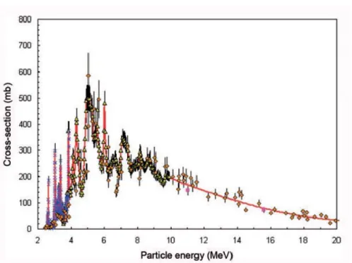
Figure 2. Excitation function for the, 18O(p,n)18F reaction……….……5
Figure 3. Relationship between 18F irradiation yield and beam energy………..….7
Figure 4. Decay of 18F………..………..9
Figure 5. Positron-electron annihilation process after collision producing 2 gamma rays of 511 keV that are emitted in opposite directions………10
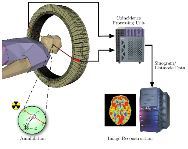
Figure 6. PET acquisition process……….12
Figure 7. Al18F radiolabeling approach for biomolecules………15
Figure 8. Chemical structures of DTPA (a), DOTA (b), NOTA-SCN-Bz--peptide and [18 F]AlF-NOTA-SCN-Bz-peptide (c), NOTA-peptide and [18F]AlF-NOTA-peptide (d), NODA-MP-peptide and [18F]AlF-NODA-MP-peptide (e)………16
Figure 9. Representation of AT1 receptor. 7TM α-helical are presented with Greek numbers (in red the helices that are important for binding)………18
Figure 10. AT1 receptor radiotracers. [11C]MK-996 (a), [11C]L-162,574 m- O11CH3 and [11 C]L-159,884 p- O11CH 3 (b), [11C]KR31173 (c), [99mTc]Tc-Losartan (d), [125I]I-Candesartan (e), R=-CH-[18F]F-(CH 2)2CH3 α-[18F]F-Irbesartan, R=-(CH2)3[18F]F δ-[18F]F-Irbesartan (f), [18F]FV45 (g) [18F]FEtLos (h), and [18F]AMBF 3Los (i) ………..20
Figure 11. DaSilva’s group AT1 receptor radiotracers. [11C]Methyl-Losartan R= -CH2O11CH3 and [11C]Methyl-EXP3174 R= -COO11CH3 (a), [18F]FPyKYNE-Losartan (b), [11C]TH4 R= =O and [11C]Methyl-Candesartan R= -OC 2H5 (c), and [18F]fluoropyridine-Candesartan (d)………23
xiii
Figure 12. Semi-automatic system for 18F-purification and production of concentrated
[18F](AlF)2+………..………26
Figure 13. Automatic synthesis module for a two-step radiofluorination. 18F-purification and 18O
recovery (A), azeotropic distillation and radiofluorination of prosthetic group (B), prosthetic group coupling reaction (C), semi-prep HPLC purification (D) and re-formulation and final filtration (E)………28
Chapter 2
Figure 1. Composition of calcitonin gene-related peptide (CGRP) (A) and adrenomedullin
receptor subtypes 1 (B) and 2 (C). Calcitonin-like receptor (CLR) is a seven transmembrane protein and the receptor activity-modifying protein (RAMP) is a component that varies in subtypes..……….………..………44
Figure 2. Adrenomedullin SPECT and PET radiotracers. Proof of concept: [125I]I-rAM(1-50) (a),
[99mTc]Tc-DTPA-AM (b), [99mTc]Tc-Cys16,21-AM (c), [99mTc]Tc-PulmoBind (d), and [18F]AlF-DFH17
(e)………48
Figure 3. Whole body images of [99mTc]Tc-PulmoBind activity (30-75 min post-injection) of in
Sprague-Dawley rat (a), mongrel dog (b), and human (c)………54
Figure 4. Whole body images of tracer activity (30-40 min post-injection, left panels) and
time-activity curves (TAC, right panels) of [18F]AlF-DFH17 in Sprague-Dawley rat (a), mongrel dog (b),
and rhesus monkey (c)……….58
Chapter 3
Figure 1. Chemical structure of DFH17, a NOTA-peptide derived from a pegylated analog of the
xiv
Figure 2. Typical elution profile of fluorine-18 from a MP-1 cartridge. After rinsing with 3 mL of
water followed by a bolus of 375 µL of saline, concentrated fluorine-18 was eluted with additional fractions of 0.9% NaCl (10 µL)……….81
Figure 3. Formation of [18F](AlF)2+ at different concentrations of AlCl
3 (n=3 per concentration)...82
Figure 4. Complexation assay with pentadentate NOTA chelator (n=3 per molar ratio, n.s. = no
significant difference, p=0.09)……….83
Figure 5. Radiolabeling of [18F]AlF-DFH17 (Scale-up) at different concentrations of AlCl 3 with
ethanol 50% (v/v) (n=3 per concentration)………84
Figure 6. Stability of [18F]AlF-DFH17 in saline at two different radioactive concentrations. Ascorbic
acid as radioprotectant (n=3 per time point)……….85
Figure 7. (a) Maximal intensity projections (MIP) images for PET/CT imaging of [18F](AlF)2+ after
1 h scan. (b) [18F]AlF-DFH17 after 15 min and (c) 1 h scan……….86
Figure S1. Analytical characterization of DFH17 (a) RP-HPLC and (b) MALDI-TOF…………..95 Figure S2. Semi-automatic system for 18F-purification and production of concentrated [18F]AlF.
Fluorine-18 is trapped onto a MP-1 cartridge, rinsed with 3 mL water, washed and eluted with 375 µL and 10 µL of 0.9% NaCl respectively and then semi-automatically dispensed (Multiposition valve) into small reactions vials containing 5 µL of 2 mM AlCl3 in sodium acetate buffer (0.1 M, pH
4). The solutions are incubated at RT for 10 min to form [18F](AlF)2+………..96
Figure S3. Radio ITLC-SG in citric acid pH 1.6 of (a) [18F]F- and (b) [18F]AlF as control. Down
panel, [18F](AlF)2+-to-NOTA molar ratios (c) 1:1, (d) 1:3 and (e) 1:5……….97
Figure S4. HPLC chromatograms of crude reaction mixture of [18F]AlF-DFH17 molar ratio 1:3
(Al18F)2+:DFH17. (a) Radiosynthesis in aqueous solution (37 MBq of [18F](AlF)2+). (b)
xv
50% (v/v) (1480 MBq of (Al18F)2+). Note: 1 represents the unbound activity [18F](AlF)2+ and 2 the
radiolabeled peptide [18F]AlF-DFH17……….98
Figure S5 Radiochemical purity of [18F]AlF-DFH17 determined by radio ITLC-SG (a) [18F]F- and
(b) [18F] (AlF)2+ as control. (c) and (d) [18F]AlF-DFH17 by ITLC-SG and RP-HPLC
respectively………..100
Figure S6. Biodistribution in organs of interest of (a) free [18F](AlF)2+ and (b) [18
F]AlF-DFH17………..101
Chapter 4
Figure 1. Representative analytical HPLC chromatograms of [18F]7. Co-injection with authentic
standard (7) in green (tR=7.78 min) (a). Radioactive signal at QC time (time 0) in red and 4 h
post-synthesis in black (tR =7.80 min) (b)……….122
Figure S2-1. Representative analytical HPLC chromatograms of [18F]6 at different temperatures.
(tR [18F]6=18.4 min and by-product [18F]fluorobenzene, tR=12.6 min)……….130
Figure S3-1. Representative semi-Prep HPLC radiochromatogram of purification of products
[18F]6 and [18F]7………...131
Figure S3-2. Zoomed HPLC chromatogram of [18F]7 with UV (254 nm) and radiation
detection………..132
Chapter 5
Figure 1. General mechanism of Sonogashira cross-coupling reaction adapted to the [18
xvi
Annex for Chapter 5
Figure S1. Multiple-sequence alignment of AM receptor sub-components CLR (left) and RAMP2
(right) of rat, dog, primate and human. The study was performed with the multiple sequence alignment tool: Clustal omega available at EMBL-EBI website. CLR sub-component of AM receptor it is conserved among species since the percent of identity is high (~90%). In contrast, RAMP2 varies considerably from rat to human (68% vs 100%), showing high differences in the identity of their primary sequence. This variation of RAMP2 is the responsible of contrasted inter-species [18F]AlF-DFH17 uptake observed by PET imaging………..161
Figure S2. Colum switch HPLC for radiometabolite analysis. C1 Capture column (Oasis HLB)
and C2 Analytical column (Phenomenex Luna C18). Flow rates: C1, 1 mL/min and C2, 2 mL/min………..162
Figure S3. Representative analytical HPLC chromatograms displaying radiometabolites in dog
plasma at 6, 12, 30, and 60 min post-injection. No [18F]AlF-DFH17 metabolites or parent
compound were detected in dog plasma at all time
points………164
Figure S4. Elution profile of [18F]FB-Candesartan in a C18 Sep-Pak Plus (360 mg) with ethanol
as mobile phase. Two mL ethanol was used to eluted of [18F]FB-Candesartan with a subsequent
solvent evaporation step at 45 oC under nitrogen in order to obtain a concentrated formulation of
the tracer………..165
Figure S5. Analytical HPLC chromatograms of chemical impurities of [18F]FB-Candesartan (Top)
and best HPLC separation condition (Bottom) with co-injected authentic compound FB-Candesartan (tR=19.09 min). Phenomenex Luna C18 (2) column (250×4.6 mm, 10μm) (2 mL/min,
Water 0.1% TFA/CH3CN 0.1% TFA 55:45). Impurity at tR=14.9 min was successfully separated
xvii
(tR=17.5 min and tR=20 min) were eluted along with FB-Candesartan (tR=19.09 min) which is still
xviii
List of Schemes
Chapter 1
Scheme 1. β+ decay process……….9
Scheme 2. Electron capture process………...9
Chapter 3
Scheme 1. Al18F-complexation assays with pentadentate NOTA chelator………..76
Scheme 2. Radiosynthesis of [18F]AlF-DFH17……….…77
Chapter 4
xix
List of Tables
Chapter 1
Table 1. Most typically used positron emitters isotopes and their cyclotron production
conditions……….2
Chapter 2
Table 1. Radiotracers for Molecular imaging of AM receptors………..………62
Chapter 3
Table S1 Results of quality control for the final formulation of [18F]AlF-DFH17 (Representative
batch)………..99
Chapter 4
Table 1. Optimization of [18F]6 reaction conditions………....124
xx
List of abbreviations
[18F]AlF F-18 Aluminum fluoride
[18F]FB F-18 fluorobenzyl
[18]FIB F-18 fluoroiodobenzene
%ID percent of the injected dose ACE angiotensin converting enzyme ACEI ACE inhibitor
AlF Aluminum fluoride AM Adrenomedullin Ang Angiotensin ARBS AT1R blockers
AT1R Angiotensin type 1 receptor
Bq Becquerel
CGRP Calcitonin gene-related peptide Ci Curie
CLR Calcitonin receptor-like receptor CT Computerized tomography
CTEPH Chronic thromboembolic pulmonary hypertension DFH12 N4-chelator derivative of adrenomedullin fragment 22-52
DFH17 NOTA-chelator derivative of adrenomedullin fragment 22-52 DMF Dimethylformamide
DMSO Dimethylsulfoxide
DOTA 1,4,7,10-Tetraazacyclododecane-1,4,7,10-tetraacetic acid DTPA Diethylenetriaminepentaacetic acid
xxi ED Effective dose
EDTA 2,2',2'',2'''-(Ethane-1,2-diyldinitrilo)tetraacetic acid EOB end of bombardment
EOS End of synthesis
ESI-MS Electrospray ionization mass spectrometry EtOAc Ethyl acetate
eV Electron-volt FB Fluorobenzyl
FDA Food and drug administration FIB Fluoroiodobenzene
FOV field of view
GPCR G protein-coupled receptor hAM (1-52) Human adrenomedullin
hAM (22-52) Human adrenomedullin fragment 22-52 HF heart failure
HPLC high-performance liquid chromatography HRMS high-resolution mass spectrometry IC50 Half maximal inhibitory concentration
ICL Intracellular loop K222 Kryptofix 222 MA Molar activity
MAA Albumin marco-aggregates
MALDI-TOF Matrix Assisted Laser Desorption Ionization - Time of Flight MAP Mean arterial pressure
MRI Magnetic resonance imaging NMR Nuclear magnetic resonance
xxii
NODA-MPAA 2,2′-(7-(4-(2-((2-aminoethyl)amino)-2-oxoethyl)benzyl)-1,4,7-triazonane-1,4-diyl)diacetic acid
NOTA 2,2′,2”-(1,4,7-triazacyclononane-1,4,7-triyl)triacetic acid NOTA-NHS NOTA mono-N-hydroxysuccinimide
PEG Polyethylene glycol
PET positron emission tomography PH Pulmonary hypertension
QC quality control
QMA Quaternary methyl ammonium rAM (1-50) Rat adrenomedullin RAS renin angiotensin system RBF Renal blood flow
ROI Regions of interest
SAR structure-activity relationship
SCN-Bz-NOTA 2-S-(4-isothiocyanatobenzyl)-1,4,7-triazacyclononane-1,4,7-triacetic acid SPECT Single-photon computerized tomography
TAC time-activity curve
TBAHCO3 Tetrabutylammonium hydrogenocarbonate
TEA Triethylamine TFA Trifluoroacetic acid THF Tetrahydrofuran
TLC thin layer chromatography TM transmembrane
xxiii
Acknowledgements
This thesis is dedicated to my family, supervisors, colleagues, friends, institutions
and all those who made these five years worthwhile.
1
Chapter 1: Introduction
2
1.1 Cyclotron.
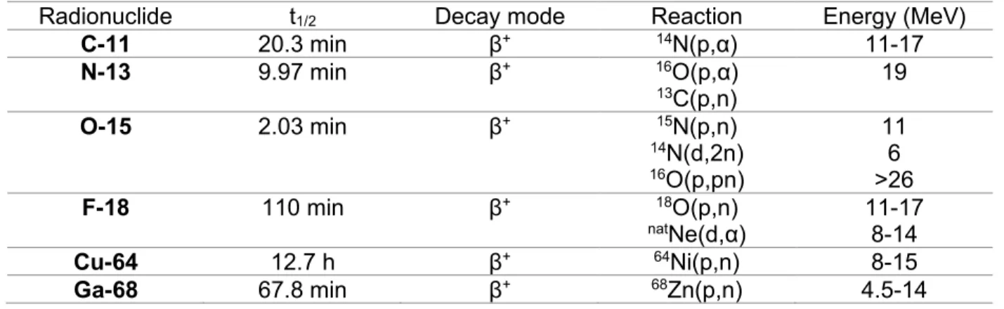
Cyclotron-produced radionuclides, and their corresponding radiopharmaceuticals, are extremely valuable in basic medical research, disease diagnosis and radiotherapy treatment. Practically, most used PET radioisotopes (Table 1) are currently produced by a medical cyclotron.
Table 1 Most typically used positron-emitting isotopes and their cyclotron production conditions.
Radionuclide t1/2 Decay mode Reaction Energy (MeV)
C-11 20.3 min β+ 14N(p,α) 11-17 N-13 9.97 min β+ 16O(p,α) 13C(p,n) 19 O-15 2.03 min β+ 15N(p,n) 14N(d,2n) 16O(p,pn) 11 6 >26 F-18 110 min β+ 18O(p,n) natNe(d,α) 11-17 8-14 Cu-64 12.7 h β+ 64Ni(p,n) 8-15 Ga-68 67.8 min β+ 68Zn(p,n) 4.5-14
The cyclotron was invented by Ernest O. Lawrence and Milton S. Livingston in 1934 to accelerate particles (i.e. protons and deuterons) until achieving a high kinetic energy [1, 2]. Then these particles can be used to bombard other nuclei at a particular energy in a target to produce short-lived positron-emitting isotopes that are then incorporated in a molecule (called radiotracer or radiopharmaceutical) for PET or SPECT imaging.
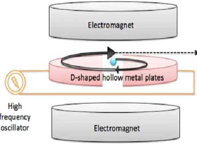
The cyclotron operating principle is based on the fact that the motion period of charged particles in a uniform magnetic field is independent of the velocity of the particle (Figure 1) [3, 4]. Briefly, the four systems (vacuum, ion source, acceleration force and beam control) are necessary to generate a particle beam capable of creating radionuclides. By combination of these four systems in the cyclotron, particle beam produced via the ion source is moving inside semi-circular
3
dees (or D-shaped metal plates) which are placed inside vacuum chambers exposed to a static electromagnetic field perpendicular to the electrode plane. The particle beam is kept under vacuum to avoid interactions with other particles and to maintain the beam focused. Then, the charged particle beam is accelerated using a high radiofrequency alternating voltage, which is applied between two hollow "D"-shaped sheet metal electrodes. The presence of a magnetic field causes the particles to curve their path in a circular shape and the electric field to correct the direction to accelerate them. After each semi-revolution, the particles move in a quasi-spiral orbit outward from the center to the rim of the dees acquiring a high kinetic energy (up to its possible maximum), due to pushes created by the reverse of radiofrequency voltages [5].
Figure 1. Basic scheme of a cyclotron.
Considering the maximum proton energy, cyclotrons are classified by type I (up to 10 MeV), type II (20 MeV) and type III (45 MeV). Nowadays, most cyclotrons accelerate negative ions due to the greater simplicity of the beam extraction process. If the particles are negatively charged, extraction is carried out by stripping electrons off the negatively charged ions and allowing the magnetic field to reverse the curvature of the resulting positively charged particle paths and to transport the particles out of the cyclotron [6]. For example, hydride ions formed by
4
the ion source are accelerated and then transformed into protons by charcoal thin foils to posteriorly incise in the target producing the nuclear reaction [7, 8].
1.2 Production of
18F.
1.2.1. Targetry
Fluorine-18 has been produced by a variety of nuclear reactions, using both nuclear reactors and accelerators. The target materials used can be either in the solid, liquid or gas phase. Solid targets of enriched 6LiCO
3 have been used for production of 18F. In this case, separation of 18F from the solid target is performed by absorbing the [18F]fluoride on alumina or ion exchange
resins, followed by thorough washing step before [18F]fluoride is eluted under basic conditions [9].
Gas targets, such as enriched 18O and 20Ne are also used for the production of 18F- via 18O(p,n)18F
and 20Ne(d,α)18F nuclear reactions [10]. Both target systems produce carrier-added 18F gas (18F 2)
suitable for electrophilic radiofluorination reactions intended for applications where high molar activity (MA) is not required [11].
No-carrier-added [18F]fluoride at high MA is currently produced using a liquid target of
enriched [18O]water in a chamber made of inert metal, such as silver, titanium, niobium or
tantalum. [18F]fluoride produced following 18O(p,n)18F reaction is the most commonly production
way for PET studies [12]. Taking this into account, we will be focused on this single form of production in the following sub-chapters.
5 1.2.2. Production cross-section
Accelerated charged particles interact with a target nucleus producing a nuclear reaction, ultimately leading to a stable or radioactive nucleus. A nuclear reaction is characterized by a cross-section, a geometrical quantity given in barn (10-24 cm2), representing the total probability
that radionuclide can be formed and will then decompose along a particular decay. The reaction cross-section occurs in function of energy along with other factors including the thickness of the target. Considering the cross-section, the beam current determines the amount of radionuclide that can be produced in a given cyclotron at various energy levels and the levels of contamination with other radionuclides that may be present in the target material. [7, 13]. As consequence, the nuclear reaction cross-section data are needed in radioisotope production programs mainly for optimization of radionuclide production routes.
6
In recent years, the18O(p,n)18F reaction on highly enriched 18O-targets (particularly as
H218O) has been extensively used. This nuclear reaction has relatively low threshold energies
(0-20 MeV), and its maximum yield occurs at less than 10 MeV (Figure 2) [14]. Taking into account that the18O(p,n)18F reaction is endoergic (Q value of -2.43 MeV), the minimum kinetic energy that
the bombarding proton should possess for this reaction to be energetically possible is of 2.57 MeV. Thus, selecting proton energies higher 3 MeV maximizes the probability of occurrence of this reaction [15]. However, energy values above 10 MeV are usually selected since F-18 production yield will be much higher (as will be explained in next section).
1.2.3. 18F production beam
Proton beam energy and the beam current along with the target volume are important practical factors in the production of 18F that determine how much [18F]fluoride can be produced
in a given amount of time [16]. As depicted in Figure 3, there is a relative dependence between [18F]fluoride yield and proton beam energy for a theoretical thick target [17]. As can be seen, the
production yield of [18F]fluoride (from the 18O(p,n)18F nuclear reaction) rises nearly linearly from 5
to 15 MeV and gradually, and at a lower rate above 15 MeV. This theoretically indicates that cyclotrons with proton energies higher than 15 MeV will produce at least 50% more F-18 compared to a 10-11 MeV cyclotron for the same beam current. However, taking into account the (p,n) reaction cross-section (Figure 2), energies above 18 MeV are not really necessary for the routine production of [18F]fluoride since the nuclear reaction will occur at lower probabilities.
Moreover, the beam current of a cyclotron also has an influence in how much radioisotope can be produced at a given energy. Theoretically speaking, the production yield is directly proportional to the beam current but in practice, the beam current is also determined by the ability to remove the heat produced in a target by a beam. Most medical cyclotrons are capable of
7
generating a beam with a current of at least 50 μA. Typically, a target with a given operating pressure has an optimum current at which the production rate of the desired amount of radionuclide is the highest [17].
Figure 3. Relationship between 18F irradiation yield and beam energy [17].
1.2.4 18F transfer and recovery
[18F]fluoride from the 18O enriched water target is typically transferred to the synthesis unit,
where the 18F is extracted from the irradiated aqueous solution and the 18O-enriched water is
recovered. Transport of the water containing the 18F is usually carried out using small bore plastic
tubing. The most common materials for this tubing are polyethylethylketone, tefzel or polyethylene. These materials are designed to contain very low amounts of 19F reducing the
formation of the unlabeled final radiopharmaceutical containing natural fluoride and therefore, without being detrimental to the MA of the final radiofluorinated compound [18]. There is a clear need of a reliable production of high MA radiopharmaceuticals that is particularly important for saturable receptor/transporter ligands. As PET is a true tracer technique (imaging at nM concentrations), receptor occupancy of radiolabeled tracers should be kept below about 1%, in
8
order to visualize easily-saturable receptors and also to avoid possible pharmacological/toxic effects [19].
With the purpose of reducing the excess of water in radiofluorination nucleophilic reactions [18F]fluoride is usually recovered from enriched water using several different techniques. Among
them, ion exchange resin or quaternary methyl ammonium (QMA) anion exchange resin offers not only separation of [18F]fluoride and efficient recovery of expensive enriched [18O]water [20],
but also scavenging of many ion contaminants from the target or foil that can alter radiofluorination reactions [21]. Due to the presence of oxygen-16 in the target water (usually at 2% levels, with
18O enriched water at 98%), nitrogen-13 (t
1/2 = 10 min) is also formed within the target by the (n,α)
nuclear reaction. Nitrogen-13 constitutes a radionuclide impurity and can appear as nitrate, nitrite, nitrogen and/or ammonia [22]. Hence major part of 13N will decay during the synthesis time.
Importantly, 13N-labeled cation impurities are commonly eliminated in the QMA elution in where
nitrogen-13 cations are eluted while 18F- is trapped in the resin.
1.3 Decay of
18F.
Nuclei that are “neutron deficient” or “proton rich” can decay by positron emission accompanied by the emission of a neutrino. After emission of a positron-particle, the daughter nuclide has an atomic number that is one unit less than the parent. Also, as consequence of a positron emission, a mass equivalent of two electrons are created by the conversion of a proton to a neutron (Scheme 1) in where 1.02 MeV is needed to create these two particles. Taking this into account, positron emission takes place only when the energy difference between the parent and daughter nuclides is equal to or greater than 1.02 MeV.
9
+
+
⎯→
⎯
n
+e
p
0 1 1 0 1 1Scheme 1. β+ decay process
A particular β+ decay process may also happen when a nucleus has a smaller N/Z ratio
compared to the stable nucleus. This process is called electron capture (Scheme 2), in which an electron is captured from the extra nuclear electron shells, transforming a proton into a neutron and emitting a neutrino. For this process to occur, the energy difference between the parent and daughter nuclides is usually, but not necessarily, less than 1.02 MeV. Nuclides having an energy difference greater than 1.02 MeV may also decay by electron capture. As larger the energy difference gets, higher the positron decay will be [13].
+
⎯→
⎯
+
+e
n
p
1 0 0 1 1 1Scheme 2. Electron capture process
Fluorine-18 decays (Figure 4) largely by positron emission (97%) with a half-life of 109.77 min and relatively low energy (maximum 0.635 MeV) thus the emitted positron has short mean range (2.39 mm in water)[23].
10
At the end of the path of β+ particles (0-10 mm, depending on its kinetic energy and density
of the tissue), positrons combine with electrons and are thus annihilated, each event giving rise to two photons of 511 keV that are emitted in opposite directions. These photons are referred to as annihilation radiations (Figure 5) and this phenomenon is the basis of the positron detection system (also known as coincidence detection system) [7].
Figure 5. Positron-electron annihilation process after collision producing two gamma rays of 511
keV that are emitted in opposite directions
1.4 PET imaging.
Positron emission tomography (PET) is a non-invasive imaging modality that uses the principle of coincidence detection of two annihilation photons (Figure 5) to measure radiotracer
11
distribution within tissues. This information, when combined with assumptions based on physiology or biochemical models, can be used to assess biological processes in vivo.
In a PET camera, when opposing 511 keV gamma rays reach simultaneously two detector crystals, a flash of light (scintillation) is produced and due to the photoelectric effect, released electrons then magnified their energy in a photomultiplier tube [24] (Figure 6). By processing the information of millions of annihilations using mathematical techniques, a map of the activity distribution within the field of view over time is generated producing a dynamic image and time-activity curve (TAC) for various regions of interest (ROI) in the subject (Figure 6) [25]. Combining the maximal positron range (which is unique for each radioisotope) with the crystal detector properties and its photomultiplier tube, determine the final resolution of PET images (typically 4-6 mm). Recently, the spatial resolution of newer clinical systems (mainly dedicated for brain imaging) has been improved up to 2.5 mm and for small animal PET imaging systems, resolutions of 0.8-1.5 mm can be achieved [26].
PET is usually used in combination with other imaging modalities, such as radiography, computed tomography (CT), or magnetic resonance imaging (MRI). The use of PET in combination with these anatomy-based imaging technique offers advantages of a simultaneous assessment of both anatomical and functional processes. These hybrid imaging techniques provide unique opportunities in diagnosing diseases and evaluating its treatment which is the base of future personalized medicine [27].
12
Figure 6. PET acquisition process
1.5 AM receptor radiotracers.
Many molecular imaging agents have demonstrated potential utility in the diagnostic of pulmonary vascular disease but very few have been tested in human subjects. Nonetheless, a non-invasive SPECT/PET imaging modality is currently unavailable to provide direct information on the pathophysiology processes affecting pulmonary vasculature (i.e. endothelium, adventitia). Recent studies have developed agents targeting angiotensin-converting enzyme, norepinephrine transporter and adrenomedullin receptors for imaging of pulmonary vasculature [28]. An extensive review of the state-of-art in SPECT and PET imaging of adrenomedullin receptors is provided in Chapter 2.
13
1.6 Chelation chemistry
Chelation is called to formation process in where two or more coordinate bonds are formed between a polydentate ligand and a single central atom. Chelators are usually organic compounds containing donor atoms such as O, N and S which share their electron pairs with transition elements (mainly metals) to form a stable coordination compound with a particular coordination number and geometry [29].
In radiochemistry, biological and radioactive drug components are often coupled through the use of bifunctional chelators with the dual purpose of radiometal complexation and bioconjugation. An ideal chelator for radiopharmaceutical preparation should efficiently bind the radiometal in high complexation yields, in aqueous medium, at low temperature and short reaction time. It is important that complexation with the radiometal be stable in vivo in order to prevent the release of the free radiometal (kinetic inertness) and non-selective binding imaging. Macrocyclic chelators possess a geometric constrain that partially organize metal ion binding sites by decreasing entropic losses upon the metal complexation. In contrast, acyclic chelators follow a more drastic change in physical and geometry orientation in order to arrange donor atoms to coordinate the metal ion, undergoing a significant decrease in entropy which is thermodynamically unfavorable. For this reason, most chelation of radiometals takes place using macrocyclic chelators which are known to form a stable entity and a more kinetically inert complexes than acyclic chelators [30].
14 1.6.1 Aluminum [18F]Fluoride
Aluminum [18F]Fluoride is an attractive method for radiofluorination by chelation of
peptides and proteins. Recently, this emerging approach has been used to label peptides in aqueous solutions increasing the reaction yield, facilitating the purification and reducing considerably the total synthesis time [31].
Aluminum as cation exist most commonly at oxidation state +3, but some compounds are known at lower oxidation states of +1 and others at +2 in gas phase. Following the Pearson classification, Al3+ behaves as a Lewis acid thus forming stable coordinates with hard Lewis bases
containing electron pair donors such as H2O, ROH, RNH2, OH-, F-, Cl-, etc [32]. Among more
than sixty metal ions, Al3+ binds to F- the most strongly (>670 KJ/mol) with the exception of Sc3+.
Known molecular geometries of aluminum with F- undergoes tetrahedral, trigonal bipyramidal and
octahedral complexes such as (AlF4)-, [AlF4(OH)]2- and (AlF6)3- with four, five and six coordination
number respectively [33]. Previous studies revealed that AlF-binary complex formation is a pH-depending process. When the pH is too high, aluminum would form insoluble hydroxide complexes. At contrary, if the pH it is too low, then the preferred fluoride species in the equilibrium would be HF. Also, at lower fluoride concentrations, aluminum appears as monofluoride species like (AlF)2+ that could form a stable complex with a suitable chelator [34].
McBride et al. [35] were the first to explore the potential of Al18F-method in
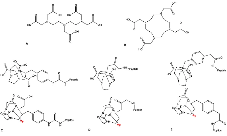
radiopharmaceutical chemistry (Figure 7). As a proof-of-concept, the authors succeeded to complex Al18F to a DTPA-conjugated peptide in high yields (Figure 8a). However, by using this
15
applications. The conclusion drawn from this initial study was the needs of finding a thermodynamic stable and kinetic inert Al18F-ligand that remains intact for several hours in vivo.
Figure 7. Al18F radiolabeling approach for biomolecules. Found in Smith, G et al. [36]
1.6.2 NOTA
NOTA-chelator (Figure 8c) was first reported in the mid-80s as small version of DOTA (Figure 8b), in the search of a more stable chelator for lanthanides as potential MRI contrasting agents [37, 38]. NOTA is a hexadentate chelate with three nitrogen and three oxygen donor atoms, that tend to form stable coordination bond with d-orbitals of transition metals of smaller atomic radii. Thus, it is probably the most successful chelator for use with 67/68Ga and 64Cu due to
its favorable radiolabeling conditions and excellent in vivo stability.
In previous studies, Al3+ was found to form highly stable octahedral complexes with NOTA
towards acid catalyzed dissociation by 27Al-NMR spectroscopy [39]. Also, the Al18F complex has
pentadentate binding to NOTA, with the sixth binding site of the aluminum filled with the fluoride ion. Thus, the SCN-Bz-NOTA (Figure 8c) modified peptide IMP449 was successfully radiolabeled
16
with Al18F in 5-20% yield. After demonstrating stability in serum for 4h, the product was able to
bind its receptor by PET imaging using nude mice bearing the human colon cancer xenograft LS174T [40].
Figure 8. Chemical structures of DTPA (a), DOTA (b), NOTA-SCN-Bz-peptide (top) and the
[18F]AlF-NOTA-SCN-Bz-peptide conjugate (bottom) (c), NOTA-peptide (top) and the [18
F]AlF-NOTA-peptide conjugate (bottom) (d), NODA-MP-peptide (top) and the [18
F]AlF-NODA-MP-peptide conjugate (bottom) (e)
Further experiments evidenced that efficient and stable binding of radiometals by chelates is highly influenced by the chelate structure. D’Souza et al. [41] studied the solid-state X-ray structure of Al18F in an improved NOTA chelator: 1,4,7-triazacyclononane-1,4-diacetate motif with
17
experimentally demonstrated that N3O2-type chelators can from stable mononuclear octahedral
complexes with aluminum containing a single coordination site for binding fluoride. Therefore, pentadentate ligands such as NOTA-NHS ester (Figure 8d) or NODA-MPAA (Figure 8e) are desired for high complexation yields, leaving only one binding site open for the fluorine-18 ion.
1.7 AT
1receptors.
The octapeptide hormone Ang II plays a central role in the regulation of blood pressure. As a part of the Renin-Angiotensin System, Ang II acts mainly by stimulating the AT1Rs which are
expressed in the kidneys, adrenal glands, heart, brain, and vasculature [42]. Previous studies suggested that there are alterations in the expression levels of the AT1Rs associated with
cardiovascular and renal diseases such as hypertension, cardiac and renal failures; but the regulatory mechanism that determines AT1R expression and disease progression is not well
understood yet [43].
AT1R is part of the seven-transmembrane (7TM) superfamily of G protein-coupled
receptors (GPCRs). New advances in the determination of the crystal structures of AT1R confirms
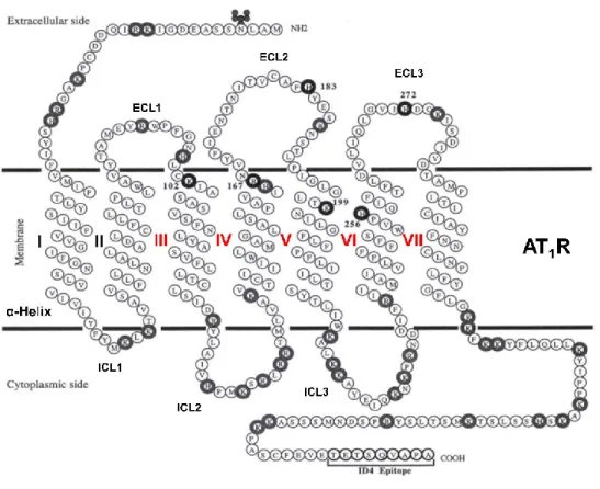
the postulates 7TM α-helical architecture with three extracellular loops (ECL1-3) and three intracellular loops (ICL1-3) (Figure 9) in where critical ligand binding residues (Arg167, Tyr35 and
Thr84) for the so-called "sartan" family of selective AT
1R blockers (ARBs) have been identified
18
Figure 9. Representation of AT1 receptor. 7TM α-helical are presented with Greek numbers (in
red the helices that are important for binding). Adapted from Noda et al. [44].
1.8 Candesartan.
Candesartan is an ARB approved by FDA in 1998 as a treatment of hypertension. This compound selectively binds to AT1R giving a long-lasting blockage and suppression of the
response by Ang II. It displays a higher binding affinity (IC50 0.6 nM) compared to other selective
AT1R antagonists (Losartan, IC50 20 nM and Valsartan, IC50 2.7 nM) [45]. Thus, a
radiopharmaceutical based on the parent structure of Candesartan with similar pharmacological binding properties could exhibit some potential as a molecular imaging agent of AT1 receptors.
19
1.9 AT
1receptor radiotracers.
AT1R radioligands have been previously developed for SPECT and PET imaging but so
far only few of them are based on Candesartan. The Johns Hopkins PET group developed the first AT1R PET radioligand [11C]MK-996 by N-benzoylation with [α-11C]benzoyl chloride in 1995
(Figure 10a). The compound was produced with an average MA of 1162 mCi/μmol in 38 min including reformulation [46].
Close structural analogues [11C]L-162,574 and [11C]L-159,884 were also prepared in low
yields by Johns Hopkins PET group (Figure 10b). High AT1R binding was obtained in canine
adrenals and renal cortex demonstrating the tracer potential to examine in vivo AT1R binding in
dogs. Nonetheless, [11C]L-159,884 was rapidly metabolized in humans [47] limiting its future
applications. This group also produced the [11C]KR31173 (Figure 10c) in low yields (at high MA
>7000 mCi/μmol) and without remarkable brain uptake. Transient regional AT1R up-regulation
was detected with [11C]KR31173 in the infarcted area by small‐animal PET imaging [48].
Verjans et al. [49] reported the radiolabeling of Losartan with 99mTc for SPECT imaging.
Losartan was attached with an N4-type chelator and four PEG groups as linkers (Figure 10d). No
data were shown for radiolabeling yield or MA. Compared to controls, nearly 2.4-fold higher AT1R
binding was observed in the infarct region of a mouse with permanent coronary artery ligation. However, the authors concluded that it is necessary to engineer new probes with better targeting characteristics for realization of potential clinical application.
20
Figure 10. AT1 receptor radiotracers. [11C]MK-996 (a), [11C]L-162,574 m- O11CH3 and [11
C]L-159,884 p- O11CH
3 (b), [11C]KR31173 (c), [99mTc]Tc-Losartan (d), [125I]I-Candesartan (e),
R=-CH-[18F]F-(CH
2)2CH3 α-[18F]F-Irbesartan, R=-(CH2)3[18F]F δ-[18F]F-Irbesartan (f), [18F]FV45 (g),
[18F]FEtLos (h), and [18F]AMBF
3Los (i).
Sanad et al. [50] reported the radioiodination of Candesartan with iodine-125 and its biological evaluation as potential tracer for cardiovascular disorder detection (Figure 10e). Using the chloramine-T method for radioiodination, the radiochemical yield of [125I]I-Candesartan was
21
98%. No data were reported for MA. The authors stated that heart uptake of [125I]I-Candesartan
was higher than [125I]I-Valsartan and [99mTc]Tc-Valsartan at same time post-injection. While the
authors concluded that [125I]I-Candesartan can be easily and safely used in myocardial SPECT
imaging, it should be noted that nuclear properties of iodine-125 (half-life 59.43 days and gamma energy 35.5 keV) are not ideal for clinical applications. The authors suggest that further clinical trials are required to evaluate the potential of [125I]I-Candesartan as cardiac SPECT imaging
agent. However, due to the ideal gamma-ray characteristics, selection of the I-123 labeled Candesartan derivative would have been a better choice for SPECT imaging.
Two F-18 derivatives of Irbesartan were reported by Hoffman et al. [51] in 2018 (Figure
10f). Radiofluorinations at α- and δ-positions of tosyl precursors were carried out in 11%
radiochemical yield and 94% radiochemical purity. Considering the results of in vitro competition studies, α-F-Irbesartan (IC50 6.6 nM) was evaluated in rats and pigs by PET imaging. In vivo
studies revealed that α-[18F]F-Irbesartan accumulated in all relevant tissues expressing AT 1Rs
such as heart, kidney and lung. The tracer uptake in these organs was proven to be specific to AT1R due to reduction of uptake following blocking studies with Irbesartan (5 mg/kg). However,
PET imaging in healthy heart was difficult due to low cardiac retention and high liver uptake. The study concluded that more work is needed to further explore its applicability for broad uses. Following the radiofluorination approach of α-[18F]F-Irbesartan, the 18F Valsartan-derivative FV45
(Figure 10g) was synthesized in 21.8 % yield and more than 99% radiochemical purity. No data were reported for MA. However, PET studies in rats revealed a fast and clear kidney uptake that was selectively blocked by a pre-treatment with valsartan. The authors concluded that [18F]FV45
had almost identical AT1R binding affinity as its lead compound Valsartan, facilitating the
22
Recently, two tracers based on Losartan were labeled with F-18 using two different methods [53]. [18F]Fluoroethyl-Losartan (Figure 10h) was radiolabeled by 18F-fluoroakylation of
Losartan potassium using the prosthetic group 2-[18F]fluoroethyl tosylate in low yield (2.7%), low
MA (38 Ci/mmol) and high radiochemical purity (>95%). [18
F]Ammoniomethyltrifluoroborate-Losartan ([18F]AMBF
3Los) (Figure 10i) was prepared following 18F-19F isotopic exchange in 11%
radiochemical yield and MA of 2900 Ci/mmol. Higher binding affinity was found with AMBF3Los
(Ki=7.9nM) in a competition assay using AT1R-expressing membranes. Then, the tracer was
evaluated in rats via PET imaging with AMBF3Los blocking revealing specific uptake in the kidney
cortex. Taking into account their findings, the authors concluded that [18F]AMBF
3Los might be
considered a valuable PET imaging tracer for monitoring AT1R expression in several diseases.
1.9.1 Our AT1R Losartan derivative radiotracers
DaSilva’s group reported the synthesis of [11C]methyl-Losartan, [11C]methyl-EXP3174,
and [18F]FPyKYNE-Losartan, (Figure 11a and b) in good yields, high radiochemical purity and
reasonable synthesis time [54-57]. [11C]methyl-Losartan and [11C]methyl-EXP3174 were
synthesized by O-[11C]methylation of Losartan and its active metabolite EXP3174 respectively.
Both radiotracers displayed high kidney-to-blood ratios which was further confirmed to correspond to [11C]methyl-EXP3174 specific binding for AT
1R. Regardless the high selectivity for AT1 over
AT2 and MAS receptors, [11C]methyl-Losartan exhibited the presence of radiometabolites in the
kidney with similar pharmacological binding profile to the parent tracer which is detrimental for AT1R quantification by PET imaging. This study concluded that [11C]methyl-EXP3174 displayed
favorable uptake kinetics and binding profile to AT1Rs supporting further studies to assess its full
23
Figure 11. DaSilva’s group AT1 receptor radiotracers. [11C]Methyl-Losartan R= -CH2O11CH3 and
[11C]Methyl-EXP3174 R= -COO11CH3 (a), [18F]FPyKYNE-Losartan (b), [11C]TH4 R= =O and
[11C]Methyl-Candesartan R= -OC
2H5 (c), and [18F]fluoropyridine-Candesartan (d).
[18F]FPyKYNE-Losartan ([18F]fluoropyridine-Losartan) was synthesized via click chemistry
using a two-step, double reactor TRACERlab® FX N Pro GE Healthcare module in 7-14% overall
process yield. MA of 200‐4200 mCi/μmol were achieved. PET studies in rats and pigs displayed high uptake in the AT1R-rich kidneys with good contrast compared to surrounding tissues.
Blocking studies exhibited high binding selectivity for renal AT1R over AT2R. In addition,
24
compounds suggesting a potentially advantageous metabolic profile. However, in order to prevent saturation of receptors and be able to image cardiac AT1Rs, the MA needs to be above 7000
mCi/μmol [59].
1.9.2 Structure-activity relationship studies (SAR) justifying Candesartan labeled derivatives
Based on reported SAR studies by Kubo et al. [60], large prosthetic groups can be introduced at the imidazole 7-position of Candesartan without changing its binding properties and pharmacological functions. Coupling a prosthetic group at this position requires a chemical “inert” bond to enzymatic cleavage. C-C bond in vivo cleavage by enzymes does not occur with unfunctionalized methylene carbons, olefins, and aromatic molecules and requires oxidation reactions at two adjacent carbon atoms following a multi-step mechanism [61]. [11
C]methyl-Candesartan and its des-ethyl derivative [11C]TH4 (Figure 11c) were synthesized in high yields
of 50-70% from [11C]methyl iodide and with specific activities of 300-1500 mCi/μmol. Even when
both tracers displayed high uptake in the kidney, [11C]TH4 exhibited reduced selectivity for AT 1R
compared to [11C]methyl-Candesartan [54]. Initial in vivo PET modeling performed with
[11C]methyl-Candesartan evidenced its ability to detect changes in AT
1R density and/or affinity in
the rat kidney. However, the quantification and modeling demanded corrections due to the presence of the hydrophobic metabolite [11C]TH4 bound to renal AT
1R [62].
Few weeks ago, our group published the synthesis of the novel tracer [18
F]fluoropyridine-Candesartan (Figure 11d) labeled with the [18F]FPyKYNE prosthetic via the copper-catalyzed
azide-alkyne cycloaddition at the imidazole 7-position in 10% yield and high radiochemical purity (≥97%) [63]. Using an improved methodology, the prosthetic group [18F]FPyKYNE was completely
separated from its nitro precursor allowing the production of the tracer in MA greater than 10 000 mCi/μmol. Ex vivo biodistribution and autoradiography studies revealed specific binding of
![Figure 3. Relationship between 18 F irradiation yield and beam energy [17].](https://thumb-eu.123doks.com/thumbv2/123doknet/2036513.4478/33.918.245.709.273.545/figure-relationship-f-irradiation-yield-beam-energy.webp)


![Figure 7. Al 18 F radiolabeling approach for biomolecules. Found in Smith, G et al. [36]](https://thumb-eu.123doks.com/thumbv2/123doknet/2036513.4478/41.918.226.676.194.470/figure-al-f-radiolabeling-approach-biomolecules-smith-g.webp)