Université de Montréal
The role of the kinetochore in chromosome segregation during Meiosis I
Par Evelyne Turrin
Programme de biologie moléculaire, Faculté de médecine
Mémoire présenté en vue de l’obtention du grade de Maîtrise en biologie moléculaire, option Générale
Décembre 2019
Université de Montréal
Unité académique - Programmes de Biologie Moléculaire, Faculté Médecine
Ce mémoire intitulé
The role of the kinetochore in chromosome segregation during Meiosis I
Présenté par
I
Résumé
La ségrégation des chromosomes est un processus complexe permettant la division égale du matériel génétique entre les cellules filles. Contrairement aux cellules somatiques, ce processus est sujet à des erreurs dans les cellules germinales telles que les ovocytes. Lorsque des erreurs surviennent lors de la ségrégation des chromosomes durant la méiose cela peut conduire à une aneuploïdie. L’aneuploïdie est la présence d’un nombre incorrect de chromosomes dans une cellule et est connue pour causer l’infertilité et des arrêts de grossesses chez l’humain. L’incidence de l’aneuploïdie augmente avec l’âge maternel (1).
Le kinétochore est une structure cellulaire impliqué dans la ségrégation des chromosomes. Il est composé de plus de 100 protéines et se situe entre les microtubules et les centromères. Les microtubules se lient aux kinétochores, et ces derniers s’attachent sur les centromères afin de séparer les chromosomes homologues durant la méiose et les chromatides des sœurs pendant la mitose (1–3). Dans les cellules somatiques, cette structure est bien connue (2). Pourtant, moins d’informations sont connues à dans l’ovocyte de mammifère en développement au cours de la méiose I (3,4).
Ce projet vise à étudier le rôle du kinétochore durant la ségrégation des chromosomes dans l’ovocyte de souris en développement. Plus spécifiquement, l’assemblage, le désassemblage, la dynamique et la tension des protéines du kinétochore seront évalués. Ce projet permettra de mieux comprendre le rôle du kinétochore durant la méiose I, ses implications durant la séparation des chromosomes, et éventuellement ses implications dans l’aneuploïdie.
Mots-clés : Kinétochore - Ovocyte - Ségrégation des chromosomes - Méiose I - Aneuploïdie -
II
Abstract
Chromosome segregation is an intricate process in dividing genetic material equally between daughter cells. This process, unlike in somatic cells, is error prone in germ cells like the oocyte. When errors occur during meiosis in segregating chromosomes, aneuploidy results when the cell has an incorrect number of chromosomes. This can result in infertility and birth defects in human reproduction. The incidences of aneuploidy are also seen to increase with increasing maternal age (1).
The kinetochore is a cellular structure at the heart of chromosome segregation. It is composed of more than 100 proteins and is located between the microtubules and the centromeres. The microtubules attach onto the kinetochores, which themselves attach onto the centromeres, in order to pull the homologous chromosomes apart during meiosis and the sister chromatids during mitosis (1–3). Much is known about this multi-protein structure in somatic cells (2). Yet, very little is known about this in the developing mammalian oocyte during Meiosis I (1,3,4). This project aims to investigate the role of the kinetochore in chromosome segregation in a developing mouse oocyte. More specifically, kinetochore protein assembly, disassembly, dynamics and tension will be assessed. This project will achieve a better understanding of the kinetochore’s role in Meiosis I, its implications in chromosome segregation in a developing mouse oocyte, and how it may be involved in aneuploidy.
Keywords: Kinetochore - Oocyte - Chromosome Segregation - Meiosis I - Aneuploidy -
III
Table of Contents
• Résumé- I • Abstract- II
• List of Figures- VI • List of Abbreviations- VII • Acknowledgements- IX • Remerciements-X
1. Chapter 1- Introduction- pg. 1
a. Chromosome segregation: mitosis vs meiosis- pg. 1 b. Aneuploidy- pg. 3
c. The kinetochore and its functions- pg. 6 d. The kinetochore and its proteins- pg. 7
i. The kinetochore and its proteins: the inner kinetochore- pg. 7 ii. The kinetochore and its proteins: the outer kinetochore- pg. 9
iii. The kinetochore and its proteins: the loosely associated proteins- pg. 10 e. Kinetochore assembly- pg. 10
f. The kinetochore and signaling cascades- pg. 11
g. The kinetochore and its attachments to the microtubule- pg. 13 h. The kinetochore and its control factors- pg. 15
i. Kinetochore disassembly- pg. 16
j. The kinetochore and distance: the concept of delta- pg. 17 k. The kinetochore and force- pg. 19
2. Chapter 2- The rationale and hypothesis- pg. 25 a. The rationale- pg. 25
b. The hypothesis- pg. 25
3. Chapter 3- Materials and Methods pg. 26 a. Methods- pg. 26
i. Dissection- pg. 26
ii. Fixation and antibody staining- pg. 28 iii. Microinjection- pg. 3
IV
1. Making mRNAs that will be translated to proteins that are fluorescently tagged- pg. 32
2. Making mRNAs: transformation and inoculation procedure- pg. 32 3. Making mRNAs: DNA mini prep- pg. 32
4. Making mRNAs: linearize DNA- pg. 33
5. Making mRNAs: extraction via DNA mini prep- pg. 33 6. Making mRNAs: IVT (in-vitro transcription) reaction- pg. 34 7. Making mRNAs: Poly(A) tailing reaction- pg. 34
8. Making mRNAs: RNA cleanup- pg. 34 iv. Preparing oocytes to measure delta- pg. 35
v. Confocal microscopy- p. 35 vi. ImageJ and FIJI analysis- pg. 37
1. How protein intensity is measured- pg. 37 2. How delta is measured- pg. 37
4. Chapter 4- Results- p. 39
a. Results aim 1: Outer kinetochore protein assembly differs in mammalian oocyte to that in somatic cells- pg. 39
i. Fixed assay demonstrates differing Hec1 protein assembly in mammalian oocyte than that in somatic cell- pg. 39
ii. Live assay demonstrates similar kinetochore protein assembly in the mammalian oocyte to that found in fixed assay- pg. 43
b. Results aim 2: The kinetochore in the mammalian oocyte is a compliant structure- pg. 45
i. Fixed assay demonstrates potential Hec1 kinetochore protein compliance in the mammalian oocyte- pg. 45
5. Chapter 5- Discussion- pg. 50
a. Aim 1: protein assembly discussion- pg. 50 b. Aim 2: delta discussion- pg. 53
V 7. References- pg.59
VI
List of Figures
1. Figure 1.1: Organization of kinetochore proteins in the cell- pg. 7
2. Figure 1.2: Organization of inner and outer kinetochore proteins in the cell- pg. 10 3. Figure 1.3: Movement of the kinetochore- pg. 20
4. Figure 3.1: Fixation protocol for oocytes- pg. 29
5. Figure 3.2: How to measure delta in ImageJ software- pg. 38 6. Figure 4.1: CENP-A protein assembly- pg. 40
7. Figure 4.2: CENP-C protein assembly- pg. 40 8. Figure 4.3: Hec1 protein assembly- pg. 41
9. Figure 4.4: Kinetochore protein assembly from fixed data: protein intensity vs time- pg. 43 10. Figure 4.5: Kinetochore protein assembly from live data: protein intensity vs time- pg. 45 11. Figure 4.6: Delta assay- pg. 46
12. Figure 4.7: Measuring delta in reference to CENP-B: distance vs time- pg. 47 13. Figure 4.8: Stretch in the oocyte kinetochore- pg. 48
VII
List of Abbreviations
APC/C- anaphase promoting complex/cyclosome AurA- Aurora Kinase A
AurB- Aurora Kinase B AurC- Aurora Kinase C BSA- bovine serum albumin
cAMP- cyclic adenosine monophosphate
CCAN- constitutive centromere associated network CDK1- cyclin dependent kinase 1
CENP- centromeric protein
CLASP- cytoplasmic linker associated proteins CycB- cyclin B
GVBD- germinal vesicle breakdown
HJURP- Holiday junction recognition protein IBMX- 3-siobotyl-1-methyl-xanthine
IVF- in-vitro fertilization IVT- in-vitro transcription
MAPs- microtubule associated proteins MPF- M-phase promoting factor
MTOCs- microtubule organizing centres NEDB- nuclear envelope breakdown PBS- phosphate buffered saline PFA- paraformaldehyde
VIII PGS- preimplantation genetic screening
PGT- preimplantation genetic testing
PGT-A- preimplantation genetic testing for aneuploidy PMSG- pregnant mare serum gonadotropin
PMP- protein metallophosphatases RE- restriction enzyme
IX
X
Remerciements
Thank you to CRCHUM, the Greg Fitzharris Lab, and Université de Montréal for funding and equipment. Thank you to CHUSJ, the lab of Dr. Jean-Sebastien Joyal, and McGill for my undergraduate experience in preparing me for my Master’s. Most importantly, thank you to my family and friends; Mom, Sabrina, Bruno, Simona, Leah, my fitness family, and everyone else who has supported me on this journey.
1
Chapter 1- Introduction
Chromosome segregation: Mitosis vs Meiosis
Chromosome segregation is an intricate, complicated, and important cellular process (5).
Chromosome segregation, in somatic cells, is the process of dividing the genetic material equally from the parent cell into the resulting two daughter cells (5). In order for this to occur without errors, three things are required: something to identify each chromosome, something to attach the DNA to other cellular players to be able to be moved around, and something to create the force necessary to move around the DNA (2). These cellular players are the centromere, the
kinetochore, and the microtubule, respectively (2). When chromosome segregation occurs correctly, chromosomes will move into the middle of the cell along the centre line termed the metaphase plate (6). As this occurs, microtubules emanate from the centrosomes to attach onto the kinetochores (6). These centrosomes have centrioles that help with microtubule
polymerization (7). The formation of the spindle stems from the centrioles (1). This is organized spatially to be on either end of the cell and this will allow the microtubules to attach onto the chromosomes (7). In somatic cells, microtubules attach onto the kinetochores upon nuclear envelope breakdown (NEBD) (8). The kinetochores make direct contact with the centromeres, a structure in the middle of the chromosomes in humans (or at the end of chromosomes in mice) that hold the sister chromatids together and a site of recognition for the kinetochore to attach onto (1,6). Cohesin proteins are also responsible to keep the sister chromatids together prior to their separation (8). The cohesin proteins also provide tension to the kinetochores in opposite response to what the kinetochore feels at the microtubules; the microtubules are also generating a pulling force (7). The microtubules emanate from either end of the centrosomes, coming from spindle poles on opposite ends of the cell. This is termed bipolar attachment (6). When
proceeding from metaphase to anaphase, the microtubules attached onto the kinetochores will pull apart the sister chromatids to either pole of the cell (1). Chromatids are held together by cohesin and this cleavage occurs when the separase protein is able to cleave cohesin; separase performs its action at anaphase (1). Telophase and cytokinesis then occur and form two identical daughter cells with equal amount of DNA material (9). This process of chromosome segregation is seen to occur in somatic cells (2,9).
2 The process of meiotic chromosome segregation occurs differently to what is observed in mitotic chromosome segregation. The first difference being sister chromatids are separated during mitosis whereas homologous chromosomes are separated during Meiosis I, and then followed by sister chromatids being co-segregated after the separation of the homologous chromosomes (3,7). The same terminology applies to kinetochores; in mitosis, sister kinetochores are pulled to either side whereas in Meiosis I, homologous kinetochores are pulled to either side of the cell (7,10). The oocyte is larger compared to the somatic cell; mouse oocytes are roughly 80μm in diameter and human oocytes are 130μm in diameter (7). Oocytes undergo two divisions, Meiosis I and Meiosis II, to produce an oocyte that has half the genetic material than what it began with (1). Prior to entering meiosis, oocytes are arrested in prophase where they grow in size (7,12). Oocytes are ready for meiosis when they have a germinal vesicle (GV); this is when the DNA is compacted into a circular shape in the middle of the cell (7,13). Both Meiosis I and II result in asymmetrical cell division, where a large oocyte is formed and a smaller polar body is also formed that later dies (1,14). In Meiosis I, homologous chromosomes are separated that result in two daughter cells. Following in Meiosis II, sister pairs are separated, resulting in haploid cells that have half the genetic material to the parent cell (1,7). In order for the homologous
chromosomes to separate in Meiosis I, microtubules that come from the same pole will attach onto sister kinetochores (3). This differs from somatic cells where microtubules come from opposite poles to pull apart the sister chromatids (2).
There are similarities and differences between cellular structures and proteins between the oocyte and somatic cell. Cohesin proteins that hold the chromatid arms together are present in both somatic and meiotic cells (7,10,15). Somatic cells contain centrosomes with centrioles that will nucleate microtubules. Mammalian oocytes do not contain either of these (7). This means that spindle pole location is not pre-determined, and spindle assembly and microtubule
polymerization occur without a centrosome in the oocyte (1,7). Mouse oocytes contain multiple clusters of microtubule-organizing centres (MTOCs) to overcome this challenge (1,7). This is because mouse oocytes contain acentrosomal spindles, and the use of MTOCs are needed for bipolarity to occur and a bipolar spindle to form (8). MTOCs only cluster in mouse oocytes and do not cluster in human oocytes (7).
3 To recapitulate, the oocyte undergoes an intricate process in Meiosis I and Meiosis II in order to form the oocyte that will later be fertilised to become an egg. In Meiosis I at anaphase I and telophase I, an asymmetrical division occurs, where a larger, secondary oocyte is formed and the primary polar body is formed that later dies (1). NEBD or GVBD occurs where the DNA
becomes condensed, the chromosomal ring in the middle becomes no more and the microtubules begin to polymerize (7,12). Afterwards, Meiosis II occurs where a haploid oocyte will result, with the secondary polar body forming and dying in conjugation (1).
Meiosis I is timed in both human oocytes and mouse oocytes (1,3,8). Mice oocytes are a well-established model for studying human oocytes. Both species undergo GVBD, where the nuclear envelope dissolves (1). Once this occurs, then spindle formation and migration occur (1). The placement of the spindle is important because this ensures asymmetric cell division. Size of the oocytes and timing of oocyte maturation differ in humans to mice. Mouse oocytes are smaller in size (60-70μm in diameter) and human oocytes are larger (100-120μm in diameter) (1,17). In mice, it takes approximately 20 minutes for GBVD to complete, whereas in humans, it takes approximately 1 hour for GVBD to complete. In mice, it takes 3 to 4 hours after GVBD to form the bipolar spindle and microtubules and kinetochores are only stably attached 6 to 8 hours after GVBD (8). In humans, it takes 16 hours for the spindle to form and up to 20 hours for the kinetochore to make a stable attachment (7,18). It is also observed that some cellular structures tend to lose their structure and geometry with increase in female biological age. This increases the chance to observe a phenomenon termed aneuploidy (1,3,4).
Aneuploidy
The process of chromosome segregation should occur with high fidelity, ensuring proper
distribution of genomic material. Yet, this process is prone to errors in the oocyte during meiosis (7). The oocyte is a large cell with a large cytoplasm. It undergoes an asymmetric cell division, with an improperly functioning spindle assembly checkpoint (1,14). The purpose of this
asymmetry is to have one large egg capable to be fertilized as the end result (1,14). When chromosome segregation errors occur, a phenomenon termed aneuploidy results. Aneuploidy is when the incorrect number of chromosomes are present in the cell. In contrary, an euploid cell is when there is the correct number of chromosomes present in the cell (1). Aneuploidy is
4 predominately observed in cancer and infertility. In cancer, uncontrolled cell division occurs with improper checkpoints and it is shown that aneuploid cells are found in tumours and aid in their growth (1,19). Aneuploidy is also associated with an increase risk in infertility and birth defects; the risk of aneuploidy increases with increased maternal age (1,21). The mouse is an important model for studying aneuploidy, as 1 to 2 year old mouse oocytes show similar artefacts to that of older women with aneuploidy (4,20,22).
The concept of aneuploidy has been at the centre of debate for a number of years (23). It is also observed for genetic screen tests in determining if an embryo is healthy for implantation into the women, such as in in-vitro fertilization (IVF) treatments (24). These genetic screen tests like pre-implantation genetic screening (PGS) are under debate (23,25). Here, embryologists look for euploid embryos to discard those that are aneuploid (25,26). Now, PGS is termed PGT-A (preimplantation genetic testing aneuploidy) (27,28). The euploid embryos are deemed healthy with a high chance for successful pregnancy (23,24). Yet, most embryos are mosaic, having a mix of euploid and aneuploid cells (29). Some experts state these mosaic embryos should not be implanted and others say that mosaic embryos have the potential to result in successful
implantation (23,27,28). The human oocytes and embryos discarded from fertility clinics and used for research are deemed unhealthy, but demonstrate potential to develop into healthy embryos and complete Meiosis I faithfully (3,29,30). This debate is ongoing and provides societal context for a scientific problem.
Aneuploidy can result due to poor microtubule attachments onto kinetochores, poor kinetochore attachments, misalignment of chromosomes along the metaphase plate, and loss of cohesin (1,4). There are two main types of oocyte chromosome segregation errors: sister chromatids separating too early and non-disjunction of chromosomes (1,31). The first type of error occurs due to loss of cohesion between the sister chromatids (in Meiosis II) or loss of cohesion between sister
homologous chromosomes (in Meiosis I) (1,4). The errors in Meiosis II occur more frequently than the errors in Meiosis I (7,31,32). The second error, chromosomal non-disjunction, occurs when a pair of sister chromatids displace to the wrong cell during cell division due to defects in spindle assembly (1). Both types of errors are observed in mice and in human oocytes (1,31). Oocytes are highly specialized in their function to undergo fertilization (7). The cellular factors that oocytes possess impact why this cell type is error prone in its cell division (7). In human
5 oocytes, it is observed that the spindle goes from apolar to bipolar while the microtubules
assemble onto it. This bipolarity of the spindle is difficult to maintain and fluctuations of the spindle result in apolar or multipolar attachments, leading to chromosome segregation errors (7,10,15). Human oocytes lack MTOC clusters that are found in mouse oocytes and hence human oocytes are more error prone than mouse oocytes (7,30). These spindle errors are one cause of aneuploidy (7,10).
The gradual loss of cohesin is another cause of aneuploidy. Cohesin holds sister chromatids together and is dismantled by separase at anaphase (1,7,8). It is believed that mammalian oocytes begin with the set number of cohesin proteins prior to oocyte establishment and loss of cohesin occurs with increasing maternal age (1,22,33). This results in chromosome segregation errors (1,22). In older mice, a loss of cohesin has been observed (1,34,35). There was also found an increased rate of chromosome misalignments in older mouse oocytes (4,22). The majority of chromosome misalignments were bioriented and this increases the chance of chromosome segregation errors (4). In older mouse oocytes, there are also defects in spindle and microtubule assembly (1,36,37). Loss of cohesion is not solely responsible for aneuploidy and impacts minimally defective microtubules during Meiosis I (4).
Kinetochore defects and increased separation are also observed with increased maternal age (3). The young mouse kinetochore in the oocyte appears clustered whereas the kinetochore pair in older mice with aneuploidy appears separated (4,34,35). Also, older oocytes require more time to create proper kinetochore-microtubule attachments than younger oocytes (4). This is likely attributed to loss of cohesin proteins in aged oocytes (4,34,35). This loss of cohesion in older oocytes in Meiosis I coincides with the different kinetochore geometry observed (4). The protein Meikin is needed to ensure that sister kinetochores stay together. In older mice, there is less cohesin and Meikin, hence sister kinetochores are separated (3,34,38). Kinetochores from older mouse oocytes make less stable microtubule attachments and fewer robust end-on attachments compared to kinetochores from younger mice (2,4,8). Kinetochores in human oocytes
undergoing Meiosis I appear similar to Meikin-deficient mice because these kinetochores appear to be separated due to less cohesin in older human oocytes (3,34,38). This loss contributes to aneuploidy (1).
6 Kinetochore-microtubule attachment errors occur independently of kinetochore geometry and contribute to aneuploidy (4). It was observed in human oocytes that inter-kinetochore distance increases with age independently of pre-existing fertility problems, and is part of aging. Yet, this may also contribute to aneuploidy (3). The kinetochore separation in human oocytes is greater than that observed in mice and contributes to why human oocytes are more prone to aneuploidy than mouse oocytes (3,7,30). The kinetochore is an integral structure to chromosome segregation and will be discussed in detail.
The kinetochore and its functions
The kinetochore is a cellular structure located between the microtubules and the chromosomes, and is where the microtubules attach onto in order to pull apart the chromosomes during chromosome segregation (2). The kinetochore attaches directly onto the centromeres, that hold the homologous chromosomes together during Meiosis (3,4). The kinetochore has 3 main functions. First, it is where the microtubules attach onto in order to pull the chromosomes apart during chromosome segregation. Second, it is partly responsible for force generation in pulling apart and moving the chromosomes (39). Third, it serves as a signaling hub between the chromosomes and the microtubule to ensure proper chromosome segregation (2,6). More specifically, it helps control the spindle assembly checkpoint (SAC) to ensure proper microtubule-kinetochore attachment (2,6,39).
The kinetochore has been studied in many model organisms including Saccharomyces cerevisiae, the yeast, maize, the mouse and in humans (3,39). There are similarities and differences of the kinetochore between these models. The yeast kinetochore only binds to one microtubule at a time, making it an easy model to study (39). During Meiosis I, the kinetochore of the budding yeast and the mouse appear fused together, whereas kinetochores in the human oocyte appear as distinct units and are able to make individual attachments onto microtubule spindles (3,38,40). Reasons why this occurs are less understood, but this contributes to the high incidence of aneuploidy observed in human oocytes (3). The MTOCs in mice cluster together, making the kinetochores that it attaches onto closer together in space and the lack of MTOCs in humans can impact the kinetochore separation (1,3,7,8). Differences between the model
7
The kinetochore and its proteins
The kinetochore is very well studied in somatic cells and limited in oocytes (2,6,8). The
kinetochore is a dynamic structure that in vertebrate cells is composed of more than 100 proteins (2). Present at each kinetochore are many copies of each of these proteins (2,39). The
kinetochore proteins can be subdivided into 3 protein groups each with a distinct function; the inner kinetochore proteins, the outer kinetochore proteins, and the regulatory proteins that are loosely associated to the kinetochore (Fig.1.1) (2,3). The following groups of proteins are found in the somatic cell and in the oocyte. Homologues of these proteins exist in other species
(2,3,8,39,40).
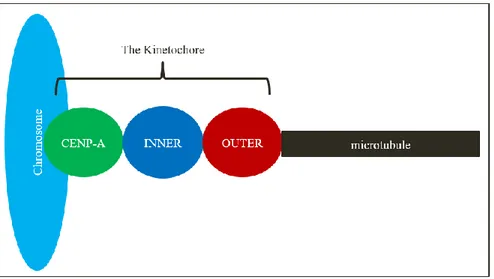
Figure 1.1: Organization of kinetochore proteins in the cell- CENP-A and other CCAN proteins are the closest to the chromosomes. The inner kinetochore proteins are located between the centromeric proteins and the outer kinetochore. The outer kinetochore makes direct contact to the microtubule (2).
The kinetochore and its proteins: the inner kinetochore
The inner kinetochore proteins make direct attachments onto the centromeres to connect the DNA and the kinetochore (2). The placement of the inner kinetochore proteins is required for the remainder of the kinetochore to assemble (2). A subgroup of the inner kinetochore proteins is called the constitutive centromere associated network (CCAN). The CCAN is composed of 16 kinetochore proteins that can be divided into 5 complexes and is always associated with the centromeres during the cell cycle (6,41). These protein complexes are C,
CENP-8 T/W/S/X, CENP-L/N, CENP-H/I/K/M, and CENP-O/P/Q/R/U (6,48). The first 4 complexes mentioned are necessary or chromosome segregation to take place (6).
CENP-A is centromeric protein A. CENP-A is a histone H3 variant located at the centromeres that is able to form nucleosomes (2,42). In nucleosomes, CENP-A takes the place of H3 at the end of the kinetochore and forms a nucleosome in complex with histone H2A/B and histone H4 (6,42). CENP-A is required for the kinetochore proteins to assemble at the centromeres (2). CENP-A is deposited onto the centromeres via acetylation by histone H4 Lys5 and Lys12 and is then monomethylated by histone H4 Lys20 in order for the inner kinetochore and other CCAN proteins to assemble onto CENP-A (6). The methylation serves as an epigenetic marker for the CCAN to recognize CENP-A in its assembly (6). In this way, CENP-A also functions as an epigenetic marker for the other kinetochore proteins to localize properly (2,6,43,44). CENP-A deposition begins at G1 in vertebrates. CENP-A will only be deposited on active centromeres completed by the histone chaperone HJURP (Holiday junction recognition protein) and the Mis18 complex (2,45,46). Mis18 will signal HJURP to the centromeres, that will then bring CENP-A onto the nucleosomes (2,45). During this time at G1, CENP-C is already present at the centromeres in somatic cells and serves as the receptor for Mis18 so CENP-A can accumulate (2,47). This ensures one location on the centromere for CENP-A to be deposited to provide one location where the kinetochore can attach to (2).
CENP-C is an important inner kinetochore protein that aggregates towards the kinetochore base and is needed to ensure that CENP-A is deposited at the centromeres (2,42,49). CENP-C binds directly to CENP-A and is an important link between the centromere and the inner kinetochore (Fig.1.2) (2,6,50).
CENP-T-W-S-X binds to DNA and is needed for stable attachment between the kinetochore and the centromere (2,50). Similar to CENP-C, this complex is also required to link the centromere to the outer kinetochore, in order for the outer kinetochore to be under the influence of the
microtubules (2). CENP-T does not bind to CENP-A like CENP-C, but binds to centromeric DNA (6). CENP-T/W/S/X and CENP-A are responsible and required for the interaction between the kinetochore and the DNA (50). Both CENP-C and CENP-T arms of outer kinetochore
9 kinetochore proteins assemble and organize dictates how the outer kinetochore proteins assemble onto the inner kinetochore platform in space (39).
The kinetochore and its proteins: the outer kinetochore
The outer kinetochore proteins interact directly with the microtubules and are responsible for the connection between the inner kinetochore proteins and the microtubules (Fig.1.2) (2). The outer kinetochore dictates organizing events that occur at the microtubule plus-end and are partly responsible for generating force within the kinetochore. It is also suggested it may play a role in ensuring checkpoint activity by helping control the SAC and in correcting misattachments between the microtubule and the kinetochore (2,39,42).
The outer kinetochore is composed of three independent protein complexes that when combined form the KMN network (2). These protein complexes are the KNL-1 complex (KNL1C), the Mis12 complex (Mis12C), and the Ndc80 complex (Ndc80C) (2). KNL1C is composed of Zwint-1 and KNL1 (6). Mis12C is composed of Mis12, Dsn1, Nsl1, and PMF1 (Nnf1) (6,52). Ndc80C is composed of Spc24, Spc25, Nuf2, and Ndc80 (Hec1) (6). Unlike the CCAN that is constantly present at the centromeres, these outer kinetochore proteins are recruited onto the inner kinetochore proteins during mitosis (6). Ndc80C is the only outer kinetochore complex to form the connection between the inner kinetochore proteins and the microtubules via Nuf2 and Hec1 (2,6,42). The Mis12C binds to both the Ndc80C and KNL1C (6). KNL1C communicates with signaling proteins like Bub1 part of the SAC and phosphatases like protein phosphatase 1 (PP1) (6). More specifically, KNL1C is where Bub1 will bind to (42). Each outer kinetochore protein complex has its own function.
CENP-C and CENP-T are seen to recruit the outer kinetochore proteins onto the CCAN via two different pathways (2,6). In somatic cells, CENP-C recruits Mis12C and KNL1C during
interphase (6,53). Ndc80C is the last outer kinetochore protein complex to be recruited and this occurs after NEBD (6). Unlike KNL1C and Mis12C, Ndc80C is not found in the nucleus during interphase (6,41). The CENP-T pathway for outer kinetochore protein recruitment differs highly compared to that of the CENP-C pathway (6). CENP-T will directly bind to Ndc80C without requiring KNL1C or Mis12C (6,54). In the CENP-C pathway, it is essential for KNL1C and Mis12C to bind prior to Ndc80C (6,54,55). This suggests that the CENP-C and CENP-T
10 CENP-C and CENP-T are required for proper interactions of the outer kinetochore to the
microtubules (2,50,56,57).
Figure 1.2: Organization of inner and outer kinetochore proteins in the cell- CENP-A is in green, inner kinetochore proteins (CENP-C and CENP-T/W/S/X) are in blue, and outer kinetochore complexes (Ndc80C, Mis12C and KNL1C are in red). Inner and outer kinetochore proteins make direct attachments to one another, and Ndc80C makes interactions with the microtubules (2).
The kinetochore and its proteins: the loosely associated proteins
The loosely associated kinetochore proteins are in control of signaling cascades and help in the communication between the kinetochore and the microtubules, the centromere, and the rest of the cell (2,6). Kinetochore assembly and disassembly is a well controlled process in somatic cells and these proteins that include Ska1 (spindle-associated and kinetochore-associated) complex, Bub1, Mad1 and Mad2 in vertebrates play an important role (2,6). Ska1 is needed for Ndc80C to bind to the depolymerizing end of the microtubule when force is being applied (2). Kinases like Aurora B (AurB) and the cyclin-dependent kinase 1 (CDK1) are highly involved in kinetochore assembly and disassembly in somatic cells and oocytes (8,10,41).
Kinetochore assembly
Kinetochore assembly and disassembly is well controlled in somatic cells. At the start of mitosis, the kinetochore remains detached from the microtubules (39). After NEDB, inner kinetochore
11 proteins rapidly assemble onto the centromeric proteins and within 20 minutes, outer kinetochore proteins assemble onto the inner kinetochore proteins (2). The inner kinetochore always
assembles prior to the outer kinetochore in somatic cells of vertebrates, and the outer kinetochore always disassembles prior to the inner kinetochore (2,6,42). After inner kinetochore protein assembly, KNL1C and Mis12C assemble first at S phase, followed by Ndc80C assembly at the end of G2 (41,58). At anaphase, the KMN network comes off from the inner kinetochore platform (41).
During mitosis, kinetochore assembly occurs in a specific order in somatic cells. First, the histone H3 variant CENP-A remains constant during mitosis. Second, inner kinetochore proteins CENP-C and CENP-T raise by 50% within 10 minutes of nuclear envelope break down and decrease 50% within 10 minutes of anaphase (41). Third, the outer kinetochore assembles. Before NEBD, Mis12C is present at centromeres, but reaches its maximum levels during mitosis (41,58). The majority of Ndc80C accumulates after prophase and is incorporated into the nucleus at prophase (41). Disassembly of the outer kinetochore is well timed, beginning 6 minutes prior to anaphase onset and disassembly terminated within 20 minutes post anaphase (41).
In oocytes, kinetochore assembly is less understood and timing differs to that of somatic cells. Signaling cascades have been studied in the oocyte, but kinetochore assembly in the oocyte has been less investigated (3,4,8). It appears that the human kinetochore in meiosis has similar architecture to that of the human kinetochore in somatic cells with the inner and outer kinetochore being distinct parts (3).
The kinetochore and signaling cascades
The kinetochore is under control of many signaling cascades in the cell, the main one being M-phase promoting factor (MPF), that is composed of Cyclin B (CycB)-CDK1 (cyclin-dependent kinase 1) (6,60). CycB-CDK1 also controls when cells begin mitosis, and is the main kinase during M-phase (1,6,61). It is observed in somatic cells that as the level of CDK1 increases, so does the frequency of kinetochore protein assembly; the level of CDK1 activity controls the rate at which the mammalian kinetochore assembles (1,2,41). CDK1 levels increase rapidly in the mitotic cell, allowing for the kinetochore to assemble without delay (1,61). The assembly of CENP-C and CENP-T onto the mammalian centromeres is controlled by phosphorylation by
12 CDK1 seen at interphase (2). Phosphorylation by CDK1 at CENP-T is required for the
interaction between CENP-T and the Ndc80C, in order to have proper contact between the inner and outer kinetochore arms (6). CDK1 does not only phosphorylate kinetochore proteins but phosphorylates other loosely attached proteins in order for them to bind to the kinetochore (6). CDK1 will phosphorylate the spindle-associated and kinetochore-associated (Ska) complex in order for it to bind to the end of Ndc80C to allow Ndc80C to interact with the microtubule (6,62,63). The phosphorylation driven primarily by CDK1 encourages the protein-protein interactions necessary for proper kinetochore protein communication and assembly (2).
CDK1 in oocytes exhibits different patterns of expression than in the somatic cell. In the somatic cell, CDK1 levels stay constant during mitosis and decrease after anaphase. In the oocyte, CDK1 activity increases slowly during Meiosis I and will peak at metaphase 6 hours post GVBD (1,8). This activity coincides with stable kinetochore microtubule interactions (8). Since CDK1 is dependent upon CycB, CycB levels in oocytes follow the same trends as CDK1 in the mouse oocyte (8). Also, CDK1 activity is required for stable end-on attachments between microtubules and kinetochores in oocytes (8). This is why CDK1 activity is gradual in oocytes because it puts off stable attachments for later on in meiosis, avoiding segregation errors (8). The slow
accumulation of CDK1 also ensures slow kinetochore attachments and assembly (1). Yet, correctly oriented chromosomes could lack proper attachments to kinetochore-microtubules, since this assembly can occur even as delayed into late Meiosis I (1,64). It is thought that this slow assembly reduces errors in the oocyte by ensuring proper attachments (1,8).
Aurora B kinase (AurB) is also involved in kinetochore protein assembly. AurB aids in the interaction between CENP-C and the outer kinetochore by phosphorylating Dsn1, a protein of the Mis12C and allowing this outer kinetochore complex to localize to CENP-C (6). AurB is more involved with kinetochore protein disassembly by correcting for misattachments between the kinetochore and microtubule (1,2,10,42). AurB encourages turnover of the microtubule at improper attachments between the microtubule-kinetochore interface (1,65). AurB will recognize incorrect attachments and will phosphorylate outer kinetochore complexes Ndc80C and the Dam1 and Ska1 complex (2,58,62,66). This phosphorylation inhibits protein interactions between the kinetochore and microtubule and allows proper kinetochore-microtubule
13 complex (CPC) and is able to act specifically at the inner centromere, only phosphorylating improperly attached kinetochores (2,6,67).
AurB behaves the same in mouse oocytes during Meiosis I as that in somatic cells during mitosis. Upon phosphorylation, it destabilizes the interactions between the kinetochore and the microtubule (8). Oocytes possess another homolog of Aurora kinase called Aurora Kinase C (AurC). In mammalian oocytes, AurC has similar functions to AurB (1). Yet, why two versions of the kinase are required in oocytes remain less understood (1,68). AurA also phosphorylates misattachments during Meiosis I, as seen in mitosis (7,69,70). AurB/C acts differently in meiosis in oocytes than does AurB in mitosis in somatic cells (7). Upon correct attachment in mitosis, AurB phosphorylation decreases whereas even with correct attachment in meiosis, AurB/C phosphorylation levels remain high (7,69). This is because since the kinetochore is being pulled in towards the centromere, it moves closer to the inner centromere where AurB/C is located (7). This is when phosphatase PP2A acts during metaphase to counteract this kinase and to stabilize attachments (7). Attachment of stable kinetochore-microtubule attachments is slow during meiosis. AurB/C activity is high until the spindle achieves bipolarity, and after Aurora activity decrease while CDK1 levels slowly increase (7,64). The main kinases that control kinetochore assembly and disassembly are CDK and the Aurora Kinases (7,8,41). Yet, other kinases are also involved in ensuring correct microtubule-kinetochore attachments, like Bub1, Mps1, and Polo-like Kinase 1 (Plk1) (2).
The kinetochore and its attachments to the microtubule
The kinetochore can make correct attachments to the microtubule via lateral attachments when the kinetochore binds to the microtubule from the side or end-on attachments when the
kinetochore binds to the plus end of the microtubule (2). End-on attachments are stronger and more stable than lateral attachments (2,42). Ndc80C creates the kinetochore-microtubule attachments that often begin as lateral and then switch to the stable end-on attachment before proceeding with mitosis (39,71,72,73).
Kinetochore-microtubule attachments are also seen to contribute to aneuploidy. Young mice oocytes have more proper polar attachments than oocytes from aged mice, with older mice showing fewer end-on attachments (4,35,74). This demonstrates that timing of proper
14 attachments is malfunctioning in older oocytes and contributes to misattachments seen in
aneuploidy (4).
The kinetochore can make different attachments to the microtubules depending where the oocyte is in its maturation. In Meiosis I, sister kinetochores make connections to the same pole and are conserved as a pair to divide the homologous chromosomes to its respective side, termed amphitelic attachments (1,75). In Meiosis II and in mitosis, sister kinetochores attach to
microtubules coming from spindle poles on opposite ends of the cell to divide sister chromatids (1). Yet, correct kinetochore-microtubule attachments do not always occur and incorrect
attachments can result. Some of these incorrect attachments include merotelic attachments, syntelic attachments, monotelic attachments, and no attachment (2,3,39). Merotelic attachments occur when one kinetochore makes connections to microtubules coming from both poles of the spindle poles (2,39). In somatic cells, merotelic attachments are not detected by the SAC and lead to chromosome segregation errors (4). When this occurs, a pull can be felt by the
chromosome coming from both poles during chromosome segregation at anaphase. Hence, this improper attachment can lead to improper segregation and chromosome lag (1). Merotelic attachments also occur when the kinetochore-microtubule attaches to a spindle that fails to undergo bipolarization (7,10,15). Unlike in mitosis where kinetochore microtubule attachments are corrected prior to metaphase, kinetochore-microtubule attachments in meiosis can have merotelic attachments into metaphase and these need to be corrected (7,64,74,76). Merotelic and lateral attachments increase in older mouse and human oocytes that have less cohesin and cohesion (3,4,34,35). These misattachments can also occur due to a change in kinetochore geometry in older oocytes (4,77).
Merotelic attachments are not recognized by the SAC (spindle assembly checkpoint), but are corrected by tension sensors at the kinetochore (4,7,77). As AurB/C levels decrease, the increase in tension is sensed and merotelic attachments switch to end-on stable attachments (7). This is thought to occur upon the dephosphorylation of the substrates after AurB/C activity is completed (7,64).
The second misattachment, syntelic attachments (monopolar attachments) occur when both sister kinetochores make connections to one spindle pole (2,39,50). The third misattachment,
15 attachment (3). This is observed when an attachment occurs prematurely or when improper error-correction occurs (3,64). The fourth misattachment, unattached occurs when one or both sister kinetochores do not connect to the microtubule (2,50). In somatic cells, a correct bi-oriented attachment needs to occur between the sister kinetochores and the microtubules (2). The kinases ensure correct kinetochore-microtubule attachments for the cell to progress to anaphase; when errors are detected, the cell cycle is halted until correct attachments are made (2). This occurs with oocytes where in Meiosis I, amphitelic attachment is achieved in order to be able to properly segregate chromosomes to either end during anaphase (1).
In human oocytes during Meiosis I, the kinetochore can have different attachments to the microtubule. Sister kinetochore pairs can have one kinetochore make a connection to a
microtubule or sister kinetochore pairs make connections to microtubules at the same time (3). The later attachment ensures that each kinetochore makes connections to a separate spindle (3). The homologous chromosomes can make attachments to spindles in Meiosis I in two ways, so the kinetochore pair in the human oocyte can act independently of each other (3). Hence, in human oocytes, there are 4 places where the kinetochore can attach onto independent of each other; making bi-orientation more difficult to achieve (3,64). This contributes to how error prone Meiosis I is (3).
The kinetochore and its control factors
Two control factors of M-phase progression include the SAC and the anaphase promoting complex/cyclosome (APC/C), which are both involved in Meiosis I (78). Anaphase is delayed upon SAC activation when incorrect kinetochore-microtubule attachments are sensed by the SAC at the kinetochore-microtubule interface (2,7,79). Correct attachments must occur before proceeding with the cell cycle to ensure correct chromosome segregation will take place (2,8). The proteins of the SAC include Bub1, BubR1/Mad3, Bub3, Mad1, Mad2, and Mps1. The SAC is a group of proteins that are part of the regulatory proteins that help the kinetochore to
communicate with the rest of the cell (2,79). These SAC proteins aggregate to the kinetochore when improper attachments occur (1,80). This SAC signal is activated upon Bub1
phosphorylation and this will allow Bub1 to bind to Mad1 (6). The SAC also operates by the use of Mad2 in somatic cells that binds to kinetochores when there are misattachments between the kinetochore and the microtubule (2,81,82). Mad2 will form the mitotic checkpoint complex
16 (MCC) by binding with Mad3 and BubR (2,83). MCC then binds to inactive Cdc20 and this will prevent anaphase. Cdc20 will activate the APC once bound to it, once correct attachments occur and the cell will continue to anaphase (2,81,82). This is one way that the kinetochore behaves as a signaling hub for communication with the cell in ensuring proper chromosome segregation (2). In oocytes, the SAC also functions during Meiosis I, though at low intensity. It is found to be inactivated in late MI, even when misaligned chromosomes and misattachments between the kinetochore and microtubule are present (8,84,85). The SAC in oocytes cannot sense minor misattachments or misaligned chromosomes and allows errors to persist in mice and human oocytes by not inhibiting the APC/C (1,84,85). This is thought to occur due the oocyte’s large size. (1).
The way kinetochore assembly and signal transduction occurs in the oocyte during meiosis differs greatly to that in the somatic cells during mitosis (8). Sister kinetochores in meiosis need to attach to one pole only to ensure that homologous kinetochores will be pulled to either end of the cell; this separation of homologous kinetochores is needed for separation of homologous chromosomes (7,86). It is also observed that the stable attachment between kinetochores and microtubules is different between mitosis and Meiosis I. In mitosis, stable kinetochore and microtubule attachments occur after NEBD when the kinetochores are bioriented (7,10). In meiosis, the stability increases steadily well into metaphase, when the kinetochores are switching from apolar to bipolar (7,8,84,85). This activity coincides with CDK1 levels in the cell (7,8,41). As well, though the SAC is well functioning in somatic cells, this error correction method presents itself with less fidelity in meiotic cells (1,8). Therefore, kinetochore assembly and signaling transduction is well known in the somatic cell, but needs to be investigated in oocytes (8).
Kinetochore disassembly
Phosphatases are used to disassemble the kinetochore and continue the cell cycle (2). Yet, how the kinetochore disassembles is less understood than how the kinetochore assembles in somatic cells (6). The kinetochore only begins to disassemble upon high levels of the APC/C at anaphase onset (41). It is known that the CycB-CDK1 levels are also seen to dictate the rate at which the kinetochore disassembles. CycB-CDK1 activity decreases upon the degradation of CycB (6). It is
17 observed that a combination of decreasing CDK1 levels and the action of protein phosphatases are required for the outer kinetochore to begin in its disassembly (41). This cues the end of mitosis and kinetochore disassembly. These phosphatases include the protein phosphatase 1 (PP1) and protein phosphatase 2A (PP2A) (6). PP1 opposes the activity of AurB by
dephosphorylating the substrates of AurB, like Dsn1 of the Mis12C (6,87). This occurs at anaphase when AurB moves from the inner centromere to along the spindle (6). This is how the CENP-C-Mis12C interaction between the inner and outer kinetochore terminates. The PP2A phosphatase will dephosphorylate the substrates phosphorylated by CDK1, specifically the CENP-T-Ndc80C interaction between the other inner and outer kinetochore arm (6). This dephosphorylation is essential in order for outer kinetochore protein disassembly to occur (41). Ndc80C is the outer most component of the outer kinetochore in contact with the microtubule, and in order to obliterate this interaction, dephosphorylation must occur (41). Outer kinetochore protein disassembly occurs independently of activity at the microtubule, and is governed by phosphorylation and dephosphorylation patterns (41). In somatic cells, CDK1 levels are seen to peak right after NEBD, and will stay constant until after anaphase where they drop (6,41). In oocytes, the role of kinetochore disassembly is less known, but CDK1 levels appear to control kinetochore assembly. CDK1 levels in the oocyte accumulate slowly after prophase and peak at metaphase(1,8). Also, Protein Kinase C (PKC) in the oocyte plays a role in controlling anaphase onset, and is involved in kinetochore disassembly (60). Yet, kinetochore disassembly and its other protein regulators in the oocyte are much less understood (1,4,8)
The kinetochore and distance: the concept of delta
The kinetochore is a flexible structure that is dynamic and compliant, with the distance between kinetochore proteins changing over time (6,42,88). This distance that separates kinetochore proteins is termed delta (42). It is observed in somatic cells that the inner kinetochore proteins undergo the most displacement whereas the outer kinetochore proteins appear noncompliant (42). This was deduced by using anticancer drugs like taxol that inhibits microtubule
polymerization prematurely activating the SAC (42,82,89). When cells were treated with taxol at metaphase when the kinetochore was under the most tension from the microtubules, this allowed the kinetochore to be viewed under no tension (42,88). The kinetochore proteins were observed to approach one another, whereas under partial influence from the microtubule they remain
18 separated (42). This demonstrates that the kinetochore is a compliant and dynamic structure (42). Kinetochore compliance in oocytes remains to be investigated (3,4)
Measurements of delta in somatic cells describe the stiffness or compliance of the kinetochore proteins. It was observed in somatic cells that kinetochore proteins change their delta throughout mitosis, with emphasis during metaphase when maximum tension is observed and chromosomes oscillate at the metaphase plate (42,88). It was found in somatic cells that only CENP-A and CENP-C are compliant proteins during metaphase oscillations of the chromosomes, whereas all other kinetochore proteins remain noncompliant (42). Oscillations occur when chromosomes change rapidly between poleward and antipoleward movement due to microtubule influence. Due to the oscillations, either the kinetochore can be noncompliant and stiff or the kinetochore can be compliant and move along with the oscillations (42,90,91).
The Ndc80C was found to be a stiff complex in somatic cells. Under treatment of taxol, Ndc80C remained noncompliant, unlike the inner kinetochore proteins (42). The Ndc80C remained fixed at metaphase even when under high tension from the microtubules in relation to the inner
kinetochore (42). This result was surprising since Ndc80C helps to transduce the force derived from the microtubules (42,92). Nocodazole is another drug used to obliterate the microtubules, similar to Taxol. This occurs by nocodazole binding to 𝛽-tubulin and preventing polymerization (42,93). This drug was used depolymerize microtubules to view Ndc80C under no tension and it was observed that the Ndc80C proteins became closer together (42). This suggests that the Ndc80C may be flexible from within, but as a whole the structure does not move (42).
In somatic cells, CENP-C was the inner kinetochore protein observed to be the most compliant. The remainder of the CCAN was found to be noncompliant during metaphase oscillations with Ndc80C, and including complexes KNL1C and Mis12C (42). Under taxol treatment, it was observed that CCAN proteins such as CENP-A, CENP-C, and CENP-T all moved towards the Ndc80C, rather than towards the centromere (42). Components of the KNL1C and the Mis12C were also seen to move during Taxol treatment (42). A possibility for this occurrence is the existence of two arms to the kinetochore; CENP-C attaching to Mis12C, KNL1C and Ndc80C, and CENP-T-W-S-X attaching to Ndc80C (2,42). The CENP-C arm with the KMN network is compliant and more flexible whereas the CENP-T arm with Ndc80C remains stiff and
19 centromere, compared to the KMN network arm which remains in place. This insinuates an uncoupling of the kinetochore arms under this treatment and that the kinetochore arms may act independently of each other. Overall, it was found that the link between the centromeres and the CCAN was compliant, whereas the link between the CCAN and to the outer kinetochore is noncompliant in somatic cells (42).
The kinetochore delta measurements in somatic cells was completed, but has yet to be done in oocytes. The oocyte has different CDK1 level activity than in somatic cells and is possible that delta will differ in the oocyte compared to the somatic cell (1,8). One preliminary study observed the distances between the meiotic human sister kinetochores increase with age (3,34,64,94). Also, the intra-kinetochore distance from the inner to the outer kinetochore increases with age in relation to cohesion loss (3,34). The kinetochore distance is greater in human oocytes compared to mouse oocytes in relation to aneuploidy (3). Yet, the distances surrounding the meiotic kinetochore is understudied and worth investigation (3).
The kinetochore and force
The term force has multiple applications in different fields. It is a vector, where it has both speed (magnitude) and a direction. In physics terms, F=ma, where ‘m’ is mass in kilograms and ‘a’ is acceleration in m/s2. A force upon an object can create or destroy, make bonds or break bonds,
and can create or release tension (50,95). The kinetochore, and not solely the microtubules, is partly responsible in generating the force to pull apart the chromosomes during chromosome segregation in somatic cells (50,88). The kinetochore is under both passive and active force within itself; the active forces create the force and the passive forces are observed at points of friction (88). The kinetochore is able to slide over itself and is able to polymerize and
depolymerize (50,88).
Force at the kinetochore has two main functions. The first is to signal correct attachments of the kinetochores to the microtubules and the second is to pull apart the chromosomes for
chromosome segregation to occur (50). The kinetochore transduces the force that is derived from the microtubules, in order to correctly orient and pull apart the paired sister chromatids in mitosis (50,88). The kinetochore is under different amounts of force during cell division due to the varying length of microtubules during the cell cycle (50). The greatest amount of force and
20 tension observed at the kinetochore is during metaphase when opposite spindle poles make connections to the sister chromatids to align the chromosomes in a bioriented manner (42,50,88,96). Two main movements can occur at the kinetochore. Poleward movement
predominately occurs at anaphase when the kinetochore is being pulled toward the opposite pulls of the cell (50,88). Antipoleward movement occurs when the kinetochore is being moved away from the cell membrane extremities and towards the centre of the cell (50,88). Both of these movements affect the intra-kinetochore and interkinetochore distances (50). Oscillating between these two movements occur when the chromosomes are lined at the metaphase plate (88). During these oscillations, there is also force coming from the chromosomes as they are getting ready to separate their sister chromatids (50,96). It was observed that during poleward movement, the distances between the kinetochore proteins decrease and the kinetochore compresses within itself, due to active forces at the kinetochore (88). When the microtubule is depolymerizing, active forces are produced at the kinetochore and this creates a pull of the chromosomes towards the poles (88). The opposite is observed upon polymerization of the microtubule, and friction is created at the kinetochore due to passive forces as a result. This passivity is generated by
antipoleward movement, and the sliding motion of the kinetochores going over the microtubules (88,97,98). Mechanical compliance of the kinetochore allows the kinetochore to respond
physically to a force acting upon it, either via compression or stretching (88).
Figure 1.3: Movement of the kinetochore- The kinetochore can move in an antipoleward fashion towards the chromosome, or in a poleward fashion towards the microtubules (50).
21 Microtubules are responsible for pulling apart the chromosomes via force derived from their polymerization and depolymerization. The kinetochore is also another force transducer; it is responsible for generating the force from the microtubules onto the chromosomes to pull them apart. (39,88). This interaction between the kinetochore and the microtubule is dynamic throughout the cell cycle. In somatic cells, beginning at prometaphase, microtubules bring
kinetochores to the metaphase plate where the centromere-kinetochore-microtubule attachment is made. Following this at anaphase, the kinetochores still attached to the centromeres pull apart the homologous chromosomes to either end of the cell with the force generated by the microtubule (2). The kinetochore begins its attachment to the microtubule laterally, but converts to the more robust end-on attachment when pulling the chromosomes apart during chromosome segregation. (2,4,8). Upon making end-on attachments, the force to pull apart the chromosomes can come almost all from the microtubules depolymerizing. This allows the kinetochore to maintain the connection between the centromere and the microtubule and not generate all the force (2,99). The force required to pull apart the chromosome derive mainly from the microtubules upon their depolymerization. When microtubules depolymerize, this process is also called catastrophe and it can generate a significant force up to 65pN (50,100). The kinetochore must find a way to use this force with control and decrease the rate of the microtubule; by forming precise attachments and modulating the tension (50,101–103). Also, the kinetochore is able to withstand a high amount of force and remains stable under high tension (50). The kinetochore can withstand up to 700pN of force, when the typical force to break a bond is 100pN, demonstrating its robustness (50,104). The kinetochore is able to diffuse the force from the microtubules to its proteins in the arms of the outer-inner kinetochore, allowing the proteins to experience the force at the same time (50). Ndc80C is the outer kinetochore protein complex that attaches to the microtubule; 10 to 20 Ndc80C proteins can attach to one end of a microtubule, making for a robust interaction and stable way to diffuse the force (50,105–107). In humans, 20 microtubules can be attached to one kinetochore via its plus end, demonstrating the stability of the kinetochore (39,42). This occurs as the microtubules undergo polymerization and depolymerization (39).
Several motor proteins generate force at the microtubule-kinetochore interface including the kinetochore motors dynein and kinesin (50). The spindle poles and kinetochore can also generate a force; not solely the microtubules (50,88,108,109). When incorrect attachments of the
22 kinetochore-microtubule arise, the forces from the kinetochore can aide in moving the
chromosomes by regulating tension and help control the microtubule in its movements (50). The kinetochore also uses its force and the force from the microtubule to correctly guide the
chromosomes spatially (2). In humans, the kinetochore works with the microtubules in order to move the chromosomes over a distance of 5μm while experiencing forces of 100pN
(39,103,110).
Proteins termed MAPs (microtubule-associated proteins) act at the kinetochore to help promote polymerization and depolymerization of the microtubules (39). These proteins are the
cytoplasmic linker associated proteins (Clasp1 and Clasp2) and the kinesin-like proteins (Kif2c and Kif18a), respectively (2,111,112). These proteins aid the kinetochore by helping control microtubule dynamics, hence allowing the kinetochore to use its force and the force coming from the microtubules to correctly guide the chromosomes in their placement and segregation (2). More specifically, Clasp proteins encourage polymerization and inhibit depolymerization of the microtubule (42,112). Other motor proteins include the CENP-E motor protein and the CENP-F coiled- coil protein (39,42). CENP-E is a type of kinesin protein dependent upon Bub1 and is partly responsible for ensuring correct kinetochore-microtubule attachments. Bub1 is needed for CENP-E to localize to kinetochores (42,113–115). CENP-F, like CENP-E also requires Bub1 to localize to the kinetochore (42,58,113). Many of the motor proteins are redundant in their function. It is thought that since the kinetochore is robust with many attachments, using these redundant proteins helps to ensure the kinetochore functions properly (39).
The kinetochore is able to sense when misattachments occur at the microtubule-kinetochore interface. This is due to tension sensed at the kinetochore; improper attachments produce less tension at the kinetochore than do proper attachments (50,88). The kinetochore can sense the distance between its pairs (interkinetochore distance) and within a kinetochore, the inner and outer kinetochore distance (intrakinetochore distance) (50,116). The kinetochore assembly is a timely process and the distance between kinetochore proteins in a single kinetochore can be measured over time. Therefore, the distance between inner kinetochore components and outer kinetochore components is coupled and when misattachments occur, these distances become uncoupled (50,88,116). This is a way for the kinetochore to verify its assembly (50). The
23 and from the microtubule (50,88). This is well observed in somatic cells, and more needs to be investigated in oocytes (7,50,88).
AurB is an important kinase able to correct for misattachments between the kinetochore and the microtubule by sensing tension between these two cellular components (50,67,117). When AurB senses a misattachment, or lack of tension, its substrates are under a high amount of
phosphorylation (50,51,117). This phosphorylation weakens the affinity of the outer kinetochore to the microtubule and keeps the kinetochore unattached to the microtubule, awaiting a proper attachment (39,50).
When correct biorientation is achieved, the substrates of AurB become dephosphorylated. This is when distance and spatialization have an impact (50,67,117). AurB is located at the inner
centromere region whereas the majority of its substrates are located at the outer kinetochore (42,50). When proper attachment and tension is achieved, the outer kinetochore becomes further away from the inner kinetochore/centromere (50). This distance prevents AurB from
phosphorylating its substrates; hence tension and force also play a role in ensuring correct kinetochore-microtubule attachments (50). When the activity of AurB is decreased, the tension is stabilized and so are the attachments between the kinetochore and the microtubule (8). In monopolar attachments, there is less tension between the kinetochore and microtubule
attachments than observed in proper bipolar attachments (15,39,65). When this loss of tension occurs due to improper attachments, other error correction mechanisms ensue. How this lack of tension activates AurB is less understood, but is a way in correcting improper attachments (39). In oocytes, the same was observed where AurB plays a role in tension and ensuring kinetochore-microtubule attachments during MI (8). Yet, AurB/C is less sensitive in its activation and inactivation in meiosis than in mitosis. Phosphorylation of AurB stops in mitosis as tension persists and the substrates of the outer kinetochore go further away from AurB (7). In oocytes, it phosphorylation by AurB/C onto kinetochore substrates still persists, even when the kinetochore becomes stretched (1,64). This is an inherent problem. AurB/C prevents improper kinetochore-microtubule attachments and encourages this binding turnover (1,65). In oocytes, even when maximum tension of the chromosome and kinetochore persist, microtubule-kinetochore turnover could also persist if phosphorylation by AurB/C keeps continuing (1,64). If turnover keeps occurring, then reattachments can occur, leading to mis-attachments when wanting to attach
24 again, and hence segregation error. This is another reason why the oocyte is prone to
25
Chapter 2- The Rationale and Hypothesis
The Rationale
The kinetochore, its assembly, and its signaling cascades have been extensively studied in the somatic cell (2). Yet, much less is known about the kinetochore in the developing mammalian oocyte in regards to its assembly, proteins, and signaling cascades (1,7,18). There are many differences between the oocyte and the somatic cell in regards to kinetochore assembly and dynamics. First, CDK1 levels are different in the mammalian oocyte compared to that in somatic cells (1,8). Second, the oocyte has another homolog of Aurora kinase than somatic cells (1,39). Third, the oocyte posses an error prone chromosome segregation and is subject to aneuploidy more than the somatic cell (1,3,4). The kinetochore is at the centre of chromosome segregation and may be involved in aneuploidy (3,4). This project aims to investigate the kinetochore in developing mammalian oocytes during Meiosis I, and achieve a better understanding of this structure and its potential role in aneuploidy. The kinetochore appears to be flexible and compliant in somatic cells and oocyte findings will be compared to known somatic data (42).
The Hypothesis
In regards to the oocyte kinetochore, it is hypothesized that oocyte kinetochore protein assembly will undergo different timing and kinetics compared to what is observed in the somatic cell. It is also hypothesized that the kinetochore’s flexibility, compliance, and delta will differ in the mammalian oocyte during Meiosis I than what is observed in somatic cells during mitosis. This project is divided into two aims that focus on: 1) kinetochore protein assembly and 2)
kinetochore protein dynamics. The first aim of this project hopes to look at the kinetochore protein assembly and observe where along Meiosis I this compares and contrasts with actions in mitosis. The second aim of this project will observe kinetochore compliance in the mouse oocyte and compare that to the somatic cell.
26
Chapter 3- Materials and Methods
Methods
The aims of this project were completed via 3 major sets of experiments: First, viewing
kinetochore assembly in fixed oocytes via staining with antibodies. Second, viewing kinetochore assembly in live oocytes via microinjection with mRNAs that will be translated to proteins that are fluorescently tagged. Third, fixing oocytes and staining with antibodies after microinjection to measure delta of kinetochore proteins.
Dissection
Prior to dissection, 5 collection dishes (Culture Dish; Thomas Scientific #9380D40) must be prepared. 2 dishes with 5mL each of IBMX:M2 media 1:1000, (M2 medium; Sigma-Aldrich® #M7167 and IBMX (3-isobotyl-1-methyl-xanthine) (IBMX; Sigma-Aldrich ® # I5879) and placed on the heat pad set to 37℃. 1 dish with 20μL drops of IBMX:M2 in a flower pattern, covered with mineral oil to be placed on the heat pad (Mineral Oil (light oil/neat); Sigma-Aldrich® #M8410). 1 dish with 20μL drops of IBMX:M16 (1:1000) in a flower pattern (M16 medium; Sigma-Aldrich® #M7292) and 1 dish with 20μL of M16 medium to be placed in the incubator, set to 37℃ with 5% CO2. IBMX is a drug that is a non-selective phosphodiesterase
inhibitor. It is able to halt meiosis by keeping cAMP concentrations high in the oocyte, keeping the oocyte under meiotic arrest (118).
Young CD-1 female mice aged 3 to 4 months (Charles-River #022CD1) were injected with 0.2mL pregnant mare serum gonadotropin (PMSG) (PMSG Genway Biotech; GWB-2AE30A), two days prior to sacrifice (4). Injections of PMSG were administered via syringe with a 27gauge needle and injected subcutaneously (1mL syringe; BD Biosciences #309659 and Precision Guide Needle 27G; BD Biosciences #305109). Injections were performed by holding the back and neck of the mouse with one hand, flipping the mouse so that the stomach is facing up, and injecting in the lower abdomen close to the legs. PMSG is a gonadotropin and causes the ovaries to undergo superovulation; this promotes folliculargenesis and increased oocyte formation. Injections were given Day 1 in the afternoon (between 16:00-18:00) (119–121).
27 On Day 3 in the morning (07:00-10:00), CD-1 female mice were sacrificed for their ovaries. Sacrifices and dissections were performed in the animal facility. Mice were placed in a
biological safety hood within a chamber for which to manipulate the mice. The ventilation box was connected to two tubes; one supplied oxygen and the other isoflurane. The mice began in the box with only oxygen supplied to them, and slowly, the isoflurane concentration was increased to 3-4%. This anesthetized the mice. They were only removed from the box once a reflex test was administered to confirm anesthesia, and sacrificed by neck dislocation. Post dislocation, a reflex text is administered, and then dissection to extract the ovaries can commence.
The mouse was flipped onto its back and ethanol was sprayed on its stomach to wet the hair and prevent it from contaminating the ovaries. The skin was cut at the pelvic line and opened up to expose the peritoneal cavity. This lining was cut to expose the stomach, intestines, ovaries and fallopian tubes. The intestinal organs were pushed up towards the upper chest to expose the fallopian tubes and ovaries. The fallopian tubes were pulled up to locate the ovaries. The fallopian tube was cut at the base of the ovaries to isolate both ovaries from the mouse. The ovaries were placed in a 1.5mL Eppendorf tube (Sigma-Aldrich® #Z336769) filled with IBMX:M2 (1:1000).
The ovaries in IBMX:M2 medium were dissected under the microscope without a heat pad. In between manipulations, the culture dishes were placed back on the heating pad (if containing M2) or back in the incubator (if containing M16). Under a dissecting microscope, the ovaries were placed in Dish #1 with 5mL IBMX:M2. The fat was stripped away from the ovaries via tweezer manipulation (Ideal-Tek, 5.SA). The fat was left in Dish #1 and the ovaries were placed in Dish #2 containing 5mL IBMX:M2. Tweezers in one hand held the ovary in place and in the other hand was a syringe with a needle 27gauge needle. The syringe with the needle was used to scratch away the ovary to dissect it and release the oocytes from within it.
Oocytes covered in a surrounding layer of granulosa cells must be transferred from Dish #2 to Dish #3 containing the IBMX:M2 drops. This was completed via mouthpipetting. With a Bunsen burner, the tip of a glass pipette was molded into just about the size of a diameter of a mouse oocyte. This was confirmed under the microscope. Once this was completed, a P1000 filtered tip was added to it, which was then added to the rubber mouth pipette (Aspirator Tube; Sigma-Aldrich® #A5177-5EA). The plastic holder of the aspirator was placed against the lips where an air flow controlled the rate at which and how many oocytes were taken in. Prior to taking