OATAO is an open access repository that collects the work of Toulouse
researchers and makes it freely available over the web where possible
Any correspondence concerning this service should be sent
to the repository administrator:
[email protected]
This is an author’s version published in:
http://oatao.univ-toulouse.fr/25561
To cite this version:
Bonou, Sidoine and Sagbo, Etienne and Aubry, Clémentine and
Charvillat, Cédric
and Ben-Nissan, Besim and Cazalbou, Sophie
Conversion of snail shells (Achatina achatina) acclimatized in Benin to
calcium phosphate for medical and engineering use. (2019) Journal of the
Australian Ceramic Society, 55 (4). 1177-1186. ISSN 2510-1560
Official URL: https://doi.org/10.1007/s41779-019-00334-6
https://doi.org/10.1007 / s41779-Q 19-00334-6
Conversion of snail shells (Achatina achatina) acclimatized in Benin
to calcium phosphate for medical and engineering use
S. A. S Bonou 1 • E. Sagbo 1 • C. Aubry2'3 • C. Charvillat2 • B. Ben-Nissan4 • S. Cazalbou 2
Abstract
Most methods for producing calcium phosphates involve synthetic calcium and phosphates sources. However, it has recently been proposed that calcium phosphate can be produced with bio-based calcium sources such as nacre, coral, and cuttlefish bones. One specific source of bio-based calcium is found in the Achatina snail shell, which becomes a waste product after flesh consumption. The present work aimed to assess the effectiveness of Achatina snail shells and to study the conversion kinetics in both acid and alkaline environment rich in phosphate ions. It was observed that in acidic conditions, the calcium released by the dissolution of the aragonite precipitates with the phosphate ions ofreaction medium induces brushite formation which is rapidly converted into monetite. In alkaline conditions, calcium released from aragonite reacts with surrounding phosphates and car bonate ions and induces carbonated apatite precipitation. Regardless of the source of calcium used in the presence of phosphate, the conversion is carried out according to complex phenomena that involve topotactic transformation or dissolution-precipitation mechanisms.
Keywords Calcium phosphate • Calcium carbonate • Snail shell • Chernical conversion • XRD • FTIR
Introduction
Historically, natural products such as wood, gold, and coral were some of the first materials used to repair human bones. Today, these products remain a source ofinspiration for mod ern researchers in the field of biomaterials. Several research initiatives have focused on exploring new possible materials for the production of calcium phosphate [1-7]. One prornising biosourced material is coral. lndeed, coral shows promise due to its porous structure which is similar to cancellous bone, its chernical composition which provides the calcium ions nec essary for the precipitation of neoformed bone, and its
r8I S. Caz.albou
sophie. caz.albou@univ tlse3.fr
1 Laboratoire de Chimie Inorganique et de l'Environnement (LACJE) FAST, UAC, Cotonou, Bénin
2 CIRJMAT Carnot lnstit ute, UPS INPT CNRS UMR 5085,
University ofToulouse, Toulouse, France
3 Laboratoire de Génie Chimique, UMR 5503, INPT, Université de Toulouse 3, Toulouse, Franc e
4 Department ofChernistry and Forensic Science, University of Technology Sydney, Broadway, NSW 2007, Australia
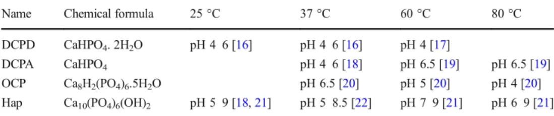
bioactive ions such as magnesium and strontium that promote the activity of osteoblasts responsible for bone regeneration [8-11 ]. lt is the reason why many articles treat on the use of coral in bone repair surgery and recent studies have focused on the conversion of calcium carbonate coral to calcium phos phate to promote bone regeneration [6, 12-15]. Regardless of the source of calcium used in the presence of phosphate, the conversion is carried out according to complex phenomena that involve topotactic transformation or dissolution reprecipitation mechanisms that Jead to the formation of dif fe ren t phases: monetite (DCPA), brushi te (DCPD), octacalcium phosphate (OCP), hydroxyapatite deficiency (DHA), hydroxyapatite (HA), or tricalcium phosphate (TCP). The phases formed then depend on the experimental conditions. The main conditions influencing precipitation of calcium phosphate are phosphate and calcium concentrations, pH, temperature, ionic strength, the presence of other ions, and reaction duration. Table 1 summariz.es the various calcium phosphates formed, talcing into account the temperature and pH parameters.
Recently, Achatina achatina snails have been considered a prornising material with which calcium phosphate can be pro duced. As such, the current study explored the possibility of converting Achatina snail shells, which constitute a "noble"
waste, to calcium phosphate using the simply wet chemical method. Moreover, the kinetics of conversion was investigat-ed. The morphology and evolution of the crystalline phases were analyzed over a period of 24 h, taking into account the reaction media used and in particular their pH.
Materials and methods
Material
The raw materials used for the current study were the shells of Achatina achatina snails. After washing and rinsing with dis-tilled water, the shells were crushed in a mortar and then crushed in a mill. Then, the powder was sieved in a 1-mm sieve. The diameter of the resulting powder was less than 1 mm. Figure1shows the stages of powder production.
Methods
Conversion ofAchatina snail shells to calcium phosphate In order to follow the conversion of snail shells to calcium phosphate, two solutions containing phosphate were prepared to obtain phosphate acid and alkaline media. The two solu-tions of reagent (acid reagent A: H3PO4and alkaline reagent
B: (NH4)2HPO4) were prepared either by mixing 5 mL of
orthophosphoric acid (85% by weight) or by dissolving 6.67 g of diammonium hydrogen phosphate in 325 mL of deionized water. Then, 6 g of snail shell powder was added to the prepared solutions, stirred at 540 rpm, and maintained at 80 ± 0.5 °C for 24 h. The Ca/P solution ratios were chosen so that under given pH conditions (acidic or alkaline pH), the
sub-saturation of solutions avoids the simultaneous precipita-tion of two phosphocalcic phases and allow to follow the evolution of the conversion of the snail shells.
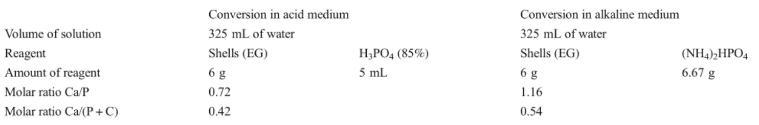
Thus, at acidic pH, the Ca/P = 0.72 ratio was lower than those of the brushite or the monetite (Ca/P = 1). Similarly, at alkaline pH, the ratio Ca/P = 1.16 was lower than those of OCP (1.33) and HA (1.667) which preferentially precipitate at these pH values. At 0.5 h, 1.5 h, 2.5 h, 3.5 h, 4.5 h, and 24 h, an equal volume of the suspension was taken from the reaction medium. The samples were filtered, washed with deionized water, and dried at 37 ± 0.5 °C for 15 h. The products obtained from con-versions carried out in the acid medium were denoted EG-A 0.5 h, EG-A 1.5 h, EG-A 2.5 h, EG-A 3.5 h, EG-A 4.5 h, and EG-A 24 h. The products obtained from conversions carried out in the alkaline medium were denoted EG-B 0.5 h, EG-B 1.5 h, EG-B 2.5 h, EG-B 3.5 h, EG-B 4.5 h, and EG-B 24 h. Details of the composition of the reaction media and the corresponding ratios Ca/P and Ca/(P + C) are shown in Table2.
Phase analysis by XRD
Phase analysis of the products was carried out via the X-ray powder diffraction method using D8 ADVANCE (BRUKER), under Cu Kalpha1 + 2 = 0.15418 nm radiation, from 20 to 80° (2 theta), steps of 0.02°, 2 s/step, divergence slit 0.3°, Sollers slit 2.5°, and detector linear LynxEye (2.73°). The phase quan-tification was done using Rietveld analysis performed by Topas software (3.0v).
Morphology by SEM
The observations of the morphology of unconverted and con-verted snail shells were made with a scanning electron
Fig. 1 Photos of some steps of powdering snail shells Table 1 The characteristics of
calcium phosphate phases formed in different physicochemical conditions
Name Chemical formula 25 °C 37 °C 60 °C 80 °C
DCPD CaHPO4. 2H2O pH 4 6 [16] pH 4 6 [16] pH 4 [17]
DCPA CaHPO4 pH 4 6 [18] pH 6.5 [19] pH 6.5 [19]
OCP Ca8H2(PO4)6.5H2O pH 6.5 [20] pH 5 [20] pH 4 [20]
microscope (SEM, Leo 435 VP, LEICA Microsystems (Cambridge, U.K.)). The acceleration voltage was set at 5 kV, the probe current at 50 pA, and the diaphragm at 30μm. The samples were metalized with silver (9 nm) prior to observation.
FTIR analysis
The synthesized calcium phosphate powders were ground in an agate mortar and thoroughly mixed with KBr (FTIR Grade). Three milligrams of powder sample was mixed with 300 mg of KBr powder (1%w/w). Transparent pellets were prepared in a stainless steel die by applying a uniaxial load of a 6.89-MPa pressure (Carver press). The FTIR spectra were acquired by transmission, in the wavenumber range 400– 4000 cm 1using a Nicolet 5700 spectrometer (64 scans, with a resolution of 4 cm1). The spectra were subsequently ana-lyzed with the OMNIC 8 software (Thermo Nicolet). Dataavailability Available Online at www.austceram.com/cat-egory/journal/.
Results and discussions
Results
Characterization of unconvertedAchatina achatina shells TGA and chemical composition The results of chemical com-position, shown in Table3, were obtained by X-ray fluores-cence using the fusion method based on the realization of pearls.
On the TGA thermogram, O.M. is used to nameBorganic matter^ which was present in the raw shell powder.
Main weight loss events registered during heat treatment can be assigned to water loss (20–180 °C), inter-crystalline (180–400 °C), and intra-crystalline (400–600 °C) organic matter loss and emission of CO2(> 600 °C) during carbonate
decomposition [22, 23] (Fig.2). From the results of TGA analysis, the organic matter in snail shells was evaluated at 2.12% (w/w). The mineral composition of the EG shells is
shown in Table3. Results show the presence of trace elements of Mg2+and Si2+which are particularly interesting for bio-medical application due to their ability to enhance bone cells proliferation and favor mineralization [24,25].
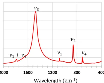
Fourier transform infrared spectroscopy and XRD analysis The indexing of the represented diffractogram, by comparison of the peaks obtained with those of JCDPS database, shows that mineral phases of snail shells are calcium carbonates with aragonite as dominant phase (98.6 M%) completed with trace of calcite (1.4 M%) (JCPDS files [00-041-1475] and [00-047-1743] respectively) (Fig.3). Due to the small amount of cal-cite, the characteristic bands of calcite (1404 cm 1 (ν3),
872 cm 1(ν2), and 712 cm 1(ν4)) are coincident with those
of aragonite (1490 cm 1(ν3), 856 cm 1(ν2), and 713 cm 1
(ν4)) and therefore, the FTIR spectrum shown in Fig.4
pre-sents a characteristic spectrum of aragonite. Small variations in absorption bands positions were registered between pure aragonite and aragonite from snail shells. Indeed, characteris-tic absorptions bands of pure aragonite at 1490 cm 1(ν3),
856 cm 1(ν2), and 713 cm1(ν4) were shifted to 1478 cm 1
(ν3), 860 cm 1(ν
2), and 712 cm 1(ν
4). The small variations
in absorption bands positions can be attributed to the presence of snail shells of organic matter (2.12%w/w) and trace ele-ments such as magnesium, potassium, iron, aluminum, sili-con, and sodium (less than 1%w/w) [26–32].
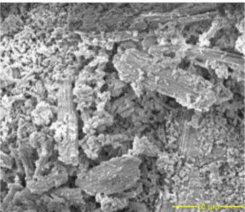
Morphology by SEM The microarchitecture of the samples was determined using SEM analysis. The SEM images of pow-dered snail shells highlight small needle crystals, some of which are bonded longitudinally in the same direction (Fig.
Table 2 Composition of acid and alkaline reaction media and corresponding Ca/P and Ca/(P + C) ratios
Conversion in acid medium Conversion in alkaline medium
Volume of solution 325 mL of water 325 mL of water
Reagent Shells (EG) H3PO4(85%) Shells (EG) (NH4)2HPO4
Amount of reagent 6 g 5 mL 6 g 6.67 g
Molar ratio Ca/P 0.72 1.16
Molar ratio Ca/(P + C) 0.42 0.54
Table 3 Chemical composition of mineral elements of EG shells by fusion method of XRF
Cation elements Mineral analysis, % (w/w)
Ca 56.52 Mg 0.61 Si 0.16 Al 0.12 Na 0.10 Fe 0.07 K 0.05
5). It is likely that these are linked by organic matter. For other powdered snail shells, the parallel organization was not appar-ent, but instead appeared randomly distributed, which is likely due to the grinding of the powder.
Characterization ofAchatina achatina shells converted to calcium phosphate
pH measurements pH measurement solutions are made using the 765 laboratory pH meter (Knick, Germany) calibrated with two buffer solutions (acetic buffer: pH = 4 and ammonia buffer: pH = 9 at 25 °C) measurements.
During the conversion reaction of calcium carbonate to calcium phosphate, the pH was monitored and the results are shown in Fig.6. For both EG-A and EG-B, the first pH value was measured immediately after pouring the reagent then stirred into the aqueous suspension containing the shell pow-der. For EG-A (acid medium), immediately after adding
H3PO4, the pH of suspension decreased to 1.78. The pH
ki-netics presented in Fig. 6can be divided into three distinct phases. The first phase consisted of a rapid increase in the pH from 1.78 to 3.03 for the first 0.5 h of the reaction. Then, the pH decreased to 2.73 after 2.5 h. Finally, in the third phase, the pH gradually increased to 3.5 at 24 h. These changes were related to the gaseous release of carbon dioxide (CO2) and the
dissolution of acid carbonate phases from aragonite during its conversion to calcium phosphate. Over time, the reduction of the carbonates present in the reaction medium led to a gradual increase of the pH.
The results for EG-B (alkaline medium) contrasted those of EG-A. Indeed, for EG-B, the pH reached 8.27 immediately after adding (NH4)2HPO4and gradually increased to 9.3 after
24 h. This gradual increase of pH can be explained by the
Fig. 2 TGA analysis
Fig. 3 Diagram of X ray diffraction ofAchatina achatina snail shells before conversion
400 800 1200 1600 2000
Wavelength
(cm
1)
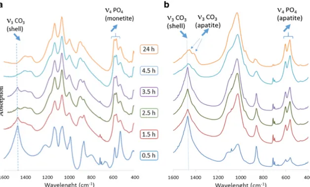
releasing of carbonate ions when some of them were exchanged with phosphate ions during the reaction in the alkaline solution. The carbonate ions released in the aqueous medium reacted with water to generate hydroxide ions. These hydroxide ions are responsible for the pH increase during the reaction. FTIR The chemical constituents of the pulverized shells for all time measurements in all experiments were analyzed by FTIR. The results are presented in Fig.7a, b. Regardless of the re-agent used, the conversion of powdered snail shells to calcium phosphates started immediately after addition of the reagents and mixing the precursor at 80 °C. For EG-A, 30 min after introduction of H3PO4, the FTIR spectrum showed
character-istic bands of aragonite (ν3CO3at 1478 cm 1,ν2at 860 cm 1,
andν4at 712 cm 1) as well as those of brushite (DCPD) (ν3
PO4at 1132 and 1060 cm 1
,ν1PO4at 984 cm 1
, andν4PO4
at 526 cm1[33]). After 1.5 h, the intensity of the carbonate bands decreased and the phosphate bands of the brushite gave way to the vibration bands of the phosphates characteristic of monetite (DCPA) (ν3PO4at 1128 and 1064 cm 1,ν1PO4at
992 cm 1, and ν4 PO4at 576 and 563 cm 1[33]). As the
reaction occurs, a decrease in the intensity of the carbonate vibration bands was observed. After 24 h, the FTIR spectrum no longer shows carbonate vibration bands and only the phos-phate bands of monetite constituted the spectrum. For EG-B, an immediate decrease in the intensity of the carbonate bands of aragonite coupled with an increase in the phosphate bands of hydroxyapatite (ν3PO4at 1092 and 1040 cm 1,ν1PO4at
962 cm 1, andν4PO4at 601 and 561 cm 1[33]) was
record-ed. Importantly, the amount of aragonite converted to hy-droxyapatite during the first 2.5 h of the reaction and de-creased over time. From 4.5h, the CO3bands of aragonite
decreased to reveal the CO3bands of the substituted
hydroxy-apatite. Thus, the vibration bands at 1450 and 880 cm 1can be attributed to type A carbonates (substituted for OH ) and the bands at 1412 and 873 cm 1to type B carbonates (substituted for PO43 ) [34, 35]. Indeed, apatite is a very Bwelcoming^
structure in which a large number of ionic substitutions can occur. During the conversion of aragonite into apatite, the ionic species present have the possibility of integrating the freshly formed apatitic phase. Thus, some of the carbonate ions from aragonite integrate the apatite structure resulting in the formation of carbonate apatite. After 24 h of treatment, the reaction did not seem to be complete, as only 87% of the pulverized shells were converted into carbonated apatite. XRD Figure 8 shows the X-ray diffraction diagrams and Table4displays the results of the phase quantifications pres-ent during the conversion process for the powders treated in an acidic environment in the presence of H3PO4(EG-A) (also see
Fig.8a). Table5displays the results of the phase quantifica-tions present during the conversion process for the powders treated in an alkaline environment in the presence of (NH4)2HPO4(EG-B) (also see Fig.8b).
For the EG-A samples (obtained with the orthophosphoric acid), the X-ray diagrams show the evolution of three crystal-line phases.
The aragonite whose intensity of their characteristics peaks decreased during the duration of the conversion; the brushite is formed in the first step of the conversion process and then transforms to monetite which is the final product obtained at the end of reaction.
The results shown in Table5clearly demonstrate that the first product formed in the immediate moments of the reaction was brushite. This may be due to the calcium brought by the aragonite and phosphate within the reaction medium. The sec-ond phase of conversion involved the transformation of the brushite into monetite. After 24 h, the conversion of aragonite, composed of snail shells, was complete, and the only detect-able product was monetite.
Regarding the EG-B samples (obtained with (NH4)2HPO4),
Fig.8shows a gradual decrease of diffraction peaks of calcium carbonates, whereas there was an increase of the intensity of peaks attributed to hydroxyapatite. Table5 demonstrates the
Fig. 5 SEM image ofAchatina snail shells
Fig. 6 pH kinetics during the reaction of conversion of calcium carbonate to calcium phosphate
gradual conversion of aragonite to apatitic calcium phosphate. After 24 h, 87% of the aragonite had been converted to hy-droxyapatite. Combined with the results observed from FTIR, and the width of the diffraction peaks, the current results con-firm that the apatitic structure formed is not pure hydroxyapatite but poorly crystallized carbonate apatite. These results are sim-ilar to previous findings that observed conversions with coral-line structures and sea snail shells [6,12–14,36].
Morphology
The morphologies of the products formed after 1.5 h and 24 h of reaction are shown in Fig.9. The results reveal the changes
in morphology over time for both the acid and alkaline reac-tion medium.
For EG-A, after 1.5 h of conversion in an acid medium at 80 °C, the freshly formed crystals were large thin plates affixed to massive crystals with flat surfaces. However, after 24 h, they showed a general granular morphology with no regular shaped crystalline components observed.
For EG-B, products obtained after 1.5 h of the conversion process in alkaline environment have morphology of petal-like crystals. Over time, the size of the petals decreased and their organization gave rise to an entangled structure formed of small badly defined platelets. Ducan et al. attribute this change of morphology to differences in synthesis conditions, differences of reaction medium, and different free ions
Fig. 7 a FTIR spectra of snail shells converted in orthophosphoric acid phosphate solution. b FTIR spectra of snail shells converted in ammonium phosphate solution
Table 4 Quantification for EG A experiment showing the amount of present phases and pH of the reaction medium Quantification of products obtained in orthophosphoric acid phosphate solution
Time (h) Ratio (% by mass) of formed phases (XRD) ± 0 3% Physicochemical
parameters Aragonite, CaCO3
(JCPDS 00 041 1475)
Calcite, CaCO3 (JCPDS 00 047 1743)
Brushite, CaHPO4, 2H2O (JCPDS 00 009 0077) Monetite, CaHPO4 (JCPDS 01 070 0360) pH of medium 0 98.6 ± 0.3 1.4 ± 0.3 0 ± 0.3 0 ± 0.3 1.78 ± 0.01 0.5 28.7 ± 0.3 0 ± 0.3 71.3 ± 0.3 0 ± 0.3 3.03 ± 0.01 1.5 14.2 ± 0.3 0 ± 0.3 0 ± 0.3 85.8 ± 0.3 2.77 ± 0.01 2.5 2.9 ± 0.3 0 ± 0.3 0 ± 0.3 97.1 ± 0.3 2.73 ± 0.01 3.5 1.6 ± 0.3 0 ± 0.3 0 ± 0.3 98.4 ± 0.3 2.81 ± 0.01 4.5 2.5 ± 0.3 0 ± 0.3 0 ± 0.3 97.5 ± 0.3 2.90 ± 0.01 24 0.6 ± 0.3 0 ± 0.3 0 ± 0.3 99.4 ± 0.3 3.50 ± 0.01
concentration in media [37], all of which were present in the current experiments.
Discussion
The present work demonstrates that calcium carbonate, which is present in snail shells, can be converted to var-ious calcium phosphate phases. These changes were shown to depend on the reaction medium and, in particu-lar, the pH. Mekmene et al. [38] previously demonstrated that the main factor influencing the crystalline structure of calcium phosphate precipitates is pH. Indeed, when pH
was left to drift after the addition of calcium, brushite is found to be the most prevalent crystalline phase in all precipitates.
However, when pH levels are kept constant, no brushite is seen in precipitates, and there is instead the presence of low crystallized calcium-deficient apatite [38]. Although the operating conditions and the reaction media used in the present study differ from those used by Mekmene et al., the current results agree with those ob-tained in their work.
In considering the conversion mechanisms that can lead to the formation of phosphocalcic phases, two mech-anisms are possible: dissolution-reprecipitation reactions
Fig. 8 a XRD patterns of snail shells converted in orthophosphoric acid phosphate solution. b XRD patterns of snail shells converted in ammonium phosphate solution
Table 5 Quantification for EG B experiment showing the amount of present phases and pH of the reaction medium Quantification of products obtained in ammonium phosphate solution
Time (h) Ratio (% by mass) of formed phases (XRD) ± 0.3% Physicochemical parameters
Aragonite, CaCO3 (JCPDS 00 041 1475) Calcite, CaCO3 (JCPDS 00 047 1743) Hydroxyapatite, Ca10(PO4)6OH2 (JCPDS 00 009 0432) pH 0 98.6 ± 0.3 1.4 ± 0.3 0 8.27 ± 0.01 0.5 83.2 ± 0.3 0 ± 0.3 16.5 ± 0.3 8.36 ± 0.01 1.5 53.2 ± 0.3 0 ± 0.3 46.8 ± 0.3 8.62 ± 0.01 2.5 40.0 ± 0.3 0 ± 0.3 60 ± 0.3 8.69 ± 0.01 3.5 38.1 ± 0.3 0 ± 0.3 61.9 ± 0.3 8.74 ± 0.01 4.5 38.3 ± 0.3 0 ± 0.3 61.7 ± 0.3 8.75 ± 0.01 24 12.9 ± 0.3 0 ± 0.3 87.1 ± 0.3 9.30 ± 0.01
or solid-state topotactic ion-exchange reaction mecha-nisms [12, 39, 40]. When the transformation involves ion-exchange mechanisms, the reaction produces pseudo-morphs with crystals quite similar to the original crystals. However, the precipitation of a new phase leads to a com-plete modification of the organization and shape of the crystals which it is necessary to relate with the operating conditions of synthesis. Indeed, they determine the com-petition which can exist between the dissolution rate of the sources of calcium (here aragonite), the rate of diffu-sion of calcium ions, and the rate of precipitation [18,39]. Moreover, it is important to consider how kinetic factors determine the likelihood of the formation of particular crystal phases. The formation of HAP (as deficient or substituted apatite) is much slower than that of DCPD, and during simul-taneous phase formation, a larger portion of the kinetically favored phase may be observed, even though it has a smaller thermodynamic driving force. Therefore, the balance between kinetic and thermodynamic factors is important in determin-ing the likelihood of precursor formation durdetermin-ing calcium phos-phate precipitation [6].
In this current work, the observed morphology of products from converted powdered snail shells suggests that the trans-formation to calcium phosphate occurs by dissolution phe-nomena of the early phase and then reprecipitation of the new one. Like Mekmene et al. [38], the current results suggest that the reaction mechanisms, and the final products resulting from the reactions, are strongly influenced by pH.
Orthophosphoric acid phosphate solution
Immediately after introducing snail shells into the acid medi-um, the pH increase was associated with a rapid dissolution of the calcium carbonate phases, which was followed by brushite precipitation. The brushite formation induced a pH change accentuated by the simultaneous release of gaseous carbon dioxide and dissolution of acidic carbonates. This was follow-ed by the dissolution of freshly precipitatfollow-ed brushite and the precipitation of monetite which is the most stable phase under these conditions of pH and temperature. The gradual decrease of carbonate resulting from the dissolution of aragonite, coupled with the continued formation of monetite, led to a gradual increase in pH. Under these conditions, the solubility and rapid dissolution rate of the present phases (aragonite and calcite, then brushite) led to dissolution-reprecipitation chain reactions until a thermodynamically more stable system was obtained. The successive and rapid processes that led to the conversion of aragonite into monetite may explain the struc-tural modifications of the crystals observed during the conversion.
Ammonium phosphate solution
In the alkaline medium, the formation of hydroxyapatite be-gan immediately after the addition of the ammonium phos-phate and the resulting pH increase. Under these conditions (pH close to 8), hydroxyapatite is the most thermodynamically
EG A 1.5h EG B 1.5h
EG A 24h EG B 24h
Fig. 9 SEM images showing morphology of converted products after 1.5 h and 24 h of reaction
stable phase. The ability of the apatite phases to receive other ionic species than those constituting stoichiometric hydroxy-apatite (sometimes accompanied by the creation of lacunae at the calcium and/or hydroxide sites) induced the precipitation of a carbonated apatite. The CO3ions then integrated the
apatite structure by replacing some of the PO4ions which
constituted the skeleton of the structure (B-CO3) and a part
of the OH ions located in the tunnels of apatite (A-CO3). The
presence of CO3ions in the phosphate sites and the hydroxide
sites of apatite was confirmed by the presence of vibration bands at 1450 and 1412 cm1in the FTIR spectra. Over time, the amount of carbonate ions increased and the formed apatite phase evolved towards a better-crystallized phase.
Conclusion
In this work, we explored the possibility of converting Achatina achatina snail shells to calcium phosphate using the simply wet chemical method. Moreover, we studied the kinetics of the conversion of different crystalline present phases. The current results show thatAchatina achatina snail shells can be converted to calcium phosphate. If the conver-sion started immediately after introducing shell powder into the reaction media, the conversion efficiency and the final phases obtained were dependent on the reaction media, which influenced solubility and the dissolution rate of phases in this experiment. When the reaction was carried out in an acid medium (in the presence of H3PO4), the rapid dissolution of
the calcium carbonates immediately induced brushite precip-itation which constituted a transitional phase, after which dis-solution finally reprecipitated as a more stable phase: monetite. When the conversion was carried out in an alkaline medium (in the presence of (NH4)2HPO4), the progressive
dissolution of aragonite led to the formation of poorly crystal-lized carbonate apatite. Then, the released carbonates integrat-ed the phosphate sites (type B carbonates) as well as the tun-nels of the apatitic structure (type A carbonates).
References
1. Westbroek, P., Martin, F.: A marriage of bone and nacre. Nature. 392, 861 862 (1998)
2. Tadic, D., Epple, M.: A thorough physicochemical characterisation of 14 calcium phosphate based bone substitution materials in com parison to natural bone. Biomaterials.25(6), 987 994 (2004) 3. Rocha, J.H.G., Lemos, A.F., Agathopoulos, S., Kannan, S., Valerio,
P., Ferreira, J M.F.: Hydrothermal growth of hydroxyapatite scaf folds from aragonite cuttlefish bones. J Biomed Mater Res A.77, 160 172 (2006)
4. Lemos, A.F., Rocha, J.H.G., Quaresma, S.S.F., Kannan, S., Oktar, F.N., Agathopoulos, S., Ferreira, J.M.F.: Hydroxyapatite nano
powders produced hydrothermally from nacreous material. J Eur Ceram Soc.26, 3639 (2006)
5. Guo, Y.P., Yao, Y.B., Ning, C.Q., Guo, Y.J , Chu, L.F.: Fabrication of mesoporous carbonated hydroxyapatite microspheres by hydro thermal method. Mater Lett.65(14), 2205 2208 (2011) 6. Ni, M., Ratner, B.D.: Nacre surface transformation to hydroxyapa
tite in a phosphate buffer solution. Biomaterials.24, 4323 (2003) 7. Ben Nissan, B., Milev, A., Vago, R.: Morphology of sol gel derived
nanocoated coralline hydroxyapatite. Biomaterials.25, 4971 (2004) 8. Patat, J.L., Guillemin, G.: Natural coral used as a replacement bio
material in bone grafts. Ann Chir Plast Esthet.34(3), 221 225 (1989)
9. Damien, E., Revell, P.A.: Coralline hydroxyapatite bone graft sub stitute: a review of experimental studies and biomedical applica tions. J Appl Biomater Biomech.2, 65 73 (2004)
10. He, L.Y., Zhang, X.M., Liu, B , Tian, Y., Ma, W.H.: Effect of mag nesium ion on human osteoblast activity. Braz J Med Biol Res. 49(7), e5257 (2016)
11. Tadier, S., Bareille, R., Siadous, R., Marsan, O., Charvillat, C., Cazalbou, S., Amedee, J., Rey, C., Combes, C.: Strontium loaded mineral bone cements as sustained release systems: compositions, release properties, and effects on human osteoprogenitor cells. J Biomed Mater Res B Appl Biomater.100(2), 378 390 (2012) 12. Macha, I.J., Grossin, D., Ben Nissan, B.: Conversion of marine
structures to calcium phosphate materials: mechanisms of conver sion using two different phosphate solutions. Key Eng Mater.696, 36 39 (2016)
13. Macha, I.J., Boonyang, U., Cazalbou, S., Ben Nissan, B., Charvillat, C., Oktar, F.N., Grossin, D.: Comparative study of coral conversion, part 2: microstructural evolution of calcium phosphate. J Aust Ceram Soc.51(2), 149 159 (2015)
14. Choi, G., Karacan, I., Cazalbou, S., Evans, L., Sinutok, S., Ben Nissan, B.: Conversion of calcified algae (Halimeda sp) and hard coral (Porites sp) to hydroxyapatite. Key Eng Mater.758, 157 161 (2017)
15. Roy, D.M., Linnehan, S.K.: Hydroxyapatite formed from coral skeletal carbonate by hydrothermal exchange. Nature.247(5438), 220 222 (1974)
16. Johnsson, M.S.A., Nancollas, G.H.: The role of brushite and octacalcium phosphate in apatite formation. Crit Rev Oral Biol Med.3, 61 82 (1992)
17. Marshall, R.W., Nancollas, G.H.: The kinetics of crystal growth of dicalcium phosphate dehydrate. J Phys Chem.73(11), 3838 3844 (1969)
18. Elliott, J.C.: Structure and chemistry of the apatites and other calci um orthophosphates. Elsevier, Amsterdam (1994)
19. YoungJae, K., Seon Yong, L., Yul, R., Jinhyeok, L., Juyeun, K., Yongwoo, L., Junseok, B., Young Jae, L.: Optimizing calcium phosphates by the control of pH and temperature via wet precipita tion. J Nanosci Nanotechnol.15, 10008 10016 (2015)
20. LeGeros, R.Z.: Calcium phosphates in oral biology and medicine. Monogr. Oral Sci.15, 1 201 (1991)
21. Chow L.C., Solubility of calcium phosphates. (eds.) Octacalcium phosphate., monogr. Oral Sci. Basel, Karger, vol 18. (2001), 94 111 22. Rodriguez Navarro, A., Cabral de Melo, C., Batista, N., Morimoto, N., Alvarez Lloret, P., Ortega Huertas, M., Fuenzalida, V.M., Arias, J.I., Wiff, J.P., Arias, J.L.: Microstructure and crystallographic tex ture of giant barnacle (Austromegabalanus psittacus) shell. J Structur Biol.156, 355 362 (2006)
23. Radishi, N.A., Mohamed, M., Yusup, S.: The kinetic model of calcination and carbonation of Anadara Granosa. Int J Renew Energy Res.2(3), 497 503 (2012)
24. Wu, C., Xiao, Y., Chang, J.: Silicate based bioactive ceramics for bone regeneration application. In: Wu, C., Chang, J., Xiao, Y. (eds.) Advanced bioactive inorganic materials for bone regeneration and
drug delivery, pp. 25 46. CRC Press (Taylor & Francis Group, Boca Raton (2013)
25. Burmester, A , Willumeit Römer, R., Feyerabend, F.: Behavior of bone cells in contact with magnesium implant material. J Biomed Mater Res B Appl Biomater.105(1), 165 179 (2017)
26. Andersen, F.A., Breevi, L.J.: Infrared spectra of amorphous and crystalline calcium carbonate. Acta Chem Scand.45, 1018 1024 (1991)
27. Fernandez, M.S., Valezuela, F., Arias, J.I., Neira Carrillo, A., Arias, J.L.: J Struct Biol.196, 187 196 (2016)
28. Narasimhulu, K.V., Lakshamana Rao, J.: EPR and IR spectral stud ies of the seawater mussel Mytilus conradinus shells. Spectrochim Acta A.56, 1345 1353 (2000)
29. Su, C., Suarez, D.L.: In situ infrared speciation of adsorbed carbonate on aluminum and iron oxide. Clay Clay Miner.45(6), 814 825 (1997) 30. Addadi, L., Raz, S., Weiner, S.: Taking advantage of disorder: amorphous calcium carbonate and its roles in biomineralization. Adv Mater.15, 959 970 (2003)
31. Coleyshaw, E.E., Crump, G., Griffith, W.P.: Vibrational spectra of the hydrated carbonate minerals ikaite, monohydrocalcite, lansfordite and nesquehonite. Spectrochim Acta A Mol Biomol Spectrosc.59(10), 2231 2239 (2003)
32. Gueta, R., Natan, A., Addadi, L., Weiner, S., Refson, K., Kronik, L.: Local atomic order and infrared spectra of biogenic calcite. Angew Chem Int Ed.46, 291 294 (2007)
33. Rey, C., Combes, C., Drouet, C., Grossin, D.: Bioactive ceramics: physical chemistry. In: Ducheyne, P., Healy, K., Hutmacher, D., Grainger, D.E., Kirkpatrick, J. (eds.) Comprehensive Biomaterials, pp. 187 221. Elsevier (2011)
34. Borkiewicz, O., Rakovan, J., Cahill, C.L.: Time resolved in situ studies of apatite formation in aqueous solutions. Am Mineral. 95(8 9), 1224 1236 (2010)
35. Rey, C., Collins, B., Goehl, T., Dickson, I R., Glimcher, M.J.: The carbonate environment in bone mineral: a resolution enhanced Fourier transform infrared spectroscopy study. Calcif Tissue Int. 45(3), 157 164 (1989)
36. Şahin, Y., Gündüz, O., Bulut, B., Özyeğin, L., Gökçe, H., Ağaoğulları, D., Chou, J., Kayalı, E., Ben Nissan, B., Oktar, F.: Nano bioceramic synthesis from tropical sea snail shells (tiger cow rie Cypraea Tigris) with simple chemical treatment. Acta Phys Pol A.127(4), 1055 1058 (2015)
37. Duncan, J., MacDonald, J.F., Hanna, J.V., Shirosaki, Y., Hayakawa, S., Osaka, A., Skakle, J.M.S., Gibson, I R.: The role of the chemical composition of monetite on the synthesis and properties ofβ tricalcium phosphate. Mater Sci Eng C.34, 123 129 (2014) 38. Mekmene, O., Quillard, S., Rouillon, T., Bouler, J.M., Piot, M.,
Gaucheron, F.: Effects of pH and Ca/P molar ratio on the quantity and crystalline structure of calcium phosphates obtained from aque ous solutions. Dairy Science & Technology, EDP sciences/ Springer.89(3 4), 301 316 (2009)
39. Wang, L., Nancollas, G.H.: Calcium orthophosphates: crystalliza tion and dissolution. Chem Rev.108(11), 4628 4669 (2008) 40. Lima, C.B A., Airoldi, C.: Topotactic exchange and intercalation of
calcium phosphate. Solid State Sci.6(11), 1245 1250 (2004)
Publisher’s note Springer Nature remains neutral with regard to juris dictional claims in published maps and institutional affiliations.