Open Archive Toulouse Archive Ouverte (OATAO)
OATAO is an open access repository that collects the work of some Toulouse
researchers and makes it freely available over the web where possible.
This is
an author'sversion published in:
https://oatao.univ-toulouse.fr/23119Official URL :
https://doi.org/10.1016/j.jse.2017.05.018
To cite this version :
Any correspondence concerning this service should be sent to the repository administrator:
Pham, Thuy-Trang and Accadbled, Franck and Abid, Abdelaziz and Ibnoulkhatib, Aissa
and Bayle-Iniguez, Xavier and Wargny, Matthieu and Sales de Gauzy, Jérôme Gartland
types IIB and III supracondylar fractures of the humerus in children: is Blount's method
effective and safe? (2017) Journal of Shoulder and Elbow Surgery, 26 (12). 2226-2231. ISSN
1058-2746
OATAO
Gartland types IIB and III supracondylar fractures
of the humerus in children: is Blount’s method
effective and safe?
Thuy-Trang Pham, MD
a,*, Franck Accadbled, MD, PhD
a, Abdelaziz Abid, MD
a,
Aissa Ibnoulkhatib, MD
a, Xavier Bayle-Iniguez, MD
b, Matthieu Wargny, MD
c,
Jérôme Sales de Gauzy, MD
aaService de chirurgie orthopédique et traumatologique pédiatrique, Hôpital des Enfants, CHU Toulouse, Toulouse, France bService de chirurgie orthopédique et traumatologique, Hôpital Pierre Paul Riquet, CHU Toulouse, Toulouse, France c
Service d’Epidémiologie, CHU Toulouse, Toulouse, France
Background: Blount’s method is controversial for the treatment of Gartland types IIB and III
supracon-dylar fracture of the humerus (SCFH) in children. The purpose of this study was to evaluate the clinical and radiologic outcomes and the failure and complication rates.
Methods: All types IIB and III SCFH treated with Blount’s method from 2003-2013 were included in
this retrospective single-center study. Clinical assessment was performed according to Flynn criteria. Baumann angle, anteversion angle, anterior humeral line, and humeroulnar angle were measured for radiographic assessment.
Results: Among 447 children with types IIB and III SCHF, 339 were treated according to Blount’s method.
There were 173 boys (51%), and the mean age was 6.3 years (1-14 years); 71% were type III. Mean time to surgery was 5.7 hours. According to Flynn criteria, results were satisfactory in 91% of cases. No com-partment syndrome was encountered. There were 16 (4.7%) secondary displacements requiring surgical revision. Five (1.9%) children developed a cubitus varus deformity. At latest follow-up, the mean Baumann angle was 74.7° (95% confidence interval, 74.1-75.3), the mean anteversion angle was 39.9° (95% con-fidence interval, 39.5-40.3), the anterior humeral line was normal in 87.6% of cases, and the mean humeroulnar angle was 8.7°.
Conclusion: Blount’s method is appropriate to manage types IIB and III SCFH, provided anatomic and
stable reduction is obtained.
Level of evidence: Level IV; Case Series; Treatment Study
Keywords: Blount’s method; supracondylar fracture; Gartland IIB and III; closed reduction; children; elbow
Supracondylar fracture of the humerus (SCFH) is the most frequent fracture of the elbow in children. Extension type rep-resents 96% of SCFH.8Closed reduction and immobilization
of the elbow in flexion were popularized by W.P. Blount in 1954 in his classic textbook, Fractures in Children.5 The
Ethical approval was waived by the Institutional Review Board. *Reprint requests: Thuy-Trang Pham, MD, Service de chirurgie orthopédique et traumatologique pédiatrique, Hôpital des Enfants, CHU Toulouse, 31059 Toulouse Cedex 9, France.
fracture is stable in flexion only if the posterolateral perios-teum is intact.
The management of displaced SCFH type IIB and type III according to the Wilkins-modified Gartland classifica-tion is difficult because of the frequent swelling that may cause vascular compression or even compartment syndrome and in-stability when the posterolateral periosteum is torn.13,17,25
Blount’s method was condemned in France in the 1960s after the report from Lagrange and Rigault because of the high risk of compartment syndrome in case of malreduction.17
The method was later reintroduced thanks to the shorter delays in treatment, allowing less swollen elbows.
Most authors recommend pin fixation to prevent compart-ment syndrome and to improve stability.6,11,18,19,22,27
However, complications can occur with surgical treatment, including pin track infections, joint stiffness, neurologic injuries, and secondary displacement.6,24
Our hypothesis was that Blount’s method is adequate for types IIB and III SCFH, provided stable and satisfactory reduction is obtained. The aim was to eval-uate the clinical and radiologic outcomes, failure rate, and complications.
Materials and methods
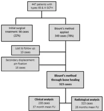
This was a single-center retrospective consecutive series. All extension-type IIB and III SCFH treated with Blount’s method from 2003-2013 were included. Among 447 children with Gartland type IIB or III SCFH, 98 (22%) were treated surgically and 349 (78%) were treated with Blount’s method.
We retrospectively reviewed the hospital records of the study cohort, including personal data, preoperative clinical examinations and associated lesions, time from injury to surgery, operative notes, postoperative evaluations, duration of immobilization, presence of complications, need for further surgery, and clinical assessment at final follow-up visit. Patients returned for clinical examination and radiographs in 57% of cases.
Clinical evaluation and overall rating at latest follow-up were performed according to Flynn criteria12(Table I). Anteroposterior
and lateral radiographs of the elbow were analyzed using Baumann angle and distal humerus anteversion angle postoperatively, at 1 week, at the time of bone consolidation, and at latest follow-up. Humeroulnar angle was measured at latest follow-up.
Statistical analysis was performed using Statistique R version 2.14.1 software (The R Foundation for Statistical Computing, Vienna, Austria). Theχ2statistic was used for qualitative variables and Student
t-test for quantitative variables. Results are displayed with raw values
and percentages for qualitative variables and with means, medians, standard deviations, and interquartile ranges for quantitative vari-ables. P was considered significant if< .05.
Description of Blount’s method
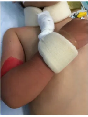
Under general anesthesia, the patient was positioned supine with the affected limb placed on the image intensifier. Closed manipu-lation consisted of traction, pronation or supination, and then elbow flexion, respectively. Elbow flexion was maintained at about 120° by a collar and cuff bandage (Fig. 1) for 4 weeks. In case of an un-stable reduction in elbow flexion, pin fixation was performed. If closed reduction was unsuccessful, open reduction and pin fixation were performed. Radial pulse and capillary refill time in the fingertips were checked, and pulse oximetry at the middle finger was moni-tored systematically immediately after reduction in elbow flexion. Immediate postoperative anteroposterior and lateral radiographs of the elbow were performed systematically (Fig. 2). Parents were given basic care and observation guidelines. Clinical and radiologic control was scheduled in the outpatient clinic within 10 days from hospi-tal discharge. No physiotherapy was prescribed. Sport activities were allowed after 3 months.
Results
The records of 447 children with Gartland type IIB or III SCFH were analyzed, of whom 98 (22%) were treated sur-gically with pin fixation because of the following reasons: open fracture in 12 cases (12%), failed closed reduction in 28 cases (28.5%), instability of the fracture in elbow flexion in 20 cases (20.5%), and distal ischemia in elbow flexion because of edema in 38 cases (39%).
Table I Flynn criteria for grading of outcome
Result Rating Cosmetic factor
Loss of carrying angle (°) Functional factor Loss of motion (°) Satisfactory Excellent 0-5 0-5 Good 6-10 6-10 Fair 11-15 11-15 Unsatisfactory Poor >15 >15
The lower of the ratings is the overall rating, and an elbow that has a varus deformity is automatically graded poor.
A total of 349 patients were treated with Blount’s method: 243 type III (71.7%) and 96 type IIB fractures (28.3%). There were 173 boys (51%) and 166 girls (49%) with a mean age of 6.3 years (1-14 years). The left side was affected in 197 cases (58.1%) and the right in 142 (41.9%; dominant side in 44.4% of cases). The fracture was sustained on the occa-sion of a sports injury in 207 cases (61%), a fall from standing height in 74 cases (22%), a household accident in 40 cases (11,7%), a motor vehicle accident in 5 cases (1.5%), and an undocumented cause in 13 cases (3.8%). Average time to man-agement was 5.7 hours (23 minutes–20 hours). Associated lesions were recorded in 127 cases (Table II).
Mean operative time (reduction+ collar and cuff bandage) was 11.6 minutes (95% confidence interval [CI], 11.11-12.07). Mean hospital stay was 1.6 days (95% CI, 1.49-1.64). Mean immobilization time in a collar and cuff bandage was 26.2 days (95% CI, 25.42-27.02). According to 1 sur-geon’s preference, 96 patients (28%) had an additional immobilization in a 90° elbow flexion cast for 16.6 days on average (95% CI, 15.55-17.7).
Ten patients were lost to follow-up. Sixteen patients had a secondary displacement managed with repeated reduction and pin fixation 7 days postoperatively, leaving 323 patients
for radiologic analysis at 45 days of follow-up. There were 238 patients who were clinically examined at a mean 36.8 months of follow-up (2.1-134.9 months) (Fig. 3).
Clinical outcome
Results were satisfactory in 97% of cases (excellent or good in 95% of cases) according to Flynn criteria (Table III). In-cluding the 16 patients who underwent surgical revision and who were considered to have poor outcome, results were sat-isfactory in 91% of cases (n= 231/254).
Mean range of motion of the elbow in flexion-extension was 140.3° (100°-160°), with mean flexion of 138.2° (95% CI, 137.5-138.8) and mean extension of+2.1° (95% CI, 1.3-2.9). Mean loss of extension compared with the contralateral side was 9.8° (5°-30°). Range of motion in pronation-supination was
Figure 2 (A) Preoperative elbow radiograph, lateral view: Gartland type III supracondylar fracture of the humerus. (B) Elbow
radio-graphs, anteroposterior and lateral views, immediately after reduction and immobilization in elbow flexion.
Table II Preoperative associated lesions
Preoperative associated lesions No.
Ipsilateral fractures (3.5%) 12
Buckle fracture distal radius 5
Salter-Harris type II physeal injury distal radius 4
Forearm fracture 3
Cutaneous (18.9%) 64
Severe swelling and bruising 53
Gustilo type I open fracture 7
Subcutaneous bone extremity 4
Neurologic (13.3%) 45
Median nerve 13
Anterior interosseous nerve 19
Radial nerve 12
Radial and ulnar nerves 1
Vascular (1.8%) 6
Pulselessness resolved after fracture reduction 6
Figure 3 Flow chart of the cohort. SCFH, supracondylar
frac-ture of the humerus; FU, follow-up.
1 447 pa · ents wlth 1 types 118 & 111 SCfH 1 1
Initial surgical Blounrs method treatment: 98 cases applied
(22%) 349 cases (78%) 1 Lost o follow up: 10 cases 1 Secondary displacement: pin fixa ion 16 cases Blount's method through bone healing
323 cases
ainkal analysis Radiological analysis
238 cases
--
323 casessymmetric. Mean humeroulnar angle was +7.3° (95% CI, 6.73-7.81).
Five patients (1.9%) had cubitus varus with a clinical car-rying angle of−10° (−5° to −15°), loss of the humeroulnar angle of 17° (10°-20°) compared with the contralateral side, and Baumann angle of 93.6° (80°-104°). None reported a func-tional or cosmetic complaint. All patients with initial nerve palsy had fully recovered at latest follow-up. Parents were very satisfied in 98.8% of cases.
Seven patients had fair results, 6 functionally (restricted range of motion) and 1 cosmetically (loss of carrying angle). Mean range of motion in the 6 functionally impaired pa-tients was 124° (100°-140°). Mean loss of extension compared with the contralateral side was 17.5° (10°-25°). One patient had a painful elbow affecting daily activities. This 14-year-old boy had a type III fracture complicated by periarticular ossifications causing severe stiffness (range of motion of 25°). Despite surgical release 3 months postoperatively, his elbow remained relatively stiff (range of motion of 100°) and painful at latest follow-up. The patient with a fair cosmetic result had a loss of carrying angle of 20° without functional consequences.
Radiologic results
Baumann angle was 75.7° (95% CI, 75.3-76.2) postopera-tively and 74.7° (95% CI, 74.1-75.3) at latest follow-up (P= .00025). Mean distal humerus anteversion angle was 39.7° (95% CI, 39.3-40.1) postoperatively and 39.9° (95% CI, 39.5-40.3) at latest follow up (P= .30).
At latest follow-up, 20 patients (6.4%) had an abnormal Baumann angle, of which 17 were above 81° (mean, 84.9°; 95% CI, 80.25-89.53) and 3 were below 64° (mean, 60°). An-teversion angle was above 40° (mean, 45.1°; 95% CI, 44.3-45.8) in 63 patients (19%). Mean humeroulnar angle was 8.7° at latest follow-up (95% CI, 7.84-9.62).
Secondary displacement
There were 36 cases of secondary displacement (10.6%) at the first-week visit. Sixteen (4.7%) were managed with sur-gical revision. A continued conservative treatment of the remaining 20 patients with secondary displacement was chosen by the surgeon, who considered that it was not clinically
significant. This matter was always explained to the patient and family, who accepted this decision.
Of the 20 patients managed with conservative treatment, 3 (15%) had a fair outcome and 1 had a poor outcome (5%) according to Flynn criteria. Three patients had cubitus varus with a Baumann angle above 90°, of whom 2 had an exces-sive anteversion. One patient had a fair outcome with 15° loss of motion.
Complications
Five patients (1.5%) presented with a skin sore at the wrist and 3 (0.9%) at the elbow, necessitating local care with dress-ings. One patient (0.3%) had his bandage loosen after a fall. There was 1 case of ulnar nerve palsy due to excessive tight-ness of the bandage, which recovered spontaneously when the bandage was removed. Two patients suffered from a type I complex regional pain syndrome. No compartment syn-drome was noted.
Discussion
Remodeling of SCFH is mild as the chondroepiphysis of the distal humerus provides only 20% of the longitudinal bone growth. The aim of treatment is therefore to maintain ana-tomic reduction to allow normal function and range of motion, along with satisfactory cosmesis. The recommended method for treatment must be as simple as possible and reliable while bearing a low risk of complication. Most publications advo-cate pin fixation for SCFH types IIB and III, with various preferred constructs.6,11,18,19,22,27
Blount’s method, initially described in 1954, relies on the continuity of the posterior periosteum, which provides the nec-essary stability to maintain the reduction in elbow flexion.5,7
Application to types IIB and III remains controversial because of the risk of compartment syndrome and fracture instabil-ity. The author did not recommend the method in case of neurovascular compromise or marked swelling.5
We ex-tended the use of the method to types IIB and III SCFH, except for unstable or unreducible fractures and in case of vascular compromise persisting after fracture reduction. Neurologic deficit did not influence our decision. Severe swelling was a relative contraindication, at the discretion of the surgeon in charge.
The posterior periosteum is torn in about 50% of type III SCFH.17Yet, Blount’s method is based on an intact
posteri-or periosteum. In other wposteri-ords, about half of type III cases are eligible for this method of immobilization. The current series demonstrated that 66% of such fractures were suc-cessfully managed using Blount’s method, whereas Akakpo-Numado et al reported 70% and Williamson and Cole reported 60%.2,26
Clinical results were satisfactory in 91% of cases accord-ing to Flynn criteria, which is consistent with the literature. De Gheldere and Bellan,9 in a series of 74 children,
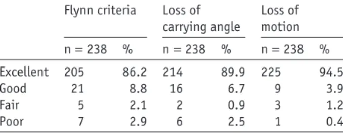
Table III Clinical outcome according to Flynn criteria
Flynn criteria Loss of carrying angle Loss of motion n= 238 % n= 238 % n= 238 % Excellent 205 86.2 214 89.9 225 94.5 Good 21 8.8 16 6.7 9 3.9 Fair 5 2.1 2 0.9 3 1.2 Poor 7 2.9 6 2.5 1 0.4
reported 94% excellent and good results according to Flynn criteria for type II and 73% for type III. Results of type III were influenced by the direction of displacement: fractures remained stable in 88% of posterior displacements, 58% of posteromedial displacements, and only 36% of posterolat-eral displacements.9 Kinkpé et al observed 100% stable
fractures and 100% good results in 67 type III fractures. Time to treatment (mean time to referral, 30 hours; mean time to treatment, 46 hours) did not influence anatomic and clinical results.15
We did not proceed with a comparative study as our in-dications for Blount’s method and pin fixation were different. However, Sigge et al have reported better results with Blount’s method than with pin fixations.21
Kennedy et al obtained similar results in both groups and concluded that immobili-zation in elbow flexion was effective when stable reduction was obtained in Gartland types II and III SCFH. No corre-lation existed between the type of treatment and poor results. However, surgical treatment was associated with the risk of superficial infection.14
Compartment syndrome incidence varies from 0.1% to 0.3%.3The combination of the SCFH with ipsilateral forearm
fracture represents a major risk factor. Blakemore et al re-ported 3 cases (7%) among 33 such combined lesions.4
Ipsilateral forearm fracture and marked swelling did not con-traindicate Blount’s method in our series. None of our patients sustained a compartment syndrome, yet this complication is the main argument against Blount’s method in the literature. Secondary displacement occurred in 36 cases (10.6%), of which 16 (4.5%) were managed with repeated reduction and pin fixation. Nonetheless, 16 of the remaining 20 patients had excellent results according to Flynn criteria, 3 had cubitus varus and remained asymptomatic at latest follow-up, and 1 patient had a 15° loss of elbow motion. Clavert et al re-corded 7 secondary displacements in a 120-case series (5.8%), of which 6 involved type IV fractures and 1 involved a type III according to the Lagrange and Rigault classification.17
Kinkpé et al applied Blount’s method to 67 Gartland III SCFH and described only 2 (3%) secondary displacements.15
Akakpo-Numado et al had a 25% rate of secondary displacement, also with Blount’s method.2
Our surgical revision rate was similar to those of pin fix-ation series from the literature.6,11,18,19,22,27Moreover, Blount’s
method has several advantages over pin fixation: it is simple and cheap, with a low risk of infection and low risk of nerve injury, and it avoids another procedure for pin removal. The absence of circular immobilization facilitates postoperative care and observation.
Cubitus varus was observed in 5 (1.9%) of our patients vs. 3% to 58% of cases in the literature.10,16In 4 cases, this
deformity resulted from a wrong indication or wrong man-agement: 3 presented with a secondary displacement managed conservatively, and 1 had an insufficient initial reduction (40% rotation). Blount’s method should not be used in case of an insufficient fracture reduction because of the low bone re-modeling potential at the distal humerus. Close observation
is necessary, and surgical revision should be decided in a timely manner in case of secondary displacement. Cubitus varus results from a medial angulation at the fracture site, with or without rotation deformity, rather than from a growth dis-turbance at the distal humerus.11,23It has been thought in the
past that only cosmesis and a lesser degree of function were at stake in cubitus varus. Williamson and Cole obtained 95% excellent results with Blount’s method despite 22.7% cubitus varus.26
However, long-term complications, such as ulnar nerve palsy and posterolateral instability of the elbow, are now clearly admitted.1,20
Six patients (2.5%) had a poor clinical result according to Flynn criteria, with a mean elbow range of motion of 124° (100°-140°) and a mean loss of extension of 17.8° (10°-25°). One presented with periarticular ossifications and was affected in his daily activities. This patient was aged 14 years at the time of the fracture, and initial pin fixation should have been selected.
We acknowledge some limitations to this study, related to its retrospective and noncomparative nature. Data analyses relied on patients’ records in only 57% of cases.
Conclusion
We were able to use Blount’s method to successfully treat 78% of presented cases of type IIB and III supracondy-lar humeral fractures in children. However, among the original cohort, 8.5% of the patients showed distal isch-emia when placed in elbow flexion, and 10.7% did not maintain fracture reduction. In such situations, pin fixa-tion is warranted. In carefully selected cases, Blount’s method is a reasonable option for treating type IIB and III supracondylar humeral fractures in children. Close moni-toring is necessary.
Disclaimer
The authors, their immediate families, and any research foundations with which they are affiliated have not re-ceived any financial payments or other benefits from any commercial entity related to the subject of this article.
References
1. Abe M, Ishizu T, Shirai H, Okamoto M, Onomura T. Tardy ulnar nerve
palsy caused by cubitus varus deformity. J Hand Surg Am 1995;20:5-9.
2. Akakpo-Numado GK, Mal-Lawane M, Belouadah M, Kabore B, Lefort
G, Daoud S. [Management of Lagrange and Rigault stage IV extension type supracondylar fracture of the humerus in children]. Rev Chir Orthop
Reparatrice Appar Mot 2005;91:664-70.http://dx.doi.org/10.1016/
s0035-1040(05)84471-x
3. Battaglia TC, Armstrong DG, Schwend RM. Factors affecting forearm
compartment pressures in children with supracondylar fractures of the
humerus. J Pediatr Orthop 2002;22:431-9.http://dx.doi.org/10.1097/
4. Blakemore LC, Cooperman DR, Thompson GH, Wathey C, Ballock RT. Compartment syndrome in ipsilateral humerus and forearm fractures in children. Clin Orthop Relat Res 2000;376:32-8.
5. Blount WP. Fractures in children. Baltimore: Williams & Wilkins; 1954.
p. 26-42.
6. Brauer CA, Lee BM, Bae DS, Waters PM, Kocher MS. A systematic
review of medial and lateral entry pinning versus lateral entry pinning for supracondylar fractures of the humerus. J Pediatr Orthop 2007;
27:181-6.http://dx.doi.org/10.1097/bpo.0b013e3180316cf1
7. Clavert JM, Lecerf C, Mathieu JC, Buck P. [Retention in flexion of
supracondylar fracture of the humerus in children. Comments apropos of the treatment of 120 displaced fractures]. Rev Chir Orthop Reparatrice Appar Mot 1984;70:109-16.
8. Damsin J, Langlais J. Fractures supracondyliennes. Symposium sur les
fractures du coude chez l’enfant (sous la direction de J.-C. Pouliquen). Rev Chir Orthop Reparatrice Appar Mot 1987;73:421-36.
9. De Gheldere A, Bellan D. Outcome of Gartland type II and type III
supracondylar fractures treated by Blount’s technique. Indian J Orthop
2010;44:89.http://dx.doi.org/10.4103/0019-5413.58612
10. Dowd GS, Hopcroft PW. Varus deformity in supracondylar fractures
of the humerus in children. Injury 1979;10:297-303.http://dx.doi.org/
10.1016/0020-1383(79)90047-0
11. Flynn JC. Displaced supracondylar fracture of the humerus in children:
technique of closed reduction and percutaneous pinning. Oper Tech Orthop 1993;3:121-7.
12. Flynn JC, Matthews JG, Benoit RL. Blind pinning of displaced
supracondylar fractures of the humerus in children. Sixteen years’ experience with long-term follow-up. J Bone Joint Surg Am 1974; 56:263-72.
13. Gartland JJ. Management of supracondylar fractures of the humerus in
children. Surg Gynecol Obstet 1959;109:145-54.
14. Kennedy JG, El Abed K, Soffe K, Kearns S, Mulcahy D, Condon F,
et al. Evaluation of the role of pin fixation versus collar and cuff immobilisation in supracondylar fractures of the humerus in children. Injury 2000;31:163-7.
15. Kinkpé CV, Dansokho AV, Niane MM, Chau E, Sales de Gauzy J,
Clement JL, et al. Children distal humerus supracondylar fractures: the Blount Method experience. Orthop Traumatol Surg Res 2010;96:276-82.
http://dx.doi.org/10.1016/j.otsr.2009.12.010
16. Labelle H, Bunnell WP, Duhaime M, Poitras B. Cubitus varus deformity
following supracondylar fractures of the humerus in children. J Pediatr Orthop 1982;2:539-46.
17. Lagrange J, Rigault P. Les fractures de l’extrémité inférieure de l’humérus
de l’enfant. Rev Chir Orthop Reparatrice Appar Mot 1962;48:334-414.
18. Maity A, Saha D, Roy DS. A prospective randomised, controlled clinical
trial comparing medial and lateral entry pinning with lateral entry pinning for percutaneous fixation of displaced extension type supracondylar fractures of the humerus in children. J Orthop Surg Res 2012;7:6.
http://dx.doi.org/10.1186/1749-799X-7-6
19. Mazda K, Boggione C, Fitoussi F, Penneçot GF. Systematic pinning
of displaced extension-type supracondylar fractures of the humerus in children. A prospective study of 116 consecutive patients. J Bone Joint Surg Br 2001;83:888-93.
20. O’Driscoll SW, Spinner RJ, McKee MD, Kibler WB, Hastings H,
Morrey BF, et al. Tardy posterolateral rotatory instability of the elbow due to cubitus varus. J Bone Joint Surg Am 2001;83-A:1358-69.
21. Sigge W, Behrens K, Roggenkamp K, Würtenberger H. [Comparison
of Blount’s sling and Kirschner wire fixation in the treatment of a dislocated supracondylar humeral fracture in childhood]. Unfallchirurgie 1987;13:82-90.
22. Skaggs DL, Cluck MW, Mostofi A, Flynn JM, Kay RM. Lateral-entry
pin fixation in the management of supracondylar fractures in children. J Bone Joint Surg Am 2004;86-A:702-7.
23. Smith L. Deformity following supracondylar fractures of the humerus.
J Bone Joint Surg Am 1960;42-A:235-52.
24. Taniguchi Y, Matsuzaki K, Tamaki T. Iatrogenic ulnar nerve injury after
percutaneous cross-pinning of supracondylar fracture in a child. J Shoulder Elbow Surg 2000;9:160-2.
25. Wilkins K. Fractures and dislocations of the elbow region. In: Rockwood
CA, Wilkins KE, King RE, editors. Fractures in children. 3rd ed. Philadelphia: JB Lippincott; 1991. p. 509-828.
26. Williamson DM, Cole WG. Treatment of selected extension
supracondylar fractures of the humerus by manipulation and strapping in flexion. Injury 1993;24:249-52.
27. Woratanarat P, Angsanuntsukh C, Rattanasiri S, Attia J, Woratanarat
T, Thakkinstian A. Meta-analysis of pinning in supracondylar fracture
of the humerus in children. J Orthop Trauma 2012;26:48-53.http://