OATAO is an open access repository that collects the work of Toulouse
researchers and makes it freely available over the web where possible
Any correspondence concerning this service should be sent
to the repository administrator:
tech-oatao@listes-diff.inp-toulouse.fr
This is an author’s version published in:
http://oatao.univ-toulouse.fr/27619
To cite this version:
Mazerolle, Paul and Meresse, Thomas and Gangloff, Dimitri and Kolsi, Karim
and Dupret Bories, Agnès
Vascularized lymph node transfer with submental
free flap. (2020) European Annals of Otorhinolaryngology, Head and Neck
Diseases, 137 (1). 73-77. ISSN 1879-7296
Vascularized lyn1ph node transfer with subn1ental free
flap
P. Mazerolle
a,
•
. T. Meresse
b, D. Gangloff
b, K. Kolsi
b, A. Dupret-Bories
c• Oiirurgie ORL et Cervico-Faciale, Institut Universitaire du Cancer de Toulouse Oncopole, CHU Larrey, 24, chemin de Pouvourville, 31059 Toulouse. France • Chirurgie Plastique et Réparatrice, Institut Universitaire du Cancer de Toulouse Oncopole, Institut Claudius Regaud, 1, avenue Irène Joliot-Curie, 31059 Toulouse, France
'Chirurgie ORL et Cervico-Faciale, Institut Universitaire du Cancer de Toulouse Oncopole, Institut Claudius Regaud, 1, avenue Irène Joliot-Curie, 31059 Toulouse, France
ARTICLE INFO ABSTR ACT Keywords:
Lymphoedema
Vascularized lymph node transfer Submental flap
Limb lymphoedema is common in patients who have undergone inguinal or axillary lymph node dis section. Lymphoedema seriously impacts the patient's quality of life by inducing adipogenesis, fibrosis and repeated episodes of lymphangitis and cellulitis. Following failure of compression therapies, several curative or symptomatic surgical options have been proposed over recent decades, such as liposuction or lymphovenous anastomosis.
Vascularized lymph node transfer techniques have recently been described, with promising results.
Vascularized lymph node transfer with submental free flap appears to be the most reliable of these technique, associated with the lowest morbidity. The flap harvesting technique presents several specific differences compared to conventional submental free fla p. Agood knowledge of neck anatomy is essential and multimodal and multidisciplinary management is often required.
ln the light of a case report, we describe the basic submental free flap technique for vascularized lymph node transfer for the treatment of lower limb lymphoedema. This technique may be used by head and neck surgeons performing flap harvest, as well as plastic surgeon surgeons or gynaecologists ensuring long-term management of these patients.
1. Introduction
Lower limb lymphoedema is common after inguinal lymph node
dissection, especially in patients who have received adjuvant radio
therapy, as interruption of the main lymphatic drainage pathways
leads to interstitialaccumulation ofprotein-rich lymph in the lower
limbs. This mechanism leads to chronic inflammation with adipo
genesis and fibrosis, or even repeated infections (lymphangitis or
cellulitis) with a major impact on the patient's quality of life (1 ].
The only treatments currently proposed for lower limb lym
phoedema are purely symptomatic, designed to reduce the
patient's symptoms of swelling and discomfort by means of
lymphatic drainage massages or compression therapy. Following
failure of these treatments, several surgical options have been pro
posed, such as liposuction or lymphovenous anastomoses
(2)
.
Vascularized lymph node transfer techniques have recently
become more popular. In particular, lymph node transfer to
the ankle by means of a submental free flap has provided
• Corresponding author.
E-mail address: Dupret-bories.agnes@iuct-oncopole.fr (P. Mazerolle). https://doi.org/10.1016/ j.anorl.2019.11.004
encouraging results
(3.4)
. The transferred lymph nodes capture
interstitial lymph, which then enters the venous circulation via the
flap pedicle
(5 ]
.
However, dissection of this flap requires a good knowledge of
the anatomy of the submental and submandibular region to ensure
reliable flap harvesting, while avoiding damage to adjacent nerves
and vessels or cosmetic sequelae.
2. Technique
2.1. Descriptive anatomy
The submental artery is a constant branch of the facial artery,
arising between 5 and 6.5 centimetres from the origin ofthis artery,
deep in the submandibular gland (
Fi
g
. 1
). It travels deeply under
neath the basilar edge of the mandible, above the mylohyoid
muscle, then above or through the anterior belly of the digastric
muscle. It terminates in the submental region, where it anasto
moses with the contralateral submental artery. Ouring its course,
it gives rise to several septocutaneous perforating branches also
supplying the adipose tissue containing submental (la) and sub
mandibular (lb) lymph nodes. Retrograde dissection of the facial
Fig. 1. Surgical anatomy of the submandibular gland. a: facial artery; b: facial vein;
c: submental pedicle; 1: marginal mandibular branch of the facial nerve (VII); 2: hypoglossal nerve (XII); asterisks: lymph nodes.
artery reveals a loop situated posterior to or in the body of the submandibular gland and continuing as far as the external carotid artery, where it arises deeply underneath the posterior belly of the digastric muscle.
Venous return is ensured by the submental vein, draining into the facial vein, according to a satellite course of the artery. The facial vein drains inconstantly into the thyrolinguofacial venous trunk and then into the internal jugular vein, via a more superficial course than that of the facial artery, generally above the posterior belly of the digastric muscle.
During dissection, particular attention must be paid to the marginal mandibular branch of the facial nerve, which forms a loop by crossing over the facial artery in the superficial submandibular region[6]. The hypoglossal nerve, situated in the deep submandibu-lar region, underneath the posterior belly of the digastric muscle, adjacent to the origin of the facial artery, must also be carefully identified.
2.2. Flap landmarks and design
It is recommended to perform preoperative neck ultrasound to identify the side with the greatest number and the largest lymph nodes in areas Ia-Ib, in contact with the facial pedicle.
The patient is placed in a conventional head and neck surgery position: head in hyperextension, slightly turned towards the con-tralateral side. Passage of the facial artery over the mandibular margin can be identified by palpation or Doppler ultrasound, and indicates the superior limit of dissection. Cutaneous perforators derived from the submental artery can be identified by Doppler ultrasound, particularly when a large skin paddle is required. A horizontal elliptical or spindle-shaped skin paddle centred on the submental region is raised, with a maximum transverse long axis of 15 cm and a vertical axis of 10 cm, ensuring sufficient laxity to allow one-stage closure of the flap harvest site. The superior limit of the flap must be situated flush with the basilar edge of the mandible in order to limit cosmetic sequelae. The incision is extended laterally in a skin fold as far as the mandibular angle.
Facial nerve monitoring using an electrode placed over the men-talis muscle is recommended in order to prevent any risk of injury to the marginal mandibular branch of the facial nerve.
2.3. Flap harvest
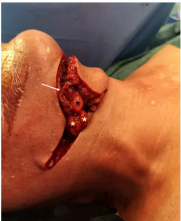
The incision extends as far as the platysma, starting in the lateral part of the skin paddle (Fig. 2). Careful dissection flush with the deep surface of the platysma muscle must be performed from above downwards in order to widely expose the submandibular region.
Fig. 2. Appearance of the flap after dissection. Asterisks: lymph nodes larger than
1 cm in areas Ib (black asterisk) and IIa (yellow asterisk). White arrow: submental pedicle.
Identification of the marginal mandibular branch of the facial nerve underneath the plane of the superficial cervical fascia may be difficult and time-consuming. However, this step is essential, as this small nerve branch must be carefully dissected so that it can be retracted to allow safe access to the submandibular region.
The anterior border of the sternocleidomastoid muscle must be released in order to identify the jugular vein and carotid artery. The posterior belly of the digastric muscle, indicating the posterior limit of the flap must then be dissected, taking care to preserve a maximum of adipose tissue within the flap (areas IIA and posterior Ib).
The facial artery and vein are then dissected over the inferior edge of the mandible. The submental pedicle arises about 1 cm underneath the basilar edge of the mandible. After identifying the submental artery, the facial pedicle can then be ligated distally to the origin of this pedicle. The submental artery is then dissected distally starting at its origin, by continuing the skin incision of the superior part of the flap. The anterior belly of the digastric muscle can be included to ensure a stronger flap. Meticulous haemosta-sis of the deep muscle perforating branches must be performed at this stage. All of the adipose tissue as far as the basilar edge of the mandible must be included to ensure a maximum of lymph nodes in the flap and to avoid injuring the septocutaneous perfo-rating vessels. The inferior incision of the skin paddle of the flap is then completed as far as the suprahyoid muscle plane, completely releasing the medial extremity of the flap.
Finally, the facial artery and facial vein are dissected via a retro-grade approach, in contact with the submandibular gland, allowing the inclusion of all adipose tissue of the submandibular (Ib) and subdigastric (IIa) areas. This dissection is continued as far as the internal jugular vein and external carotid artery, where the facial pedicle can be ligated flush with these vessels. Particular care must be taken when dissecting the subdigastric course of the facial artery, as the hypoglossal nerve crosses the facial artery in this region.
Fig. 3. Medial retromalleolar recipient site before (left) and after (right) submental flap placement. Note the slightly congested appearance of the flap after placement despite
tension-free skin closure.
The submental flap therefore comprises lymph node areas Ia (underneath the skin paddle), Ib (without the submandibular gland) and IIa. The submandibular gland is consequently almost totally released and only remains adherent by its inferior edge and partially by its deep surface. The donor site is then closed by sutures after placing a suction drain.
2.4. Preparation of the flap recipient site and flap placement The preferred flap recipient site is a medial retromalleolar site with anastomoses onto the posterior tibial artery (Fig. 3). An S-shaped incision is made and large subcutaneous dissection is performed to create a compartment for insertion of the flap. The recipient vascular pedicle is readily identified in the medial retro-malleolar region. End-to-end anastomoses are performed after release of the posterior tibial vessels from their sheath. Tension-free closure is essential to avoid compromising venous drainage of the flap. A non-suction drainage system is placed at the flap recip-ient site. When necessary, a large calibre venous anastomosis with the long saphenous vein at the anterior part of the incision can be performed.
2.5. Postoperative care
Close clinical surveillance must be ensured for the first 48 hours according to local protocols. The main risk is that of impaired venous drainage due to excess tension in the recipient zone. In the presence of a congested or “venous” appearance of the flap, tension must be immediately decreased by releasing sutures or even revis-ing anastomoses in the operatrevis-ing room. Elevation of the limb helps to promote venous return. Drainage systems are removed starting on the second postoperative day depending on the volume drained. Weight bearing can be resumed on the day after the operation. Pro-phylactic anticoagulation is continued for two to three weeks, until the patient has regained satisfactory mobilization. The use of a com-pression stocking on the operated side depends on the quality of venous return of the flap and may need to be continued until normal venous return has been restored.
2.6. Case report
To illustrate this procedure, we present the case of a 49-year-old man managed by vascularized lymph node transfer with submental free flap for secondary lymphoedema of the right lower limb, one year after resection of a right inguinal melanoma associated with inguinal and pelvic lymph node dissection followed by adjuvant radiotherapy (48 Gy) with good cancer control, as he was consid-ered to be in remission following this therapeutic sequence.
He progressively developed lymphoedema of the lower limb, first observed at the third postoperative month, which became progressively worse despite compression therapy and lifestyle
changes. After presentation and discussion of the various treatment options with the patient, it was decided to perform vascularized lymph node transfer with submental free flap, according to the technique described above.
The postoperative course was uneventful despite a con-gested appearance of the flap immediately after the operation, although the flap maintained good capillary refill and skin trophicity. Reduction of lymphoedema was observed and expe-rienced by the patient by the second postoperative day, with 30% reduction of lymphoedema one week after the operation, possibly enhanced by perioperative bedrest. The patient was discharged on the tenth postoperative day with unrestricted mobilization. At 5 months, the patient presented a marked reduc-tion of limb circumference, essentially in the distal part of the lower limb, decreasing from 45 cm to 37 cm 20 cm below the knee and from 61.5 cm to 60.8 cm 20 cm above the knee (Fig. 4 A and B). The patient also reported a marked reduc-tion of his symptoms. Lymphoscintigraphy performed 5 months postoperatively demonstrated lymphatic drainage on the medial aspect of the right ankle via the transferred lymph nodes (Fig. 4C and D).
3. Discussion
Although the submental free flap has been described for a long time, harvesting of this flap with the inclusion lymph nodes must comply with a number of rules to ensure functional success of the procedure on lymphoedema, as reduction of lymphoedema is directly dependent on the number of lymph nodes present in the flap[7]. Harvesting of the submental free flap remains difficult and anatomically demanding, particular in order to preserve adjacent nerves, and must be performed by a surgeon experienced in neck surgery, regardless of his or her primary specialty.
Creation of a submental skin paddle presents a number of advan-tages. In addition to allowing surveillance of flap vitality, this skin paddle is often necessary to allow tension-free skin closure of the recipient site, a zone with limited elasticity often asso-ciated with marked fibrosis of the cutaneous and subcutaneous tissues as a result of chronic lymphoedema. Skin is also known to be richly endowed in lymphatic capillaries and may conse-quently contribute to better results in terms of drainage of the lymphoedema via the dermis and the lymph nodes of the flap. This skin paddle is generally removed at a second operation for cosmetic purposes, after resolution of the lymphoedema. In the future, flap harvest techniques without a skin paddle could be use-ful to minimize the cosmetic sequelae of flap donor and recipient sites.
According to two recent reviews of the literature of the various reported lymph node transfer free flaps, the advantage of the sub-mental flap is the length of its pedicle (about 5 cm), with a large diameter artery (about 3 mm), constant lymph nodes with a mean
Fig. 4. Appearance of the lower limb preoperatively (A) and 5 months postoperatively (B). Lymphoscintigraphy performed 5 months postoperatively showing lymphatic
drainage of the right lower limb on the medial aspect of the right ankle via the transferred lymph nodes (C, D).
of 3 lymph nodes included in the flap, i.e. an equivalent number of lymph nodes to that provided by an inguinal flap, the flap most commonly used at the present time in this indication. The submen-tal flap also avoids the risk of secondary lymphoedema of the donor site, the main iatrogenic risk associated with inguinal or axillary flaps. However, harvesting of a submental free flap is associated with a risk of injury of the marginal mandibular branch of the facial nerve and cosmetic sequelae related to the visible neck scar[8,9].
Only limited data are available in the literature concerning the intermediate and long-term results of the submental flap in this indication. According to two recent studies, the estimated suc-cess rate, defined by improvement of lymphoedema, is between 70 and 100%[8,9]. No consensus has been reached concerning patient selection or the choice of donor and recipient sites for lymph node transfer techniques, due to the low level of proof currently available (retrospective studies based on small series) and the poorly eluci-dated mechanisms of action of transferred lymph nodes. Lymph node transfer in the head using a supraclavicular flap has also been described, but the results of the procedure are more uncertain due to the inconstant presence of lymph nodes in this zone (area Vb)
[10]. No comparative study has been conducted between vascu-larized lymph node transfer and lymphovenous anastomoses, the main alternative technique for the treatment of lymphoedema.
Although this technique primarily concerns head and neck sur-geons, the indication for vascularized lymph node transfer must be discussed with practitioners specialised in the management of lymphoedema, which remains multimodal in the context of a global treatment strategy[11]. Two-team surgery in this clinical setting also allows a gain of time by operating on the two sites concomi-tantly, in addition to the combined expertise of the head and neck and plastic surgeons.
4. Conclusion
Vascularized lymph node transfer is an increasingly popular technique, which raises great hopes for the treatment of lym-phoedema. The submental free flap is now one of the preferred techniques in this setting, but it requires a good knowledge of head and neck surgery in order to ensure safe and reliable flap harvest-ing. It therefore constitutes a multidisciplinary procedure, in which the patient’s preference and motivation play a predominant role.
Disclosure of interest
The authors declare that they have no competing interest.
Acknowledgements
Fanny Cros, Pauline Jeanneton.
References
[1]Beesley V, Janda M, Eakin E, Obermair A, Battistutta D. Lymphedema after gyne-cological cancer treatment: prevalence, correlates, and supportive care needs. Cancer 2007;109(12):2607–14.
[2]Allen RJ, Cheng M-H. Lymphedema surgery: patient selection and an overview of surgical techniques. J Surg Oncol 2016;113(8):923–31.
[3]Tourani SS, Taylor GI, Ashton MW. Vascularized lymph node transfer: a review of the current evidence. Plastic and Reconstructive Surgery 2016;137(3):985–93.
[4]Patel KM, Lin C-Y, Cheng M-H. A prospective evaluation of lymphedema-specific quality-of-life outcomes following vascularized lymph node transfer. Ann Surg Oncol 2015;22(7):2424–30.
[5]Cheng M-H, Huang J-J, Wu C-W, Yang C-Y, Lin C-Y, Henry SL, et al. The mechanism of vascularized lymph node transfer for lymphedema: natural lym-phaticovenous drainage. Plast Reconstruct Surg 2014;133(2):192e–8e.
[6]Righini CA, Petrossi J, Reyt E, Atallah I. An original submandibular approach technique sparing the cervical branch of the facial nerve. Eur Ann Otorhino-laryngol Head Neck Dis 2014;131(2):143–6.
[7]Gustafsson J, Chu S-Y, Chan W-H, Cheng M-H. Correlation between quan-tity of transferred lymph nodes and outcome in vascularized submental lymph node flap transfer for lower limb lymphedema. Plast Reconstruct Surg 2018;142(4):1056–63.
[8]Pappalardo M, Patel K, Cheng M-H. Vascularized lymph node transfer for treatment of extremity lymphedema: an overview of current controver-sies regarding donor sites, recipient sites and outcomes. J Surg Oncol 2018;117(7):1420–31.
[9]Scaglioni MF, Arvanitakis M, Chen Y-C, Giovanoli P, Chia-Shen Yang J, Chang EI. Comprehensive review of vascularized lymph node transfers for lymphedema: outcomes and complications. Microsurgery 2018;38(2):222–9.
[10]Steinbacher J, Tinhofer IE, Meng S, Reissig LF, Placheta E, Roka-Palkovits J, et al. The surgical anatomy of the supraclavicular lymph node flap: a basis for the free vascularized lymph node transfer. J Surg Oncol 2017;115(1):60–2.
[11]Kung TA, Champaneria MC, Maki JH, Neligan PC. Current concepts in the surgical management of lymphedema. Plast Reconstruct Surg 2017;139(4):1003e–13e.