DEVELOPMENT OF LC-MS/MS METHODS FOR THE ANAL YSIS OF REACTIVE METABOLITE PROTEIN T AR GETS
DISSERTATION PRESENTED
AS PARTIAL FULFILLMENT OF THE DOCTORATE IN CHEMISTRY
BY
MAKAN GOLIZEH
Avertissement
La diffusion de cette thèse se fait dans le respect des droits de son auteur, qui a signé le formulaire Autorisation de reproduire et de diffuser un travail de recherche de cycles supérieurs (SDU-522 - Rév.0?-2011 ). Cette autorisation stipule que «conformément à l'article 11 du Règlement no 8 des études de cycles supérieurs, [l'auteur] concède à l'Université du Québec à Montréal une licence non exclusive d'utilisation et de publication de la totalité ou d'une partie importante de [son] travail de recherche pour des fins pédagogiques et non commerciales. Plus précisément, [l'auteur] autorise l'Université du Québec à Montréal à reproduire, diffuser, prêter, distribuer ou vendre des copies de [son] travail de recherche à des fins non commerciales sur quelque support que ce soit, y compris l'Internet. Cette licence et cette autorisation n'entraînent pas une renonciation de [la] part [de l'auteur] à [ses] droits moraux ni à [ses] droits de propriété intellectuelle. Sauf entente contraire, [l'auteur] conserve la liberté de diffuser et de commercialiser ou non ce travail dont [il] possède un exemplaire.»
DÉVELOPPEMENT DE MÉTHODES LC-MS/MS POUR L'ANALYSE DE PROTÉINES CIBLES DES MÉTABOLITES RÉACTIFS
THÈSE PRÉSENTÉE
COMME EXIGENCE PARTIELLE DU DOCTORAT EN CHIMIE
PAR
MAKAN GOLIZEH
This Ph.D. program was a umque opportunity for me to gam a deeper understanding of bio-analytical mass spectrometry while enjoying a continuous sense of satisfaction through constant learning, improvement and productivity. This was, of course, not possible without the support of a number of people and organizations that, in the past four years, have effectively contributed to the content and context of my research. Therefore, I wish to cordially thank, first and foremost, my supervisor, Professor Lekha Sleno, for accepting me as a graduate student, training me with patience and precision, giving me the opportunity to engage multiple research projects in an independent yet coached manner, and for supporting me in ail the aspects of my professional and persona! life; all the former and present members of Professor Sleno's team, particularly Dr. André LeBlanc, Leanne Ohlund, Yasmin Boukhedimi, Maxime Sansoucy, Biao Ji, and Ghazaleh Moghaddam for being excellent co-workers and unforgettable friends; our resourceful undergraduate interns, Julie-Anne Jauffrit, Christina Schneider and Lucie Huart, for their assistance; Professor Isabelle Marcotte and Professor Steve Bourgault, for patiently accompanymg my research and providing helpful comments; Professor Karen Waldron for kindly accepting to read and evaluate this thesis; Professor Huu Van Tra for being a supportive program director; the staff of the Departrnent of Chemistry for always being nice and cooperative, particularly Sonia Lachance, Marie-Josée Crevier, Odette Desrosiers, Sylvie Lemieux, Isabelle Rheault, Mathieu Maurin-Soucy and Jacqueline Hue Tieu; the Faculty of Science and UQÀM for providing a positive and constructive environment; Groupe de Recherche Axé sur la Structure des Protéines (GRASP) and Fondation UQAM, for graduate student scholarships; Ministère de l'éducation du Québec, for additional financial support; and finally, my brother, Ashkan, for always being understanding, caring and supportive before and during my Ph.D. studies.
LIST OF FIGURES ... · ... ix
LIST OF TABLES ... xiv
LIST OF ABBREVIATIONS ... xvi
LIST OF SYMBOLS AND UNITS ... xxii
RÉSUMÉ ... xxiv
ABSTRACT ... xxv
CHAPTER ONE: INTRODUCTION ... 1
1.1 MOLECULAR TOXICOLOGY OF REACTIVE METABOLITES ... 2
1.1.1 Biologie Oxidation and Oxidative Stress ... 3
1.1.2 Xenobiotic Metabolism ... 7
1.1.3 Adduct Formation and Prote in Targets ... 15 1.2 EXPERIMENTAL TECHNIQUES: MS-BASED PROTEOMICS ... 18
1.2.1 Proteomics Technology ... 19 1.2.2 LC-MS/MS ... 26
1.2.3 Multidimensional Separation in MS-based Proteomics ... 34
1.3 CHALLENGES AND RESEARCH DESIGN ... 40
1.3.1 Preparation ofLiver Microsomal Samples ... 40
1.3.2 Data Processing Workflow ... 46
1.3.3 Research Outline ... 47
CHAPTER TWO: OPTIMfZED PROTEOMIC ANALYSIS OF RAT LIVER MICROSOMES USING DUAL ENZYME DIGESTION WITH 2D-LC-MS/MS ... 49
2.1 ABSTRACT ... 50
2.2 INTRODUCTION ... 50
2.3 .1 Materials ... 55
2.3.2 Sample Preparation ... 56
2.3.3 Digestion Conditions ... 56
2.3.4 Strong Cation Exchange Fractionation ... 57
2.3.5 Reversed-Phase UHPLC-MS/MS ... 58
2.3.6 Data Processing ... 60
2.4 RESULTS AND DISCUSSION ... 61
2.4.1 Single-Enzyme Digestion ... 62
2.4.2 Sequential Dual-Enzyme Digestion ... 65
2.4.3 Paralle1 Dual-Enzyme Digestion ... 66
2.4.4 Analysis of Identified Pro teins ... 70
2.5 CONCLUSIONS ... 78
2.6 SUPPORTING INFORMATION ... 79
CHAPTER THREE: MULTIDIMENSIONAL LC-MS/MS ANALYSIS OF LIVER PROTEINS IN RAT, MOUSE AND HUMAN MICROSOMAL AND S9 FRACTIONS ... 80 3.1 ABSTRACT ... 81 3.2 INTRODUCTION ... 81 3.3 EXPERIMENTAL ... 84 3.3.1 Materials ... 84 3.3.2 Sample Digestion ... 84 3.3.3 Protein Fractionation ... 85 3.3.4 Peptide Fractionation ... 86
3.3.5 RP-UHPLC-MS/MS Analysis of Peptide Fractions ... 87
3.4 RESULTS AND DISCUSSION ... 88
3.4.1 Method Optimization ... 89
3.4.2 2D-LC-MS/MS Using Protein-Level Fractionation ... 91
3.4.3 2D-LC-MS/MS Using Peptide-Leve! Fractionation ... 93
3.4.4 3D-LC-MS/MS (Combined Protein-and Peptide-Leve! Fractionation) ... 94
3.4.5 Tryptic versus Peptic Digestion ... 94
3.4.6 Comparison of the Four Workflows ... 98
3.4. 7 Rat Proteome Results ... 1 00 3.4.8 Cross-Species Comparison ofLiver Proteins ... 105
3.5 CONCLUSIONS ... 112
3.6 ACKNOWLEDGEMENTS ... 113 3.7 SUPPORTING INFORMATION ... oooooooooooooooo .. ooooooooooooooooooooooooooo113 CHAPTER FOUR: DATASET FROM PROTEOMIC ANALYSIS OF RAT, MOUSE, AND HUMAN LIVER MICROSOMES AND S9 FRACTIONS ... 114
4.1 ABSTRACT ... 0 0 ... 0 0 ... 0 0 .... 0 ... 0 ... 0 ... 0 ... 0 115 402 VALUE OF THE DATA ... oo ... oooooooooooooooooooooooo ... oo.ooooo .. ooooooooooo .. oo.oooo115 403 SPECIFICATIONS TABLE ... 00 0000 00 .. 00 ... oo·· .... oo ... oo ... oo .. ···oo·oo·· 116
4.4 EXPERIMENTAL DESIGN, MATERIALS AND METHODS.oo .. ooooooooooooooooo.117 405 ACKNOWLEDGEMENTS ... oo ... 0 0 ... oo .oo oo. 0 oo. 00 0 00 ... 00 0 0 0 0 0 0 00 0 .. 0 0 0 0 0 000 0 0 0 00 0000 000 0 00 119 4.6 SUPPORTING INFORMATION oo ... oo ... oo ... oo ... oo .. oo ... oo ... 120
CHAPTER FIVE: IDENTIFICATION OF ACETAMINOPHEN ADDUCTS OF RAT LIVER MICROSOMAL PROTEINS USING 2D-LC-MS/MS ... 00.00 ... 122
5.1 ABSTRACT oooooooooo•oo···oo .. o.oooo .. oo .. oo··o··ooooo .. o ... oo .... o .... o .. oooOoo ... oo.o .. oooo .. o .. o ... 123 502 INTRODUCTION ··oo···o···o•oo ... oooo .. ooooooooooooooooooooooooooooooooooooooooooooooooooooo 123 503 EXPERIMENTAL ... oooo···ooooooooo ... oo.oooooooooooooooooooo .. o.oooo···o···o···o···ooooooooooooo .. o.126
5.3.1 Chemicals and Reagents ... 126
5.3.2 Drug Metabolism ... 126
5.3.3 Protein Digestion ... 127
5.3.4 SCX Chromatography ... 127
5.3.5 Microsomal Trapping Experiments with Standard Peptides ... 128
5.3.6 RP-UHPLC-MS/MS Analysis ... 128
5.3.7 Data Analysis ... 129
5.4 RESULTS AND DISCUSSION ... 131
5 .4.1 Proteome Analysis of the Microsomal Samples ... 131
5.4.2 Peptide Spectral Matching ... 132
5.4.3 Statistical Differentiai Ana1ysis ... 132
5.4.4 Peak-Pair Finding ... 133
5.4.5 Differentiai Analysis Combined with Diagnostic Ion Screening ... 133
5.4.6 Data Verification and Comparison of Data Mining Workflows ... 134
5.4.7 APAP Covalent Binding Target Proteins ... 141
5.5 CONCLUSIONS ... 146
5.6 ACKNOWLEDGEMENTS ... 146
5.7 SUPPORTING INFORMATION ... 146
CHAPTER SIX: COVALENT BINDING OF 4-HYDROXYNONENAL TO MATRIX METALLOPROTEINASE 13 STUDIED BY LIQUID CHROMATOGRAPHY-MASS SPECTROMETRY ... 147
6.1 ABSTRACT ... 148
6.2 INTRODUCTION ... 149
6.3.1 Materials ... 151
6.3.2 Incubation of Recombinant Human MMP-13 with HNE ... 152
6.3.3 Specimen Selection, Chonclrocyte Culture and Treatment ... 152
6.3 .4 Immunoprecipitation ... 153
6.3.5 Protein Fractionation by Ion Exchange Chromatography ... 153
6.3 .6 Prote in Digestion ... 154
6.3.7 LC-HR-MS/MS Analysis ... 155
6.3.8 LC-MRM Analysis ... 155
6.3.9 Data Processing ... 156
6.4 RESULTS AND DISCUSSION ... 157
6.4.1 Covalent Modification ofMMP-13 by HNE ... 157
6.4.2 Reactivity of HNE Modification Sites ... 164
6.4.3 Targeted MRM-Based Assay ... 166
6.4.4 Analysis of Cell Culture Samples ... 166
6.5 CONCLUSIONS ... 174
6.6 ACKNOWLEDGMENTS ... 175
6.7 SUPPORTING INFORMATION ... 175
CHAPTER SEVEN: SUMMARY AND CONCLUSIONS ... 176
7.1 SUMMARY OF FINDINGS ... 177
7.2 LIMITATIONS AND PERSPECTIVES ... 184
APPENDIX A ... 194
APPENDIX B ... 200
APPENDIX C ... 205
APPENDIX D ... 215
1.1 Chemical structure of flavin a.denine dinucleotide (F AD) and nicotinarrtide adenine dinucleotide (NAD) cofactors involved in biologie redox reactions ... 4 1.2 Key play ers of oxidative stress and potential molecular targets ... 6 1.3 Summary of the acetaminophen metabolic pathway ... 11 1.4 Relationship between xenobiotic metabolism and reactive metabolite-induced
toxicity ... 12 1.5 Covalent binding of malondialdehyde (MDA) with DNA leading to the formation of potentially mutagenic (modified) guanine (M1 G), adenine (M1A), and cytosine (M1C) ... 16 1.6 Liver metabolism of trimethoprim, formation of a reactive iminoquinone methide (an a,B-unsaturated diimine) metabolite, and its subsequent covalent binding to hepatic pro teins ... 17 1.7 Simplified depiction of the four major domains of "omics" research and their primary molecular classes of interest ... 20 1.8 Different steps involved in a typical experimental workflow in a gel-based (a) or gel-free (b) proteomics approach ... 21 1.9 Steps involved in bottom-up (a) and top-down (b) proteomics analysis ... 22 1.10 Peptide fragmentation via CID and structure of b- and y-type product ions often
used for de nova peptide sequencing or peptide spectral matching ... 23 1.11 A typical SILAC quanti ta ti on workflow ... 25 1.12 Major components of a typical mass spectrometer. ... 27 1.13 General scheme of AB Sciex TripleTOF™ 5600 mass spectrometer, the hybrid QqTOF instrument used in this dissertation ... 33 1.14 Selectivity of HILIC, cation exchange (CX), anion exchange (AX) and RP chromatography for peptides as a function of the ir charge and polarity ... 3 7
1.15 Chemical structure of mixed-mode compound-specific/class-specific sorbents used in Oasis HLB, MAX, MCX SPE cartridges showing their hydrophilic interaction (a), hydrophobie interaction (b), anion exchange (c), and cation exchange ( d) sites ... 39 1.16 Amino acid sequence and approximate topology of Escherichia coli water channel aquaporin Z and its transmembrane regions ... 42 1.17 Solubilizing agents tested for the analysis of liver microsomal proteins were selected from different classes ... 44 1.18 Workflow of an in vitro experiment using liver microsomes to study protein covalent binding ... 45 1.19 Research design in the development of analytical methods for the identification of reactive metabolites target pro teins ... 48 2.1 Representative UV chromatogram ( detected at 220 nm) from SCX fractionation and total ion chromatograms from LC-MS/MS ana1ysis of one SCX fraction showing survey TOF-MS and sum of dependent MS/MS scans from trypsin and pepsin digestions ... 59 2.2 Venn diagram representing the proportion of identified proteins for single enzyme digestions as a function of the total number of proteins from trypsin, pepsin and G1u-C data sets combined ... 64 2.3 2D map of isoelectric point and GRA VY scores for the proteins identified in this study as a function of molecular weight ... 72 2.4 Number of transmembrane domains (TMD) of identified integral membrane proteins (IMP) under tryptic, peptic and parallel separately treated tryptic-peptic digestions and under three different strategies of tryptic-peptic parallel dual digestion ... 73 2.5 Number of confident peptides per protein (A) and overall protein sequence coverage (B) from en tire data set (1 095 pro teins in total) ... 74 2.6 GO annotations for identified rat liver microsomal proteins based on their molecular function and biological processes ... 75
2. 7 A subset human analogs of identified pro teins reported to be involved in metabolic processes analyzed for the presence of known human protein-protein interactions using InnateDB ... 77 3.1 MDLC-MS/MS proteomic analysis workflow ... 89 3.2 LC-UV traces at 220 nm for protein-level fractionation from mixed-bed WCX/WAX (CATWAX), tandem dual-colunm WCX-WAX and WAX-WCX, stand-alone WCX and WAX separations for RLM and RLS ... 92 3.3 LC-UV traces at 220 nm from the SCX peptide-leve! fractionation of RLM and RLS sam pies digested by trypsin or pep sin ... 93 3.4 Venn diagrams representing the number ofidentified proteins in RLM and RLS using ID-, 2D- and 3D-LC-MS/MS workflows ... lOO 3.5 Statistical distribution of isoelectric point and GRA VY score as a function of
MW for the pro teins identified in RLM and RLS ... 103 3.6 Gene ontology annotations based on molecular function (A) and biological
process (B) for the pro teins identified in RLM and RLS ... 104 3.7 Statistical distribution of liver proteins in rat, mouse and human in terms of
isoelectric point and GRA VY score as a function of MW ... 107 3.8 Gene ontology annotations based on molecular function (A) and biological process (B) for the liver proteins (from microsomes and S9) in rat, mouse and human ... 108 3.9 Venn diagrams depicting the number ofshared and unique proteins identified in rat, mouse and human liver microsomal (LM) and liver S9 (LS) proteins ... 109 3.10 Venn diagrams depicting the number of shared and unique pro teins identified in rat, mo use and hum an li ver sam pies (microsomes and S9 fractions) ... 110 3.11 Percent alignment of proteins unique to rat, mouse or human liver microsomes or S9 fraction ... 110 3.12 Percent alignment of proteins unique to human with those unique to rat and mouse ... lll
5.1 Covalent binding of NAPQI to the free thiol group of cysteine residues in pro teins ... 125 5.2 (a) UV trace at 220 nrn from SCX chromatography of the APAP-treated rat
li ver microsomes digest; (b) representative total ion chromatogram from
UHPLC-RP-LC-MS/MS analysis of SCX fraction #11 of the APAP-treated sample ... l30 5.3 High-resolution MS/MS spectra (m/z 80-300 range) from APAP-modified (a) glutathione, (b) ILISDFGLCK, (c) QACLFK, and (d) LQQCPFEDHVKL peptides ... 135 5.4 Data analysis workflow for the identification of AP AP-modified peptides incorporating peptide spectral matching (A), differentiai analysis combined with diagnostic ion screening (separate SCX fractions) (B), statistical differentiai analysis based on LC-MS signal intensities (SCX fractions combined) (C), and peak-pair finding between control and treated samples (D) ... 136 5.5 Putative role of the identified modified proteins in the previously suggested mechanism for APAP-induced hepatotoxicity ... 145 6.1 Sequence annotation of hurnan MMP-13 (UniProt accession P45452) and the identified HNE modification sites ... 158 6.2 Mechanism of reaction for the nucleophilic addition of histidine and cysteine to HNE ... l60 6.3 HNE adducts CID-generated diagnostic products corresponding to (a) cleaved dehydrated HNE, (b) neutralloss of HNE, (c) cleaved irnmonium ion of HNE-modified histidine, and ( d) cleaved dehydrated irnrnonium ion of HNE-modified histidine ... 160
6.4 MS/MS spectra from the His-48-containing peptide (
+
3 charge state) inunmodified (a) and HNE-modified (b-e) forms ... 161 6.5 Graphie representation of rnodified/unrnodified peak area ratios under different HNE concentrations and incubation times at [HNE]
=
200 J..LM ... l656.6 Evidence ofHNE binding to MMP-13 by immunoprecipitation ... 169 6.7 Overlaid extracted ion chromatograms of digested peptides containing HNE-modifications obtained from HNE-treated rhMMP-13 (A) and IP-purified MMP-13 from cultured chondrocytes (B) ... 173 7.1 Workflow for the bioinformatics analysis of rat li ver microsomal pro teins ... 178 7.2 MD-LC approaches employed to maximize peptide separation efficiency for proteomics analysis of rat li ver microsomes and S9 fractions ... 180 7.3 A typical data-dependent acquisition (DDA) cycle with dynamic exclusion .. 181 7.4 The analytical approach developed throughout this dissertation for identification of reactive metabolite protein targets using LC-MS/MS ... 183 7.5 Biotin-coupled reagents for selective enrichment of peptides using affmity separation incorporating iodoacetamide (a) and maleimide (b-e) moieties for specifie conjugation with the thiol functionality of cysteine-containing peptides ... 188 7.6 Sample preparation strate gy for affinity enrichment of AP APyne-modified
peptides using click chemistry ... 189 7.7 Graphical comparison between horizontal and vertical selection of precursor ions in DDA (a) and DIA (b ) ... 192
1.1 Radical and non-radical oxygen metabolites ... 5
1.2 Human cytochrome P450 enzymes and their substrates ... 8
1.3 Selected hu man cytochrome P450 xenobiotic substrates ... 9
1.4 Selected xenobiotic conjugation reactions ... 10
1.5 Most common classes of electrophilic reactive metabolites ... 14
1.6 Comparison of the most common mass analyzers ... 30
1.7 Proteases commonly used in bottom-up proteomics ... .41
2.1 Comparison between different digestion conditions for analysis of rat liver microsomes by 2D-LC-MS/MS ... 63
2.2 Comparison between sequence coverage (%)and number of confident peptides for three selected pro teins ... 69
3.1 Improvements achieved by optimization of the IDA-MS/MS parameters ... 91
3.2 Comparative results from the analysis of rat liver microsomes (RLM) and S9 fractions (RLS) using the four fractionation approaches (trypsin, pepsin, and the two enzymes combined) ... 96
3.3 Replicate analysis of tryptic and peptic digests for each LC-MS based approach ... 97
3.4 Number and % identified pro teins and IMPs unique to and shared between tryptic and pep tic digestions ... 98
3.5 Proteomic analysis of rat, mouse and human liver microsomes and S9 fractions using the optimized 2D-LC-MS/MS method ... 106
4.1 Processed data from proteomic analysis of rat, mouse, and human liver microsomes and S9 fractions ... 121
4.2 Dataset identifiers of the mass spectrometry data obtained from the analysis of rat, mouse, and human liver microsomes and S9 fractions on the public proteomics repositories ... 121
5.1 Number of putative AP AP-modified peptides detected by each data mining workflow and percent shared between different workflows ... 137 5.2 List of the identified AP AP adducts of RLM pro teins ... 139
6.1 Distinct identified peptides encompassing HNE-modification sites within the
MMP-13 structure ... 163 6.2 Optimized m/z values for the precursor and product ions and collision energies for MRM ana1ysis of unmodified and HNE-modified peptides detected for MMP-13 ... 167
6.3 Distribution of the identified proteins and peptides in the CATW AX fractions
collected from hu man chondrocyte cultures ... 170 6.4 List of the identified pro teins (1% FDR) in human chondrocyte cultures enriched by immunoprecipitation ... 170 6.5 LC-MS/MS data obtained from the analysis of human chondrocyte cultures to confirm HNE-modification sites ofMMP-13 ... 172 7.1 List of suggested modifications and extensions to this work for future studies
ABC
ACN ADR APAP APCI API APPI ASKl ATP AX CATWAX CE CI CIDCMC
cox
cv
ex
CYP DADAB
DBS DDADDM
DENN DIA A TP -binding cassette acetonitrileadverse drug reaction
N-acety 1-p-aminopheno 1 ( acetaminophen) atmospheric-pressure chemical ionization atmospheric-pressure ionization
atmospheric-pressure photoionization apoptosis signal-regulating kinase 1 adenosine triphosphate
anion exchange
mixed-bed weak cationlweak anion exchange capillary electrophoresis
chemical ionization
collision-induced dissociation critical micelle concentration cyclooxygenase coefficient of variation cation exchange cytochrome P450 differentiai analysis N,N-dimethyl-4-aminoazobenzene dynamic background subtraction data-dependent acquisition n-dodecyl ~-D-maltoside
differentially expressed in neoplastic versus normal cells data-independent acquisition
DILI
DMEMDOTA
dRDTT
EC
D
ECM
ElELISA
ER
ERLIC
ESI
ETC
ETD
ExPASy
FADFADP
FAST A
FBS
FC-14FDR
FMO
FT-ICR
GC
GDP
GE
GELFrEE
GlueGO
drug-induced liver injury
Dulbecco's modified Eagle's medium
1 ,4, 7,1 0-tetraazacyclododecane-1 ,4, 7,1 0-tetraacetic a cid
deoxyribose dithiothreitol
electron-capture dissociation
extracellular matrix
electron ionization
enzyme-linked imrnunosorbent assay endoplasmic reticulum
electrostatic repulsion-hydrophilic interaction chromatography electrospray ionization
electron transport chain electron-transfer dissociation
expert protein analysis system
flavin adenine dinucleotide
flavin adenine dinucleotide phosphate
fast-ail
fetal bovine serum
n-tetradecyl phosphocholine
false discovery rate
flavin-containing rnonooxygenase
Fourier-transform ion cyclotron resonance gas chromatography
guanosine diphosphate
gel-electrophoresis
gel-eluted liquid fraction entrapment electrophoresis glucose
GPCR GRAVY GSH GST GTP
HILIC
HLB
HLM
HLSHNE
HPLC
HRHSAB
lAM ICAT IDA IDE IEF IL-1~ IMP IP IRMPD iTRAQ JNKLC
MADD MALDI MAPK MAXG-protein coupled receptor grand average of hydropathy glutathione
glutathione S-transferase guanosine triphosphate
hydrophilic interaction liquid chromatography hydrophilic-lipophilic balanced
human liver microsomes human liver S9 fraction 4-hydroxy-2-(E)-nonenal
high-performance (pressure) liquid chromatography high-resolution
hard and soft acids and bases 2-iodoacetamide
isotope-coded affinity tag
information-dependent acquisition integrated development environment isoelectric focusing
interleukin-1 beta
integral membrane protein immunoprecipitation
infrared multi-photon dissociation
isobaric tags for relative and absolute quantitation c-Jun N-terminal kinase
liquid chromatography
MAPK-activating death domain protein matrix-assisted laser desorption ionization mitogen-activated protein kinase
MCX MDA MDLC MeCAT MGO MGSTl MLM MLS MMP MPO MRM MS MSA MS/MS MudPIT MWCO NAD NADP NAPQI NCBI NMR NOX NSAID NU5M OA ONE PANTHER PAPS PIM
mixed-mode cation exchange malondialdehyde
multidimensionalliquid chromatography metal-coded affinity tag
methylglyoxal
microsomal glutathione S-transferase 1 mouse liver microsomes
mouse liver S9 fraction matrix metalloproteinase myeloperoxidase
multiple-reaction monitoring mass spectrometry
multiple sequence alignment tandem mass spectrometry
multidimensional protein identification technology molecular weight eut-off
nicotinamide adenine dinucleotide
nicotinamide adenine dinucleotide phosphate N-acetyl-p-benzoquinone imine
National Center for Bioteclmology Information nuclear magnetic resonance
NADPH oxidase
non-steroidal anti-inflammatory drug NADH-ubiquinone oxidoreductase chain 5 osteoarthritis
4-oxo-2-(E)-nonenal
protein analysis through evolutionary relationships 3 '-phosphoadenosine-5 '-phosphosulfate
PMF PMP POE PPI PSD PS-DVB PSM PTGS PTM PUFA QET RA ReT OF RLM RLS RNS ROS RP
sc
x
SDC SDS SDS-PAGE SFC SILACSIM
SOD SPE SRM SULTpeptide mass fmgerprinting
peripheral membrane protein polyoxyethylene
protein-protein interaction post-source decay
polystyrene-divinylbenzene peptide spectral matching prostaglandin G/H synthase
post-translational modification polyunsaturated fatty acid
quasi-equilibrium theory rheumatoid arthritis reflectron time-of- flight
rat liver microsomes rat liver S9 fraction reactive nitrogen species reactive oxygen species
reversed-phase
strong cation exchange
sodium deoxycholate
sodium n-dodecyl sulfate
sodium n-dodecyl sulphate polyacrylamide gel-electrophoresis
supercritical fluid chromatography
stable isotope-labeling with amino acids in cell culture
selected-ion monitoring
superoxidase dismutase
solid-phase extraction
selected-reaction monitoring
SWATH TCA TDC TFA TIC TMD TNF TOF TPDB TRAF2 Tris UDP UGT UHPLC
uv
WAXwc x
XAO XICsequenhal window-acquisition of all theoretical mass spectra trichloroacetic acid
time-to-digital converter trifluoroacetic acid total ion chromatogram transmembrane domain tumor necrosis factor time-of-flight
target protein database
TNF receptor-associated factor 2 tris(hydroxymethyl)aminomethane uridine diphosphate UDP-glucuronyltransferase ultra-high-pressure HPLC ultraviolet
weak anion exchange weak cation exchange xanthine oxidase
A
AngstromB magnetic sector
o
c
degree centigrade (Celsius) cc cubic centirnetereps cycle per second
Da Dalton
Mo potential difference
E total energy
E0 potential
Eex excess energy
Eint internai energy
g gram, gravity (g-force)
h hour
IJ chemical hardness
kE rate constant of a gas-phase unirnolecular reaction liter M rnolar rn meter 1..1. rn1cron mm minute MW molecular weight mlz rnass-to-charge N nurnber of electrons
n nurnber of analytical replicates v number of vibrational states
NaN not a number
p peak capacity
pl isoelectric point
ppm part per million
ps1 poundspersquare inch
Q
quadrupoleq radiofrequency-only quadrupole
rh recombinant human
rpm round per minute
RT
retention times
total degree of freedoms second
s-I per second
SIN
signal-to-noisev
Voltv/v volume per volume
w/v weight per volume
w/w weight per weight
endogènes subissent les transformations métaboliques en formant des« métabolites réactifs
»
qui peuvent réagir avec des biomolécules, altérer leurs fonctions moléculaires et affecter des processus biologiques. Le foie joue un rôle prédominant dans le métabolisme, et les protéines hépatiques sont souvent ciblées par les métabolites réactifs causant potentiellement l'hépatotoxicité. L'identification de ces protéines est donc essentielle pour mieux comprendre les mécanismes reliés à cette toxicité. Plusieurs essais ont été effectués afin d'étudier les liaisons covalentes sur les protéines et identifier leurs cibles potentielles. Toutefois, la faible abondance des protéines modifiées, les problèmes techniques, et l'absence de méthodes appropriées pour analyser des échantillons biologiques ont été les défis limitant le progrès de cette recherche. Parmi les techniques analytiques employées en bioanalyse, la spectrométrie de masse (MS) est devenue la méthode de choix en raison de sa capacité exceptionnelle d'acquérir des grandes quantités de données qualitatives et quantitatives à partir de mélanges complexes. La spectrométrie de masse en tandem (MS/MS) couplée à la chromatographie liquide (LC) a permis 1 'analyse efficace des échantillons protéiques avec une grande sensibilité dans la recherche protéomique.La thématique principale de cette thèse a été le développement d'une approche analytique pour identifier les protéines cibles des métabolites réactifs et leurs sites de modifications par LC-MS/MS. Cette recherche a été organisée en trois phases, selon un des principaux défis posés dans ce type d'analyse : (1) préparation d'échantillon, (2) détection par LC-MS/MS, et (3) traitement des données. Premièrement, la méthode analytique a été optimisée pour l'analyse protéomique dans les fractions S9 et microsomales de foie de rat, souris, et humain. Puis, des microsomes de rat ont été incubés avec l'acétaminophène, formant un métabolite réactif, afin de générer des adduits protéiques, puis analysés par la méthode développée en utilisant une stratégie de traitement de données multi-étage. Une approche plus ciblée a été également établie afin d'étudier la modification covalente de métalloprotéase matricielle 13 par le produit de peroxydation lipidique, 4-hydroxynonénal.
Mots-clés : liaison covalente, métabolites réactifs, LC-MS/MS, microsomes de foie de rat, acétaminophène, métalloprotéase matricielle 13, 4-hydroxynonénal
undergo metabolic transformations to form "reactive metabolites". These species can covalently bind to biomolecules, including proteins, altering their molecular functions affecting biological processes. The liver plays a predominant role in metabolic transformations and hepatic proteins are often targeted by reactive metabolites potentially leading to hepatotoxicity. Identification of these proteins is therefore important to understand the mechanisms involved in this type of toxicity.
Several previous attempts have been made to study protein covalent binding reactions and identify their potential targets. However, low abundance of the modified proteins, technological issues, and lack of appropriate methods for efficient analysis of complex biological samples are challenges that have limited the progress of this research. Amongst the analytical techniques employed in bioanalysis, mass spectrometry (MS) has become the method of choice due to its incredible capacity to acquire large amount of qualitative and quantitative information from complex
mixtures. Tandem mass spectrometry (MS/MS) in combination with liquid
chromatography (LC) has enabled efficient analysis of protein samples with high sensitivity fostering new possibilities in proteomics research.
The main theme of this dissertation was to systematically develop an analytical approach to identify reactive metabolite protein targets and their sites of modification using LC-MS/MS. This research was designed in three phases to address the main challenges posed in this type of analysis: (1) sample preparation, (2) LC-MS/MS detection, and (3) data analysis strategies. The analytical method was first optimized for proteomics analysis of rat, mouse and human liver S9 and
rnicrosomal fractions. Next, rat liver microsomes were incubated with
acetaminophen, known to form a reactive metabolite, to produce protein adducts, which were then subjected to the developed method and identified using a multi-stage data processing work:flow. A more targeted approach was also established to study covalent binding of matrix metalloproteinase 13 by the lipid peroxidation product, 4-hydroxynonenal.
Keywords: covalent binding, reactive metabolites, LC-MS/MS, rat liver microsomes,
INTRODUCTION
Most compounds which a living organism is exposed to are changed upon
excre6on. The sophisticated molecular machinery of the cell encompasses a myriad
of highly specialized mechanisms to cope with different groups of chemicals that
bind to or cross its plasma membrane. These biotransformations however, are not
necessarily flawless, and under certain conditions, can jeopardize cell homeostasis
and survival.
Scientific and technological breakthroughs in the last decades gave rise to
novel areas of research that enabled a clearer understanding of biological
mechanisms. Bioanalytical chemistry, or bioanalysis, is a sub-discipline of analytical
chemistry that concentrates on qualitative and quantitative analysis of small and large
molecules in biological systems using efficient instrumental techniques such as
chromatographie and electrophoretic separation, ligand binding assays, mass
spectrometry (MS), and nuclear magnetic resonance (NMR). The expanding role of
bioanalytical chemistry in academie and industrial environrnents has made it an
important area of research for scientists in chemistry and biochemistry. The
exceptional capacity of bioanalytical techniques in providing high-quality data on the
chemical composition of complex matrices have, in recent years, facilitated numerous
scientific endeavors that depend on accurate measurements in biological samples,
This introductory chapter will briefly discuss the chemistry of intrinsically reactive products of biotransforrnation, known as the "reactive metabolites", which are the major focus of this dissertation, followed by a short overview on bioanalytical techniques used in this work. The chapter will be concluded by discussing current challenges in this area and how they are addressed in this thesis.
1.1 Molecular Toxicology of Reactive Metabolites
Metabolism usually produces "non-toxic" metabolites, which are relatively
polar and can readily be excreted from the living organism and thus detoxified.
However, certain xenobiotics and endogenous compounds form highly reactive
metabolites, wbich can interact with vital intracellular macromolecules, and result in
toxicity. In the 1940s and 1950s, James and Elizabeth Miller demonstrated for the
first time that N,N-dimetbyl-4-aminoazobenzene (DAB), a hepatocarcinogen in rats,
would covalently bind to proteins and nucleic acids through a process they called
"metabolic activation". They also demonstrated tbat covalent binding of these species
was an essential part of the carcinogenic process (Conney, 200 1).
Norrnally, reactive metabolites can be detoxified via a series of enzymatically -assisted covalent reactions to endogenous scavenger molecules known as the
"conjugation reactions". However, overabundance or excessive reactivity may help
these chemicals skip or saturate designated conjugation mechanisms to trigger undesired reactions within the tissue depending on the nature of the reactive species and the physiology of the organism (Attia, 201 0).
The following section briefly discusses major intracellular sources of the
reactive metabolites, their formation and fate within the organism, the chemistry of
1.1.1 Biologie Oxidation and Oxidative Stress
Oxygen is the vital element of aerobic life in which oxidation of organic
compounds is exploited for the release of free energy. However, not only is 02 fatal
to anaerobie bacteria, due to its high chemical reactivity, excess oxygen is also toxic
to aerobic organisms. Even at non-toxic concentrations, aerobic organisms must deal
with the toxic consequences of partial oxidation-reduction reactions, that is the
formation of reactive oxygen-derived compounds (Joseph y, 1997).
An oxidation-reduction, or redox, reaction is of the general form
which can be written as two separate yet dependent and complementary half-reactions
involving an electron-donor (reductant) and electron-acceptor ( oxidant) species:
The direction of a redox reaction depends on which spec1es bas a greater
tendency (or potential, E0) to accept the available electrons. The potential difference
(Mo) of a redox system is an important thermodynamic factor, and determines if a
redox reaction can proceed spontaneously.
In living organisms, redox reactions are catalyzed by "oxidoreductases", a
large class of enzymes th at utilize pyridine- or flavin-cofactors (Figure 1.1) to trans fer
electrons from one molecule to another. Oxidoreductases are generally classified into
four groups (oxidases, dehydrogenases, hydroperoxidases, and oxygenases) and
reduce or oxidize a wide range of organic functional groups and inorganic elements
Redox reactions are also the core of the electron transport chain (ETC), the
essential component of photosynthesis and cellular respiration in prokaryotic and
eukaryotic cells. In eukaryotes, the ETC transfers electrons from NADH to Oz
(Mo
=
1.1 V) through three large protein complexes: NADH-ubiquinoneoxidoreductase (Complex I), ubiquinone-cytochrome c oxidoreductase (Complex III),
and cytochrome c oxidase (Complex IV). This electron transfer creates an
electrochemical proton gradient across the inner mitochondrial membrane that drives
adenosine triphosphate (A TP) synthesis (Murray et al., 2009).
e
X :
NH
2
0 1 N ""<:::,O=r
-O
~
<
N
1N
)
1 H Ho=
P
1-o6
H H ~ HO OH H2c 1 H-C-OH 1 H-C-OH 1 H- C- OH 1 H2C 1 1 # .<9N
H
H
,cxx
N
x;
N
yo
H3C N 0Figure 1.1 Chemical structure of flavin adenine dinucleotide (F AD) (left) and
nicotinamide adenine dinucleotide (NAD) (right) cofactors involved in biologie redox
reactions
The final stage of the ETC involves a four-step reduction of molecular oxygen
(Oz) to water:
OH
The above reduction reaction bas a very large positive ~Eo value and is therefore thermodynamically favorable in the presence of almost any biochemical reductant. In the cell, the transfer of four electrons to 02 involves iron and copper ions, and is accomplished in a controlled and concerted manner; however, the release of intermediary products (superoxide, peroxide, hydroxyl radical) cannot be fully avoided (Josephy, 1997). Oxygen reduction intermediates are extremely reactive and
immediately react with other species to give rise to radical and non-radical
metabolites, including "reactive oxygen species (ROS)" and "reactive nitrogen
species (RNS)", representing the major sources of oxidative and nitroxidative stress.
A list of the most important reactive oxygen metabolites is given in Table 1.1 (Kohen
and Nyska, 2002).
Table 1.1 Radical and non-radical oxygen metabolites
Radical Non-radical Oxygen bi-radical (02••) Superoxide ion (02·-) Hydroxyl (HO•) Peroxyl (ROO•) Alkoxyl (RO•) Nitric oxide (NO•)
Hydrogen peroxide (H202)
Organic peroxide (ROOH)
Hypochlorous acid (HOC!)
Ozone (03)
Aldehydes (RCOH) Singlet oxygen (102)
Peroxynitrite (ONOOH)
Oxidative stress can be induced either directly by "primary ROS" or indirectly
via partial oxidation of biomolecules, such as lipids and carbohydrates. The products
of these reactions, which are usually classified under "secondary ROS", are also
ROS include malondialdehyde (MDA), 4-hydroxy-2-(E)-nonenal (HNE),
4-oxo-2-(E)-nonenal (ONE), and 2-oxoaldehydes such as glyoxal (GO) and methylglyoxal (MGO). The chemistry of these reactions will be discussed in more details in Chapter 6. Free radical chain reactions, such as autoxidation reactions, metal ions ( especially
iron, copper, and cobalt), and certain oxidoreductases, including NADPH oxidase,
xanthine oxidase, superoxide dismutase, and myeloperoxidase are other key players
of ROS formation and oxidative stress. Figure 1.2 summarizes chemical species,
enzymes and the mechanisms involved in these processes (Chondrogianni et al., 2014). H10 + 02 ETC
1
Caralase :.\IPO SOD \'OX and X.AOw
T
--c-r--+c
i
~
...J
... .
Autoxidation·oH+ -oH ---..:. Proreins, R'IA, D\'A
u
,;,,
c.,,~~~
1 _ _ _ _ _ _ _ _ _ _ _~
r
~~-~~
r·~~~~·
r
-,
1i
0 0 0 0 1:
Ro
·
1Roo
·
--+--+--+ H Jl A H oH o / : 1 :\IDA K\t: GO and :\!GO 1 1 1 1 Secouda1TROS 1 1----~---JFigure 1.2 Key players of oxidative stress and potential molecular targets.
ETC: electron transfer chain; GO: glyoxal; HNE: 4-hydroxy-2-(E)-nonenal; MDA: malondialdehyde; MGO: methylglyoxal; MPO: myeloperoxidase; NOX: NADPH oxidase; ROS: reactive oxygen species; SOD: Superoxidase dismutase; XAO: xanthine oxidase. M2+ is a bivalent metal ion.
Living organisms have adapted to continuously control and tightly regulate the presence of oxygen metabolites. This is extremely important for maintaining vital cellular and biochemical functions and any interference or imbalance leading to an uncontrolled state can cause excessive oxidative stress and cellular damage.
1.1.2 Xenobiotic Metabolism
The Unified Medical Language System classifies any chemical substance that is foreign to the biological system including natural products, drugs, environmental agents, etc. as a "xenobiotic".1
In animais, xenobiotics enter the body through digestion, inhalation, or intracutaneously, and exit via urine, faeces, sweat or breath. Most environmentally persistent xenobiotics are hydrophobie, thus can easily cross biological membranes. For the same reason, these compounds cannot be removed by ultrafiltration of the blood in the kidneys. Hydrophobie xenobiotics can sometimes be extracted by the li ver and excreted into faeces through bile; yet the majority of these species tend to be passively readsorbed and thus cannot be effectively eliminated. This problem is solved by metabolic transformation of lipophilic xenobiotics into more water-soluble conjugates (Josephy, 1997).
One of the liver's mam physiological functions is the metabolism of xenobiotics into hydrophilic metabolites through a series of oxidation, reduction, or bydrolysis (phase I) followed by conjugation reactions (phase II). Cytochrome P450 (CYP) enzymes play a predominant role in the phase I metabolism of a large variety of chemical compounds (Williams, 2006). CYPs represent a group of heme-containing proteins that bind two atoms of oxygen, to form a molecule of water
1
together with the production of a metabolite, which is generally more polar than the original substrate, through the following reaction:
CYP
RH+ 02 +NADPH+W
CYPs often catalyze hydroxylation, dealkylation, oxidation, ring-opening, and
reduction reactions and, based on the nature of the electron transfer proteins, are
usually classified into mitochondrial, microsomal, and bacterial P450 systems
(Hanukoglu, 1996). ln human, more than 50 CYP isoforrns have been isolated among
which the CYPl, CYP2 and CYP3 families are known to be more involved in
xenobiotic metabolism, althougb most CYPs also catalyze metabolic conversion of a
variety of endogenous compounds such as fatty acids, steroids, vitamins, bile acids
and hormones. In higber animais, CYPs are localized in nearly all organs, especially
the liver, small intestine, skin, nasal epithelia, lung and kidney. However, the highest
concentration bas been found in the liver (300 pmol/mg microsomal protein) and the
intestinal epithelia (20 pmollmg microsomal protein) (Martignoni et al., 2006).
Table 1.2 and Table 1.3 summarize human CYPs based on their substrates
(Ioannides, 2008).
Table 1.2 Human cytochrome P450 enzymes and their substrates
Substrates CYP involved
Fatty Acids 4All, 4B1, 4F12, 212
Eicosanoids 4F2, 4F3, 4F8, 5A1, 8Al
Vi ta mins 24Al, 26A1, 26B1, 27B1
Steroids 7Al, 7B1, 8Bl, 11Al, llBl, 11B2, 17Al, 19A1, 21A2, 27Al,
39Al, 46Al, 51Al
Xenobiotics 1Al, 1A2, lBl, 2A6, 2Al3, 2B6, 2C8, 2C9, 2C18, 2C19, 2D6, 2El,
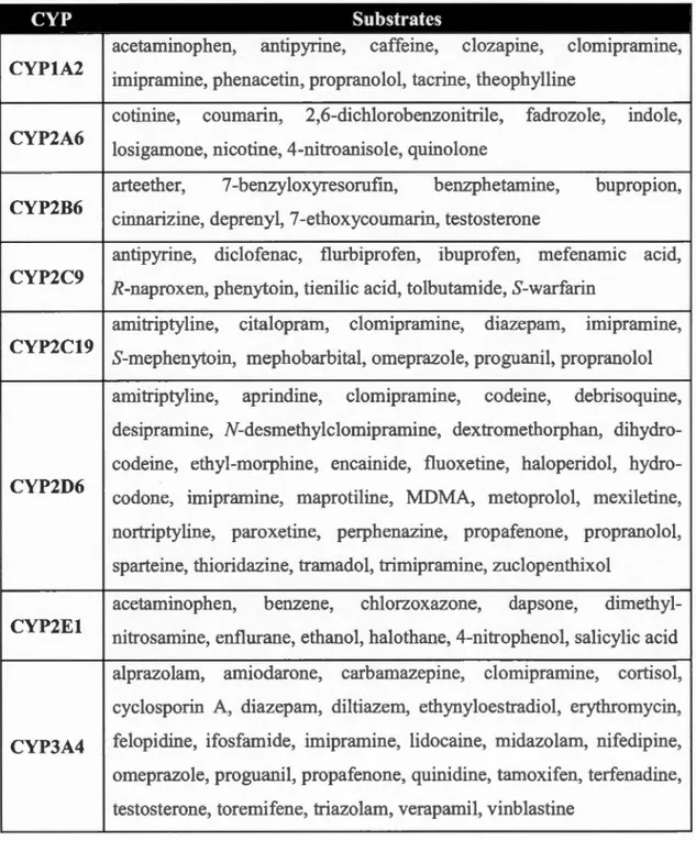
Table 103 Selected human cytochrome P450 xenobiotic substrates CYP Substrates CYP1A2 CYP2A6 CYP2B6 CYP2C9 CYP2C19 CYP2D6 CYP2El
acetaminophen, antipyrine, caffeine, clozapine, clomipramine, imipramine, phenacetin, propranolol, tacrine, theophylline
cotinine, coumann, 2,6-dichlorobenzonitrile, fadrozole, indole, losigamone, nicotine, 4-nitroanisole, quinolone
arteether, 7 -benzyloxyresorufin, benzphetamine, bupropion, cinnarizine, deprenyl, 7 -ethoxycoumarin, testosterone
antipyrine, diclofenac, flurbiprofen, ibuprofen, mefenamic acid, R-naproxen, phenytoin, tienilic acid, tolbutamide, S-warfarin
amitriptyline, citalopram, clomipramine, diazepam, Imipramme, 0 0 0 S-mephenytoin, mephobarbital, omeprazole, proguanil, propranolol amitriptyline, aprindine, clomipramine, codeine, debrisoquine, desipramine, N-desmethylclomipramine, dextromethorphan, dihydro-codeine, ethyl-morphine, encainide, fluoxetine, haloperidol, hydro-codone, imipramine, maprotiline, MDMA, metoprolol, mexiletine, nortriptyline, paroxetine, perphenazine, propafenone, propranolol, sparteine, thioridazine, tramadol, trimipramine, zuclopenthixol
acetaminophen, benzene, chlorzoxazone, dapsone, dimethyl-nitrosamine, enflurane, ethanol, halothane, 4-nitrophenol, salicylic acid alprazolam, amiodarone, carbamazepine, clomipramine, cortisol, cyclosporin A, diazepam, diltiazem, ethynyloestradiol, erythromycin, CYP3A4 felopidine, ifosfamide, imipramine, lidocaine, midazolam, nifedipine, omeprazole, proguanil, propafenone, quinidine, tamoxifen, terfenadine, testosterone, toremifene, triazolam, verapamil, vinblastine
Phase II biotransformations modify the structure of xenobiotics by conjugating endogenous metabolites, often to increase their water-solubilityo Moreover, sorne of
the functional groups used in these reactions, such as the carboxylate terminal glucuronate conjugates, are recognized by specifie organic anion active transporters in the liver and kidney, facilitating excretion into the bile and urine, respectively. The majority of Phase Il conjugations occur via electrophilic addition of an "activated donor" to nucleophilic centers in xenobiotic substrates, including oxygen in hydroxyl and nitrogen in amine groups. Glutathione (GSH), on the contrary, reacts with electrophilic centers of the xenobiotic or its reactive metabolite. Table 1.4 lists the
most common xenobiotic conjugation reactions, their associated enzymes and
subcellular localizations (Joseph y, 1997; Badenhorst et al., 20 13).
Table 1.4 Selected xenobiotic conjugation reactions
Reaction Enzyme Cellular Component
Glucuronidation UDP-glucuronyltransferase Micros omal
Sulfation Sulfotransferase Cytosolic, Microsomal
Methylation Methyltransferase Cytosolic, Microsomal
Acetylation Acety 1 transferase Cytosolic
Glycine conjugation Glycine N-acyltransferase Cytosolic, Microsomal
Glutathione conjugation Glutathione S-transferase Cytosolic, Microsomal
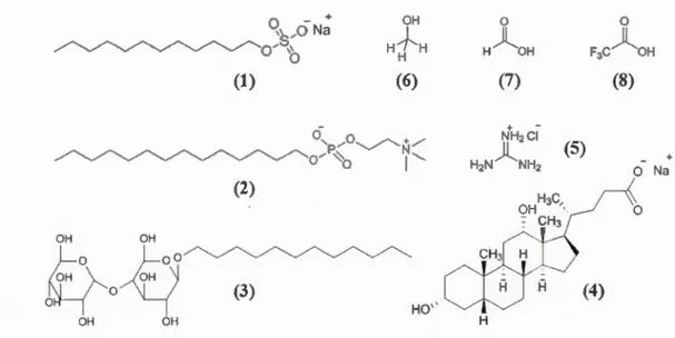
Depending on the organ involved, xenobiotic metabolism can sometimes involve multiple biotransformation routes. Figure 1.3 shows the metabolic pathway for N-acetyl-p-aminophenol (AP AP), also known as paracetamol or acetaminophen. As illustrated, a therapeutic dose of APAP is primarily metabolized in the liver via glucuronylation and sulfation and then excreted into bile or blood by ATP-binding cassette (ABC) transporters. However, a small portion of the drug undergoes a phase I transformation to N-acetyl-p-benzoquinone imine (NAPQI), a reactive
metabolite, which can subsequently be conjugated with GSH, enzymatically
AB CCJ, G4 APAP-S0 3 25-36 % SULT lAI , IA J , IA4 , 2Al , l EI A B CC2 , G2 PAPS NH , e
~
)
?e~N--r7
O-S-O-P-O-C H 2 NJl ~ Il Il ~ 0 '--) N 0 0)-1"
0 OH cf -~ -cf Il 0 bl ood ..---< bile H3 c ~-t-.. ___/\__ f=o HO~N H APAPH~~
0H~
~
-0-C>=o---H
-0
-e0
? ) ?o01)
HO OH 0 ~ fi NH UGT lAI , I A6, o-g-o-g-o-Ç---0 ---j A P AP -G l u c IA9 ,2 81 5 ~ UDP-Giuc OH O H 47 -6 2 % GSTP I , Tl , MI 0 e oocyJ~ NH 3.
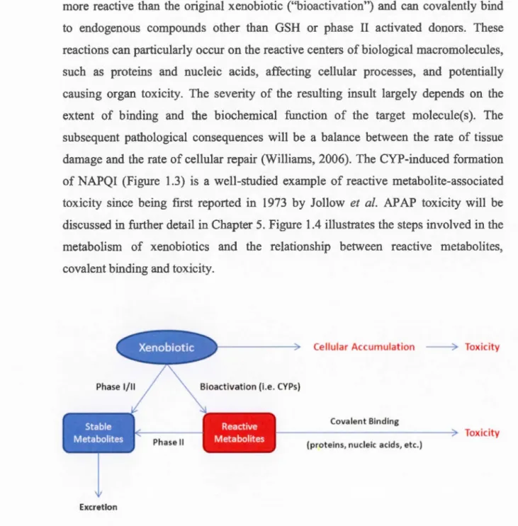
A PAP GSH o lig o p e pt1d ease HO 9-APAP-Cy s 5-8% (s •A H3 N eco Figure 1.3 Summary of the acetaminophen metabolic pathway. The enzymes and intermediates involved as weil excretion routes for each synthetic conjugate are also shown. ABC: ATP -binding cassette transporter ; AP AP: acetaminophen CYP: cytochrome P450 ; Glue : glucose; GSH: glutathione; GST : glutathione S-transferase ; NAPQI: N-acetyl-p -benzoquinone imine ; PAPS: 3' -phosphoadenosine-5'-phosphosulfate; SULT : sulfotransferase; UDP : uridine diphosphate ; UGT glucuronyltransferase.Phase l and II biotransformations often serve as an efficient detoxification pathway; bowever, sorne CYP-catalyzed reactions may generate metabolites that are more reactive than the original xenobiotic ("bioactivation") and can covalently bind to endogenous compounds other than GSH or phase Il activated donors. These reactions can particularly occur on the reactive centers of biological macromolecules, such as proteins and nucleic acids, affecting cellular processes, and potentially causing organ toxicity. The severity of the resulting insult largely depends on the extent of binding and the biochemical function of the target molecule(s). The subsequent pathological consequences will be a balance between the rate of tissue damage and the rate of cellular repair (Williams, 2006). The CYP-induced formation of NAPQI (Figure 1.3) is a well-studied example of reactive metabolite-associated toxicity since being first reported in 1973 by Jollow et al. APAP toxicity will be discussed in further detail in Chapter 5. Figure 1.4 illustrates the steps involved in the metabolism of xenobiotics and the relationship between reactive metabolites, covalent binding and toxicity.
Cellular Accumulation ----7 Toxicity
Phase 1/11 Bioactivation (i.e. CYPs}
Covalent Binding
---~ Toxicity
Phase Il (proteins, nudeic acids, etc.)
Excretion
Figure 1.4 Relationship between xenobiotic metabolism and reactive metabolite-induced toxicity
Theoretically, both the nucleophilic and electrophilic reactive metabolites can
undergo covalent binding reactions and induce toxicity; however, in nature, the
reactions involving reactive electrophiles occur more frequently due to the lack of
intrinsic highly electrophilic groups in biomolecules. Instead, nucleophilic centers are significantly present in pro teins ( cysteine, lysine, histidine, arginine, serine,
threonine, selenocysteine, asparagine, aspartate and glutamate residues), nucleic acids (nitrogenous bases) and other compounds such as GSH. Therefore the study of reactive metabolites often involves the chemistry of biologically-occurring
electrophiles (Gersch et al., 20 12).
Based on the mechanism of their reaction with nucleophiles, electrophilic
metabolites can be classified into three major groups: (1) Michael acceptor systems,
(2) ring-strained scaffolds, and (3) other electron-deficient systems. Table 1.5 lists the most common classes of reactive electrophiles under each category with selected xenobiotics known to form examp1es of each class (Zhou et al., 2005; Kalgutkar, 2011).
According to the "electronic theory of acids and bases", electrophiles spontaneously react with nucleophiles, if energetic parameters are met. However, the
selectivity of such a reaction mainly depends on the size and electron density of the
species involved. From this perspective, an electrophile-nucleophile reaction is a Lewis acid-base reaction and its product results from the addition of two complete chemical entities, therefore often referred to as an "acid-base complex" or "adduct" (House, 2008). The selectivity of electrophilic (reactive) metabolites, the mechanisms
of their reactions, and the types of adducts formed lie within an interesting research
Table 1.5 Most conunon classes of electrophilic reactive metabolites
Reactive Electrophile Structure Example(s)
Michael Acceptor Systems
a,p-Unsaturated carbonyl
~
0
carbamazepine, L-dopa, tolcaponea,p-Unsaturated imine
~NH
acetaminophen, lidocainea,p-Unsaturated diimine =N "=NH trimethoprim
Ring-Strained Scaffolds
Epoxide
)
o
bergamottin, diallyl sulfone,Arene oxide
(>
o
capsaicin, imipramine, raloxifeneOtber Electron-Deficient Systems
Nitroso 1 N=O erythromycin, pbenacetin,
sulfarnetboxazole 1
Diazene N=N isoniazid
1
\Œ=<
Iminium ion N- aminopyrine, haloperidol, mirtazapine
1 (±)
Diazonium ion N::N hydralazine
1
Nitrenium ion ,....--(±) N, aristolochic acids, clozapine
Isocyanate -N=C=O sulofenur, tolbutamid, troglitazone
e
S-oxide 0 1 tienilic acid
~s<±l
Aldehyde/ketone
)=
o
diclofenac, indomethacinAcyl halictes
)=
o
chloramphenicol, halothane1.1.3 Adduct Formation and Protein Targets
A covalent binding reaction on a protein does not automatically lead to toxicity. On the contrary, the efficacy of sorne drugs, such as penicillin, aspirin and omeprazole, relies on their ability to covalently bind to their protein targets (Williams, 2006). Nonetheless, irreversible chemical modification of a biomolecule by a reactive metabolite can affect its biological function and the cellular processes involving this molecule. It is therefore important to identify potential targets of a reactive metabolite to assess the extent and direction of such potential biological consequences.
Hard and soft acids and bases (HSAB) theory explains that hard electrophiles generally react with hard nucleophiles and soft species prefer the soft counterparts.
In 1983, Parr and Pearson explained the concept of "chemical hardness" ('1) as the second derivative of the total energy of a chemical system (E) with respect to changes in the number of electrons (N) at a fixed nuclear environment (z).
By this definition, "hard" species are small, have a high electron density, and are weakly polarizable, whereas "soft" species are large, have a low electron density, and can be readily polarized.
Applying HSAB theory to reactive metabolites, hard electrophiles, including cations, epoxides, and aldehydes, should react with hard nucleophilic centers such as the amine functional groups in DNA/RNA nucleobases and lysine residues in proteins. Soft electrophiles, such as Michael acceptor systems, would prefer soft nucleophiles, which include cysteine residues in proteins and glutathione. Free radicals can also react with lipids to initiate lipid peroxidation chain reactions. It is
-however important to consider that, in addition to the chemical hardness, the microenvironment (pKa, hydrophobicity, etc.) of a nucleophilic center in the tertiary
structure of the target molecule can also affect an adduction reaction (Park et al.,
2005).
As discussed earlier, toxicological response of a covalent binding reaction
largely depends on the biological role of the target molecule and the cellular
processes affected by the target's structural alteration. These responses usually include (1) mutagenicity, teratogenicity or carcinogenicity, (2) necrotic or apoptotic cell death, and (3) hypersensitivity reactions (Williams, 2006). For example, the
endogenous lipid peroxidation product, malondialdehyde (MDA), is known to
covalently bind to guanine, adenine, and cytosine residues in DNA structure
demonstrating mutagenic properties in bacterial and mammalian cells and
carcinogenicity in rats (Figure 1.5) (Mamett, 2000). Phase l transformation of
5-aminosalicylic acid (also known as mesalazine), a non-steroidal anti-inflammatory
drug (NSAID), by myeloperoxidase generates a quinone intermediate, which then binds to hemoglobin Jeading to hypersensitivity (Liu et al., 1995). Similarly, the metabolism of severa! other drugs such as dapsone, carbamazepine, trimethoprim, and olanzapine results in covalent modification of neutrophil proteins leading to agranulocytosis (Zhou et al., 2005).
) l
H
HO
)L
H
DNA 0 01
1
0 HN HNeN
:SN>
~N
N:)
A
N
t )
O
AN
N
N
l
N
N
1 1 dR dR dR MDA M1G M1A M1CFigure 1.5 Covalent binding of malondialdehyde (MDA) with DNA leading to the
formation of potentially mutagenic (modified) guanine (M1G), adenine (M1A), and
cytosine (M1C). dR: deoxyribose.
A comprehensive list of reactive metabolite in vivo target
proteins can be found on Target Protein Database (TPDB) VIa http://tpdb.medchem.ku.edu:8080/protein database (Hanzlik et al., 2007). Figure 1.6 illustrates liver metabolism of trimethoprim, a bacteriostatic antibiotic, and the
resulting covalent binding reaction of hepatic pro teins (Lai et al., 1999).
OCH3
H
3
C
O~NVNH
2
1 1 ~~ MPO HJCO # .6 N H202, Cl· NH2 trimethoprim CYP IA2, 3A4t
OCH3H
3
C
O
~"""
7NfNH _ _ _____ H CO # # .6N 3 HS NH2Figure 1.6 Liver metabolism of trimethoprim, formation of a reactive iminoquinone
methide (an a,~-unsaturated diimine) metabolite, and its subsequent covalent binding
to hepatic proteins. CYP: cytochrome P450; MPO: myeloperoxidase.
Approximately 1000 xenobiotics are known to induce hepatotoxicity (Chen et al., 2014). More than half of the TPDB entries are liver protein targets. The liver
plays a key role in the metabolism of xenobiotics and thus is exposed to high concentrations of reactive metabolites, making it an important target for covalent
binding reactions. Although these reactions do not always lead to hepatotoxicity,
results in regulatory actions including denied approval, use restrictions, or post-market withdrawal. lt is also believed that severa! adverse drug reactions (ADRs) are also related to drug-induced organ toxicity (Williams, 2006). ldentifying potential covalent target proteins can shed light on the biological mechanisms involved in a drug's ADRs and thus has become an important area of research in pharmacology and molecular toxicology. The following section will briefly discuss the methods developed in recent decades for systematic study of proteins in biological samples including techniques particularly adapted for covalent binding investigations.
1.2 Experimental Techniques: MS-Based Proteomics
The word "protein" was coined by Swedish chemist Jons Jacob Berzelius, and introduced as a new class of organic substances by Dutch chemist Gerardus Johannes Mulder, who carried out the first elemental analysis of proteins in 1838 (Hartley, 1951). ln 1845, Justus von Liebig, the German organic chemist, found that proteins, regardless of their slight difference in solubility under different conditions, are largely similar in their chemistry, and play an important role in biological processes in the body (Carpenter, 1986).
Analysis of proteins in biological systems has al ways been a challenge. Heat and pH sensitivity, extremely large molecular weights, and relatively high water-solubility make them difficult targets for traditional analytical techniques. Pioneering work on separation methods by Frederick Sanger paved the road for the development of protein and DNA/RNA sequencing, eventually bringing him two Nobel prizes. Tedious and time-consuming protein sequencing was later greatly improved by the development of phenylisothiocyanate sequencing chemistry by Edman in 1949, and its automation in 1967 (James, 1997).
The proteomics era began with the invention of sodium dodecyl sulphate
polyacrylamide gel-electrophoresis (SDS-PAGE) (Kenrick and Margolis, 1970), and two-dimensional gel-electrophoresis (2D-GE) (O'Farrell, 1975; Klose, 1975) in the 1970s. 2D techniques significantly enhanced protein analysis resolving power and sensitivity, and enabled electro-blotting for protein identification with antibodies or Edman sequencing. However, low rate of detection of membrane pro teins due to their
poor water-solubility, limited reproducibility, and problems with spot identification in complex mixtures were major drawbacks of initial gel-based methods (James, 1997).
The development of bio-analytical mass spectrometry and the parallel growth of protein databases, in late 1990s and early 2000s (Aebersold and Mann, 2003; Washbum et al., 2001), greatly enhanced the performance and throughput of traditional proteomics methods. The following section will briefly describe the state-of-the-art proteomics technology and use of mass spectrometry to address numerous challenges in protein analysis in biological samples.
1.2.1 Proteomics Technology
Proteomics is the large-scale study of proteins to obtain a global, integrated view of cellular processes at the protein leve! (Blackstock and Weir, 1999). The term "proteomics" was first introduced by Peter James (1997) from the Swiss Federal Institute of Technology to make an analogy with "genomics", the large-scale study of genes. The aim of a proteomics study was, at first, limited to describing protein
expression changes under biological perturbations such as a specifie disease state or
drug treatment (Anderson and Anderson, 1998). Today, boundaries of proteomics
have largely expanded to include qualitative and quantitative analysis of proteins, their functions, structure, and interactions with other molecules in a cell, within a
From a "systems biology" perspective, while genomics deals with the analysis
of the complete set of DNA within an organism, and transcriptomics can be defined
as the study of all RNA molecules and their relationships with protein biosynthesis,
proteomics is the intermediary that links these areas to metabolomics, being the study
of cellular processes involving small molecules known as metabolites (Mann et al.,
2013). The relationship between different levels of "omics" research and the type of
molecules normally involved in each area of study are depicted in Figure 1. 7.
L _ _ _ _
G
__
e
n_o~In_j_c
s
_
·
__~~
. .~~L~
_ _ _ _ _ _D_N~A---~
~_T_r_a_ns_c_0~p_to_rru_._c
s
__~
. . . .~~L~
_ _ _ _ _ _RN
_
~
~
A---
~
L _ _ _ _P
_
ro
_
t
_
e
;
~
n
_,_i
c
_
s
__~--
. .·~~L~
_____
P
_
ro
_
;
~
e
i_n_s
____~
' ' ' 'Il 'V L _ _ _ M_e_t_ab_o_lo_m __ ic_s __~
. . . .~~L~
_ _ _ _ M_e_ta_b_o_li_te_s __~
Figure 1.7 Simplified depiction of the four major domains of "omics" research and
their primary molecular classes of interest
Initial proteomics analyses utilized gel-electrophoresis for separation and
identification of proteins using manual (Sanger) sequencing. Modem methods however are usually based on automated gel-free approaches. Gel-based analysis includes protein separation followed by an in-gel protein digestion and subsequent peptide analysis using cbemical sequencing techniques, such as Edman degradation
(Edman et al., 1950), or peptide mass fmgerprinting (PMF) using matrix-assisted
referred to as gel-electrophoresis-mass spectrometry or GE-MS (Michaud and Snyder, 2002). Gel-based techniques are known for their accuracy and simplicity however, gel manipulation steps are time-consuming, and the method is not applicable to all proteins (Van Summeren et al., 2012).
Gel-free proteomics usually involves in-solution digestion of the proteins prior to peptide fractionation or separation using one or multi-dimensional liquid
chromatography (LC) coupled with atmospheric-pressure ionization (API) tandem
mass spectrometry (MS/MS). Unlike gel-based techniques, gel-free proteomics could be readily used for high-throughput analysis and is applicable to most proteins;
however, LC-MSIMS instruments are costly and require extensive method
development (Van Summeren et al., 2012). Figure 1.8 exhibits major steps involved in gel-based and gel-free proteomics analytical workflows.
(a)
Gel electrophoresis In-gel digestion MALDI-MS Data Mining
(b)
In-solution digestion LC (ID or 2D) MSl (API) Data Mining
Figure 1.8 Different steps involved in a typical experimental workflow in a ge1-based (a) or gel-free (b) proteomics approach. API: atmospheric-pressure ionization; LC: liquid chromatography; MALDI: matrix-assisted laser desorption ionization; MS:
mass spectrometry; PMF: peptide mass fingerprinting.
A proteomics experiment can be "top-down" or "bottom-up" (Walther and Mann, 201 0). Characterization of proteins by their molecular weight is known as


![Figure 1.11 A typical SILAC quantitation workflow. The heavy-to-light LC-MS peak area ratio (b/a) in the [treated + control] mixture determines the relative abundance of labeled protein(s) in the treated sample](https://thumb-eu.123doks.com/thumbv2/123doknet/3097823.87792/51.907.116.818.300.826/figure-quantitation-workflow-treated-determines-relative-abundance-treated.webp)