Study of the subcellular localization of cell cycle
regulator Cks1 and its impact on cancer
par Yipeng Yao
Département de pharmacologie Faculté de médecine
Thèse présenté à la Faculté des études supérieures en vue de l’obtention du grade de maîtrise
en pharmacologie
Décembre 2012
Faculté des études supérieures et postdoctorales
Cette thèse intitulée:
Study of the subcellular localization of cell cycle regulator Cks1 and its impact on cancer
Présenté par : Yipeng Yao
a été évalué par un jury composé des personnes suivantes :
Dr. Richard L. Momparler, président-rapporteur Dr. Sylvain Meloche, directeur de recherche Dr. Jean-François Gauchat, membre du jury
Résumé
La progression dans le cycle cellulaire est contrôlée par de vagues oscillantes de cyclines et des kinases cycline-dépendantes (Cdk). Ces kinases sont régulées positivement par l’association des sous-unités cyclines régulatrices et négativement en se liant aux inhibiteurs de Cdk. Parmi ces derniers, p27 inhibe tous les complexes cycline-Cdk quelle que soit la phase cellulaire et agit en tant que régulateur négatif principal de la prolifération cellulaire dans une variété de cellules et de tissus. Intrinsèquement, p27 phosphorylé est ubiquitiné et dégradé par le complexe SCFSkp2-Cks1. Des études génétiques de la souris, ainsi que des examens cliniques chez l’homme, ont montré que p27 est un important suppresseur de tumeur. Le gène est rarement muté. Cependant, p27 est fréquemment réprimé dans les cancers humains en raison d’une augmentation de l’expression de Skp2 et de Cks1 dans le noyau, ce qui est généralement associée à un mauvais pronostic. La localisation subcellulaire de Cks1 est donc d'une importance primordiale dans le contrôle de la prolifération cellulaire. Les résultats récents de notre laboratoire ont montré une interaction entre Cks1 et les protéines de transport nucléaire importine α1 et β3. Aussi, l’analyse de la séquence primaire de Cks1 a également révélé un signal de localisation nucléaire classique (NLS) à son extrémité C-terminale. Des mutations ont été effectuées sur le NLS suspect pour déterminer si oui ou non l'import nucléaire de Cks1 était contrôlé par cette séquence. Un inhibiteur synthétique de l’importine β a également été utilisé pour étudier l’import de Cks1 dans le noyau. Les résultats indiquent que l’extrémité C-terminale
de Cks1 est en effet un NLS puisque les mutations de Cks1 et l'inhibition de l’importine β conduisent, tous deux, à l'accumulation de Cks1 dans le cytoplasme. Ces résultats ont été utiles pour mieux comprendre le mécanisme régulant la localisation de Cks1. Toutefois, des travaux futurs sont nécessaires pour mieux comprendre l'impact de la séquestration cytoplasmique de Cks1 sur le cancer et ainsi espérer aboutir à l'identification de nouvelles cibles pharmacologiques impliqués dans la prolifération cellulaire.
Abstract
Progression through the cell cycle is controlled by oscillating waves of cyclins and cyclin-dependent kinases (Cdk). These kinases are regulated positively by association with cyclin regulatory subunits and negatively by binding of Cdk inhibitors. Among the latter, p27 inhibits all cyclin-Cdk complexes regardless of the cell cycle phase and acts as a primary negative regulator of cell proliferation in a variety of cell types and tissues. Intrinsically, phosphorylated p27 is ubiquitinated and degraded by the SCFSkp2-Cks1 complex. Mouse genetic studies and human clinical investigations have shown p27 as an important tumor suppressor, which gene is rarely mutated. However, p27 is frequently downregulated in human cancers due to an increased expression of nuclear Cks1 and this is usually associated with a poor prognosis. The subcellular localization of Cks1 is thus of primordial importance in the control of cell proliferation. Recent results from our laboratory have shown an interaction between Cks1 and nuclear transport proteins α1 and β3 importin. Analysis of the primary sequence of Cks1 also revealed a classic nuclear localization signal (NLS) at its C-terminal. Mutations have been done on the suspected NLS to determine whether or not Cks1’s nuclear import is regulated by this motif. A synthetic inhibitor of β importin has also been used to study the mechanism of Cks1 import. Results indicated that the C-terminal end of Cks1 is indeed a NLS since mutations of Cks1 and inhibition of β importin both lead to accumulation of Cks1 in the cytoplasm. These outcomes were helpful to better understand the mechanism regulating Cks1 localization.
However, future works are required to further understand the impact of cytoplasmic sequestration of Cks1 on cancer and hopefully lead to the identification of novel pharmacological targets involved in cell proliferation.
Table of Contents
1. Introduction
1
1.1. Cell cycle
1
1.2. Regulation
3
1.2.1. Phosphorylation
5
1.2.1.1. Cyclins
6
1.2.1.2. Cyclin-dependent kinases
9
1.2.2. Proteolysis
12
1.2.2.1. Skp, Cullin, and F-box complex
14
1.2.2.2. Anaphase-promoting complex
15
1.3. Cyclin/Cdk inhibitors
20
1.3.1. Ink family
20
1.3.2. Cip/Kip family
22
1.4. Cyclin/Cdk inhibitor p27
24
1.4.1. Structure
24
1.4.2. Function
25
1.4.3. Regulation
26
1.4.4. Relevance to cancer
28
1.5. Skp2
31
1.5.1. Structure and function
31
1.5.2. SCF
Skp2-complex
32
1.6. Cks1
36
1.6.1. Structure and Function
36
1.6.2. Relevance to cancer
40
2. Hypothesis & Objectives
42
3. Materials & Methods
44
3.1. Plasmids & Mutagenesis
44
3.2. Cell culture
45
3.3. Transfection & Infection
46
3.4. Antibodies
48
3.5. Co-immunoprecipitation
48
3.6. GST-pull down
49
3.7. Radioactive in vitro translation
50
3.8. Western Blot
51
3.9. Immunofluorescence
52
3.10. β Importin inhibitor
53
4. Results
55
4.1. Tandem Affinity Purification
55
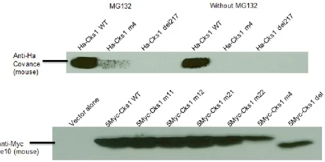
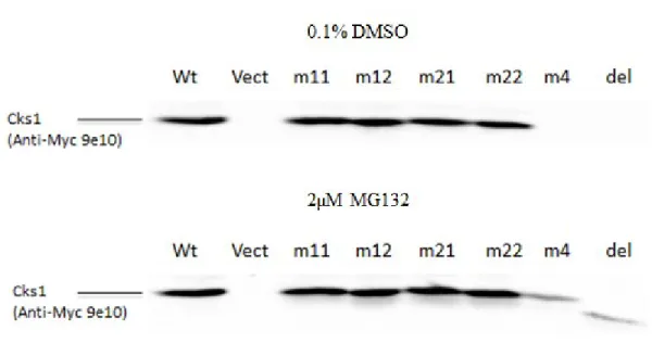
4.2. Cks1 expression
57
4.4. Subcellular localization of Cks1
63
4.5. β Importin inhibition
67
5. Discussion & Perspectives
71
5.1. Cks1 expression
73
5.2. Cks1 interactions
74
5.3. Cks1 localization
75
5.4. Effect of pharmacological β importin inhibition on Cks1
localization
76
5.5. Impact on cancer
78
List of figures
Figure 1 Cell cycle overview
4
Figure 2 Mechanisms of nuclear import and export
8
Figure 3 SCF and APC complexes regulate each other through the cell cycle
18
Figure 4 Structure of the SCF
Skp2-Cks1 complex linked to p27-Cdk2 33
Figure 5 The amino acid sequence as well as the crystallography of the
human form of Cks1
39
Figure 6 Inhibitor synthesis
53
Figure 7 Process of TAP purification
55
Figure 8 Cks1 expressions in HEK293 cells
57
Figure 9 Cks1 expression in infected Rat1 cells
58
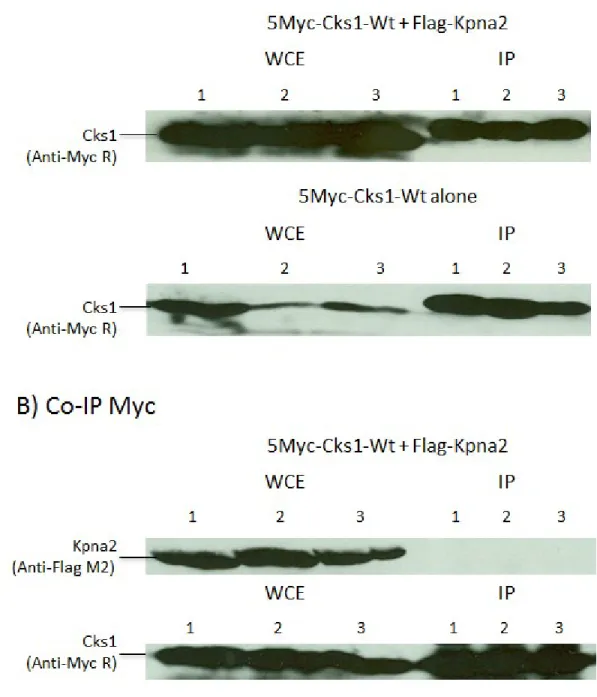
Figure 10 Co-IP results of Cks1 with Kpna2
60
Figure 11 Results of the pull down of Cks1 by α importin-conjugated
beads
61
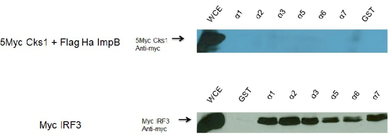
Figure 12 Cells expressing 5Myc-Cks1
62
Figure 13 Immunofluorescence analysis of Cks1 localization
64
Figure 14 Cks1-wild type and mutants localization in infected Rat1 cells
66
Figure 15 Immunofluorescence of Cks1-wt infected Rat1 cells after protein
depletion by 8 hours cycloheximide incubation
68
Figure 16 Cks1 localization after 8 h incubation of cycloheximide and
4 h DMSO or inhibitor
69
List of Abbreviations
α-imp : α importin
APC/C : anaphase-promoting complex/cyclosome ATF1 : activating transcription factor 1
β-imp : β importin BP : bait protein
β-TRCP : β-transducin repeat-containing protein CAK : cdk-activating kinase
CaPO4 : calcium phosphate
CCRK : cell cycle related kinase Cdc : cell division cycle protein CDC2L : Cdc2-like protein
CDE : cell cycle dependent element Cdk : cyclin depedent kinase
CHR : cell cycle gene homology region
Cks1 : cyclin dependent kinase regulatory subunit 1 CksHs1 : human Cks1
Cki : cdk inhibitor
Co-Ip : coimmuno-precipitation
COPS5 : COP9 constitutive photomorphogenic homolog subunit 5 DMSO : dimethyl solfoxide
DNA : deoxyribonucleic acid FBS : fetal bovine serum
FBW7 : F-box and WD-40 domain protein 7 FoxO : forkhead box class O family
Grb2 : growth factor receptor-bound protein 2 hKIS : human kinase interacting stathmin Il-8 : interleukin 8
IRES : internal ribosome entry site kDa : kilo-dalton
KID : kinase inhibitory domain
KPC : Kip1 ubiquitination-promoting complex Leu : leupeptin
LRR : leucine rich repeat
LS/MS : liquid chromatography mass spectrometry Mad2 : mitotic arrest deficient 2
MAPK : nitogen-activating protein kinase MPF : mitosis promoting factor
N>C : nuclear accumulation of specific protein N<C : cytoplasmic accumulation of specific protein
N=C : even distribution throughtout cell of specific protein NES : nuclear export signal
NF-γ : nuclear factor γ
NLS : nuclear localization signal PA : pepstatin A
PCNA : proliferating cell nuclear antigen PFA : paraformaldehyde
PI3K : phosphatidylinositol-3-kinase PKB/Akt : protein kinase B
PMSF : phenylmethylsulfonyl fluoride
RanGEF : Ran guanine nucleotide exchange factor RanGTP : Ran GTPase activating protein
Rb : retinoblastoma protein RBX1 : ring box protein 1
RNA : ribonucleic acid ROCK : rho-kinase RT : room temperature SB : sample buffer
SCF : Skp, Cullin, F-box complex shRNA : short hairpin RNA
Skp: S-phase kinase associated protein TAP : tandem affinity purification TGF-β : transforming growth factor β TPR : tetratricopeptide repeat
Ub : ubiquitin
UTR : untranslated region Van : sodium orthovanadate
À mes grands-parents, dont trois ont été enlevés par le cancer,
à mes chers parents
et aux héros en blouse de laboratoire, travaillant jour et nuit pour guérir le cancer.
Thanks
- To Dr. Sylvain Meloche for giving me the opportunity to attain my masters’
degree under his supervision and guidance.
- To Laure Voisin and Benoit Pelletier for their guidance and help throughout my
project
- To Edith Giasson, Paul Deleris, Pierre-Luc Tanguay, Stéphanie Duhamel ,
Christophe Fremin, Marc Saba El Leil, and Justine Rousseau for their experimental support and helpful advices.
- To Dr. Ricahrd L. Momparler and Dr. Jean-François Gauchat for taking time to read and correct my thesis.
- To Christian Charbonneau for his help with microscopes - To Sylvie Caron for her help with my thesis deposit. - To all other members of the lab
- To my parents Jia and Jianhua for their unconditional love and support - To Fan dan, love of my life, for her love and support
1. Introduction
1.1. Cell cycle
Cell division is a process by which one parent cell undergoes different stages in order to become two identical daughter cells each with the same genetic material as the parent cell. Most of the living organisms will divide once or many times during their life. For unicellular creatures, it serves as means of replication. For multicellular organisms, it serves many functions: growth, repair, and replacement. Prokaryotic cells replicate via either binary fission or budding. However, eukaryotic cells must go through different stepwise phases, each of which will start only after the full termination of the previous one. All of these steps are called the cell cycle.
In human, the cell cycle consists mainly of 5 phases (see Fig.1A). [1-3] The first phase or Gap 1 (G1) is where most of the normal adult cells are. They either become
quiescent (G0) and differentiate into non-dividing cells with specific functions or, upon
stimulation by mitogenic factors, activate proteins that will drive the cells into cell division. The ensuing synthesis phase (S) encompasses the important step of deoxyribonucleic acid (DNA) replication. Once past the step of G1/S transition, cells will be committed to cell
division. All chromosomes duplicate and form pairs of chromatids. The completion of DNA duplication is followed by a second gap phase (G2). The cell further increases in size,
synthesizes proteins, and assembles the necessary cellular structures to prepare for cell separation. Mitosis or M phase occurs when the nuclear envelop breaks apart and the sister chromatids separate via a system of microtubules, each attached to the kinetochore of a
chromatid and to one of the polar bodies located on opposite side of the cell. Cell cleavage or cytokinesis occurs and two identical daughter cells are created. Both cells are now in the G1 phase and the cycle starts again.
1.2. Regulation
As all biological activities are, the cell cycle is tightly regulated to prevent production of genetically damaged daughter cells and uncontrolled cell division that would ultimately lead to cancer. At G1, the restriction checkpoint first decide, according to
environmental factors, whether the cell differentiate or re-enter cell division. [4] Then, the retinoblastoma proteins (Rb), inhibitor of the transcription factor E2F, must be inactivated via phosphorylation for the cell to transit from G1 to S phase. The post-replication DNA
damage checkpoint at the G2/M transition ensures that no damaged DNA will be passed on
to daughter cells.[5] Finally, the spindle or mitotic checkpoint monitors the even segregation of chromosomes via kinetochore attachments and microtubule tensions. [6]
Cell cycle regulations mainly occur via two post-translational mechanisms: phosphorylation and proteolysis. Phosphorylation is mediated by two families of regulatory proteins: cyclins and cyclin-dependent kinases (Cdk). [1, 2, 7] The effect can be either activator or inhibitory depending on the function of the substrate or the site of phosphorylation. Proteolysis remove proteins no longer required or in excess, whether they are negative or positive regulators. [8] Ubiquitination of proteins targeted for proteolysis is marked by two ubiquitin-dependent complexes: the anaphase-promoting complex/cyclosome (APC/C) [9, 10] and the Skp, Cullin, F-box containing (SCF) complex.
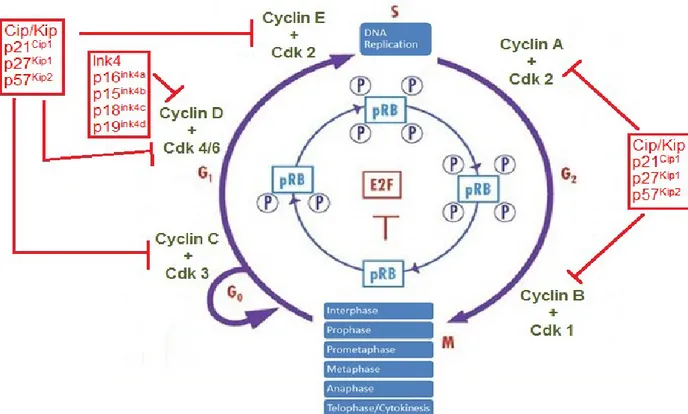
Figure 1 Cell cycle overview (adapted from {Viglietto et al. Cell Cycle 2002;1(6)})
A) Overview: Eukaryotic cells must undergo specific steps in order to produce two identical daughter cells. The mammalian cell cycle can be separated into 5 phases. Differentiated or quiescent cells are at G0. Normal adult cells are at the G1-phase. In
presence of mitogens, such as growth hormones, inhibitory phosphorylation of retinoblastoma protein occurs, which release the transcription factor E2F, leading to cell growth and replication of its DNA at the S-phase. Once past the G1/S transition, the cell
is committed to finish the cell cycle. At the second gap phase G2, the cell grows further
in size and produce proteins required for mitosis. Mitosis or M-phase is where the cell cleaves into 2 identical daughter calls each containing one copy of the duplicated chromosomes. Mitosis is further separated into six steps, each describing a step of DNA separation. Finally, cytokinesis is the separation of cell cytoplasm via membrane cleavage.
B) Cyclin/Cdks: regulatory cyclins and Cdks form holoenzymes that regulate the progression of cell cycle. Each cyclin bind to specific Cdks and each cyclin/Cdk complex modulate a specific part of the cell cycle. Cyclin C/Cdk3 promote quiescence exit in presence of mitogens. Cyclin D/Cdk4 and Cyclin D/Cdk6 up-regulate transcription of cell cycle required genes. Cyclin E/Cdk2 mediate the G1/S transition via
formation of prereplication complex and other proteins required for DNA replication. Cyclin B/Cdk1 is the mitotic cyclin complexes that drive the cell to enter into mitosis. C) Cyclin/Cdk inhibitors: Inhibition of cell cycle is mediated by two families of cyclin/Cdk
inhibitors, the Ink4 and Cip/Kip families. Ink4 inhibitors are named due to their abilities to inhibit specifically Cdk4 associated complexes. Its members are p16, p15, p18 and p19. Cip/Kip inhibitors, however, are involved in regulating all cyclin/Cdk complexes. Cip/Kip members are p21, p27 and p57.
1.2.1. Phosphorylation
Phosphorylation refers to the addition of one or more phosphate group (PO43-) to a
protein. Phosphates are sterically bulky and negatively charged moiety. Therefore, their addition can alter a protein’s biochemical properties as well as its structure and activities. The conformational and property changes can then create docking sites to mediate protein-protein interactions, modify signal sequences on protein-proteins to regulate their subcellular localization, and activate enzymes by bringing their active sites into proper position. [13] Phosphorylation can activate or inhibit a protein depending on its function and phosphorylation site. Effects are often amplified due to pathway mechanisms. Thus, reversible phosphorylation is an important mechanism of control of cellular enzymatic
activities. Protein kinases phosphorylate while protein phosphatases dephosphorylate. In the cell cycle, most proteins, required for the activation of genes involved in DNA synthesis and replication, are regulated by those mechanisms. The Rb protein is an example of a protein whose activity is inactivated by phosphorylation.
1.2.1.1. Cyclins
Cyclins are a family of proteins first discovered by Evans et al. when studying sea urchin eggs’ cell division. [14] They are so named because of the cyclic variation in their concentration during the cell cycle. These regulatory proteins are furthermore grouped into different classes on the basis of expression timing, amino acid sequences, and structure. [15] However, all cyclins share a region of homology called the cyclin box present up to twice on the protein depending on its class. [1, 16] This conserved sequence of about 100 amino acids long is responsible for the binding and activation of Cdks. Cyclins are the regulatory subunits of Cdk-holoenzymes. [17] Therefore, most cyclins must associate with specific catalytic cofactors (Cdks) to perform checkpoint regulatory functions. [18] Several cyclins have been found to take parts in other regulatory complexes, such as cyclin F which compose the F-box protein of the SCF complex. [19]
There are in total 12 classes of cyclins [16] : A, B, C, D, E, F, G, H, I, J, K, and L. However, only the cyclins A, B, C, D, and E are involved in regulating the cell cycle phase transition via cyclin/Cdk complexes. (see Fig.1B) During G0, cyclin C is transcriptionally
activated in response to mitogens. [2] By binding and activating Cdk3, the cyclin C/Cdk3 complex will then mediate the cell cycle re-entry of quiescent cells. [20, 21] At G1, Cyclin D
acivation is mitogen-induced. [17] The formation of cyclin D/Cdk4 and cyclin D/Cdk6 complexes will in turn phosphorylate Rb, their main substrate, which acts on centrosome duplication, mitochondrial function, and cytoskeletal modeling. [22] Cyclin D has also been shown to promote transcriptions, independent of Cdks, by acting on promoter regions. [23] Cyclin D/Cdk4/6 also activate indirectly cyclin E by sequestering cyclin/Cdk inhibitor p21 and p27. During G1, Cyclin E is known to primarily bind and activate Cdk2, but interaction
with Cdk1 and Cdk3 have also been demonstrated. [24] The cyclin E/Cdk2 holoenzyme is
essential for G1/S phase transition due to its role in E2F activation, prereplication complex
formation, centrosome duplication, histone biosynthesis, and endoreplication. [25] Recent studies by Geng et al. revealed cyclin E’s role in loading of the minichromosome maintenance helicase into the DNA replication complex in a cyclin-independent manner.
[24] A- and B-types cyclins are called mitotic cyclins due to their role in S and M phases. [26]
Cyclin A concentration accumulates in late G1 phase, through S and G2 phase, and rapidly
decreases in M phase. [26] Once activated, it forms complexes with Cdk2, and drives DNA synthesis by interacting with a subunit of DNA polymerase δ, the proliferating cell nuclear antigen. [27, 28] B cyclins are synthesised during S phase and are retained in the cytoplasm by binding to the nuclear export protein Crm1 via its nuclear export signal. [29] (for mechanism of nuclear import/export, see Fig.2) Binding of the cyclin to Cdk1 forms the M-phase-promoting factor (MPF), essential for the triggering of mitosis. [30] During mitosis, MPF,
activated by cyclin A, enters the nuclear and mediate the dissolution of nuclear envelope and the separation of chromatids. [1, 31]
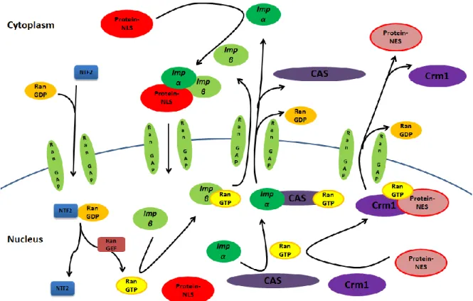
Figure 2 Mechanisms of nuclear import and export (adapted from {Pemberton and Paschal,
Traffic 2005; 6})
Nuclear import: cytoplasmic proteins containing the nuclear localization signal are recognized by specific α importin or karyopheryn (Imp-α) and β importin (imp-β). The heterodimerization of imp-α and imp-β on the NLS site of cargo protein mediate its entry into the nucleus through nuclear pore complexes.
Nuclear export: nuclear proteins containing the nuclear export signal (NES) is recognized by the exportin or karyopherin Crm1 (Crm1), either directly or indirectly via adaptor protein. Crm1 mediate the nuclear export of cargo proteins in cooperation with RanGTP.
Ran is a guanosine triphosphate (GTP) binding protein involved in release of importins from cargo proteins after import, nuclear export of unbound β, export of unbound imp-α in cooperation with exportin CAS and export of Crm1-bound proteins. The energy, released from hydrolysis of Ran-bond GTP to guanosine diphosphate (GDP) by the GTPase-activating protein (RanGTP), is utilized in transport across the nuclear membrane. Inactive RanGDP is reactivated by Ran Guanine nucleotide Exchange Factors (RanGEF).
1.2.1.2. Cyclin-dependent kinases
Cyclin-dependent kinases are a family of small proline directed serine/threonine kinases, all ranging from 34-40 kiloDalton (kDa). [32, 33] This kinase family is highly conserved through evolution. In an experiment, Lee and Nurse replaced the yeast’s Cdc2 gene (a Cdk1 homologue in fission yeast) with human Cdk1 gene. The yeast was able to progress through the cell cycle. [34] Also, recent studies have shown a high complementarity between the Cdks. In mammalian cells, due to the numerous compensatory mechanisms, Cdk1 alone with its’ partner cyclin A and B is able to drive the cell cycle. [35]
In humans, there are up to twenty different Cdks (1-20) classified to date. [2] These include 11 classical CDKs (Cdk1–11), two recently studied kinases (Cdk12 and 13), several proteins named based on the presence of a cyclin-binding element (PFTAIRE and PCTAIRE proteins) and a few Cdks-likes with similar sequences, such as CDC2-like kinases (CDC2L) or cell cycle-related kinases (CCRK). [36] Among them, only Cdk1, 2, 3,
4, and 6 are involved in the regulation of cell cycle progression at different checkpoints. The Cdks accomplish a variety of functions in numerous mechanisms such as DNA transcription and modification (Cdk11), mRNA processing, telomere elongation (Cdk1), activation of other Cdks (Cdk7), cell differentiation, senescence (Cdk5), etc. [2, 32, 37]
Crystallography revealed that Cdks form a tertiary structure containing a small amino-terminal lobe and a larger carboxyl-terminal lobe between which ATP bind to donate its γ-phosphate during phosphorylation. However, a large, flexible T-loop or activation loop blocking the binding of protein substrate at the entrance of the active-site cleft and incorrectly positioned amino acid side chains prevent Cdk activity. [33] Indeed, as
mentioned above, most Cdks are inactive on their own. For activation, binding of its cyclin partner and phosphorylation of a threonine near the active site by a Cdk-activating kinase are both required. [33, 38] Thus, the availability of the specific corresponding cyclin is by itself a regulation mechanism. Upon holoenzyme formation, 2 alpha helices on the Cdk, the PSTAIRE helix and the L12 helix, interact with the bound Cdk and change position and structure for the reconfiguration of the active site to allow binding of ATP. Once activated, the cyclin/Cdk complexes will phosphorylate their substrates on a specific consensus sequence of 4 amino acids long: [S/T*]PX[K/R]. [39] “S/T” is the serine or threonine on which the phosphate group attaches. “P” stands for proline. “X” is any polar amino acid. “K” is for lysine. “R” is for arginine.
Cdk1 is the first Cdk studied, also known in yeast as either Cdc28 or Cdc2. [40, 41] There are numerous alternatively spliced isoforms produced for the same gene. This kinase
forms the catalytic subunit of the highly conserved MPF complex along with cyclin B. MPF activates proteins needed for mitosis. A-cyclins also associate with Cdk1 and regulate its activity. The phosphatase Cdc25 is responsible for removal of the inhibitory phosphates on Thr(14) and Tyr(15), while Wee 1 is responsible for their phosphorylation. [42] Cdk2 has two alternatively spliced variants and binds to both cyclin E and A. [43] Cdk2 binds to G1
-phase cyclin E to mediate G1/S transition. Activated Cdk2 also interacts with Smads, which
are signal transduction proteins activated in response to transforming growth factor β (TGF-β), to inhibit cell cycle arrest. [44] In the S phase its association with cyclin A recognizes DNA damage and initiate DNA repair during DNA synthesis. The main function of cyclin C-associated Cdk3 is to promote cell cycle reentry via Rb phosphorylation. [20] Cdk3 also works downstream of Rb by activating directly E2F to promote S-phase entry. [45] Recent studies by Zheng et al. demonstrated Cdk3’s non-cell cycle regulatory function in cell transformation, via its interaction with the activating transcription factor 1 (ATF 1). [46] Type 4 Cdks bind with D-cyclins to regulate the G1/S
progression. Along with Cdk2, it regulates Smads during G1/S transition. [44] Cdk4 is also
involved in controlling the subcellular localization of BRCA1, a nuclear phosphoprotein that function as tumor suppressors by activating apoptosis. [47] Cdk 6 binds to D-cyclins in mid G1 phase, prior to Cdk2 activities, and drives the cell into S phase. [48] The main
function of cyclin D/Cdk6 is in the phosphorylation of Rb protein. Slomiany et al. observed that primary mouse astrocytes infected with retroviral RCAS-Cdk6 resulted in drastic morphological changes. [49] When further investigated, changes in patterns of gene
expression, changes in the actin cytoskeleton and enhanced motility have been noted. These changes are all part of the process of cellular differentiation.
1.2.2. Proteolysis
Proteolysis is defined by the directed degradation or digestion of proteins by cellular enzymes into simpler, more soluble substances such as peptides and amino acids. This degradation is performed by either a simple protease or the complex proteasome machinery. Cell cycle regulating proteolysis is mediated by ubiquitination enzymes that label the degradation-targeted protein with multiple ubiquitins (Ub). [12, 50] The tagged protein is then directed to the 26S proteasome, which will catalyze the breakdown of peptide bonds.
Ubiquitins are small proteins (~8 kDa) that target proteins for degradation. First, an E1 ubiquitin-activating enzyme activates Ub in an ATP-dependent way. The activated Ub is then transferred to an E2 ubiquitin-conjugating enzyme. The latter one, attached to an E3 ubiquitin-ligase, mediates the covalent bounding of ubiquitin’s carboxyl terminal to a lysine residue on the target protein via an isopeptide bond. This step is repeated multiple times leading to the formation of a polyubiquitin chain in which each ubiquitin is linked to a specific lysine of the previous Ub. [12, 50] The number of ubiquitins forming the chain determines the affinity of the modified substrate protein for the proteasome and also the timing of degradation. Studies have shown that 4 Ubs is the minimal targeting signal. [51]
Once the marked protein binds to the proteasome, unfolding and deubiquitination occurs.
[52] The substrate is then translocated to the core proteolytic sites, which cleaves the protein
into short polypeptides by catalyzing nucleophilic attacks on threonine residues. [53] Upon peptides release, the Ubs are recycled for future ubiquitination. During the degradation of the cyclin/cdk comlplexes, the catalytic subunit cdk is also released, intact. [54]
Since the length of the polyUb chain is E3-ligase specific and can affect the affinity of proteasome binding, the type of E3 involved in the ubiquitination-dependent degradation pathway is of primordial importance. In humans, multiple E1, E2 and E3 enzymes have been identified, each have a specific subset of ubiquitination partners. E3-ligases are classified into 4 major groups on the basis of their subunit composition: HECT-types, RING-finger-type, U-box-type and PHD-finger-type. [50, 55] In cell cycle, two polymeric E3 ubiquitin-ligases are implicated in the proteolysis pathway, both of which are of the RING-type class. The SCF complex is active from late G1 to early M phase; it is primarily
involved in the G1/S and G2/M transitions. On the other hand, APC/C’s expression is
required from mid-M phase to S phase to induce progression and exit of mitosis. [9, 50] SCF and APC/C complexes expression overlap and control each other in form of positive and negative feedbacks. (see Fig.3) At early mitosis, SCFβ-TRCP ubiquitinates APCCdc20 inhibitor Emi1/2; thus, activating the latter. Then in G1, APC/CCdh1 recognizes
Skp2 and mediates its proteolysis, leading to low SCF level and accumulation of cyclin/cdk inhibitors. This results in an increase of cyclin/Cdk which phosphorylates Cdh1 and mediates its inhibition. [50]
1.2.2.1. Skp, Cullin, and F-box complex
The SCF complex is formed of 4 subunits: Ring Box protein 1 (RBX1), CUL1, S-phase kinase-associated protein (Skp1), and F-box protein. The RBX1 protein contains a zinc binding domain, called the RING finger, which binds to the E2 ubiquitin-conjugating enzyme. CUL1 is from the cullin protein family that functions as a scaffold protein linking RBX1 to Skp1. Skp1 is the adaptor protein that links the substrate receptor protein to the CUL1-RING complex. The receptor responsible for tagged substrate recognition is called an F-box protein. Consequently, Skp1 is also called an F-box binding protein. [56]
RBX1 and Skp1 are highly conserved. In some cases, they can be replaced by related proteins without affecting the activity rate of SCF. [33] In fact, the rate of ubiquitination by SCF is not controlled by the core RBX1-CUL1-Skp1 complex, but rather by the substrate’s affinity for its corresponding F-box protein. The binding affinity is influenced by both the phosphorylation of target protein at specific site or sites and by the type of F-box receptor involved.
F-box proteins contain an approximately 40 amino acids long motif, called F-box due to its presence on cyclin F when discovered, which functions as a site of substrate recognition domain. [56-58] There are over seventy different F-box proteins identified in humans, each of which recognizes a specific subset of target proteins. [33, 59] They are divided into 3 classes, based on their protein-protein interaction domains. Fbw interacts with their WD-40 domains while Fbl uses leucine-rich repeats and Fbx contains various other protein-protein interaction domains (zinc-finger, proline-rich,
carbohydrate-interacting, Sec7, cyclin box, calponin homology and Traf-domain-like). [11, 60, 61] Only three of the F-box receptor proteins are implicated in SCF-dependent cell cycle regulation: S-phase kinase-associated protein 2 (Skp 2), F-box and WD-40 domain protein 7 (FBW7), and β-transducin repeat-containing protein (β-TRCP).
Skp 2 recognizes and promotes ubiquitination of various regulatory proteins such as the transcription factor E2F-1, the RNA elongation factor Cdk 9, and the negative cell cycle regulators, p27, p21 and p57; thus, promoting cell cycle progression. On the opposite, FBW7 promotes the degradation of positive regulators such as the growth factor receptor Notch 4, and the transcription factors Myc and c-Jun. Both Skp2 and FBW7 target cyclin E, one on the free form and the other one on the Cdk 2-bound form. β-TRCP has various functions during the cell cycle since it regulates IκB, the inhibitor of the transcription factor NF-κB, and β-catenin, involved in the Wnt signaling pathway; as well as Wee1, inhibitor of Cdk1, and Cdc25, activator of Cdks. [59]
1.2.2.2. Anaphase-promoting complex
The Anaphase-promoting complex or cyclosome is the largest and most complex E3 ligase known to date. It is a polymeric complex formed from more than 11 different proteins. [8, 62] These subunits include the scaffolding cullin-homolog Apc2, the WD-40 domain protein APC4 as well as the RING-H2 finger APC11. [54] Studies have shown that a complex formed of only the core subunits, Apc2 and Apc11, is able to catalyse protein
ubiquitylation, although in a non-specific manner. [62] Sequencing of the subunits APC3, APC6, APC7 and APC8 revealed the presence of multiple tandem repeats of a conserved 34-residues motif, dubbed tetratricopeptide repeat (TPR), which is involved in protein-protein interactions mediating a wide variety of cellular functions. [63]
As the name implies, the main function of APC/C is to trigger metaphase/anaphase transition and mitosis exit. Recent studies have also found a new role in genome replication checkpoint by destroying Rad17, a promoter of DNA damage repair, to terminate cell-cycle arrest after DNA have been repaired. [10] The activities of APC/C are highly regulated with two activator proteins: Cdc20 and Cdh1; as well as 4 inhibitors: Emi1 and mitotic checkpoint complex subunits, Mad2, Mad2b and BubR1. [54, 63] As with the F-box protein subunits of SCF, each activator recognizes a subset of substrates for specific protein degradation. The ubiquitination of target protein is mediated by a small, 9 or more residues long, amino-terminal motif known as the destruction box. The consensus sequence on B-cyclins was found to be RXALGXIXN. [63] However, neither the sequence nor the number of destruction-box is conserved between proteins, considerable differences are found even in A- and B-type cyclins.
Metaphase occurs when all duplicated chromosomes align at the metaphase plate. Anaphase is where the chromatids separate and move to opposite poles of the cell. Spindle assembly checkpoint regulatory proteins, mitotic arrest deficient 2 (Mad2) and serine/threonine kinase BubR1, delay chromosomal segregation until all chromosomes are aligned and attached at their kinetochore to the opposing poles. Free kinetochores link to and inhibit the APC/C activator Cdc20 via the Mad2 protein. [64] The regulatory cyclin
B/Cdk1 complex phosphorylates Cdc20, thus increasing its affinity for APC/C. Once activated, APC/CCdc20 mediates the degradation of both mitotic A- and B-cyclins and securin. Securin inhibits the cysteine protease separase, which cleaves cohesin, a protein complex responsible for holding the sister chromatids together. [9]
The APC/C activator Cdh1 has much more versatile biological functions. Cdh1 is negatively regulated by phosphorylation. APCCdc20-degradation of cyclin B activate Cdh1 which in turn degrade various substrates including cyclin A, cyclin B and Cdc20. Expression of APCCdh1 is required from mid-M phase to S phase. [10] In late mitosis, it participates in mitosis exit by degrading proteins such as kinesin Cin8, aurora kinases A, B and C, as well as polo-like kinases Plk1-4, for chromatids separation, centrosome formation and cytokinesis. [2, 10] In G1, Cdh1 coordinates cell division and differentiation. APCCdh1 can
either degrade transcriptional suppressors of differentiating-licensing factors, Id1,2 and 4, for cellular differentiation or proceeds to degrade many proteins, Tome-1, Geminin, Cdc6, Skp2, Ets2, Rb protein, etc, to maintain high expression of cyclin/cdk inhibitors such as Wee1 and p27 and to assemble the pre-replication complex required in S-phase. [10, 65] S-phase Cdh1 let the cell progress to G2-phase after DNA repair.
Figure 3 SCF and APC/C complexes regulate each other through cell cycle (adapted from
{Frescas and Pagano, Nat Rev Cancer 2008; 8(6)})
The cross-regulation of SCF and APC/C complexes leads to progression of cell cycle. In G1, the APC/CCdh1 complex promotes degradation of cyclins and Skp2, resulting in inactive
Cdks and cell cycle arrest. Mitogenic factors increase expression and activities of cyclin/Cdk complexes, which inhibit Cdh1 via its phosphorylation. In addition to the inactivation of Cdh1, the APC/C inhibitor Emi1 binds to APC/C and prevent further degradation of Skp2, resulting in the decrease p21 and p27 concentration and increase of cyclin/Cdk activities. On the other hand, activated SCFβ-TrCP promotes the proteolysis of the phosphorylated Cdk activator Cdc25. In G2, dephosphorylation of Cdc25 and
phosphorylation of Cdk inhibitor Wee1 further contribute to activation of cyclin/Cdks. APC/CCdc20 is activated by high cyclin expression and participate in a negative feedback of cyclin regulation. Phosphorylation of Emi1 during M-phase releases it from APC/CCdh1, which resumes Skp2 and mitotic cyclins degradation. Cell cycle arrests at the G1-phase.
1.3. Cyclin/Cdk inhibitors
Cyclin/Cdk complexes are regulators of the cell cycle and are involved in many mechanisms. Therefore, their regulation is essential for cell survival. Control of cyclin/cdk expression occurs at multiple levels, from synthesis to degradation, through phosphorylation and dephosphorylation. As mentioned above, monomeric Cdks are endogenously inactive enzymes. Their activation depends on the specific binding of their partner cyclin as well as on the phosphorylation of the conserved threonine residues. Phosphorylation of T-loop by the Cdk-activating kinase (CAK), a multi-subunit complex containing cyclin H and Cdk7, activates Cdks. Wee1-mediated phosphorylation at the N-terminal inactivates Cdks. [38, 66] This activation/inactivation phosphorylation effect can be reversed by the phosphatase Cdc25. [67] Moreover, cyclin/cdk is negatively controlled via either binding of inhibitory proteins, the cdk inhibitors (Cki), or ubiquitin-dependent degradation by SCF and APC/C. Inhibition of cyclin/cdks are mediated by two families of CKIs working in coordination: the Ink4 family containing p15, p16, p18 and p19, and the Cip/Kip family with p21, p27 and p57. (see Fig.1C)
1.3.1. Ink 4 family
The Ink4 CKI family is composed of 4 structurally similar members: p16, also called ink4a, p15 or ink4b, p18 or ink4c and p19, also known as ink4d. They are named after their ability to specifically inhibit the D-cyclins dependent catalytic subunits Cdk4 and
Cdk6. [68, 69] Each ink4 inhibitory protein contains multiple ankyrin repeats, which are conserved sequences of 33 amino-acid long that form the protein-protein interaction modules [70], and are essential for recognizing only Cdk4 and 6 but not cyclin D. Ink 4 proteins are highly conserved through evolution, actually mouse ink4 inhibitors share ~90% homology with the corresponding human proteins. [71] Many cancers often carry mutation or deletion of one of the Ink4 genes.
Outside the inhibition of Cdk4 and 6, p16ink4a also act as a tumor suppressor by interacting with c-Jun N-terminal kinases to prevent cell transformation via the c-Jun pathway [72] and by contributing to oxidative stress relief in a Rb-independent way. [73] An
alternative spliced form of the p16ink4a gene, p14arf, inhibits Mdm2, which is an inhibitor of the tumor suppressor p53. [74] The p15ink4b protein is an important part of the TGF-β-mediated anti-proliferative response pathway. [75] It is able to rescue cell-cycle inhibition in p16ink4a-negative mice embryonic fibroblasts. [76] The third ink4 member, p18ink4c, suppresses tumor formation by collaborating with the phosphatase Pten [77] while working independently of the Cip/Kip inhibitor p27 [78] and tumor suppressor p53 [79]. The p19ink4d is different from the other ink4 proteins as its expression fluctuates during cell cycle and is mediated by the transcription factor E2F1, [80] thus, regulating G1 phase termination.
Inactivation of p18ink4c and p19ink4d are found to impair fertility in male mouse during spermatogenesis. [81]
1.3.2. Cip/Kip family
The Cip/Kip family contains three members: p21 also called cip1, p27 or kip1 and p57 or kip2. Unlike the Ink4s, the Cip/Kip proteins have a much broader spectrum of inhibitory targets; they can modulate and inhibit the activities of all the cyclin D-, E-, A- and B-Cdk complexes. They all share a conserved amino-terminal motif which confers them the ability to bind to both cyclins and Cdks subunits. [69, 82] However, no homology was found in the rest of their sequences. This indicates that even though each Cip/kip preforms different functions, they are still regulated via the same mechanism. [83]
The importance of p21cip1, p27kip1 and p57kip2 inhibition during cell cycle has been largely documented. Stress-caused DNA damage activates the tumor suppressor p53 protein, which will then bind, along with DEAD box RNA helicase p68 [84] , to a consensus sequence of the p21cip1 promoter, leading to its transcription. [85] Other than to mediate cell cycle arrest, p21 also competes for binding with Fen1 to the proliferating cell nuclear antigen (PCNA) to inhibit DNA replication and DNA damage repair. [86] When not required, p21 as well as p27 are cleaved by the cysteine-aspartic acid protease caspase 3, which suggests their importance in the caspase-dependent apoptosis pathway. [87] p27 is highly expressed in quiescent cells and in normal cells in the absence of mitogenic signals.
[66, 83] The main regulatory function of p27 is to prevent cell cycle entry in G
1 by inhibiting
both cyclin D/Cdk4 and cyclin E/Cdk2. Once the cell enters the cell cycle, p27 is rapidly degraded and exported to the cytoplasm. [67] Unlike the other CKIs, p57 is pivotal for the regulation of embryonic development as it’s the only CKI required during mousse
embryogenesis. In addition to cyclin/cdk binding, p57 can inhibit PCNA in similar way as p21. [88] Moreover, p57 is involved in various mechanisms: in cell signaling via Notch, in myogenesis via myoD, embryonic growth via the growth factors BMP2 and 6, and apoptosis via the p53-homolog p73. [83]
Most cell cycle inhibition actions of Cip/kip proteins are performed in the nucleus, but they also partake in various non-proliferative mechanisms in the cytoplasm. Indeed, all Cip/kip proteins have shown abilities to modulate cytoskeleton dynamics via the RhoA pathway, in which the Ras homolog GTPase RhoA is inhibited by p27. [83] While p21 is able to affect cell shape and movement by inhibiting the cytoskeleton regulator rho-kinase (ROCK) [89], p57 interacts with and translocate the serine/threonine kinase LIMK, resulting in actin fiber reorganization. [90] Loss of Cip/Kip inhibitors in cancer cell often results in increased cell invasion and metastasis.
1.4. Cyclin/Cdk inhibitor p27
Among the Cip/Kip inhibitors, one is of particular interest: the p27kip1 inhibitor. In G1 phase, it is the principal inhibitor responsive to proliferative and anti-proliferative
signals to maintain the balance between cell differentiation and cell cycle entry. p27 deregulation has been observed in various types of cancers and often correlates with poor prognostic results. [91] In recent years, p27 has been the target of many anti-cancer strategies as it is a pivotal defence mechanism against uncontrolled cell division. Therefore, understanding p27’s function and regulation mechanism is primordial.
1.4.1. Structure
p27kip1, encoded by the CDKN1B gene, is a 198 amino acids long intrinsically unstructured protein containing two major functional domain. [92] On the amino-terminal, a kinase inhibitory domain (KID), conserved between all Cip/Kip, is responsible for separate binding to cyclin and Cdks, which confers more stability to binding of complexes rather than cyclin or cdk alone. [67, 91] The sequential binding of cyclin induces the formation of a
3
10 helix which then lead to binding of Cdk. The carboxyl-terminal end of p27kip1 contains
various domains responsible for non-cell cycle related functions in the cytoplasm. It mediates interactions with GTPases RhoA and Rac, microtubule-destabilizing protein Stathmin, adaptor protein Grb2 and regulatory protein 14‑3‑3. [91, 93]
There are also various regulatory domains and motifs in p27kip1. The presence of a nuclear export signal (NES) is found near the amino-terminal end of p27. This NES helps to recruit the COP9 constitutive photomorphogenic homolog subunit 5 (COPS5) or Jab1 to the Jab1 binding domain, located around the center of the protein sequence. [67] A bipartite nuclear localisation signal is located between amino acid 152 and 168. α and β importins bind to p27 separately to mediate its nuclear import. [91] Multiple serine, threonine and tyrosine residues on p27 are sites of phosphorylation, which modulate p27 localisation, function, and degradation.
1.4.2. Function
The cell cycle inhibitor p27kip1 is mainly expressed in G0 and G1 phases. In the
absence of mitogens and/or presence of TGF-β, p27kip1 binds to cyclin E/Cdk2 complex in a sequential manner and inhibits its activities. [92] This results in maintaining of quiescence and cell cycle arrest at the G1 phase [94]. This arrest can be lifted when cyclin E/Cdk4/6 are
produced in mid-G1 and titrate p27 away from cyclin E/Cdk2. Then, as cell cycle
progresses, p27 is phosphorylated and exported out of the nucleus. Interesting to note, during embryonic development, nuclear p27 was found to promote neuronal differentiation by stabilizing neurogenin 2 via its amino-terminal. [95]
A Cdk-independent function of p27, once transported into the cytoplasm, is to modulate cell shape and motility via actin cytoskeletal rearrangement. Exported p27
co-localises with F-actin and binds to Rho GTPase RhoA and Rac1. [96] Rho-mediated signaling pathway leads in inhibition of the actin depolymerizing protein cofilin, resulting in stress fibers stabilization. Cytoplasmic p27 prevents Rho GTPase activation by the guanine-nucleotide exchange factor (GEF), thus promoting cell migration. [91] The group of Baldassarre also discovered that p27kip1 inhibits the extracellular matrix mediated cell migration, but not cell-cell adhesion. [93] This inhibition is done by binding to the microtubule-destabilizing protein stathmin via its C-terminal.
Another inhibitory function of cytoplasmic p27Kip1 is to prevent growth factor receptor-bound protein 2 (Grb2) activation by blocking its association with the guanine nucleotide exchange factor SOS. [97] Grb2 plays a role in the activation of the GTPase Ras, which is the activator of the mitogen-activating protein kinase (MAPK) signal pathway. MAPK cascade promotes transcription activation of a variety of genes involved in cell survival, mitosis, cell differentiation, etc.
1.4.3. Regulation
The regulation of p27 occurs at 3 levels: transcription regulation on the promoter region of p27, translational regulation of the mRNA and post-translational cytoplasmic retention and proteolysis. [67] In absence of growth or survival signals, protein kinase B (PKB/Akt) is inactivated. PKB/Akt is an inhibitor of transcription factors of the Forkhead box class O family (FoxO). Inhibition of PKB/Akt increases FoxO in the nucleus, which in
turn results in an increased transcription of the CDKN1B gene as well as p27 protein stability. [98] The oncogenic tyrosine kinase Bcr-Abl was found to interact with p27 inducers and prevent p27 up-regulation after growth factor deprivation or TGF- β treatment. [99] Bcr-Abl was also found to promote p27 degradation in phosphatidylinositol-3-kinase (PI3K)-dependent (i.e. PKB/Akt) and PI3K-independent ways. Furthermore, the presence of an internal ribosome entry site (IRES) in the 5’ untranslated region (5’-UTR) of p27 mRNA facilitates its translation in presence of stress. However, a U-rich region of the 5’-UTR loop has been shown to interact with the RNA-binding protein HuR, previously characterized as an inhibitor of p27 translation and down-regulator of endogenous p27. [100]
Post-translational regulation often begins with phosphorylation of p27. In general, tyrosine phosphorylation inactivates the protein while phosphorylation of serine or threonine residues leads to delocalisation and/or degradation. [101] In response to mitogens, p27 cytoplasmic retention begins with phosphorylation of two sites. While the human kinase interacting stathmins (hKIS) phosphorylates serine 10 of p27, PKB/Akt proteins phosphorylates the threonine 157 residue. [67, 102] The phosphorylated p27 is then bound by Jab-1 at the NES/Jab 1 binding domain, which serves as an adaptor for binding of p27 to the shuttle protein, exportin CRM-1, to mediate nuclear export. [103] (for mechanism of export, see Fig.2)
The transport of p27 to the cytoplasm lifts the cell from the p27kip1-induced G1
arrest and pushes it toward cell cycle. Some p27 are retained in the cytoplasm to mediate cell motility regulation while most are degraded. The regulatory 14-3-3 protein was shown
to compete with α5-importin for binding to p27 NLS to suppress the nuclear transport of threonine 157-phosphorylated p27, thus promoting cytoplasmic accumulation of p27. [104]
Different mechanisms mediate the proteolysis of p27Kip1 depending of its location (nuclear or cytoplasmic), state (free or bound) and/or phosphorylated sites. The degradation of cyclin-dependent kinase inhibitor p27Kip1 at the G1/S transition of the cell cycle by the
ubiquitin–proteasome pathway is its predominant type of negative regulation. At the G1/S
transition, cyclin E/Cdk2 complexes phosphorylate nuclear p27 on threonine 187, making it susceptible for ubiquitination by the nuclear ubiquitin ligase (E3) SCFSkp2. [105] (see Fig.4)
Studies revealed that the tyrosine kinase Src can phosphorylate p27 on both tyrosine 74 and 88, resulting in a decrease in Cdk2 inhibition of p27 but an increase in cyclin E/Cdk 2-mediated proteolysis. [106] However, if p27Kip1 is exported from the nucleus, then the cytoplasmic Kip1 ubiquitination-promoting complex (KPC) will be the one promoting proteolysis of p27Kip1. [107] The adaptor protein Grb2 can bind to exported p27 at its phosphor-tyrosine site and accelerate Jab1/CSN5-mediated degradation of p27. [108]
1.4.4. Relevance to cancer
p27kip1 has been deemed a tumor suppressor protein because its functional impairment has been implicated in tumor development in humans. Unlike other well characterized tumor suppressors, mutation of the p27 CDKN1B gene is rarely observed. [67] Most alterations of p27 are caused by down-regulation of translation and transcription,
increased proteolysis rate, sequestration by cyclin D/Cdk 4/6 complexes and cytoplasmic retention. [91] Indeed, a drastic reduction of p27 level has been observed in about 50% of all cancer types, such as breast, colon, lung, etc., and is associated with poor outcome. [66, 67] Incidentally, an up-regulation of SCFSkp2 was observed in most of the p27 deregulation-mediated tumor formations. [109]
Two different tyrosine kinase receptors-activated Ras-mediated signaling pathways cause the deregulation of p27 in malignant cells. First, Ras activates PKB/Akt, which phosphorylates both p27 and transcription factor Afx, resulting in cytoplasmic retention of p27 and lower p27 transcription. Furthermore, Ras activates MAPK signaling, leading to increased expression of cyclin D/Cdk4, thus more free cyclin E/Cdk 2 and more nuclear p27 degradation. [67] The consequence of increased degradation of nuclear p27 is the absence of cyclin/cdk inhibition, thus absence of cell cycle arrest at G1. Moreover,
cytoplasmic p27 was observed in 70% of the invasive melanomas while none in the non-invasive ones. [110] Since cytoplasmic p27 are involved in cell motility, its delocalisation to the cytoplasm contributes to cancer cell metastasis. [111]
Many factors, such as tumour differentiation, grade, size, and stage, are often used as prognostics to predict severity of a cancer and outcome of a treatment. Levels of p27 are found to be of independent prognostic significance, since reduced p27 in cancer cells have a 1.82–5.94 fold increased risk of disease recurrence or death. [111] Therefore, in cancers originated from epithelials, central nervous system and lymphoid tissues, where p27 functions are the most affected, restoration of p27 levels and/or nuclear localization may predict treatment responses.
Various anti-cancer therapies have sought to restore p27’s inhibitory functions. Studies found that a degradation-resistant form of p27 was able to induce growth arrest and apoptosis in breast cancer cells. [112] In recent years, a multitude of molecular therapies have been developed targeting growth factor receptors, including the epidermal and insulin-like growth factor families, signal transducer kinase PKB/Akt and MEK (also called MAP2K), and tyrosine kinase Bcr-Abl and Src; all of which regulate endogenous p27. [91, 111] Other therapies sought to reduce p27 degradation with use of specific proteasome inhibitors. However, to date, only the proteasome inhibitor argyrin A was shown to affect p27 selectively. [91] Other strategies are also aiming to reduce metastasis caused by cytoplasmic
p27. MiRNA-mediated inhibition of p27 translation has emerged as a novel mechanism that can reduce p27 in some human cancers. [111] Two mi-RNAs (miRNA-221 and miRNA-222) have been found to down-regulate p27 translation via the 3’UTR of its mRNA. [91]
1.5. Skp2
1.5.1. Structure and Function
Skp2 protein is called an F-box protein due to the presence of the conserved F–box motif substrate recognition domain. [56] The structural F-box domain is responsible for its substrate specificity. Skp2 is a member of the 22 F-box proteins belonging to the Fbl sub-class in which ‘Fb’ stands for F-box and ‘L’ represents the leucine-rich repeat (LRR). In fact, Skp2 counts 10 tandem leucine-rich repeats forming an arc-shaped α-β-repeat-structured protein-protein interaction domain. [11]
Skp2 (also called p45) stands for S-phase kinase-associated protein 2, because human Skp1 and Skp2 were originally discovered in association with the cyclin A/Cdk2 complex and was later shown to promote entry into S-phase. [105, 113] Both Skp2 and cyclin A possess a non-canonical interaction motif specific for their mutual binding. This association serves to directly protect cyclin A-Cdk2 from p27 inhibition through competitive binding. [114] Most of Skp2’s functions involve in triggering degradation when linked to the SCF ubiquitination complex.
1.5.2. SCF
Skp2-complex
The SCFSkp2 complex refers to the core Cul1 - Rbx1 - Skp1 linked to the substrate-recognition F-box protein Skp2. (see Fig. 4) Incorporation of Skp2 into the SCF complex confers the ability to induce ubiquitination of a variety of targets, namely the Cip/Kip cyclin/Cdk inhibitors p21Cip1, p27Kip1 and p57Kip2, cyclins D and E, transcription factors c-Myc, Foxo1 and E2F-1, as well as the pocket protein p130 and various others. [11, 109, 115]
Mostly expressed at G0, p130 form suppressor complexes with E2F factors and
inhibit all the E2F-dependent transcriptions. In addition, p130 has the ability to directly inhibit Cdk2/cyclin E and A complexes. Therefore, poly-ubiquitination of S672-phosphorylated p130 by SCFSkp2 is required to remove cells from quiescence. [116] Furthermore, studies discovered that Skp2-containing SCF complexes were able to interact directly with E2F-1 and mediate its rapid degradation at the S/G2 transition. [117] Skp2 also
mediates the ubiquitination of c-Myc, resulting in cell cycle inhibition. [118] This inhibition was largely compensated by up-regulation of c-Myc promoter gene transcription by the complex Skp2-c-Myc.
The previous interactions were only a few examples of Skp2’s role in cell cycle regulation. Despite all its other functions, ubiquitination of p27 by Skp2 is essential for cell cycle progression. At the G1/S transition, SCFSkp2 targets specifically T187-phosphorylated
p27 for proteolysis. [105] An interesting fact, unlike other known SCF substrates, the binding of p27 by SCFSkp2 requires the accessory protein Cks1. [119]
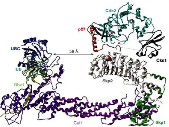
Figure 4 Structure of the SCFSkp2–Cks1 complex linked to p27-Cdk2 (adapted from
{Cardozo and Pagano, Nat Rev Mol Cell Biol 2004; 5(9)})
The E3 ubiquitin ligase SCFSkp2 is composed of a Cul1 scaffolding protein with a Rbx1 E2 binding protein and a F-box binding protein Skp1 on each end. Rbx1 recognize and bind to a E2 ubiquitin conjugating enzyme while Skp1 binds to the Skp2-Cks1-p27 complex. Cks1 interacts with the F-box protein Skp2 and greatly increases its affinity for T187 phosphorylated p27. Cks1 further strengthen Skp2-p27 binding by associating with Cdk2, which is bound to p27 to mediate its phosphorylation on threonine 187. SCFSkp2 then will transfer the ubiquitin molecule from E2 to p27. A poly-ubiquitin chain will be formed. This poly-Ubs chain targets p27 to the 26S proteasome for proteolysis and increases the proteasome’s affinity for the tagged p27.
1.5.3. Regulation and Cancer
Level of Skp1 and Cul-1 are constant throughout the cell cycle while Skp2 expression is regulated. Various transcription factors, E2F1, FoxM1, GABP, NFκB, SP1 and CBF1, all promote Skp2 gene transcription while Foxp3 has been shown to repress Skp2 expression. [115] Akt-mediated phosphorylation of Skp2 can further modulate its localisation as well as function. Skp2 accumulates from late G1 and rapidly degrade before
mitosis. [117] Poly-ubiquitination of Skp2 was found to be mediated by APCCDH1, as a result of SCF and APC/C coordinated regulation of cell cycle. [120] Phosphorylation of Skp2 on serine 64 and 72 by Cdk2 and Akt respectively prevent Cdh1 binding. [115]
An increased level of Skp2 has been observed along with reduced levels of p27 as well as increased invasion and metastasis in many types of cancer. [113, 121] The level of Skp2 mRNA can be used to predict p27 level in tumors and correlate with the tumor stage.
[122] Thus, SCFSkp2 inhibitors have been considered as a novel class of antitumor agents. Ji
et al. discovered that knock-down of Skp2 alone in cancer cells only had mild anti-tumor effects. On the other hand, disruption of Skp2-cyclin A interaction by a blocking peptide was able to induce selective cancer cell killing. [123] Further studies permitted Chen et al. to find a compound, dubbed compound A, that could prevent the incorporation of Skp2 into the SCF complex. This resulted in G1/S cell-cycle arrest as well as SCFSkp2- and
p27-dependent, caspase-inp27-dependent, programmed cell death via autophagy. [109]
Recent works by Wu et al have identified small molecular inhibitors specific to SCF-Skp2 activity using both in silico and in vitro assays. [124] These compounds
selectively blocked SCFSkp2-dependent degradation of p27 by acting on the targeted pocket formed by Skp2-Cks1. Effective cell cycle arrest at G1 or G2/M phases has been observed
in cancer cells treated with these inhibitors. Once again, this discovery underlines the importance of the Skp2-Cks1-dependent p27 degradation mechanism in cancers.
1.6. Cks1
The human Cdc28 protein kinase regulatory subunit 1 (Cks1) is encoded by the
Cks1B gene on the chromosome 1q. [125, 126] It belongs to the cell cycle regulatory Suc1 (suppressor of Cdc2 mutation)/Cks family. First discovered in yeasts, they were identified as genetic suppressors of Cdk mutations. [119, 127] Functional domain sequences are largely conserved between species. Two homologous Cks1 proteins are found in human: Cks1 (CksHs1) and Cks2 (CksHs2). They share ~81% of sequence homology, yet differ in respective folding. [128] All Cks proteins are characterized by their ability to bind to a Cdk catalytic subunit; however, only human Cks1 possesses the additional ability to mediate Skp2-p27 binding. [119] Cks2 functions in germ cells. They play a role of safeguard in DNA replication and cell differentiation. [129] Cks proteins are essential components of mitotic cyclin/Cdk complexes, mice with double knockout of Cks1 and Cks2 are non-viable.
1.6.1. Structure and Function
Cks1 is a small protein formed from only one polypeptide chain of 79 amino acids long (~9.6 kDa) and has a half-life of approximately 4 to 6.6 hours. [130-132] It is folded into four antiparallel β-strands, involved in Cdk binding, and two short α-helices, involved in Skp2 binding. [133] (see Fig.5B) Cks1 is folded in a way that there is a β-hairpin followed by an exposed α-helical hairpin on the amino-terminal and a β-hairpin on the carboxyl-terminal, resulting in a four-stranded β-sheet. [128] Cks1 contains 3 protein-protein
interaction domains for Cdk-binding, anion-binding and Skp2-binding. The anion-binding site is responsible for recognizing and binding phosphate, sulfate, or acidic residues of proteins, including phosphorylated p27. [133] (see Fig.5) A function of Cks1 is to promote binding of Cdks to partially phosphorylated proteins and mediate their poly-phosphorylation. The targets of multi-phosphorylation are often substrates of APC/C and G2- and M-phase regulators, including Cdc25 and Wee1. [127]
Moreover, Cks1 greatly increases the affinity binding of Skp2 to p27. This interaction is Skp2-specific, since it was found that ubiquitination of cyclin E by SCFSkp2 is also mediated by Cks1. [133] Skp2 associates with Skp1 of the SCF complex by its F-box
motif while its LRR domain and C-terminal binds to Skp2-binding domain of Cks1. [119] Cks1 anion-binding site recognize the phosphorylated Thr187 side chain of p27Kip1 and along with Skp2, promotes mutual binding to C- and N-terminal of p27 respectively. An invariant glutamic acid 185 of p27’s central chain inserts into the interface between Skp2 and Cks1, interacting with both. [119] Sitry et al. proposed that interaction of Skp2 with the substrate is further strengthened by the association of the Cdk-binding site of Cks1 with Cdk2/cyclin E, to which phosphorylated p27 is bound. [133] (see Fig.4) p21Cip1, p57Kip2 and p130/Rb all have been shown to be ubiquitinated and degraded via the SCFSkp2-Cks1 pathway. p57 shares a homologous C-terminal with p27 that mediate its interaction with Skp2-Cks1, while the mechanism of p21 ubiquitination still remain unclear. [119] p130/Rb is an inhibitor of Cdk2 and the transcription factor E2F, degradation of this protein is thus essential for mitotic entry. [134]
Cks1 also exert multiple p27-independent functions in the G1 phase. In yeast, Cks1
is essential for Cdk activities. [135, 136] Along with Cdc28, the yeast homolog of Cdk1, Cks1 is recruited to gene promoters to modulate a subset of genes. The ubiquitin-binding domain of Cks1 allows it to selectively recruit ubiquitylated subunits of the proteasome as a mechanism of regulation of the transcriptional process. In mammalian cells, various studies also confirmed that Cks1 partake in cdks pathways, stimulate the Cdc27 component of the APC/C complex, and increase Cdc20 expression. This latter protein is known to be involved in two microtubule-dependent processes, therefore suggesting that cytoplasmic Cks1 might play a role in cell migration. Recently, Hoellein et al. has shown genetic evidence that Cks1 association with Cdk2 instead of SCFSkp2 is regulated for the G1/S
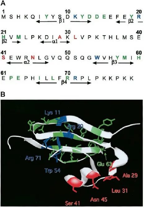
Figure 5 The amino acid sequence (A) as well as the crystal structure (B) of the human form of Cks1. (adapted from {Sitry et al., J Biol Chem, 2002, 277(44)})
Cks1 forms a single stranded polypeptide of 79 amino acids long. It has 3 distinct binding sites for interaction with anion (shown in blue), Skp2 (in red), and Cdk (in green).