Université de Montréal
Biology and characterisation of polyalanine as an emerging
pathological marker
par :
Shawn Joseph Stochmanski
Département de Neurosciences, Programme en Sciences Neurologiques Faculté des Études Supérieures et Postdoctorales
Thèse présentée à la Faculté des Études Supérieures et Postdoctorales en vue de l’obtention du grade de Ph.D.
en Sciences Neurologiques
Décembre, 2014
Université de Montréal
Faculté des Études Supérieures et Postdoctorales
Cette thèse intitulée:
Biology and characterisation of polyalanine as an emerging pathological marker
présentée par : Shawn Joseph Stochmanski
a été évaluée par un jury composé des personnes suivantes:
Muriel Aubry, président-rapporteur Guy A. Rouleau, directeur de recherche
Edward Bradley, membre du jury David L. Nelson, examinateur externe Jannic Boehm, représentante du doyen de la FESP
iii
Résumé
Dix-huit maladies humaines graves ont jusqu'ici été associées avec des expansions de trinucléotides répétés (TNR) codant soit pour des polyalanines (codées par des codons GCN répétés) soit pour des polyglutamines (codées par des codons CAG répétés) dans des protéines spécifiques. Parmi eux, la dystrophie musculaire oculopharyngée (DMOP), l’Ataxie spinocérébelleuse de type 3 (SCA3) et la maladie de Huntington (MH) sont des troubles à transmission autosomale dominante et à apparition tardive, caractérisés par la présence d'inclusions intranucléaires (IIN). Nous avons déjà identifié la mutation responsable de la DMOP comme étant une petite expansion (2 à 7 répétitions supplémentaires) du codon GCG répété du gène PABPN1. En outre, nous-mêmes ainsi que d’autres chercheurs avons identifié la présence d’événements de décalage du cadre de lecture ribosomique de -1 au niveau des codons répétés CAG des gènes ATXN3 (SCA3) et HTT (MH), entraînant ainsi la traduction de codons répétés hybrides CAG/GCA et la production d'un peptide contenant des polyalanines. Or, les données observées dans la DMOP suggèrent que la toxicité induite par les polyalanines est très sensible à leur quantité et leur longueur.
Pour valider notre hypothèse de décalage du cadre de lecture dans le gène ATXN3 dans des modèles animaux, nous avons essayé de reproduire nos constatations chez la drosophile et dans des neurones de mammifères. Nos résultats montrent que l'expression transgénique de codons répétés CAG élargis dans l’ADNc de ATXN3 conduit aux événements de décalage du cadre de lecture -1, et que ces événements sont néfastes. À l'inverse, l'expression transgénique de codons répétés CAA (codant pour les polyglutamines) élargis dans l’ADNc de ATXN3 ne conduit pas aux événements de décalage du cadre de lecture -1, et n’est pas toxique. Par ailleurs,
iv
l’ARNm des codons répétés CAG élargis dans ATXN3 ne contribue pas à la toxicité observée dans nos modèles. Ces observations indiquent que l’expansion de polyglutamines dans nos modèles drosophile et de neurones de mammifères pour SCA3 ne suffit pas au développement d'un phénotype.
Par conséquent, nous proposons que le décalage du cadre de lecture ribosomique -1 contribue à la toxicité associée aux répétitions CAG dans le gène ATXN3.
Pour étudier le décalage du cadre de lecture -1 dans les maladies à expansion de trinucléotides CAG en général, nous avons voulu créer un anticorps capable de détecter le produit présentant ce décalage. Nous rapportons ici la caractérisation d’un anticorps polyclonal qui reconnaît sélectivement les expansions pathologiques de polyalanines dans la protéine PABPN1 impliquée dans la DMOP. En outre, notre anticorps détecte également la présence de protéines contenant des alanines dans les inclusions intranucléaires (IIN) des échantillons de patients SCA3 et MD.
Mots-clés : Ataxie spinocérébelleuse de type-3 (SCA3), dystrophie musculaire oculopharyngée (DMOP), maladie de Huntington (MH), dégénération neuronale, inclusions intranucléaires (IIN), décalage du cadre de lecture ribosomique -1, ataxin-3, polyadenylate-binding protein nuclear 1 (PABPN1), huntingtin, polyglutamine, polyalanine, ATXN3, PABPN1, HTT.
v
Abstract
Eighteen severe human diseases have thus far been associated with trinucleotide repeat (TNR) expansions coding for either polyalanine (encoded by a GCN repeat tract) or polyglutamine (encoded by a CAG repeat tract) in specific proteins. Among them, oculopharyngeal muscular dystrophy (OPMD), spinocerebellar ataxia type-3 (SCA3), and Huntington’s disease (HD) are late-onset autosomal-dominant disorders characterised by the presence of intranuclear inclusions (INIs). We have previously identified the OPMD causative mutation as a small expansion (2 to 7) of a GCG repeat tract in the PABPN1 gene. In addition, we and others have reported the occurrence of -1 ribosomal frameshifting events in expanded CAG repeat tracts in the ATXN3 (SCA3) and HTT (HD) genes, which result in the translation of a hybrid CAG/GCA repeat tract and the production of a polyalanine-containing peptide. Data from OPMD suggests that polyalanine-induced toxicity is very sensitive to the dosage and length of the alanine stretch.
To validate our ATXN3 -1 frameshifting hypothesis in animal models, we set out to reproduce our findings in Drosophila and mammalian neurons. Our results show that the transgenic expression of expanded CAG repeat tract ATXN3 cDNA led to -1 frameshifting events, and that these events are deleterious. Conversely, the expression of polyglutamine-encoding expanded CAA repeat tract ATXN3 cDNA was neither frameshifted nor toxic. Furthermore, expanded CAG repeat tract ATXN3 mRNA does not contribute to the toxicity observed in our models. These observations indicate that expanded polyglutamine repeats in
Drosophila and mammalian neuron models of SCA3 are insufficient for the development of a
vi
Hence, we propose that -1 ribosomal frameshifting contributes to the toxicity associated with CAG repeat tract expansions in the ATXN3 gene.
To further investigate ribosomal frameshifting in expanded CAG repeat tract diseases, we sought to create an antibody capable of detecting the frameshifted product. Here we report the characterization of a polyclonal antibody that selectively recognizes pathological expansions of polyalanine in the protein implicated in OPMD, PABPN1. Furthermore, our antibody also detects the presence of alanine proteins in the intranuclear inclusions (INIs) of SCA3 and HD patient samples.
Keywords : Spinocerebellar ataxia type-3 (SCA3), oculopharyngeal muscular dystrophy (OPMD), Huntington’s disease (HD), neurodegeneration, intranuclear inclusions (INIs), -1 ribosomal frameshifting, ataxin-3, polyadenylate-binding protein nuclear 1 (PABPN1), huntingtin, polyglutamine, polyalanine, ATXN3, PABPN1, HTT.
vii
Table of contents
Résumé ... iii
Abstract ... v
Table of contents ... vii
List of tables ... xiii
List of figures ...xivv
List of symbols ... xv
Contribution of authors ... xxiii
Acknowledgments ...xxvi
Chapter 1 : Introduction ... 28
1.1 Trinucleotide repeat expansion disorders ... 28
1.2 Spinocerebellar ataxia type-3 ... 31
1.2.1 Clinical features ... 31
1.2.2 Imaging and neuropathological features ... 33
1.2.3 Molecular genetics ... 34
1.2.4 Ataxin-3 ... 36
1.2.5 Normal cellular and physiological roles of ataxin-3 ... 37
1.2.5.1 Involvement in the ubiquitin-proteasome pathway ... 37
1.2.5.2 Involvement in transcription regulation ... 38
1.2.5.3 Ataxin-3 interactors and protein homeostasis systems ... 39
1.2.5.4 Roles in cytoskeletal organisation and myogenesis ... 41
viii
1.2.7 Aggregation ... 43
1.2.8 Proteolysis ... 43
1.2.9 Degradation ... 44
1.2.10 Expanded ataxin-3 and disease pathogenesis ... 44
1.2.10.1 Protein aggregates and intracellular inclusions ... 45
1.2.10.2 Proteolytic cleavage and the “toxic fragment” hypothesis . 46 1.2.10.3 Localisation of expanded ataxin-3 fragments ... 47
1.2.10.4 Impaired protein degradation in SCA3 ... 48
1.2.10.5 Transcription dysregulation in SCA3 ... 50
1.3 Oculopharyngeal muscular dystrophy ... 51
1.3.1 Clinical features ... 52
1.3.2 Myopathological and neuropathological features ... 52
1.3.3 Molecular genetics ... 53
1.3.4 Polyadenylate-binding protein nuclear ... 55
1.3.5 Normal cellular and physiological role of PABPN1 ... 56
1.3.5.1 Involvement in mRNA polyadenylation ... 56
1.3.5.2 Involvement in the export of polyadenylated RNA ... 58
1.3.5.3 Involvement in transcription regulation ... 59
1.3.5.4 Intracellular localisation ... 60
1.3.6 Expanded PABPN1 and disease pathogenesis ... 61
1.3.6.1 Protein aggregates ... 61
1.3.6.2 Intranuclear inclusions ... 63
ix
1.3.6.4 Transcription dysregulation in OPMD ... 65
1.3.6.5 Involvement of the ubiquitin-proteasome pathway and molecular chaperones in OPMD ... 66
1.3.6.6 Impairment of mRNA transport and/or processing in OPMD .. ... 67
1.3.6.7 Apoptosis ... 67
1.4 Huntington's disease ... 68
1.4.1 Clinical features ... 68
1.4.2 Imaging and neuropathological features ... 71
1.4.3 Molecular genetics ... 74
1.4.4 Huntingtin ... 76
1.4.4.1 Posttranslational modification of huntingtin ... 77
1.4.5 Normal cellular and physiological role of huntingtin ... 78
1.4.5.1 Involvement in embryonic development ... 79
1.4.5.2 Involvement in cellular survival ... 80
1.4.5.3 Involvement in axonal and vesicle transport ... 81
1.4.5.4 Involvement in synaptic activity ... 82
1.4.6 Expanded huntingtin and disease pathogenesis ... 83
1.4.6.1 Protein aggregation and intranuclear inclusions ... 83
1.4.6.2 Cleavage of expanded huntingtin ... 85
1.4.6.3 Expanded huntingtin and BDNF ... 85
1.4.6.4 Transcription dysregulation in HD ... 86
x
1.4.6.6 Mitochondrial dysfunction in HD ... 90
1.5 Programmed ribosomal frameshifting – from virus to mammals ... 91
1.6 Hypothesis and objectives ... 93
Chapter 2 : Expanded ATXN3 frameshifting events are toxic in Drosophila and mammalian neuron models ... 95
2.1 Rationale ... 96
2.2. Abstract ... 98
2.3 Introduction ... 98
2.4 Results ... 99
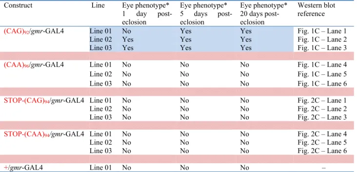
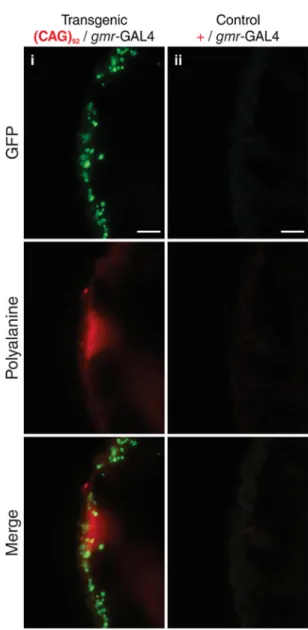
2.4.1 -1 frameshifting events are deleterious in Drosophila ... 99
2.4.2 RNA does not confer toxicity in Drosophila ... 102
2.4.3 -1 frameshifting events are deleterious in mammalian neurons ... 103
2.5 Discussion ... 104
2.6 Materials and methods ... 107
2.6.1 Transgenic Drosophila lines ... 107
2.6.2 Epon embedding and microtome preparation of sections ... 107
2.6.3 Western blot analysis ... 108
2.6.4 Drosophila immunohistochemistry ... 109
2.6.5 RT-PCR and sequencing ... 109
2.6.6 Quantitative real-time PCR ... 110
2.6.7 Constructs for organotypic slice culture ... 110
xi
2.6.9 Cortical-slice organotypic culture ... 111
2.6.10 Organotypic culture immunohistochemistry ... 112
2.7 Funding ... 112
2.8. Acknowledgements ... 112
2.9 Figures ... 113
2.10 Supplementary data ... 119
Chapter 3 : A polyalanine antibody for the diagnosis of oculopharyngeal muscular dystrophy and polyalanine-related diseases ... 123
3.1 Rationale ... 124
3.2 Abstract ... 124
3.3 Introduction ... 125
3.4 Results ... 129
3.4.1 Generation of a polyclonal antibody sensitive to polyalanine at the pathological threshold in OPMD ... 129
3.4.2 Differentiation can be made between OPMD and control patient samples ... 130
3.4.3 Alanine-containing proteins are detected in a transgenic Drosophila model of SCA3, and lymphoblastoid cells of SCA3 and HD patients ... 132
3.5 Discussion ... 132
3.6 Materials and methods ... 135
3.6.1 Production of polyalanine antibody ... 135
xii
3.6.3 Cell culture and transfections ... 136
3.6.4 Western blots ... 136 3.6.5 Human immunohistochemistry ... 137 3.6.6 Drosophila immunohistochemistry ... 138 3.6.7 Human immunocytochemistry ... 138 3.7 Funding ... 139 3.8 Acknowledgments ... 139 3.9 Figures ... 140 Chapter 4 : Discussion ... 146
4.1 Ribosomal frameshifting occurs both in vitro and in vivo ... 146
4.2 Factors that may contribute to frameshifting ... 147
4.3 -1 frameshifted products are toxic to cells ... 148
4.4 Mechanisms of translational frameshifting ... 150
4.5 RNA does not confer toxicity in our Drosophila model of SCA3 ... 152
4.6 RAN translation does not occur in our Drosophila model of SCA3 ... 153
4.7 Intrabodies for therapeutic intervention ... 154
Chapter 5 :Conclusion ... 157
List of references ... 158
xiii
List of Tables
Table 1.1 Trinucleotide repeat expansion diseases ... 30 Table 2.S.1 Transgenic Drosophila lines ... 119
xiv
List of figures
Figure 2.1 Characterisation of the ATXN3 transgenic fly lines ... 113 Figure 2.2 Analysis of the STOP transgenic Drosophila lines ... 115 Figure 2.3 Mouse organotypic culture model of ATXN3 -1 frameshifting ... 117 Figure 2.S.1 Immunohistochemical detection of -1 frameshifting in adult expCAG92 fly
heads ... 120 Figure 2.S.2 Quantitative real-time PCR analysis of transgenic Drosophila lines ... 121 Figure 3.1 Testing of Ab4340 sensitivity in hPABPN1 transfected cells ... 140 Figure 3.2 Testing the ability of Ab4340 to differentiate between OPMD patient and control
individual samples ... 142 Figure 3.3 Detection of polyalanine in a transgenic Drosophila model of SCA3, and lymphoblastoid cells of an SCA3 and HD patient ... 144
xv
List of symbols
Nucleotides: A Adenine G Guanine T Thymine C Cytosine Coding Sequences: AAG Lysine ATG Methionine CAA Glutamine CAG Glutamine CGG Arginine CUG Leucine GCA Alanine GCG Alanine GGGGCC Glycine-Proline Abbreviations: µg Microgram µm MicrometerADH Alcohol Dehydrogenase
xvi ALS Amyotrophic Lateral Sclerosis ARNm Acide Ribonucléique Messager
ARX Syndromic and Non-Syndromic X-Linked Mental Retardation ATP Adenosine Triphosphate
ATXN3 Ataxin-3 Gene
B3GAT -1,3 Glucuronyltransferase BCL B-Cell Lymphoma
BDNF Brain Derived Neurotrophic Factor
bp Base Pair
BSA Bovine Serum Albumin C57B16 C57 Black 6 Mouse
C9orf72 Chromosome 9 Open Reading Frame 72 Gene
CBP CREB-Binding Protein
cDNA Complementary Deoxyribonucleic Acid
CF Ceavage Factor
CFTR Cystic Fibrosis Transmembrane conductance Regulator
CHIP C-Terminus of Heat Shock Cognate Protein 70-Interacting Protein
CK Casein Kinase
CNS Central Nervous System
CPSF Cleavage/Polyadenylation Specificity Factor CREB cAMP-Response Element-Binding Protein CstF Cleavage Stimulation Factor
xvii C-terminal Carboxyl-Terminal
DM Myotonic Dystrophy
DMEM Dulbecco's Modified Eagle Medium DMOP Dystrophie Musculaire Oculopharyngée DNA Deoxyribonucleic Acid
DRD-2 Dopamine Receptor D2
DRD-2 Dopamine Receptor D2 Gene
DRPLA Dentatorubral-Pallidoluysian Atrophy DsRed Discosoma sp. Red Fluorescent Protein
E6-AP E6-Associated Protein
ECL Enhanced Chemiluminescence EGFP Enhanced Green Fluorescent Protein EP Equivalent Postnatal Day
ER Endoplasmic Reticulum
ERAD Endoplasmic Reticulum-Associated Degradation
expCAA Polyglutamine-encoding expanded CAA ATXN3 Trangene expCAG Polyglutamine-encoding expanded CAG ATXN3 Trangene FBS Fetal Bovine Serum
FOXL2 Blepharophimosis, Ptosis and Epicanthus Inversus Syndrome Type II FOXO Forkhead Box O
FTD Frontotemporal Dementia
FXTAS Fragile X-Associated Tremor Ataxia Syndrome GABA Gamma-Aminobutyric Acid
xviii
gmr Glass Multiple Reporter
Gp Glycoprotein
HA Influenza Hemagglutinin HAP Huntingtin-Associated Protein HBSS Hank’s Balanced Salt Solution HD Huntington’s Disease HDAC Histone Deacetylase
HIP Huntingtin-Interacting Protein hnRNP Heterogeneous Ribonucleoprotein HOXA13 Hand-Foot-Genital Syndrome HOXD13 Synpolydactyly Type II
HRP Horseradish Peroxidase Hsc Heat Shock Cognate Protein Hsp Heat Shock Protein
HTT Huntingtin Gene
ICI Intracytosolic Inclusion IIN Inclusions Intranucléaires INI Intranuclear Inclusion
InsP3R1 Type 1 Inositol (1,4,5)-Trisphosphate Receptor IP3 Inositol (1,4,5)-Trisphosphate
IT15 Interesting Transcript 15 Gene
JD Josephin Domain
xix LCL Lymphoblastoid Cell Lines lncRNA Long Noncoding RNA LSB Laemmli Sample Buffer
MAP Microtubule-Associated Protein MBNL1 Muscleblind-Like 1 MH Maladie de Huntington
MITOL Mitochondrial Ubiquitin Ligase MJD Machado-Joseph Disease
ml Millilitre
mM Millimolar
MRI Magnetic Resonance Imaging mRNA Messenger Ribonucleic Acid
mRNP Messenger Ribonucleic Acid Ribonucleoprotein NCoR Nuclear Receptor Corepressor
NEDD8 Neural Precursor Cell Expressed Developmentally Down-Regulated 8 NES Nuclear Export Signal
NGFR Nerve Growth Factor Receptor
NGFR Nerve Growth Factor Receptor Gene
NGS Normal Goat Serum
NLS Nuclear Localisation Signal NMDA N-Methyl-D-Aspartate
NR1 N-Methyl-D-Aspartate Receptor Subunit 1 Gene
xx OAT Ornithine Aminotransferase OD Oligomerisation Domains
OPMD Oculopharyngeal Muscular Dystrophy
PABPC1 Polyadenylate-Binding Protein Cytoplasmic 1 PABPN1 Polyadenylate-Binding Protein Nuclear 1
PABPN1 Polyadenylate-Binding Protein Nuclear 1 Gene
PACSIN Protein Kinase C and Casein Kinase Substrate in Neurons PAGE Polyacrylamide Gel Electrophoresis
PAP Polyadenylate Polymerase PBS Phosphate-Buffered Saline PCAF p300/CREBBP associated factor PCR Polymerase Chain Reaction PFA Paraformaldehyde
PLIC1 Protein Linking IAP to the Cytoskeleton PNS Peripheral Nervous System
PRF Programmed Ribosomal Frameshifting PSD Postsynaptic Density Scaffolding Protein RAN Repeat Associated Non-ATG
RE1/NRSE Repressor Element 1/Neuron-Restrictive Silencer Element
REST/NRSF RE1-Silencing Transcription Factor/Neuron-Restrictive Silencer Factor RIPA Radioimmunoprecipitation Assay
RNA Ribonucleic Acid
xxi
RP Ribosomal Protein
rpm Revolutions Per Minute
RRM Ribonucleoprotein-Type RNA Binding Motif RUNX2 Cleidocranial Dysplasia
SBMA Spinal Bulbar Muscular Atrophy SCA Spinocerebellar Ataxia
scFv Single-Chain Fv
SDS Sodium Dodecyl Sulfate SKIP Ski-Interacting Protein SMA Spinal Muscular Atrophy SNAP Sensory Nerve Action Potential SNP Single Nucleotide Polymorphism snRNP Small Nuclear Ribonucleoproteins SOD Superoxide Dismutase
SOX3 X-Linked Hypopituitarism SP Specificity Protein
STEP Striatal-Enriched Protein Tyrosine Phosphatase
STOP-CAA Stop Modified Polyglutamine-encoding expanded CAG ATXN3 Trangene STOP-CAG Stop Modified Polyglutamine-encoding expanded CAG ATXN3 Trangene TAFII TATA-Binding Protein-Associated Factor
TBP TATA-Binding Protein ThT Thioflavin T
xxii TrkB Tropomyosin Receptor Kinase B tRNA Transfer Ribonucleic Acid
tRNAGln-CUG Glutaminyl-Transfer Ribonucleic Acid
UIM Ubiquitin Interaction Motif UPP Ubiquitin-Proteosome Pathway
US United States
UTR Untranslated Region
VCP p97/Valosin-Containing Protein VH Variable Ig Heavy
VL variable Ig Light ZIC2 Holoprosencephaly
xxiii
Contribution of authors
Chapter 2
S.J. Stochmanski†, M. Therrien†, J. Laganière, D. Rochefort, S. Laurent, L. Karemera, R.
Gaudet, K. Vyboh, D.J. Van Meyel, G. Di Cristo, P.A. Dion, C. Gaspar and G.A. Rouleau. Expanded ATXN3 frameshifting events are toxic in Drosophila and mammalian neuron models.
Human Molecular Genetics. 2012;21:2211-2218.
Stochmanski: Study design, data generation and analysis, manuscript writing, figure production Therrien: Drosophila data generation, statistical analysis, manuscript writing and revision Laganière: Drosophila data generation and analysis
Rochefort: Construct design
Laurent, Karemera, Gaudet, Vyboh: Drosophila technical assistance Van Meyel: Drosophila expertise
Di Cristo: Organotypic culture expertise Dion: Manuscript revision
Gaspar: Study design, data generation and analysis Rouleau: Supervision and manuscript revision
Chapter 3
S.J. Stochmanski, F. Blondeau, M. Girard, P. Hince, D. Rochefort, C. Gaspar, P.A. Dion, P.S. McPhersonand G.A. Rouleau. Manuscript in Preparation.
Stochmanski: Study design, data generation and analysis, manuscript writing, figure production Blondeau: Antibody injections
xxiv Girard: Antibody technical assistance
Hince: Cell culture technical assistance Rochefort: Construct technical assistance Dion: Manuscript revision
McPherson: Antibody design and expertise Rouleau: Supervision, manuscript revision
xxv
I dedicate this work to my parents, Joseph and Gail, my sister, Laura, and my Godson, Lucas. Thank you for your understanding, patience, love and support.
xxvi
Acknowledgments
It is difficult to overstate my gratitude to my supervisor, Dr. Guy A. Rouleau. With his expert supervision, his inspiration, and his patience, I received a first-class Ph.D. experience that most students could only yearn for.
There have also been members of the Rouleau lab during my tenure that were essential to my learning and development, and to whom I owe many thanks. These include Patrick Dion and Claudia Gaspar, who not only shared with me their knowledge of trinucleotide repeat expansion diseases, but also the skills necessary to become a successful researcher. I would like to thank my conference travel companions and connections to the world of human genetics, Paul Valdmanis, Qingling Duan, Jean-Baptiste Rivière, Judith St-Onge, and Véronique Belzil; and my lab roommates Siriram Ramalingam, Aida Abu-Baker, Masoud Shekarabi, Valérie Lavastre, Jean-François Schmouth, Alanna Grant and Hannah Kaneb for the exciting conversations on molecular genetics and life in general. I am indebted to Pascale Hince, Daniel Rochefort, Martine Girard, and Janet Laganière for the teachings and technical support I received from them on my projects; and to Hélène Catoire, Emmanuelle Perrot-Audet, and Carine Daurat for all the crucial behind-the-scenes secretarial assistance. Finally, I would once again like to thank Patrick Dion for providing a review and corrections to my thesis, and encouragement while writing it.
I would like to thank my supervisory committee, consisting of Vincent Castellucci, Bernard Brais, Pierre Drapeau, and Nikolaus Heveker for all their advice.
xxvii
I also received much needed assistance from members of the department of Neuroscience at the Université de Montréal. I have to thank Richard Robitaille, Susy Daigle, and Joanne Payette.
Lastly, I would very much like to thank my family for their unwavering encouragement and support over these years, as well as Sarah Rasheed for her tremendous help and support at every step of this long journey.
Chapter 1 : Introduction
1.1 Trinucleotide
repeat expansion diseases
The expansion of trinucleotide repeat (TNR) sequences within genes is a naturally occurring phenomenon in the human genome. On rare occasions; however, these expansion events have been shown to confer severe human phenotypes. TNR diseases are often categorised into two subclasses depending on the nature of the coding sequence concerned: polyglutamine [(CAG)n] repeat expansion diseases; or polyalanine [(GCN) n] repeat expansion diseases (Table 1.1).
Polyglutamine repeat expansion diseases comprise at least nine distinct adult-onset neurodegenerative conditions, including Huntington’s disease (HD), spinal bulbar muscular atrophy (SBMA), spinocerebellar ataxia (SCA) types 1, 2, 3, 6, 7 and 17, and dentatorubral-pallidoluysian atrophy (DRPLA) (La Spada and Taylor, 2010; Orr and Zoghbi, 2007). The adult-onset disorder oculopharyngeal muscular dystrophy (OPMD), and eight other severe congenital conditions such as synpolydactyly type II (HOXD13), cleidocranial dysplasia (RUNX2), holoprosencephaly (ZIC2), hand-foot-genital syndrome (HOXA13), blepharophimosis, ptosis and epicanthus inversus syndrome type II (FOXL2), congenital central hypoventilation syndrome (PHOX2B), syndromic and non-syndromic X-linked mental retardation (ARX), and X-linked hypopituitarism (SOX3) currently account for the polyalanine repeat expansion diseases (Albrecht and Mundlos, 2005; Messaed and Rouleau, 2009).
29
Trinucleotide repeat instability depends on the nature of the repeat and its length. Polyglutamine repeat tracts are unstable in both somatic and germ cells, and the cause of their expansion likely involves one or more of the following processes: formation of unusual DNA structures and DNA slippage during lagging-strand synthesis; aberrant repair of unusual DNA mutagenic intermediates such as double-strand or single-strand breaks; or recombination within the repeats by interchromosomal strand annealing (Cleary and Pearson, 2005; Pearson et al., 2005). In contrast, polyalanine repeat tracts are mitotically and meiotically stable (Cleary and Pearson, 2005; Pearson et al., 2005), and the cause of their expansion is thought to arise from unequal crossing-over between two mispaired normal alleles (Nakamoto et al., 2002; Warren, 1997).
30 Table 1.1: Trinucleotide repeat expansion diseases
Disease Locus Gene Protein Protein
Function Repeat tract size Reference Normal Disease
Polyglutamine expansion diseases
Huntington’s disease (HD) 4p16.3 HTT Huntingtin Signaling, transcription, transport
6-34 36-121 (1993) Spinal and bulbar muscular
atrophy (SBMA) Xq12 AR Androgen receptor Steroid-hormone receptor 9-36 38-62 (La Spada et al., 1991) Spinocerebellar ataxia
type-1 (SCAtype-1)
6p22.3 ATXN1 Ataxin-1 Transcription 6-39 40-82 (Banfi et al., 1994) Spinocerebellar ataxia
type-2 SCAtype-2
12q24.13 ATXN2 Ataxin-2 RNA metabolism
15-24 32-200 (Pulst et al., 1996) Spinocerebellar ataxia
type-3/Machado-Joseph disease (SCA3/MJD)
14q32.12 ATXN3 Ataxin-3 Deubiquitinase activity,
transcription regulation
10-51 55-87 (Kawaguchi et al., 1994) Spinocerebellar ataxia
type-6 (SCAtype-6) 19p13.2 CACNA1A α1A calcium channel subunit Voltage-sensitive channel activity 4-20 20-29 (Zhuchenko et al., 1997) Spinocerebellar ataxia
type-7 (SCAtype-7)
3p14.1 ATXN7 Ataxin-7 Transcription 4-35 37-306 (Trottier et al., 1995) Spinocerebellar ataxia
type-17 (SCAtype-17)
6q27 TBP TATA box binding protein
Transcription 25-42 47-63 (Koide et al., 1999)
Dentatorubral-pallidoluysian atrophy (DRPLA)
12p13.31 ATN1 Atrophin 1 Transcription 7-34 49-88 (Koide et al., 1994)
Polyalanine expansion diseases Oculopharyngeal muscular dystrophy (OPMD) 14q11.2 PABPN1 Polyadenylate-binding protein nuclear 1 mRNA processing, transport 10 12-17 (Brais et al., 1998) Synpolydactyly type II (HOXD13)
2q31.1 HOXD13 Homeobox D13 Transcription factor 15 22-29 (Goodman et al., 1997) Cleidocranial dysplasia (RUNX2) 6p21.1 RUNX2 Runt-related transcription factor 2 Transcription factor 17 27 (Mundlos et al., 1997) Holoprosencephaly (ZIC2) 13q32.3 ZIC2 Zinc finger
protein of cerebellum 2 Transcription factor 15 25 (Brown et al., 2001) Hand-foot-genital syndrome
(HOXA13) 7p15.2 HOXA13 Homeobox A13 Transcription factor 18 24-26 (Goodman al., 2000) et Blepharophimosis,/ptosis/
epicanthus inversus syndrome type II (FOXL2)
3q22.3 FOXL2 Forkhead transcription factor FOXL2 Transcription factor 14 22-24 (De Baere et al., 2001) Congenital central hypoventilation syndrome (PHOX2B) 4p13 PHOX2B Paired-like
homeobox 2B Transcription factor 20 25-29 (Matera al., 2004) et Syndromic and
non-syndromic X-linked mental retardation (ARX) Xp21.3 ARX Aristaless-related homeobox, X-linked Transcription factor 12-16 20-23 (Stromme et al., 2002) X-linked hypopituitarism (SOX3) Xq27.1 SOX3 SRY-related HMG-box gene 3 Transcription factor 15 22-26 (Laumonnier et al., 2002)
31
1.2 Spinocerebellar
ataxia type-3
Spinocerebellar ataxia type-3 (SCA3), also known as Machado-Joseph disease (MJD), was originally described in families of Azorean descent (Nakano et al., 1972; Rosenberg et al., 1976; Woods and Schaumburg, 1972), and is currently deemed to be the most common form of SCA in the world (Ranum et al., 1995; Schols et al., 1995; Schols et al., 2004; Silveira et al., 1998). The disease is an autosomal-dominant spinocerebellar degeneration that presents a gait ataxia with pyramidal and extrapyramidal signs, peripheral amyotrophy, progressive external ophthalmoplegia, rigidity, and dystonia (Coutinho and Andrade, 1978). Cognitive deficits are not a feature of SCA3, even in advanced stages of the disease (Sudarsky et al., 1992). The age of onset has been documented to range from 4 to 70 years old, with a mean age of 40 (Carvalho et al., 2008; Coutinho, 1992), while survival time has varied from 7 to 29 years, with a mean of 21 years (Coutinho, 1992; Kieling et al., 2007). Most patients succumb to pulmonary complications and cachexia (Sequeiros and Coutinho, 1993; Sudarsky et al., 1992).
1.2.1 Clinical features
The differences in age of onset and survival time, along with the observed phenotypic variability (Nakano et al., 1972; Rosenberg et al., 1976; Woods and Schaumburg, 1972), help to illustrate the marked clinical heterogeneity associated with SCA3. To assist in the clinical classification of patients, Coutinho and Andrade (1978) characterised three distinct clinical subtypes based on the presence or absence of significant pyramidal and extrapyramidal signs. Type 1 (“Type Joseph”) identifies with an early age of onset (often before 20 years old), and a swift progression of marked pyramidal (rigidity and spasticity) and extrapyramidal (bradykinesia and dystonia) signs, along with cerebellar ataxia and external ophthalmoplegia.
32
The most common subtype, type 2 (“Type Thomas”), is characterised by an intermediate onset (20 to 50 years old), cerebellar ataxia, external ophthalmoplegia, and pyramidal signs. Finally, type 3 (“Type Machado”) presents with a later age of onset (40 to 75 years old), and is characterised by ataxia associated with peripheral alterations such as amyotrophy and motor neuronopathy. Patients that are classified as type 2 (in terms of symptoms) often progress to either type 1 or 3 in as few as four to five years, although, on occasion, some have remained in type 2 for over 20 years (Sequeiros and Coutinho, 1993). More recently, two additional subtypes have been added to the clinical classification: Type 4, the rarest subtype, which is associated with dopa-responsive parkinsonism, mild cerebellar deficits, and a distal sensorimotor neuropathy (Suite et al., 1986); and type 5, for cases resembling hereditary spastic paraplegia (Sakai and Kawakami, 1996).
Many SCA3 patients also suffer from sleep disorders thought to be resultant of the disease. Schöls and colleagues found that impaired sleep, reported as trouble falling asleep and nocturnal awakenings, was associated with older age, long-standing disease, and brainstem involvement (Schols et al., 1998). Such causes for these impairments include rapid eye movement sleep behaviour disorder (Friedman, 2002; Friedman et al., 2003), restless leg syndrome (D'Abreu et al., 2009b; Pedroso et al., 2011; Schols et al., 1998; van Alfen et al., 2001), and sleep apnea (D'Abreu et al., 2009b). Excessive daytime sleepiness is also common among patients (Friedman et al., 2003).
33
1.2.2 Imaging and neuropathological features
Neuroimaging and pathological studies performed on SCA3 patients have shown that the extent and localisation of neurodegeneration far exceeds its nomenclature. Magnetic resonance imaging (MRI) has been helpful in the diagnosis of patients, and it commonly reveals an enlargement of the fourth ventricle (Klockgether et al., 1998; Murata et al., 1998; Onodera et al., 1998). Quantitative MRI-based studies have identified atrophy of the medulla oblongata, pons, midbrain, thalamus, putamen, caudate nucleus, superior cerebellar peduncle, cerebellar vermis and hemispheres, and widespread cortical and limbic structures (D'Abreu et al., 2012; D'Abreu et al., 2011; de Oliveira et al., 2012; de Rezende et al., 2014; Klockgether et al., 1998; Murata et al., 1998; Yoshizawa et al., 2003). In addition, quantitative MRI-based studies have confirmed SCA3 patients also experience atrophy of the spinal cord, combined with anteroposterior flattening (Fahl et al., 2014; Lukas et al., 2008), and atrophy of deep white matter in the brainstem, lateral thalamus, cerebellar peduncles, and cerebellar hemispheres (Guimaraes et al., 2013; Kang et al., 2014; Lukas et al., 2006). Interestingly, the use of magnetic resonance spectroscopy has identified metabolic abnormalities in apparently normal deep white matter, suggestive of axonal dysfunction preceding atrophy (D'Abreu et al., 2009a).
The brain weight of SCA3 patients with advanced symptoms is considerably less than that of individuals with no previous history of neurological or psychiatric diseases (Iwabuchi et al., 1999). Macroscopic investigation reveals a depigmentation of the substantia nigra, as well as atrophic changes of the cerebellum, pons, medulla oblongata, medial cerebellar peduncle, and cranial nerves III to XII (Rub et al., 2003a; Rub et al., 2006; Rub et al., 2002; Rub et al., 2003b). Despite atrophy of the cerebellum, Purkinje cells and inferior olivary neurons are often spared
34
(Sequeiros and Coutinho, 1993). Neuropathological studies typically show neuronal loss of the cerebellothalamocortical and basal ganglia-thalamocortical motor loops, anterior horn cells and Clarke’s column in the spinal cord, and the following systems: visual, auditory, somatosensory, oculomotor, ingestion-related (brainstem), vestibular (brainstem), precerebellar (brainstem), dopaminergic (midbrain), cholinergic (midbrain), noradrenergic (pontine), and GABAergic (thalamus) (Gilman, 2000; Hoche et al., 2008; Iwabuchi et al., 1999; Kumada et al., 2000; Robitaille et al., 1997; Rub et al., 2008). Myelin loss is also observed, affecting cerebellar, brainstem, and spinal cord white matter, cerebellar peduncles, the medial and lateral lemniscus, and the vestibulospinal, spinocerebellar, and spinothalamic tracts (Gilman, 2000; Hoche et al., 2008; Iwabuchi et al., 1999; Kumada et al., 2000; Robitaille et al., 1997; Rub et al., 2008).
1.2.3 Molecular genetics
The SCA3 locus has been mapped to chromosome 14q32.1 (Takiyama et al., 1993), and the gene identified as ATXN3 (Kawaguchi et al., 1994). ATXN3 comprises 11 exons within a 1,776 bp coding region containing one long open reading frame (Ichikawa et al., 2001; Kawaguchi et al., 1994). The causative mutation was shown to be an expansion of a polymorphic CAG repeat within exon 10, encoding for polyglutamine in the ataxin-3 protein (Ichikawa et al., 2001; Kawaguchi et al., 1994). This repeat is nearly a pure CAG tract [(CAG)2CAAAAG(CAG)n], interrupted by a single lysine codon (AAG) near the start of the repeat (Kawaguchi et al., 1994). The length of the CAG repeat within the normal allele varies greatly, ranging from 12 to 43 (Cancel et al., 1995; Limprasert et al., 1996; Maciel et al., 1995; Matilla et al., 1995; Matsumura et al., 1996a; Ranum et al., 1995; Sasaki et al., 1995; Takiyama et al., 1995), with lengths of 14 and 23 repeats being observed most frequently (Limprasert et
35
al., 1996). Conversely, expanded alleles have CAG repeat lengths that range from 61 to 87 (Cancel et al., 1995; Kawaguchi et al., 1994; Maciel et al., 1995; Matilla et al., 1995; Ranum et al., 1995; Schols et al., 1996; Silveira et al., 1996; Takiyama et al., 1995; Takiyama et al., 1997b). Although extremely rare, intermediate size alleles (45 to 56 CAG repeat lengths) have been observed in seven individuals, and associated with disease in six (Egan et al., 2000; Gu et al., 2004; Padiath et al., 2005; Takiyama et al., 1997a; van Alfen et al., 2001; van Schaik et al., 1997). The one unaffected individual was reported to have an allele with a CAG repeat length of 51, indicating the possibility of low penetrance among intermediate size alleles in SCA3 (Maciel et al., 2001).
An inverse correlation is found between the length of the CAG repeat tract within the expanded ATXN3 allele and the age of onset for the disease, accounting for 50% to 75% of the observed variation (Maciel et al., 1995; Maruyama et al., 1995; Matsumura et al., 1996a). The length of the CAG repeat tract also determines the observed clinical subtype, with longer CAG repeat lengths conferring a more severe classification (type 1 versus type 2 or 3), and may associate with a faster disease progression (Maciel et al., 1995; Maruyama et al., 1995; Matsumura et al., 1996a). In addition, a gene dosage effect may be present in SCA3, as individuals homozygous for expanded ATXN3 alleles present with an earlier age of onset and a more rapid progression than their heterozygous peers (Lerer et al., 1996; Sobue et al., 1996).
Repeat instability of the ATXN3 gene is thought to be conferred by a single nucleotide polymorphism (SNP) immediately following the CAG repeat tract [(CAG)nC or (CAG)nG)] (Limprasert et al., 1996; Matsumura et al., 1996b). Previous work has found that expanded
36
alleles exclusively contain the (CAG)nC SNP, while both the (CAG)nC and (CAG)nG polymorphisms were seen in normal alleles from SCA3 patients and control individuals (Limprasert et al., 1996; Matsumura et al., 1996b). Interestingly, the CAG tract in normal alleles with the (CAG)nC SNP were significantly longer than in the alleles with the (CAG)nG SNP (Limprasert et al., 1996; Matsumura et al., 1996b). Furthermore, the risk for intergenerational change in the expanded allele is greater in paternal than maternal transmission (Igarashi et al., 1996; Maciel et al., 1995; Manikandan et al., 2007). It is this intergenerational instability of the expanded allele that accounts for the phenomenon of anticipation occasionally seen in families with SCA3 (Coutinho and Sequeiros, 1981; Sequeiros and Coutinho, 1993).
1.2.4 Ataxin-3
Ataxin-3 is an evolutionarily conserved protein, with ATXN3 orthologues identified in such eukaryotes as fungi, protozoans, plants, and animals (Albrecht et al., 2003; Costa et al., 2004; Linhartova et al., 1999; Rodrigues et al., 2007; Schmitt et al., 1997). In unaffected humans ataxin-3 has a molecular weight of 40 to 43 kDa, depending on the length of the polyglutamine repeat (Kawaguchi et al., 1994). It is a modular protein, located in both the cytoplasm and the nucleus, as well as mitochondria (Antony et al., 2009; Macedo-Ribeiro et al., 2009; Perez et al., 1999), and is ubiquitously expressed in cells and tissue throughout the body (Costa et al., 2004; Ichikawa et al., 2001; Paulson et al., 1997a; Schmidt et al., 1998; Trottier et al., 1998); however, levels of expression vary depending on the region (Trottier et al., 1998). The ataxin-3 protein encompasses a globular N-terminal Josephin domain (JD) with a papain-like fold, similar in structure and catalytic activity to cysteine proteases, combined with a flexible C-terminal tail containing ubiquitin interaction motifs (UIMs) and the polymorphic
37
polyglutamine tract (Albrecht et al., 2003; Goto et al., 1997; Masino et al., 2003; Scheel et al., 2003). Alternative splicing results in a C-terminal containing either two UIMs followed by the polyglutamine sequence and a hydrophobic amino acid stretch, or a C-terminal with a third UIM replacing the hydrophobic tail (Goto et al., 1997). Although both variants are detected in the brain, the three UIM variant is predominantly expressed and considered to be the more physiologically relevant isoform (Harris et al., 2010).
1.2.5 Normal cellular and physiological roles of ataxin-3
1.2.5.1 Involvement in the ubiquitin-proteasome pathway
The ubiquitin-proteasome pathway (UPP) is the principle mechanism used by cells for the catabolism of proteins. Many studies have provided evidence for ataxin-3 involvement with the UPP, in its ability to bind and cleave (deubiquitinate) polyubiquitin chains and polyubiquitinated proteins (Albrecht et al., 2003; Burnett et al., 2003; Chai et al., 2004; Doss-Pepe et al., 2003; Scheel et al., 2003). Ataxin-3 appears to function as an editor of the polyubiquitin chains added to target proteins during ubiquitination, shortening them to yield free ubiquitin instead of completely dismantling them (Burnett and Pittman, 2005; Kuhlbrodt et al., 2011; Nicastro et al., 2010; Scaglione et al., 2011; Winborn et al., 2008). Ubiquitination is the process in which one ubiquitin molecule (or a polyubiquitin chain) is covalently linked to one or more lysine residues of a target protein by an E3 ubiquitin ligase (Hershko and Ciechanover, 1998). Different linkage types confer specific functions: Lysine 48-linked polyubiquitin chains typically target proteins for proteasomal degradation (Chau et al., 1989; Finley et al., 1994); whereas lysine 63-linked chains play diverse roles in subcellular localisation (Weissman, 2001), membrane endocytosis (Mukhopadhyay and Riezman, 2007), DNA damage repair (Spence et
38
al., 1995), stress responses (Arnason and Ellison, 1994), and inflammation (Sun et al., 2004). Interestingly, ataxin-3 shows a strong preference for chains of four or more ubiquitin monomers, and lysine 48-linked polyubiquitin chains of four or more monomers are the ones involved in the targeting of proteins for proteasome degradation (Burnett et al., 2003; Chai et al., 2004; Winborn et al., 2008). Moreover, an in vitro study involving neuronal cells demonstrated that inhibiting the catalytic activity of ataxin-3 results in the accumulation of polyubiquitinated proteins (Berke et al., 2005). Collectively, these suggest that unlike the usual function of deubiqutinating enzymes to rescue target substrates from degradation, the deubiquitinase activity of ataxin-3 is associated with the delivery of the target substrates to the proteasome (Scaglione et al., 2011; Ventii and Wilkinson, 2008). In fact, ataxin-3 knockout mice show increased levels of ubiquitinated proteins when compared to their wild-type littermates (Schmitt et al., 2007), and the Caenorhabditis elegans ataxin-3 orthologue was shown to aid protein catabolism in vivo (Kuhlbrodt et al., 2011).
In certain instances, ataxin-3 is itself ubiquitinated. The protein is either mono- or oligo-ubiquitinated; however, the common form is monoubiquitinated (Berke et al., 2005; Todi et al., 2009). This posttranslational modification enhances the deubiquitinase activity of ataxin-3 toward ubiquitinated substrates and free polyubiquitin chains, independent of potential cofactors and interactors (Todi et al., 2010; Todi et al., 2009).
1.2.5.2 Involvement in transcription regulation
Another aspect of ataxin-3 function is believed to involve transcriptional regulation, likely as a transcriptional co-repressor via the modulation of histone acetylation and
39
deacetylation at selected promoters (Li et al., 2002a). Through interaction with the histone acetylase cAMP-response element-binding protein (CREB)-binding protein (CBP), p300 and p300/CBP associated factor (PCAF), ataxin-3 was shown to inhibit CREB-mediated transcription (Evert et al., 2006; Li et al., 2002a). Ataxin-3 also has the ability to inhibit p300-mediated histone acetylation by blocking access to histone actetylation sites, and to promote histone deacetylation by interacting with histone deacetylase 3 (HDAC3) and nuclear receptor co-repressor 1 (NCOR1) (Evert et al., 2006; Li et al., 2002a).
There is also evidence for ataxin-3 involvement in the cellular response to oxidative stress, as it has been shown to interact with and stabilise the forkhead box O (FOXO) transcription factor FOXO4 (Araujo et al., 2011). When cells experience oxidative stress, ataxin-3 and FOXO4 translocate to the nucleus and promote the transcription of the superoxide dismutase-2 (SOD2) gene, which in turn increases expression of the antioxidant enzyme SOD2 (Araujo et al., 2011).
1.2.5.3 Ataxin-3 interactors and protein homeostasis systems
Much work has been done to identify ataxin-3 interacting proteins, in the hope of identifying its biological functions. One such interacting protein is the ATPase p97/valosin-containing protein (VCP), which works coordinately with ubiquitinating complexes to shuttle polyubiquitinated substrates to the proteasome for degradation (Boeddrich et al., 2006; Doss-Pepe et al., 2003). The VCP/ataxin-3 complex may act to transfer ataxin-3 edited polyubiquitinated substrates directly to the proteasome or other proteasomal shuttling factors such as ubiquilin/PLIC1 and the human homologues of the yeast DNA repair protein Rad23,
40
HHR23A and HHR23B (Doss-Pepe et al., 2003; Heir et al., 2006; Kuhlbrodt et al., 2011; Wang et al., 2000). This complex may also function to regulate endoplasmic reticulum-associated degradation (ERAD), the process in which misfolded proteins in the ER secretory pathway are ubiquitintated and exported to the cytosol for proteasomal degradation (Doss-Pepe et al., 2003; Wang et al., 2006; Zhong and Pittman, 2006). It is still uncertain, however, if the VCP/ataxin-3 complex works to promote or inhibit ERAD (Wang et al., 2006; Zhong and Pittman, 2006). Interestingly, the VCP/ataxin-3 complex may also be associated with aging. Kuhlbrodt and colleagues have shown lifespan increases in C. elegans VCP and ataxin-3 double knockouts, and that the VCP/ataxin-3 complex regulates components of the insulin/insulin-like growth factor 1 signaling pathway – a pathway involved in lifespan regulation (Kuhlbrodt et al., 2011).
Another ataxin-3 interactor is C-terminus of heat shock cognate protein 70 (Hsc70)-interacting protein (CHIP) (Jana et al., 2005), an E3 ubiquitin ligase that has been linked to the pathology of several neurodegenerative diseases (Cantuti-Castelvetri et al., 2005; Howland et al., 2002; Krobitsch and Lindquist, 2000; Petrucelli et al., 2004; Qin and Gu, 2004; Shimura et al., 2004). Recent work has shown that monoubiquitinated CHIP forms an ubiquitination complex with ataxin-3, through which the deubiquitinase activity of ataxin-3 limits the length of polyubiquitin chains linked to CHIP substrates (Scaglione et al., 2011). Once the linkages have been formed ataxin-3 deubiquitinates CHIP, terminating the ubiquitination cycle (Scaglione et al., 2011). Conversely, CHIP has been shown to monoubiquitinate ataxin-3 at lysine 117 in the JD, enhancing its deubiquitinase activity (Todi et al., 2010; Todi et al., 2009).
41
Ataxin-3 also interacts with the ubiquitin-like protein neural precursor cell expressed developmentally down-regulated 8 (NEDD8), showing deneddylase activity in vitro (Ferro et al., 2007). Neddylation is a process similar to ubiquitination, in which the function of the target protein is regulated via conjugation with NEDD8. The ability of ataxin-3 to cleave isopeptide bonds between a substrate and NEDD8 provides evidence for its role in regulating neddylated complexes (Ferro et al., 2007).
Parkin, an E3 ubiquitin ligase involved in Parkinson’s disease, also shows a functional interaction with ataxin-3. In vitro, parkin is able to self-ubiquitinate, forming lysine 27- and lysine 29-linked polyubiquitin chains which are known to target substrates for lysosomal and autophagic degradation (Shimura et al., 2000). Ataxin-3 is able to deubiquitinate self-ubiquitinated parkin, and while this does not affect its stability or turnover (Durcan et al., 2011), this action may control the number and linkage type of the polyubiquitin chains attached to parkin, and thus its targeted cellular pathway (Durcan et al., 2011).
1.2.5.4 Roles in cytoskeletal organisation and myogenesis
When the ubiquitin-proteasome pathway is compromised or overwhelmed, misfolded proteins are sequestered in perinuclear inclusions termed aggresomes (Johnston et al., 1998). Ataxin-3 is thought to help regulate aggresome formation through its interactions with the aggresome/cytoskeletal organisation components tubulin, dynein, microtubules, microtubule-associated protein 2 (MAP2), HDAC6, and protein linking IAP to the cytoskeleton (PLIC1) (Burnett and Pittman, 2005; Heir et al., 2006; Mazzucchelli et al., 2009; Rodrigues et al., 2010).
42
These interactions also seem necessary for proper skeletal organisation and assembly of focal adhesions (Rodrigues et al., 2010).
Given its association with skeletal organisation, there is also evidence for ataxin-3 involvement in myogenesis (do Carmo Costa et al., 2010). In order for myoblasts to differentiate into muscle fibers, both the remodelling of the cytoskeleton and the regulation of proteins involved in integrin-mediated signalling are essential (do Carmo Costa et al., 2010). Ataxin-3 interacts with the α5 integrin subunit, repressing this proteins degradation via its role in the UPP (do Carmo Costa et al., 2010).
1.2.6 Intracellular localisation and transport
Ataxin-3 shows great mobility throughout the cytoplasm and nucleus, and its transport across the nuclear membrane is aided by a functional nuclear localisation signal (NLS), 282RKRR285, and two nuclear export signals (NES), NES77 and NES141 (Antony et al., 2009; Macedo-Ribeiro et al., 2009; Tait et al., 1998). The main mechanism for the import of ataxin-3 into the nucleus, however, seems to be the phosphorylation of three serine residues by casein kinase 2 (CK2) – serine 236 in UIM1, and serine 340 and 342 in UIM3 (Macedo-Ribeiro et al., 2009; Mueller et al., 2009). Interestingly, the translocation of ataxin-3 to the nucleus also appears to be regulated by proteotoxic stimuli such as oxidative stress and heat-shock (Reina et al., 2010). There is debate on whether CK2-dependent phosphorylation participates under these conditions (Mueller et al., 2009; Reina et al., 2010); however, evidence shows that the nuclear
43
localisation of ataxin-3 upon heat-shock requires the phosphorylation of serine 111 in the JD (Reina et al., 2010).
1.2.7 Aggregation
In vitro studies have shown that ataxin-3 has a tendency to form aggregates in a process
influenced by its N-terminal JD. Aggregation occurs through a single-step mechanism involving the self-assembly of JDs into dimers (Ellisdon et al., 2007; Ellisdon et al., 2006; Gales et al., 2005; Masino et al., 2004). These dimers then associate to form spheroidal oligomers, before elongating into classic beads-on-a-string fibrils. Ataxin-3 fibrils are SDS-soluble, Thioflavin T (ThT)-positive, and structurally resemble those of other self-associating amyloidogenic proteins. In cells, the ataxin-3 isoform bearing two UIMs (2UIM) exhibits a greater tendency to form aggregates than the three UIM (3UIM) isoform (Harris et al., 2010). Furthermore, the deubiquitinase activity of ataxin-3 is lost in fibrils, likely owing to the structural transition from α-helix to β-sheet (Masino et al., 2011b). Interestingly, the ubiquitination of ataxin-3 was shown to prevent JD self-assembly in vitro, thus preventing fibril formation and preserving its enzymatic function (Masino et al., 2011a).
1.2.8 Proteolysis
There is evidence from animal model and cell line studies that ataxin-3 is cleaved by caspases and possibly calpains (Berke et al., 2004; Colomer Gould et al., 2007; Jung et al., 2009; Wellington et al., 1998). Both caspase-1 and caspase-3 have been shown to successfully cleave ataxin-3; however, apoptotic cleavage occurs largely through the action of caspase-1, producing a polyglutamine-containing fragment in the process (Berke et al., 2004; Wellington et al., 1998).
44
Whether calpains actually participate in ataxin-3 proteolysis remains uncertain, as there is evidence for (Berke et al., 2004; Jung et al., 2009; Wellington et al., 1998) and against (Haacke et al., 2007) its involvement.
1.2.9 Degradation
The degradation of ataxin-3 has been shown to occur through both the UPP and autophagy, with the chosen method determined by the isoform involved (Berke et al., 2005; Harris et al., 2010). 2UIM ataxin-3, the less stable isoform, primarily undergoes polyubiquitination and shuttling to the proteasome for degradation. This happens through E3 ligase/shuttle protein complexes, including E4B/VCP, CHIP/heat shock protein 70 (Hsp70), and E6-associated protein (E6-AP)/Hsp70, and the endoplasmic reticulum-associated E3 ligase glycoprotein 78 (Gp78) (Jana et al., 2005; Matsumoto et al., 2004; Mishra et al., 2008; Ying et al., 2009). In contrast, the 3UIM ataxin-3 isoform is commonly degraded by macrophagy (Harris et al., 2010). Furthermore, the catalytic state of ataxin-3 may also regulate its degradation, as studies have revealed higher levels of catalytically inactive ataxin-3, which suggests slower proteasomal degradation (Todi et al., 2007).
1.2.10 Expanded ataxin-3 and disease pathogenesis
In SCA3, expansion of the polyglutamine tract in the C-terminal of ataxin-3 likely causes conformational changes in the protein, which would lead to alterations in its stability and degradation, aggregation, subcellular localisation, and molecular interactions with other proteins (Jana and Nukina, 2004). In turn, these affected properties would lead to a loss- and/or
45
gain-of-function, resulting in cellular dysfunction and the observed pathogenesis (Williams and Paulson, 2008).
1.2.10.1 Protein aggregates and intracellular inclusions
A common feature of all repeat expansion diseases is the presence of large macromolecular aggregates containing the disease protein. The initial observations made while examining SCA3 patient brain tissue were the presence of intranuclear inclusions (INIs) in disease vulnerable areas: ventral pons, substantia nigra, globus pallidus, dorsal medulla, and dentate gyrus (Paulson et al., 1997b; Schmidt et al., 1998). These INIs not only contained expanded ataxin-3, but also ubiquitin, heat-shock proteins, proteasome constituents, transcription factors, molecular chaperones, and other polyglutamine proteins (Chai et al., 1999a; Chai et al., 1999b; Chai et al., 2001; Paulson et al., 1997b; Schmidt et al., 1998). Newer techniques, however, have also identified INIs in unaffected brain areas (Rub et al., 2008; Rub et al., 2006; Yamada et al., 2002), suggesting that their presence alone does not determine the neuron’s fate (Rub et al., 2006).
More recently, expanded ataxin-3-positive aggregates have been observed in the cytosol of neurons in SCA3 brain tissue (Hayashi et al., 2003; Yamada et al., 2004), along with axons in fiber tracts known to degenerate (Seidel et al., 2010). Intracytosolic inclusions test negative for ubiquitin (Yamada et al., 2002), whereas intra-axonal inclusions are ubiquitin-positive and contain nucleoporin p62 (Seidel et al., 2010). Furthermore, intra-axonal inclusions are thought
46
to interfere with axonal transport, impairing cellular functions and promoting degeneration (Seidel et al., 2010).
Although both normal and expanded ataxin-3 form aggregates, those formed by the expanded protein occur through a two-step mechanism (Ellisdon et al., 2006). Initially, expanded ataxin-3 associates into SDS-soluble fibrils through a process similar to, but quicker than normal ataxin-3 (Ellisdon et al., 2006). In the second step, hydrogen bonding between the glutamine main- and side-chains of the polyglutamine tract induces either a β-helical turn or hairpin conformation, resulting in the formation of SDS-insoluble aggregates (Natalello et al., 2011; Seidel et al., 2010; Sikorski and Atkins, 2005). Recently, the polyglutamine tract of disease-associated proteins has been predicted to self-associate through the formation of coiled-coils, suggesting that its interaction with natural coiled-coil partners could increase aggregation (Fiumara et al., 2010; Petrakis et al., 2013).
1.2.10.2 Proteolytic cleavage and the “toxic fragment” hypothesis
As has been suggested for other polyglutamine diseases, pathogenesis resulting from the proteolytic cleavage of expanded polyglutamine protein, termed the “toxic fragment” hypothesis, may also apply to SCA3 (Tarlac and Storey, 2003; Wellington et al., 1998). In the case of ataxin-3, proteolytic cleavage was shown to generate SDS-soluble 36 kDa C-terminal fragments containing the expanded polyglutamine tract (Goti et al., 2004; Ikeda et al., 1996; Paulson et al., 1997b). These C-terminal fragments have been detected in brain homogenates from SCA3 patients and transgenic mice (Goti et al., 2004), but not from unaffected individuals (Berke et al., 2004), and were enriched in the nuclear fractions of disease vulnerable brain areas
47
(Colomer Gould et al., 2007; Goti et al., 2004). In vitro studies have further shown that ataxin-3 C-terminal fragments containing the expanded polyglutamine tracts induce a stronger aggregation and toxicity than the full-length expanded ataxin-3 protein (Breuer et al., 2010; Haacke et al., 2006; Ikeda et al., 1996; Paulson et al., 1997b).
Although there has been debate on whether calcium-dependent calpains are involved in normal ataxin-3 proteolysis (Section 1.2.8), there is increasing evidence for their involvement in the cleavage of expanded ataxin-3 and the resulting SCA3 pathogenesis (Goti et al., 2004; Haacke et al., 2007; Hubener et al., 2013; Simoes et al., 2012). Calpain-2-mediated cleavage of expanded ataxin-3 was found to produce C-terminal fragments that were prone to aggregation (Hubener et al., 2013). Furthermore, in vivo studies where calpain activity was inhibited reduced expanded ataxin-3 cleavage, aggregation, nuclear localisation, and toxicity (Haacke et al., 2007; Simoes et al., 2012). In contrast, an SCA3 transgenic mouse with its endogenous calpain inhibitor calpastatin knocked-out showed an increase in INIs and accelerated cerebellar degeneration (Hubener et al., 2013). The involvement of calpains in expanded ataxin-3 proteolysis may also explain the neuronal specificity of SCA3 pathology – calpains are calcium-dependent and require the excitation-mediated influx of calcium (Koch et al., 2011).
1.2.10.3 Localisation of expanded ataxin-3 fragments
Numerous studies have demonstrated the importance of the nucleus in the pathogenesis of SCA3 and other polyglutamine diseases, with the nuclear localisation of the expanded protein essential for disease (Schols et al., 2004; Shao and Diamond, 2007). Bichelmeier and colleagues (2007) demonstrated that C-terminal ataxin-3 fragments containing only the expanded
48
polyglutamine tract could aggregate in the nucleus or cytoplasm when coupled to a respective synthetic NLS or NES, in vitro. The INIs were shown to accumulate, whereas the ICIs were targeted for degradation (Bichelmeier et al., 2007). In transgenic SCA3 mice, artificially targeting expanded ataxin-3 to the nucleus increased levels of INIs and promoted earlier death, while forcing the nuclear export of expanded ataxin-3 reduced INIs and lessened disease symptoms (Bichelmeier et al., 2007). Although INIs are the pathological hallmark of SCA3, whether they are directly toxic or formed as a protective cellular response to cope with the toxicity of the expanded disease-proteins is still uncertain.
MITOL, a mitochondrial ubiquitin ligase, and parkin may be involved in the proteasomal degradation of expanded ataxin-3 C-terminal fragments (Sugiura et al., 2011; Tsai et al., 2003). As for full-length expanded ataxin-3, it was shown to be degraded via both the UPP and autophagy (Berger et al., 2006; Jana et al., 2005; Matsumoto et al., 2004; Mishra et al., 2008; Ying et al., 2009).
1.2.10.4 Impaired protein degradation in SCA3
As described previously (Sections 1.2.5.1 and 1.2.5.3), ataxin-3 has been shown to participate in the UPP and other protein homeostasis systems. Expansion of the polyglutamine tract in ataxin-3 could alter its normal function within these mechanisms through aberrant protein interactions and aggregation, leading to toxicity. In fact, even though there is no significant difference in the deubiquitinase activity between normal and expanded ataxin-3 (Berke et al., 2004; Burnett and Pittman, 2005), an in vitro study reported a global reduction in deubiquitinated protein in the expanded ataxin-3 model (Winborn et al., 2008). Furthermore,
49
the INIs described in SCA3 patients have contained many important proteins including ubiquitin, proteasomal components, chaperones, transcription factors, and normal ataxin-3 (Chai et al., 1999a; Chai et al., 1999b; Doss-Pepe et al., 2003; Mori et al., 2005; Paulson et al., 1997b; Schmidt et al., 1998; Takahashi et al., 2001).
Expanded ataxin-3 shows a more efficient binding of VCP, prolonging its interaction with the E4B/VCP complex and thus delaying its own degradation in the proteasome (Boeddrich et al., 2006; Matsumoto et al., 2004). Other consequences of this prolonged interaction with VCP may be the impairment of ERAD (Wang et al., 2006; Zhong and Pittman, 2006), inducing ER proteotoxic stress and subsequent degeneration, and interference with the down-regulation of neddylation (Yang et al., 2014). The CHIP/ataxin-3 interaction is also affected by expanded ataxin-3, with the expanded protein showing a six-fold increase in affinity which may target CHIP for degradation (Scaglione et al., 2011). Furthermore, despite normal and expanded ataxin-3 having similar binding affinities for polyubiquitinated parkin, expanded ataxin-3 is more efficient at cleaving its polyubiquitin chains, promoting the degradation of parkin via autophagy (Durcan et al., 2011). The resulting decrease in parkin levels may represent the Parkinson-like symptoms observed in some SCA3 patients (Buhmann et al., 2003; Gwinn-Hardy et al., 2001; Tuite et al., 1995). Additionally, aggregates in SCA3 patient brain samples were found to trap beclin-1 (Nascimento-Ferreira et al., 2011), a protein with a central function in autophagy, and whose dysfunction has been implicated in neurodegeneration (Wong and Cuervo, 2010).
50 1.2.10.5 Transcription dysregulation in SCA3
Expansion of the polyglutamine tract in ataxin-3 may also affect its proposed involvement in transcription regulation (Section 1.2.5.2). Its observed aberrant protein interactions with transcription factors and co-activators in SCA3, along with the sequestering of transcription factors to expanded ataxin-3 aggregates, suggest a role of transcriptional dysregulation in the disease pathogenesis (Evert et al., 2006; Riley and Orr, 2006). In fact, the altered transcription of several genes has been identified through analyses of brain tissue from SCA3 patients and transgenic mice, and an SCA3 neuronal cell model (Chou et al., 2008; Evert et al., 2001; Evert et al., 2003). Expanded ataxin-3 was found to down-regulate messenger RNA (mRNA) expressions of proteins involved in glutamatergic neurotransmission, intracellular calcium signaling/mobilisation or MAP kinase pathways, GABAA/B receptor subunits, Hsps, and
transcription factors regulating neuronal survival and differentiation (Chou et al., 2008). Conversely, mRNA expressions were upregulated for proteins involved in inflammation and neuronal cell death (Chou et al., 2008; Evert et al., 2001; Evert et al., 2003).
The down-regulation of mRNA expressions for proteins involved in intracellular calcium signaling/mobilisation and MAP kinase pathways is consistent with the aberrant interaction of expanded ataxin-3 with the type 1 inositol (1,4,5)-trisphosphate receptor (InsP3R1) reducing intracellular calcium levels in neurons (Chen et al., 2008; Chou et al., 2008). InsP3R1 is an intracellular calcium release channel with an important role in calcium signalling (Berridge, 1993). Interestingly, expanded ataxin-3 was also reported to alter the kinetics of voltage-gated potassium channels in neuronal cell culture and the Purkinje cells of SCA3 transgenic mice (Jeub et al., 2006; Shakkottai et al., 2011). Changes in neuronal physiology
51
may underlie the observed motor symptoms in SCA3, and likely contribute to the disease pathogenesis (Shakkottai et al., 2011).
More recently, expanded ataxin-3 was found to have a reduced ability to promote FOXO4-mediated SOD2 expression and to also interfere with the binding of FOXO4 to the
SOD2 promotor in response to oxidative stress (Araujo et al., 2011). There is also evidence for
an overall decrease in antioxidant enzyme ability in cellular models of SCA3 (Yu et al., 2009). Taken together, the resulting accumulation of reactive oxygen species and free radicals could lead to the observed mitochondrial dysfunction and eventual cell damage in SCA3 (Kazachkova et al., 2013; Laco et al., 2012; Yu et al., 2009), as has been suggested for other polyglutamine diseases (Ajayi et al., 2012; Goswami et al., 2006; Kim et al., 2003; Miyata et al., 2008).
1.3 Oculopharyngeal
muscular dystrophy
Oculopharyngeal muscular dystrophy (OPMD) was originally described in a family of French-Canadian descent (Taylor, 1915), and now has a world-wide distribution with cases reported in at least 33 countries. OPMD is an autosomal-dominant muscle disease with late-onset selective progressive ptosis, dysphagia, and proximal limb weakness (Victor et al., 1962). Although rare, some cases of autosomal-recessive inheritance have been reported (Blumen et al., 1999; Fried et al., 1975). The age of onset for OPMD is often the fifth or sixth decade of life (Bouchard et al., 1997; Brais et al., 1999), with a life expectancy for patients close to normal (Becher et al., 2001). The leading causes of death are starvation and aspiration pneumonia.
52
1.3.1 Clinical features
OPMD is a myopathy shown to affect all skeletal muscles, yet appears to spare both smooth and cardiac muscle. Muscle involvement is specific, symmetric, and its severity has been documented in the following descending order: levator palpebrae, tongue, pharynx, extraocular muscles, iliopsoas, adductor femoris, gluteus maximus, deltoids, and hamstrings (Little and Perl, 1982). Aside from the main symptoms (ptosis, dysphagia, and proximal limb weakness), affected individuals may present with facial muscle weakness, upgaze limitations, dysphonia, and tongue weakness/atrophy (Bouchard et al., 1997). Certain patients may also develop mild to severe ophthalmoparesis, occasionally causing diplopia (Tomé and Fardeau, 1994). Complete external ophthalmoplegia, however, is rare (Tomé and Fardeau, 1994). Currently, no treatment for OPMD is available.
1.3.2 Myopathological and neuropathological features
Histological studies of biopsied skeletal muscle from OPMD patients typically show changes common to most muscular dystrophies, including loss of muscle fiber, abnormal variation in fiber size, an increased number of internalised nuclei, expanded interstitial fibrous and fatty connective tissue, and autophagic rimmed vacuoles (Tome and Fardeau, 1980). Non-specific mitochondrial abnormalities have also been reported (Wong et al., 1996). The most significant ultrastructural change in OPMD is the presence of INIs in patient skeletal muscle (Brais et al., 1999; Tome and Fardeau, 1980, 1986). Electron microscopy reveals chromatin-surrounded clear zones containing tubular filaments with 8.5 nm outer and 3 nm inner diameters
53
(Tome and Fardeau, 1980). These filaments are up to 250 nm in length, unbranched, and converge to form tangles and palisades (Tome and Fardeau, 1980).
At present, the primary etiology of OPMD is considered to be myopathic, although there is mounting evidence for involvement of the peripheral and central nervous systems in the disease. Probst and colleagues were the first to indicate neurogenic changes in the peripheral nervous system (PNS) with their report of severe depletions of myelinated fiber in the endomysial nerve twigs of extraocular, pharyngeal, and lingual muscles in an OPMD patient (Probst et al., 1982). In accordance, the findings by Boukriche et al. suggest that lower motor neurons may also be involved in OPMD after biopsies performed on peroneus muscle revealed the presence of small angulated atrophic fibers and the loss of myelination, while those performed on the peroneal nerve showed signs of chronic axonal regeneration (Boukriche et al., 2002). Probst and colleagues were also the first to detail the potential involvement of the central nervous system (CNS) in OPMD with the observed loss of myelinated fibers in the cranial nerves, particularly cranial nerve III, in post mortem patient tissue (Probst et al., 1982). Additionally, Dion et al. described the presence of INIs in cerebellar neurons of an OPMD patient (Dion et al., 2005). These neurogenic changes in the PNS and CNS may lead to denervation, and ultimately contribute to the pathophysiology of OPMD.
1.3.3 Molecular genetics
The dominant OPMD locus has been mapped to chromosome 14q11.2-q13 (Brais et al., 1995), and the gene identified as polyadenylate-binding protein nuclear 1 (PABPN1); previously referred to as polyadenylate-binding protein 2 (PABP2) (Brais et al., 1998). PABPN1 consists