HAL Id: tel-02469061
https://tel.archives-ouvertes.fr/tel-02469061
Submitted on 6 Feb 2020
HAL is a multi-disciplinary open access
archive for the deposit and dissemination of sci-entific research documents, whether they are pub-lished or not. The documents may come from teaching and research institutions in France or abroad, or from public or private research centers.
L’archive ouverte pluridisciplinaire HAL, est destinée au dépôt et à la diffusion de documents scientifiques de niveau recherche, publiés ou non, émanant des établissements d’enseignement et de recherche français ou étrangers, des laboratoires publics ou privés.
2P optogenetics : simulation and modeling for optimized
thermal dissipation and current integration
Alexis Picot
To cite this version:
Alexis Picot. 2P optogenetics : simulation and modeling for optimized thermal dissipation and cur-rent integration. Optics [physics.optics]. Université Sorbonne Paris Cité, 2018. English. �NNT : 2018USPCB227�. �tel-02469061�
Université Paris Descartes
Ecole doctorale EDPIF
Neurophotonics Laboratory
2P optogenetics :
Simulation and modeling for optimized
thermal dissipation and current integration
Par Alexis Picot
Thèse de doctorat de Physique
Dirigée par Valentina Emiliani et Benoît C. Forget
Présentée et soutenue publiquement le 28 novembre 2018
Devant un jury composé de :
Dr. Valentina Emiliani (directeur de thèse) Pr. Benoît C. Forget (co-directeur de thèse) Pr. Isabelle Ledoux (rapporteur)
Dr. Ofer Yizhar (rapporteur) Dr. Anna Devor (examinateur) Dr. Samuel Grésillon (examinateur)
III
Il est certains esprits dont les sombres pensées Sont d'un nuage épais toujours embarrassées ; Le jour de la raison ne le saurait percer. Avant donc que d'écrire, apprenez à penser. Selon que notre idée est plus ou moins obscure, L'expression la suit, ou moins nette, ou plus pure. Ce que l'on conçoit bien s'énonce clairement, Et les mots pour le dire arrivent aisément.
Surtout qu'en vos écrits la langue révérée Dans vos plus grands excès vous soit toujours sacrée. En vain, vous me frappez d'un son mélodieux, Si le terme est impropre ou le tour vicieux : Mon esprit n'admet point un pompeux barbarisme, Ni d'un vers ampoulé l'orgueilleux solécisme. Sans la langue, en un mot, l'auteur le plus divin Est toujours, quoi qu'il fasse, un méchant écrivain.
Travaillez à loisir, quelque ordre qui vous presse, Et ne vous piquez point d'une folle vitesse : Un style si rapide, et qui court en rimant, Marque moins trop d'esprit que peu de jugement. J'aime mieux un ruisseau qui, sur la molle arène, Dans un pré plein de fleurs lentement se promène, Qu'un torrent débordé qui, d'un cours orageux, Roule, plein de gravier, sur un terrain fangeux. Hâtez-vous lentement, et, sans perdre courage, Vingt fois sur le métier remettez votre ouvrage : Polissez-le sans cesse et le repolissez ; Ajoutez quelquefois, et souvent effacez.
Nicolas Boileau
V
Remerciements
Il m’est aujourd’hui évident que ce qui compte le plus dans une aventure n’est ni le voyage ou la destination, mais les personnes qui nous accompagnent. C’est pourquoi je souhaiterai profiter de ces quelques lignes pour remercier toutes les personnes sans qui le travail présenté dans ce manuscrit n’aurait pu être possible.
Je souhaiterai remercier tout d’abord Valentina Emiliani et Benoît C. Forget, mes deux directeurs de thèse, pour leur soutien tout au long de cette aventure scientifique. Votre confiance m’honore et ce fût une chance de pouvoir travailler et tant apprendre à vos côtés. A travers mon implication au sein de l’association Les Cartésiens, j’ai pu être témoin des conditions de thèses de nombreux autres jeunes chercheurs, et je mesure ainsi tout particulièrement la chance qui fut la mienne de pouvoir effectuer mon doctorat au sein de ce laboratoire avec vous, tant humainement que matériellement.
Immédiatement, je pense aussi à vous Marco, Pascal, Fabrice et Nidal. Au-delà de votre soutien sans faille durant ces années, vous m’avez permis de m’épanouir et de me développer personnellement. Certains diraient même grandir. Travailler et vivre à vos côtés me manquent déjà.
J’ai une pensée toute particulière aussi pour vous Marta, Florence, Clément, Chang, Aurélien, Minh Chau, Jeanne et Stan. Chacun à sa façon, vous avez été une source joie et de stimulation tout au long de ces années. Vous avez surement de plus été les plus exposés à mes digressions sans fins et blagues en tous genres. Ce n’est certainement pas à vous que je vais apprendre que les jeux de mollets font les jambettes. Je vous remercie tout spécialement.
Je souhaiterai te remercier aussi Robert, Carolingien tout comme moi, pour ton support dans ton rôle de parrain de thèse, et en dehors. Je repense aussi à ces moments de cohabitation informatique qui furent des plus sympathiques.
Je vous souhaiterai aussi vous remercier Dimitrii, Eirini, Nicolo, Christophe, Vincent, Emiliano, Valeria, Gilles, Thomas, Imane, I-Wen, Fabio, Anthony, Hugo, Verena, Elisa,
VI Osnath, Haithem, Lyle, Marc, Rossella, Emmanuelle et Cécile. A travers nos nombreuses discussions, scientifiques ou non, votre gentillesse ainsi que le temps passé ensemble, vous m’avez permis à la fois de devenir un meilleur chercheur, et d’atteindre les objectifs que je m’étais fixé. Ces trois années se sont écoulées bien vite à vos côtés.
Il ne m’est pas possible de rédiger des remerciements sans avoir une pensée toute particulière pour vous Zuzana, Coralie-Anne, Morgane, Valentina, Flavien et Franck. Quelle aventure nous avons traversé ensemble ! J’attends la prochaine avec impatience. Votre amitié m’honore, et votre soutien tout au long de ce périple fût des plus précieux.
Qui dit aventure dit forcément Les Cartésiens. À travers cette association, j’ai eu la chance de vous rencontrer Giorgia, Anastasia, Anastasie, Christelle, Marie, Chahrazed, Léonie, Victorine, Tsevetelina, Alain, Gardy, Geoffrey, Margaux, Yasmina, Ola, Feriel, David, Jeverson, Camille, Michele et Juliette. Travailler et prendre du plaisir à vos côtés fût extrêmement enrichissant et une source de fierté pour moi. Je vous remercie tout particulièrement pour votre confiance et votre soutient. Je n’y serai pas arrivé sans vous. Je souhaiterai remercier aussi le logiciel TeamViewer, sans qui ma thèse n’aurait certainement pas été la même. A la fois source d’angoisse et de réconfort, tu ne m’as jamais déçu.
J’ai une pensée pour vous aussi, mes mollets ainsi que le Dr Michel Barbier. Comme chacun sait, on n’est pas sérieux quand on a dix-sept ans. Grâce à vous j’ai pu faire à ce moment précis le choix qui s’est avéré être le bon dix ans plus tard. Merci de me le rappeler chaque jour qui passe.
Je tiens à vous remercier aussi Marie-Claude Faure, Michel Goldmann, Gérard Louis, Anne Baudot, Marie-Agnès Sari, Mohammed Boubekri, Karine Le-Barch, Xavier Coumoul, Philippe Girard, Michele Aquino, Isabelle Fitton. Je vous remercie encore une fois aussi Benoît. Vous qui avez été mes encadrants, mes professeurs, vous m’avez aidé à développer mon sens critique, forger mes propres opinions, et appris tant de choses. Mon esprit est en grande partie la combinaison de vos enseignements, scolaires ou extras scolaires. Il en ressort plus fort.
VII Je te remercie Delphine, toi qui a su installer tant d’applications. Tu m’as de plus montré comment se tenir droit face aux difficultés. Il y a un avant et un après.
Encore merci Elizabeth pour la relecture attentive de ce manuscrit. Celle-ci t’aura permis de découvrir de nombreux mots que tu auras hâte je l’espère de réemployer lors de conversations mondaines. Je te remercie tout particulièrement aussi pour ton soutien inestimable tout au long de la rédaction de ce manuscrit. Merci, merci, et merci.
Je souhaiterai aussi remercier ma famille pour son soutien sans faille tout au long de ces trois années, et tout particulièrement mes parents pour m’avoir donné l’ambition de mes moyens, et les moyens de mes ambitions. Je mesure la chance que j’ai eu toute ma vie que vous soyez ma mère et mon père. Я счастлив, когда вы горды.
Enfin, je souhaiterai profiter de ces quelques lignes pour remercier Anna Devor, Ofer Yizhar, Samuel Grésillon et Isabelle Ledoux pour leur participation au sein de mon jury de thèse ainsi que pour leur évaluation de ce manuscrit et de mon travail. J’ai une pensée particulière pour vous Samuel Grésillon, qui avez été mon tuteur de thèse durant ces années. Merci.
VIII
Detailed Summary
Over the past 15 years, a new approach, optogenetics, has allowed the optical manipulation of neuronal activity via the use of exogenous photosensitive proteins called opsins. This field of research has led to major progress in neurosciences, opening incredible prospects for fundamental and medical research. Today, the combination of optogenetic and two-photon illumination strategies allows to control neuronal activity deep in the brain at the scale of a single neuron with a sub millisecond temporal precision. The upcoming challenge of optogenetic is to be able to manipulate with this degree of precision tens, hundreds, or even thousands of cells at a time to identify, for instance, the role of a specific microcircuit of neurons in behavior or to get closer to real physiological stimuli.
The advent of optogenetics techniques came with numerous technological advances such as the development of different illumination strategies, each with its advantages and inconvenients. At the same time, the multiplicity of now available opsins, as well as the fine tuning of their optical properties and expression methods, have opened more choices for neuroscientists. The rapid development of the field with the multiplication of tools and approaches as well as the constant rise in the ambition to tackle more complex biological questions raise the question of establishing optimal experimental conditions for each situation. Two issues are at the core of the doctoral work presented in this manuscript. First, with the multiplication of the number of target cells to simultaneously photostimulate comes the question of the total amount of the light power sent to and absorbed by the sample, and therefore the possible photodamage. In order to assess this risk and to guide optimization of the experimental conditions, we have developed and verified experimentally a simulation which calculates the spatiotemporal distribution of local heating for various illumination strategies commonly used in two-photon optogenetics. In parallel, we developed also a simulation to reproduce the dynamics of induced current during illumination. By fitting this model to electrophysiology experimental data obtained in our laboratory we were able to simulate these dynamics for the most commonly used illumination strategies, parallel and scanning.
In our model of the spatiotemporal distribution of temperature, we included the propagation of light through a scattering medium (the brain) and the light absorption which will become
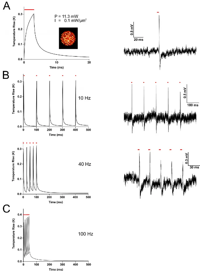
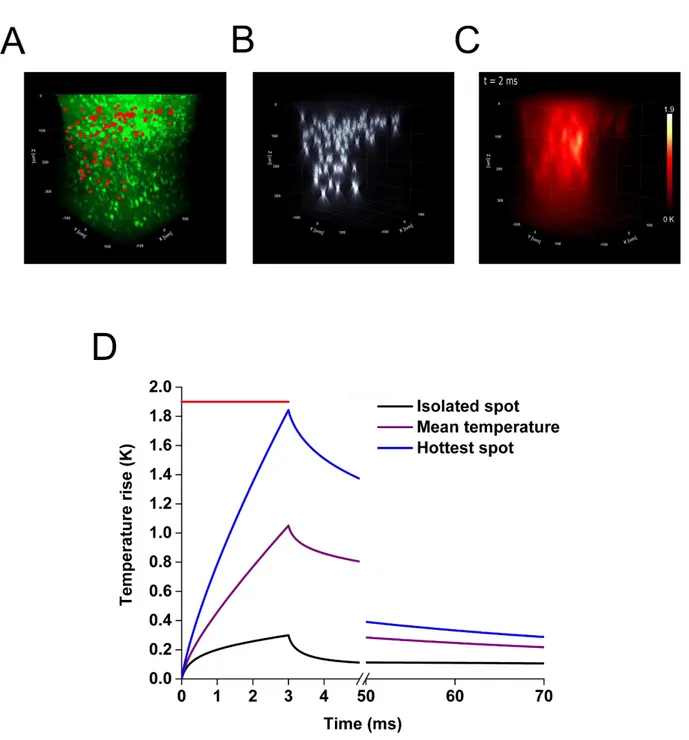
IX the local heat source. The medium is considered uniform and isotropic for heat diffusion. To validate this model, we experimentally measured the local temperature rise induced by laser heating in a phantom mimicking the optical and thermal properties of the brain. For this we used rare earths (Er, Yb) doped micro-crystals whose luminescence spectrum are temperature dependent. We were able to measure the temperature with a temporal resolution on the order of a few milliseconds and spatial accuracy of a few microns. We found these measurements in excellent agreement with the predictions of the model. We then used our model to calculate the temperature variations during a parallel activation of neurons in vivo expressing the opsin CoChR and found that the photostimulation of a cell causes an increase of the temperature of only a few tenths of a Kelvin. Then, we simulated the heating generated during the simultaneous photostimulation of 100 neurons, in a volume of 300×300×300 µm3 in order to
find the conditions (duration of illumination, distance between the spots) which would limit to 1 Kelvin in such a multiple cells photostimulation experiment. When comparing parallel and scanning illumination our simulation shows, as could be expected, an homogeneous increase of temperature within the cell in the case of the parallel stimulation, while scanning induces a significant heating at the center of the swept beam but a moderate elevation of temperature over the rest of the cell’s surface. We have also used our simulation to calculate the local heating in the experimental conditions of the most recent publications in the field of the in
vivo optogenetic, including the approaches of simultaneous scans at several depths. Finally,
our simulations can help gain insight on the optimization of the temporal sequence of photostimulation of several hundreds of target cells. Thanks to this work, the different teams working in the field of optogenetic will be able to optimize their experimental protocols, in order to ensure a controlled and safe environment for the living cells studied. To this end, a set of MATLAB functions has been made available to the scientific community that allows whoever is interested to simulate his own illumination conditions. Beyond the application to optogenetics, our simulation can be used for any photothermal study of biological tissue such as, for example, therapeutic hyperthermia or thermogenetics.
The second part of my PhD has been focused on the modeling of intracellular currents during photostimulations. Previous work has already been carried out in other laboratories, associating model of population dynamics (Markov processes in continuous time) and experimental data. Nevertheless, this work had never been associated with different techniques of illumination (parallel and scanning), in a two-photon regime, and applied to
X different opsins with various kinetic properties. We therefore implemented different models and adjusted their parameters (using conventional minimization algorithms) to fit experimental data recorded within our laboratory for three opsins, Chronos, CoChR and ReaChR. To this end, we have worked with Chinese Hamster Ovary (CHO) cells that we had modified to express the opsins of interest. We then photostimulated the cells during several seconds, with a wide range of illumination power, to obtain a representative sample of the full dynamics of opening and closing of these channels. A three-state (open - desensitized - close) model was sufficient to validate our approach but was insufficient to reproduce the full dynamics of the system, in particular to account for the bi- exponential-off transitions observed experimentally. Therefore, we implemented a four-state model, 2 “closed” and 2 “opened” states with different conductivity which is consistent with biophysical observation of the photocycle of opsins. We have shown the capacity of our model to simulate both the parallel and scanning illumination conditions.
XI
Résumé détaillé
Depuis maintenant une quinzaine d’années, une nouvelle approche, l’optogénétique, permet la manipulation optique de l’activité neuronale à l’aide de protéines photosensibles exogènes appelées opsines, naturellement présentes dans certaines algues photosensibles. Ce pan de la recherche a permis de grands progrès dans le domaine des neurosciences, ouvrant des perspectives incroyables dans le domaine de la recherche fondamentale et médicale. Aujourd’hui, la combinaison de l’optogénétique à l’illumination bi-photonique permet d’obtenir le contrôle de l’activité neuronale en profondeur à l’échelle de la cellule unique avec une précision temporelle proche de la milliseconde. Le futur de l’optogénétique est donc de pouvoir manipuler avec ce degré de précision des dizaines, centaines, voire milliers de cellules à la fois avec l’objectif de pouvoir, par exemple, éclaircir le rôle que l’activation spécifique d’un « microcircuit » de neurones a dans le contrôle du comportement.
L’avènement de ce domaine de la recherche fondamentale a été porté par et a entraîné avec lui de nombreux progrès technologiques, ainsi que le développement de différentes stratégies d’illumination comportant à la fois des avantages et des inconvénients. Parallèlement, une plus grande diversification des opsines disponibles, ainsi que le raffinement de leur méthode d’expression, ont offert encore plus de choix stratégiques pour les neuroscientifiques. Face à cette multiplication des outils, et aux ambitions sans cesse revues à la hausse pour ce domaine de recherche prometteur, il est devenu important d’être capable de déterminer les conditions optimales d’utilisation de ces techniques afin d’optimiser l’efficacité de ces approches.
Deux questions en particulier mêlant techniques d’illumination and protéines photo sensibles ont motivé le travail de thèse réalisé pendant trois ans et présenté dans ce manuscrit. Tout d’abord, la multiplication du nombre de cellules à activer simultanément pose immédiatement la question de la multiplication de la puissance lumineuse nécessaire envoyée dans l’échantillon, et rapidement celle des possibles dégâts photoinduits. Afin de mieux évaluer ces risques et proposer des pistes d’optimisation, nous avons développé et confirmé expérimentalement une simulation permettant de calculer l’amplitude et la dynamique des échauffements locaux quelle que soit la technique d’illumination utilisée aujourd’hui dans le domaine de l’optogénétique bi-photonique. Ce travail a notamment donné lieu à la publication d’un article de recherche au sein de la revue Cell Reports. Parallèlement, nous avons développé une autre simulation permettant de relier stratégie d’illumination, choix d’opsine et
XII dynamique de courant intracellulaire photo induit, en se basant sur l’exploitation de données expérimentales d’électrophysiologie effectuées au sein du laboratoire. En permettant de mieux comprendre les subtilités de chacune des techniques et protéines, nous avons ainsi pu entamer un travail d’optimisation des protocoles de photostimulation à utiliser expérimentalement. Afin de pouvoir évaluer l’échauffement local induit dans les deux grandes familles d’illumination couramment utilisées aujourd’hui (l’approche parallèle et de balayage) et d’en évaluer les risques, nous avons développé et validé un modèle optique et thermique décrivant à la fois la propagation de la lumière au sein d’un tissu diffusant comme le cerveau, et la diffusion de la chaleur photo-induite au sein de celui-ci. Nous avons validé expérimentalement ce modèle en utilisant des particules de verre dopés aux terres rares (Er, Yb), possédant un spectre de luminescence dépendant de la température. Ainsi, il a été possible de mesurer une température en excellent accord avec les prévisions du modèle, avec une résolution temporelle de l’ordre de quelques millisecondes et la précision spatiale de quelques micromètres. Ce modèle a ensuite été utilisé pour calculer les variations de température lors d’une activation parallèle de neurones in-vivo exprimant l’opsine CoChR. Il a ainsi été montré que l’activation d’une cellule provoque une augmentation de la température de seulement quelques dixièmes de Kelvin. Ensuite, nous avons simulé l’échauffement créé lors de la stimulation parallèle de 100 neurones, dans un volume de 300×300×300 µm3 et à
l’aide de notre modèle nous avons montré les conditions (durée d’illumination, distance entre les spots) qui permettent de minimiser les échauffements induits par l’optogénétique en dessous du Kelvin aussi dans le cas de l’activation de plusieurs cellules. Enfin, une comparaison des échauffements induits par les deux stratégies d’illumination (parallèle ou à balayage) a été effectuée, présentant les avantages et défauts inhérents à chacune. Comme attendu, une augmentation homogène de la température au sein de la cellule est obtenue dans le cas de la stimulation parallèle, tandis que le balayage induit un échauffement sensible au centre du faisceau balayé mais une élévation de température modérée dans les zones non illuminées de la cellule. Nous avons de plus utilisé notre simulation afin de calculer l’échauffement local des publications les plus récentes dans le domaine de l’optogénétique
in-vivo, notamment les approches de balayage simultané dans plusieurs plans. Enfin, nous avons
montré des pistes d’optimisation à exploiter pour les futures expériences impliquant plusieurs centaines de cibles photostimulées dans une fenêtre temporelle, en optimisant la distribution temporelle des illuminations. Grâce à ces travaux, les différentes équipes travaillant dans le
XIII domaine de l’optogénétique pourront optimiser leurs différents protocoles expérimentaux, afin de garantir un environnement contrôlé et sûr aux cellules vivantes étudiées. A cette fin, un jeu de fonctions MATLAB a été mis à la disposition de la communauté scientifique permettant à chacun de modéliser des conditions expérimentales spécifiques. Au-delà de cette application, ce modèle optique et thermique pourra permettre d’optimiser des stratégies d’échauffement (hyperthermie thérapeutique), d’étudier plus en détails la sensibilité des tissus cérébraux à la chaleur, ou d’améliorer les techniques de thermo génétique.
La deuxième grande thématique de ce doctorat portait sur la modélisation des courants intracellulaires lors de photostimulations. De premiers travaux avaient déjà été réalisés dans d’autres laboratoires, associant modèle de dynamique des populations (processus de Markov à temps continu) et données expérimentales. Néanmoins, ces travaux n’avaient jamais été associées à différentes techniques d’illumination (parallèle et balayage), dans un régime bi-photonique, et appliqués à différentes opsines aux propriétés cinétiques différentes. Nous avons donc entrepris de mettre en œuvre tout d’abord un modèle cinétique à trois états, puis à quatre états, à partir de données expérimentales d’électrophysiologie enregistrées au sein du laboratoire pour quatre opsines, Chronos, Chrimson, CoChR et ReaChR. Pour ce faire, nous avons travaillé avec des cellules chinoises d’ovaires de hamsters (cellules CHO) au sein desquelles nous avons induit l’expression des protéines d’intérêt. Nous les avons ensuite photostimulées durant plusieurs secondes, dans une grande gamme de puissance d’illumination, afin d’obtenir un échantillon représentatif de la dynamique complète d’ouverture et fermeture de ces canaux. Nous avons ensuite commencé par la mise en œuvre du modèle à trois états (ouvert – désensibilisé – fermé) en obtenant les paramètres de transition qui minimisent la différence entre le résultat du modèle et les données expérimentales. Pour ce faire, nous avons utilisé des algorithmes classiques d’ajustement de courbe (minimisation). Une fois ces paramètres obtenus, nous avons comparé le résultat du modèle et d’autres données expérimentales, dont le temps de photoactivation était plus faible (de l’ordre de la milliseconde). Ces travaux nous ont permis de vérifier l’efficacité du modèle à trois états pour un développement rapide, ainsi que sa capacité à prédire la dynamique de courants intracellulaire. Néanmoins, nous avons observé les limites de ce modèle à trois états, et notamment son incapacité à modéliser les transitions bi- (ou multi-) exponentielles constatées expérimentalement. Nous avons entreprise à la suite de mettre en œuvre un modèle à quatre états, basée sur de nouvelles données expérimentales, qui se montrera plus robuste
XIV aux durées d’illumination. Enfin, nous avons montré la capacité de notre modèle à simuler à la fois les conditions d’illumination parallèles et de balayage.
XV
List of abbreviations
1D/2D/3D 1P/2P/3P AP CGH CHO ChR2 Er/Yb FDTD FOV FWHM GM GPC MFP ODE ROI SLM TF : : : : : : : : : : : : : : : : :One/Two/Three Dimensions (Dimensional) One/Two/Three Photons
Action Potential
Computer-generated Holography Chinese Hamster Ovary
Channelrhodopsin-2 Erbium-Ytterbium
Finite Difference Time Domain Field Of View
Full Width Half Maximum Galvanometric Mirrors Generalized Phase Contrast Mean Free Path
Ordinary Differential Equations Region Of Interest
Spatial Light Modulator Temporal Focusing
XVI
List of contents
Remerciements V
Detailed Summary VIII
Résumé détaillé XI
List of abbreviations XV
List of contents XVI
Preamble 1
I. Neurophotonics 2
1) The neuron: a functional unit of the brain 2
2) Action potential and electrophysiology 4
3) Optogenetics for neurosciences 7
II. Single and two-photon optogenetics 10
1) Single-photon approach 10
2) Two-photon illumination: gains and challenges 11
3) Cellular targeting: from photons to action potentials 13
a) Scanning strategy 13
b) Parallel strategy 16
III. Photoinduced temperature rise: Thermal simulation and temperature measurements 20
1) Light absorption in the brain 20
a) Absorption and the “optical window” 20
b) Mono and multi photon absorption processes 21
c) Brain sensitivity to thermal variations 23
2) Publication: Temperature rise under two-photon optogenetics brain stimulation 25
3) Illumination conditions from the literature 64
a) Simultaneous spiral scanning of 10 neurons in vivo 64
XVII c) Simultaneous spiral scanning of 84 neurons in vivo at different depths 69
4) Optimization perspective for increased amount of targets 71
5) Conclusion 74
IV. Opsins dynamics and illumination strategies 76
1) Opsins kinetics diversity for optogenetics experiments 76
2) Experimental procedures 79
a) Patch-clamp recordings with 2P holographic photostimulations for three-state model 79 b) Patch-clamp recordings with 2P Gaussian photostimulations for four-state model and
scanning experiments 81
3) Photoinduced current kinetics model 84
a) Stochastic system model of the opsin 84
b) Four-state model 87
c) Extraction of the fitting parameters 89
4) Three-state model results 90
5) Four-state model results 93
6) Simulation of the photoinduced current for different illumination strategies 98
a) Description of the program 98
b) Preliminary tests of the scanning simulation with the four-state model 100
V. Conclusions and Perspectives 102
Appendix 103
Time constants calculation with the three-state model 103
Double exponential time constant calculation with the four-state model 105
References 107
Preamble
This manuscript titled 2P optogenetics: Simulation and modeling for optimized thermal
dissipation and current integration is organized in 5 chapters.
In the first chapter, after a brief introduction on the physiology of neurons and the principle of electrophysiology, I will introduce the principle of optogenetics and the main results achieved with this revolutionary approach in neuroscience.
In a second chapter I will review and describe the mostly used light delivering approaches for optogenetics brain stimulation.
In chapter III, after a short introduction on light absorption and a description of the main photodamage mechanisms, I will present the model that I have developed to simulate 3D light propagation and heat diffusion in optically scattering samples. The model with its experimental validation is described in detail in the publication A. Picot et al. enclosed in the same Chapter. In the publication, it is also demonstrated the use of the model to predict the temperature rise under the most commonly used illumination configuration for two-photon optogenetics.
In Chapter IV I will present the second part of my thesis, which has been focused on the modeling of the amplitude and temporal evolution of photocurrent under two-photon illumination for different opsins. I will compare the results achieved using a 3- and a 4-state model for a fast, a medium and a slow opsin. Lastly, I will show how knowing the parameters which describe the opsin photo-cycle is possible to predict the photocurrent traces under parallel or scanning illumination.
2
I.
Neurophotonics
The brain is one of the most fascinating parts of our body, studied for decades through a variety of methods. In order to better understand its mechanisms and organization, it has become increasingly important to consider the brain’s smaller components, such as the neurons and how they interact. In search of answers, a field of research known as “neurophotonics” has been developed, using light both to observe and interact with the brain.
1) The neuron: a functional unit of the brain
It was in 1655 that Robert Hooke became the first scientist to develop the concept of the cell as a biological unit. By composing a microscope out of an eyepiece, field lens and objective lens, Hooke analyzed plants (notably cork) and flies. He noted the plants’ and insects’ considerable organizational similarity, later naming the “cell” in 1667.
Thanks to these early observations and tools, it became possible to progress beyond simple anatomical recognition and thus to define precise cell types: the smaller components of the cell and its inner organization became attainable. Jan Evangelista Purkinje was the first to observe the cells of the nervous system in 1837, just before Theodor Schwann and Matthias Jakob Schleiden proposed in 1838 their cellular theory which defines the cell as the structural and functional unit of plants and animals. At that time, however, this particular theory was considered invalid for the nervous system.
Camillo Golgi and Santiago Ramon y Cajal went on to develop cell staining techniques which enabled finer observation of the structure of the nervous system’s cells (Figure 1) and helped Heinrich Wilhelm Waldeyer to propose his theory of the neuron as the functional component of the nervous system.
3
Figure 1: Representation of the nervous system in the cerebellum by S. R. y Cajal; from “Estructura de los centros nerviosos de las aves”, Madrid, 1905.
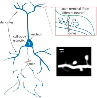
The key element of the nervous system, the neuron is an excitable cell which can receive, process, and transmit information in the form of chemical or electrical signals. These cells can be specialized in the form of sensory or motor neurons. They consist mainly of a soma cell body, with extensions called dendrites and an axon. The diameter of the soma is of the order of ten micrometers. Dendrites, meanwhile, have a diameter measuring between 0.2 and 5 micrometers and a length of between 10 and 1000 micrometers.
Figure 2: Structure of a neuron. Dendrites are small structures which receive contributions from neighboring neurons. Image: Alan Woodruff; De Roo et al., 2008.
4 Ever since antiquity, the electrical properties of catfish – depicted in bas-reliefs – have been known. They were even exploited as a therapeutic tool under the reign of Emperor Claude (41-54 AD). It is only from the seventeenth century that the electrical organs of fish were identified and dissected. Although it was important to understand that these electrical phenomena were occurring in certain specific tissues, it mattered a lot to understand that these events were not restricted to the organ itself, but at the heart of the activity of the nerves and muscles.
The excitable nature of neurons makes them particularly interesting cells. From simple observation to direct interaction, the investigation of neurons is essential for comprehension of how the nervous system operates. Towards the end of the eighteenth century, it was accepted that nerves were able to transmit an electrical signal to the muscles, thereby inducing muscle contraction. Nevertheless, in 1791, Luigi Galvani caused controversy by claiming in his Viribus electricitatis in motu musculari: Commentarius that electricity of animal origin also exists – that a frog’s biological tissue is able to generate sufficient electrical impulse through its nerves to induce muscle contraction. From this pioneering discovery stemmed the field of electrophysiology; techniques with which to study and measure currents within biological tissues – including those of the brain – would subsequently be refined.
2) Action potential and electrophysiology
Across every cell plasma membrane, ionic concentration gradients are constantly maintained by active transporters, a group of proteins ubiquitously expressed in the plasma membrane (Alberts, 2008). The existence of large K+ and Na+ gradients together with the selective permeability of the plasma membrane for different ions are at the basis of the transmembrane electric potential generation. Specific events can occur and induce a sharp and steep shift in the membrane potential reversing it from a negative value to a positive one. These shifts are called action potentials (AP) and neurons use the propagation of these variations of polarizations to trigger signals transmission, either electrical or chemical, to downstream directly connected neurons (L. Hodgkin & Huxley, 1952). AP is generated in a region close to
5 the neuronal soma and propagates along the axon up to the synapse, the connecting machinery interposed between two connected neurons.
Neurons have heterogeneous properties, and their different firing patterns are a manifestation of these dissimilarities. For instance, neocortical neurons significantly differ in their firing patterns, ranging from regular-spiking cells, which can adapt strongly during sustained stimuli, to fast-spiking cells which can endure sustained firing activity with little or no adaptation (Connors and Gutnick, 1990). In a neuronal circuit, neurons with different membrane properties and firing characteristics will produce different transformations of inputs into outputs and how neurons interact in the networks to process information. The ability to control and observe neuronal firing plays an important role in neuroscience research focused on deep understanding of brain functioning. Neuronal circuits manipulation can go from understanding the role of specific neuronal sub-populations (Boyden, 2011) to the monitoring of variations of natural neuron codes to control behavior (Dombeck et al., 2010). By activating a subpopulation of cells and monitoring the outcome elsewhere in the brain, it became possible to identify their role. Once their specificity is known, it became possible to design experiments where, by activating such a population via induction of APs, we could study specific behaviors (Grunstein et al., 2009; Houweling and Brecht, 2008; Huber et al., 2008; Li et al., 2009; Szobota et al., 2007). Additionally, as it is known that the brain is made of thousands of neurons massively connected, gaining the ability to observe action potentials will help to build connectivity maps of neurons and brain regions (Brill et al., 2016; Kohara et al., 2014; Song et al., 2005).
The first challenge of studying electrical events related to cellular physiology and ionic channels dynamics at high resolution has encouraged the development of a family of techniques (electrophysiology), which also enable to both induce and register action potentials. To measure and perturbate the state of polarization of one precise cell, it was necessary to develop techniques that would make possible to probe the electrical activity of the neurons of interest. In the last century, techniques based on the use of micro electrodes have been developed which allow intracellular or extracellular recordings with small-time resolution and robust sensitivity. Indeed, by recording directly using metal, silicon or glass electrodes, it is possible to work with a very high signal-to-noise ratio, without using reporters
6 of the neuronal electrical activity. The development of voltage registrations allowed to have a temporal precision below the millisecond, in the order of magnitude of neuronal signaling. Since their optimization in the early 1950s, extracellular recordings methods provided many advances in the understanding of the central nervous system (Humphrey and Schmidt, 1977). Extracellular microelectrodes helped to map the field potentials of single discharging neurons, answering fundamental questions about dendrites excitability (Frank and Fuortes, 1955) or discharge patterns during behavior in awake moving animals (Mountcastle et al., 1975). Moreover, extracellular recordings allowed D. Hubel and T. Wiesel to record the activity of single neurons in the primary visual cortex of anesthetized cats (Hubel and Wiesel, 1962), and to show how single neurons in this area respond to specific visual stimulus, granting them with the Nobel Prize in 1981.
While extracellular recordings led to important discoveries, the intracellular recording methods became unavoidable thanks to their very high signal-to-noise ratio. This approach, originally developed by Kenneth Cole and George Marmont in 1947 (at a time when microelectrodes were not yet available), allowed later Alan Lloyd Hodgkin and Andrew Fielding Huxley to deepen our understanding of action potentials and several ion channels (Hodgkin and Katz, 1949). Although the idea of inserting a micro-electrode into a cell was innovative, it soon came up against a significant biological constraint: the membrane of a cell is not adapted to be pierced by an external element for long periods of time. It was then necessary to develop an improvement of the techniques of clamp, which also improved the signal to noise ratio: the ”patch-clamp”, evolution of the simple clamp, took over in the late 1970s, thanks to the advances of Erwin Neher and Bert Sakmann, who perfected it reaching the capability to measure the activity of a single transmembrane molecule (Neher and Sakmann, 1976).
7 Here, a glass pipette with tip of a few micrometers wide is brought into contact with the cell membrane. It can adhere very strongly to the cell, forming a gigaseal characterized by the very high resistance (GΩ), electrically isolating the membrane portion from the rest of the cell (Ogden and Stanfield, 1981). It is filled with an ionic solution adapted to the type of experiment and the type of cell. Once this step is performed, several variants of the “patch-clamp” technique are available, depending on the research objective, ranging from the recording of single-channel currents (in “cell-attached” or “excised patch” configuration), or global currents passing through ionic channels of the cell (“whole cell” or “perforated patch”). Since then, the “patch-clamp” technique became the preferred method to both investigate the role of single channels and to monitor electrical activity of any cell type, either in vitro or in
vivo (Verkhratsky and Parpura, 2014), including induction and registration of APs.
Despite the strength of this technology, “patch-clamp" has also limitations. First, because of its invasiveness, it is very difficult to probe at the same time more than a few cells. However, brain is made of millions of neurons subdivided in multiple regions and areas, linked to complex chain of events that lead to specific behaviors and outcomes, and it became obvious that the perturbation and observation of tens, hundreds, thousands of neurons during the same experiments would help to answer more questions. Secondly, once again because of the invasiveness, patching a cell is not a reversible process and imply that it is not an adapted approach for long term studies of the same biological material. Finally, the selective inhibition of neurons remains largely inaccessible to electrophysiological approaches (Scanziani and Häusser, 2009), closing the door to the manipulation of a biological mechanism that is common in the brain. All these restrictions have challenged the scientific community to develop complementary techniques that could overcome these limitations and allow more flexible approaches for activation and registration of neuronal activity.
3) Optogenetics for neurosciences
Forty years ago, in a totally different framework, bacteriorhodopsin was discovered as a light-activated ion pump that could be found in microbial organisms (Oesterhelt and Stoeckenius, 1971). Decades later, after further researches both on bacteriorhodopsin mechanisms and variants that could be found elsewhere, membrane-bound similar proteins that allow ions flux across membrane were discovered, known as halorhodopsins (Matsuno-Yagi and Mukohata,
8 1977) and channelrhodopsins (Nagel et al., 2002, 2003). Two variations of the latest were selected for their ease of expression, conductance and photo sensitivity: Channelrhodopsin-1 and Channelrhodopsin-2 (ChR2).
Two years after the introduction of ChR2, it was demonstrated that the induction of the expression of the gene coding for ChR2 allowed researchers to elicit reliable timely controlled action potentials for the first time in mammalian neurons (Boyden et al., 2005). Since then, interest rapidly grew for proteins such as ChR2 that we will call opsins in this manuscript.
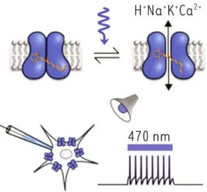
Figure 4: Activity diagram of Channelrhodopsin-2.(Tricoire 2015). Channelrhodopsin-2, a transmembrane protein, is a ion channel that opens when illuminated. Its expression inside neurons allow to induce non-invasively trains of action potentials.
These transmembrane proteins, once illuminated at a suitable wavelength, open a channel through which an ion flux passes, participating in the depolarization of the membrane in which they are expressed. If enough opsins are expressed and activated within the cell, a depolarization threshold is reached, and action potentials can be generated (as shown in the bottom right of Figure 4).
9 Even if this technique defined the optogenetics field and is now an approach of reference for intracellular currents studies, it took decades for researchers to match together the two ideas of combining light and microbial ions channels. Beyond the necessary technical challenges that were mandatory to solve, a few presumptions slowed the access to this technology. Photocurrent induced by such proteins were expected to be both too weak and not fast enough to manipulate neurons with efficiency. Additionally, because of the origin of these microbial membrane proteins, the expression of such genes was expected to be low and inducing fragility to the neuron membrane (Yizhar et al., 2011).
Once these considerations were overcome, this light-based approach that showed promises to solve patch-clamp techniques limits gave birth to a new neurosciences research domain that was named optogenetics. For some tasks, it allowed researchers to go beyond what electrodes based experiments could offer, such as the independent stimulation of multiple populations of neurons (Zhang et al., 2008), the bistable activation of neurons (Berndt et al., 2009), or the stimulation of defined second-messenger pathways (Airan et al., 2009; Kim et al., 2005). Even further, it was used to map functional connectivity (Petreanu et al., 2009), influence the neuronal dynamic circuits (Boyden et al., 2005; Cardin et al., 2009; Li et al., 2005; Zhang et al., 2007) and, finally, to control behavior (Grunstein et al., 2009; Huber et al., 2008; Szobota et al., 2007).
10
II.
Single and two-photon optogenetics
1) Single-photon approach
Wide-field single-photon (1P) approach was the first to be employed for optogenetics stimulation (Adamantidis et al., 2007; Anikeeva et al., 2012; Aravanis et al., 2007; Boyden et al., 2005; Gradinaru et al., 2007; Huber et al., 2008; Nagel et al., 2005; Zhang et al., 2007), and remains today the approach of reference for neural circuit dissection (Makinson et al., 2017; Weible et al., 2017). This technique has allowed researchers to study correlation and causal interactions in subpopulations of neurons both in vitro (Joshi et al., 2016; Morgenstern et al., 2016; Petreanu et al., 2007, 2009; Tovote et al., 2016) and in vivo (Adesnik et al., 2012; Atallah et al., 2012; Lee et al., 2012; Tovote et al., 2016).
Figure 5: Several approaches for photoactivation. With anatomic targeting, groups of neurons are illuminated with an optic fiber after injection of viral vectors in the area of interest. Under genetic targeting, only specific cell types will express the opsins thanks to regulating sequences. Adapted from (Tricoire, 2015).
With this technique, population specificity is obtained through genetic and anatomic targeting, (Figure 5), implying that precision and temporal resolution are only constrained by the channels’ and cells’ properties.
The development of strategies for opsin expression confinement in specific cell types (Beltramo et al., 2013; Cardin et al., 2009; Kuhlman and Huang, 2008), or the addition of
11 optic tools such as optical fibers (Aravanis et al., 2007; Penzo et al., 2015; Wu et al., 2014) has helped to reach deeper regions of the brain. Improvements made to 1P microscopes (such as the use of multipoint-emitting optical fibers or micro-objective-coupled fiber bundles) has even enabled the restriction of illumination to specific brain layers (Pisanello et al., 2014) or subcellular structures (Guo et al., 2009; Petreanu et al., 2009; Szabo et al., 2014; Wyart et al., 2009).
Even though 1P illumination is the technique most commonly used today, its limitations in term of penetration depth and spatial resolution have motivated the development of other approaches that could bring higher spatial precision. Indeed, when illuminating a large volume with 1P widefield technique, we cannot select with single-cell precision the neuron that we want to photoactivate. This implies that we cannot, for example, in a specific brain region, photoactivate first a defined subgroup of neurons, observe the outcome, and activate another group nearby and repeat the observation. Furthermore, all opsin-expressing neurons are stimulated at the same time, which does not allow to reproduce the spatiotemporal distribution of naturally occurring brain microcircuits activity. Also, widefield illumination does not enable performing connectivity experiment since in that case one needs to interrogate each neuron individually in its environmental context. All these reasons have motivated the development of illumination approaches giving single-cell resolution in depth. This has inspired at first to replace single-photon excitation by two-photon excitation processes.
2) Two-photon illumination: gains and
challenges
In 1P microscopy, which is based on a linear photon absorption process, an orbital electron of the protein to activate (fluorophore, opsin, …) will absorb a photon emitted by the light source. This reaction promotes the electron to an excited state from which it will relax, thereby allowing the emission of a photon of fluorescence, for example. This linear process will imply large amounts of out-of-focus neuron activation, or fluorescence. As an answer, 2P microscopy has shown itself to be highly efficient (Denk et al., 1990; Helmchen and Denk,
12 2005) and, combined with optogenetics techniques, offers a reasonable solution to the resolution issue mentioned above. With this method, two photons are required to bring the electron to an excited state. By using a high-power pulsed laser with a short pulse width (hundreds of femtoseconds), the high density of photons in the excitation area leads to the strong probability that a molecule will absorb two photons quasi-simultaneously. This condition directly implies that we have a quadratic dependence between the fluorescence (or any other non-linear process) and the light intensity, lowering substantially the out-of-focus fluorescence or neuron activation (Figure 6).
Figure 6: Localization of fluorescence signal with 1P (a panel) and 2P (b panel) excitation. On the left, fluorescein activation with a 488 nm laser source. On the right, same molecule excited with a 960 nm light source (Zipfel et al., 2003).
Importantly, the two photons must have roughly half of the photon’s amount of energy for the 1P excitation process. As a consequence, their wavelength must be twice as big. This has shifted the use of light source from the visible 1P absorption band to the near-infrared.
This aspect has helped 2P microscopy to assess its strength, opening deeper regions of brain for study. Indeed, near-infrared photons are less scattered and absorbed by the brain, as will be discussed later on. For the same reasons, three-photon microscopy (3P) has been developed with the same objectives, allowing even further access in the brain, or to go through the skull (Horton et al., 2013).
13 Therefore, 2P approaches have allowed to improve the axial resolution and penetration depth of the photoactivation techniques. Yet, being able to target with single-cell precision is not enough to guaranty the capability of the optical method to reliably activate neurons and elicit APs. Indeed, as opsins are proteins with a small single-channel conductance (40-80 fS for ChR2 (Feldbauer et al., 2009)) and a density of expression which implies a low amount of channels in the femtoliter two-photon focal volume (with a standard objective of 0.7 – 0.9 NA and a laser source in the near-infrared 800 – 1200 nm, the full width half maximum (FWHM) of this spot will be ~1 µm) (Nagel et al., 1995), the simple addition of 2P techniques is not enough to induce strong enough depolarization for APs. This challenge has spurred the development of more sophisticated 2P illumination strategies in order to be able to photoactivate a large number of channels. They can be split in two categories: the scanning and the parallel techniques.
3) Cellular targeting: from photons to
action potentials
a)
Scanning strategy
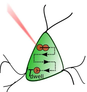
One way of increasing the number of activated channels is to scan the soma of the cell of interest with a spot. This strategy has been successfully demonstrated in cell cultures first (Rickgauer and Tank, 2009), then in brain slices (Andrasfalvy et al., 2010) and in vivo (Prakash et al., 2012). While this approach is relatively easy to be implemented, it can be limited in temporal precision as it requires to scan the laser source over the cell body or even multiple targets. For N targets, the temporal resolution T of a cycle of sequential photostimulations is given by:
𝑇 = 𝑇𝑑𝑤𝑒𝑙𝑙× 𝑁 + 𝑇𝑚𝑜𝑣𝑒× (𝑁 − 1) Eq. 1
with Tdwell standing for the time spent on a cell to photoactivate it, and Tmove the time to move
the laser beam from one cell to another. With this approach, Rickgauer and Tank in 2009 were able to generate APs with 30 ms temporal resolution (Rickgauer and Tank, 2009).
14
Figure 7: Scanning illumination of one neuron. A raster scanning pattern is showed. Adapted from (Ronzitti et al., 2017a). The raster scanning pattern is adapted to the cell morphology. The amount of lines, the direction, the scanning speed are various parameters that can be tuned in order to maximize the photoactivation efficiency.
The necessity to define a scanning protocol that will both target a significant number of channels and illuminate them within a specific time window can add constraints to the scanning approach. Indeed, a compromise must be made between the dwell time that will guarantee the opening of the channels, and the scan speed to assure that a maximum of channels will be open simultaneously to reach the action potential. Precisely as explained in Ref. (Rickgauer and Tank, 2009) :
𝐼(𝑇) 𝐼𝑚𝑎𝑥 = 𝜏 𝑇(1 − 𝑒 𝑇 𝜏) Eq. 2
where 𝐼(𝑇) is the amplitude of the photocurrent during a scan time T, 𝐼𝑚𝑎𝑥 the whole-cell
stimulated current amplitude, 𝜏 the opsin photocurrent decay constant. A scanning pattern that would be n times longer than 𝜏 would imply that the peak photocurrent would reach (1 − 𝑒−𝑛) 𝑛⁄ of the whole-cell photocurrent. Therefore, care must be given to the choice of
the couple opsin/illumination pattern to ensure that enough photocurrent could be elicited to reliably guarantee an AP. The development of slower opsins with nanoampere-scale currents such as C1V1 enabled to dramatically increase the efficiency of this approach (Packer et al., 2012; Prakash et al., 2012), reaching temporal resolution between 5 and 70 ms. However,
15 using slow opsins with scanning approaches add constraints, such as imposing limits to the achievable temporal precision of photo-evoked spikes, with significant in vivo jitter estimated at 5.6 ± 0.8 ms (Packer et al., 2015).
One way to improve temporal resolution is to underfill the back aperture of the objective, which increases both the lateral and axial spot size. Increasing the lateral size reduces the number of scanning positions, thus minimizing or even getting to zero the term Tmove,
however, it also decreases the technique’s precision, because of the increase in the axial size, as well. Temporal focusing approaches (TF) were then associated to the light activation techniques in order to compensate the deterioration of axial resolution. In such technique the pulse is temporally scrambled above and below the focal plane, which remains the only region irradiated at peak powers photoactivation (Oron et al., 2005). This allowed to fast photostimulate multiple cells in vitro and in vivo (Andrasfalvy et al., 2010; Rickgauer et al., 2014), reducing Equation 1 to T = (𝑇𝑑𝑤𝑒𝑙𝑙+ 𝑇𝑚𝑜𝑣𝑒) × 𝑁.
Recently, multiple cells photostimulation through scanning has been extended to activate tens (Packer et al., 2015) or nearly one hundred of cells at the same time at different depths (Yang et al., 2018). To do so, the optic system combines galvanometric mirrors (GM) which will move in a defined way to allow the laser source to perform this scanning pattern, and spatial light modulators (SLM) which will, by modulating the phase of the incoming light source, allow the simultaneous targeting of several cells.
Figure 8: Scanning illumination of several neurons simultaneously. A spiral pattern is here applied, with pre-defined rotation speed, amount of revolutions and size of pattern. Adapted from (Ronzitti et al., 2017a).
16 With the combination of scanning and multiplexing, the equation for the temporal resolution of photostimulation of several cells at the same time reduces to:
𝑇 =𝑑𝑠 𝑆𝑠
Eq. 3
where ds indicate the whole distance of scanning over a single cell, and Ss the scanning speed
of the system.
Lastly, another limitation for the scanning approach is the axial resolution. Indeed, requiring to work with light intensities close to saturation levels of opsin mechanisms at the focal plane, led to out-of-focus excitation above and below the cell of interest that induces significant photocurrent in the neighboring cells (Rickgauer and Tank, 2009; Ronzitti et al., 2017a).
b)
Parallel strategy
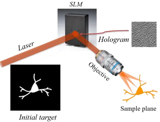
Alternatively to scanning, the number of excited channels could increase by using parallel approaches where the light propagation is modified to cover the entire target. An efficient way to reach such precise confinement for light propagation is to use computer-generated holography (CGH), as proposed in 2008 (Lutz et al., 2008; Papagiakoumou et al., 2008). Originally developed for generating multiple-trap optical tweezers (Curtis et al., 2002), the experimental scheme for CGH (Figure 9) consists in computing with a Fourier transform-based iterative algorithm (Gerchberg and Saxton, 1972) the interference pattern or phase- hologram that back-propagating light from a defined target (input image) will form with a reference beam, on a defined “diffractive” plane. The computer-generated phase-hologram is converted into a grey-scale image and then addressed to a SLM, placed at the diffractive plane. In this way, each pixel of the phase-hologram controls, proportionally to the analogous grey-scale-level, the voltage applied across the corresponding pixel of the liquid-crystal matrix, such as the refractive index and thus the phase of each pixel can be precisely modulated. As a result, the calculated phase-hologram is converted into a pixelated refractive screen and illumination of the screen with the laser beam (or reference beam) will generate at the objective focal plane a light pattern reproducing the desired template. This template can be
17 any kind of light distribution in two or three dimensions, ranging from diffraction-limited spots or spots of bigger surface to arbitrary extended light patterns (Papagiakoumou et al., 2018).
Figure 9: The iterative algorithm will process a theoretical light distribution image, often based on a fluorescence image. After computing, the interference pattern, or hologram, is obtained and addressed to the SLM. At the focal plane of the system, the initial illumination distribution is reproduced. Adapted from (Papagiakoumou et al., 2017).
Different variations of this approach have been developed over the years, achieving actuation of opsin-expressing neurons in vitro and in vivo (Bègue et al., 2013; Packer et al., 2012; Ronzitti et al., 2017b; Szabo et al., 2014), or adapted with variations such as the generalized phase contrast technique (GPC) (Papagiakoumou et al., 2010, 2013).
With parallel illumination, the temporal resolution is only limited by the illumination time and it is possible to use both fast and slow opsins. Therefore, this technique combined with fast opsins allowed to generate action potentials with millisecond temporal resolution (Bègue et al., 2013; Papagiakoumou et al., 2010), and submillisecond temporal jitter, the mean deviation of latency between the beginning of photostimulation and AP (Chaigneau et al., 2016; Ronzitti et al., 2017b). When illuminating a sample with a large excitation area, the optical axial resolution can be degraded. As an answer, CGH technique was combined with TF, allowing to preserve the axial resolution (5-10 µm) and the lateral shape of the spot after
18 hundreds of micrometers of light propagation in scattering tissues (Bègue et al., 2013; Hernandez et al., 2016; Papagiakoumou et al., 2013).
Figure 10: Parallel illumination of several neurons simultaneously. An illumination pattern is here applied, based on the shape of neurons of interest. The cells are photoactivated at the same time, recruiting as many opsins as possible. Adapted from (Ronzitti et al., 2017a).
The ambition of studying connected networks of neurons have motivated the development of 3D light generation approaches, allowing to generate complex illumination patterns (Anselmi et al., 2011; dal Maschio et al., 2017; Hernandez et al., 2016; Packer et al., 2012). Further, this ambition has spurred the development of approaches allowing to simultaneously shine multiple spatiotemporally focused spots (Accanto et al., 2017; Hernandez et al., 2016; Pegard et al., 2017; Sun et al., 2018) (Figure 10). The latest opsins development have led to somatic versions of channels that allowed to reach the single-cell precision, by restricting the expression of the channels to the soma of the neuron (Shemesh et al., 2017). Indeed, the spatial resolution of 2P photostimulation systems can be degraded due to the undesired photostimulation of neurites surrounding the cell of interest.
All of these approaches, combined with the use of high-energy amplified lasers (Accanto et al., 2017; Chaigneau et al., 2016; Ronzitti et al., 2017b; Yang et al., 2018) reaching more than 10 W at laser output and eventually highly sensitive opsins make it now possible to
19 simultaneously target hundreds of cells within mm3-size illumination volumes. But the increase of targets rises, at the same time, the question of photodamage and how to minimize it. In the next chapter, we will introduce the different kind of photodamages that we can expect from 2P photostimulations, and the thermal model that we have built in order to evaluate and minimize thermal photodamage.
20
III.
Photoinduced temperature rise:
Thermal simulation and temperature
measurements
1) Light absorption in the brain
a) Absorption and the “optical window”
In the brain, several components are responsible for light absorption. Water will be the most absorbing element, followed by chromophores such as blood components (hemoglobin, melanin, …) and proteins such as fluorophores, the latest remaining negligible (see below).
Figure 11: Absorbance of light at various wavelengths for water (solid line), oxygenated (thin dashed line) or deoxygenated hemoglobin (dots) and melanin (bold dashed line). Adapted from (Hamblin and Demidova, 2006).
21 The absorbance of each of these elements has different wavelength dependence, although as shown in Figure 11, it is possible to define an “optical window” in the range of wavelength (600 – 1200 nm) where they have a common minimum. Therefore, using excitation wavelengths in this range will allow focusing light at greater depth.
b) Mono and multi photon absorption processes
A wide range of phenomena can occur during light absorption, depending on the illumination source and its characteristics (wavelength, power, pulse duration). Given a certain wavelength, two parameters are important to define the cascade of events following the photon absorption, the irradiance and the illumination time. In particular, we focus here on the processes, which occur in the near-infrared regime, i.e. at wavelength commonly used for 2P photostimulation.
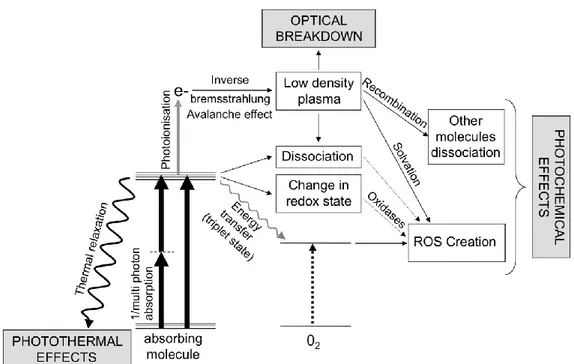
Figure 12: Cascade of events after light absorption in the brain, with near infrared light sources, without energy considerations. Adapted from (Débarre et al., 2014).
During the first step of a photon absorption, the excited molecule goes to an excited state, this is followed by a cascade of events, Figure 12. For low irradiance (below 106 W/cm2 (Boulnois, 1986)), and long illuminations (on the millisecond scale), the molecule can go back to the resting state by transferring its energy in a non-radiative way, thereby inducing local heating. Other very specific chemical reactions can be catalyzed by a long exposure to light with low irradiance. On the other hand, if the irradiance is above the previous threshold,
22 even during a very short period of time (femtoseconds to nanoseconds), there is a high probability of an inverse Bremsstrahlung avalanche effect (Vogel et al., 2005). Here, free electrons are produced in a multi-photon absorption process and they can also absorb photons when they enter into collision with other molecules, generating even more free electrons. If the light intensity is too high, this reaction gets out of control. This is the so-called avalanche effect, inducing the apparition of a plasma of electrons in the sample. For low density plasma, i. e. below the optical breakdown threshold of 1021 electrons/cm3 (Débarre et al., 2014), this will produce a family of photochemical damages inducing molecules dissociation, or the creation of reactive oxygen species (Figure 12). For plasma density above the optical breakdown threshold this will induce mechanical damages where the cells will undergo supersonic shock wave by the formation of bubbles followed by their explosion. These effects will break the tissue structures and induce cell death.
In typical optogenetics experiments using 2P and parallel illumination, a 3 ms long illumination pulse with a 10 mW mean power over a 100 µm2 cell surface is enough to elicit
an AP. This corresponds to an irradiance of 104 W/cm2 and fluence of 30 J/cm2. These conditions are clearly (see Figure 13) in the range of photothermal damage. However, since this average power illumination is obtained from a train of fs pulses, we must also consider the possibility that an individual fs pulse (whose energy is much higher than the average energy) could induce nonlinear damage. For the case of 250 fs pulses at a 500 kHz rate, each individual pulse delivers an irradiance of 8 1010 W/cm2 corresponding to a fluence 20 mJ/cm2. This is close to the threshold of nonlinear photodamage (see Figure 13) but we never observed such effects. Nonlinear photodamage have been observed at peak fluences around 0.1 J/cm2 for Chinese hamster ovarian cells (König et al., 1999), 0.5-2 J/cm2 in water (Linz et al., 2016; Noack and Vogel, 1999; Vogel et al., 2005) and 1.5-2.2 J/cm2 for porcine corneal stroma (Olivié et al., 2008).
23
Figure 13: Linear and nonlinear photodamages in biological tissues depending of the irradiance and exposure time of the light source. Several regimes can be discerned based on a level slopes that correspond to J/cm2 thresholds. Several regimes
can be discerned based on a level slopes that correspond to J/cm2 thresholds. Here we consider the peak irradiance as the
irradiance during a single laser pulse (at the femtosecond scale), as opposed to the mean irradiance calculated as the average irradiance during a repetition (at the millisecond scale) of single laser pulses. Adapted from (Boulnois, 1986).
For the scanning illumination techniques, peak irradiance can reach 1012 W/cm2. Therefore, the nonlinear photodamages should be the dominant events. In conventional multi-photon scanning imaging, due to the short dwell time and small illumination volume, heating through linear absorption can be considered a negligible source of photodamage (Débarre et al., 2014; Koester et al., 1999; Linz et al., 2016). However heating can become an important source of photodamage for repetitive scanning of large areas (Hopt and Neher, 2001; Podgorski and Ranganathan, 2016).
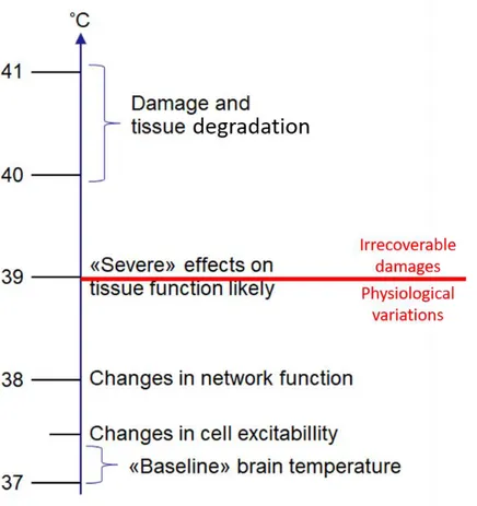
c) Brain sensitivity to thermal variations
We can classify the effects of temperature increase in two categories: physiological variations and irrecoverable damages. Relatively small temperature changes (below 2 K) can, for instance, induce modulations of the shape of APs (Hodgkin and Katz, 1949), the firing rate of neurons (Reig et al., 2010; Stujenske et al., 2015), and the channel conductance (Plaksin et al., 2018; Shibasaki et al., 2007; Wells et al., 2007) or fluctuation of synaptic responses (Andersen and Moser, 1995; Thompson et al., 1985). Furthermore, temperature variations