HAL Id: tel-02275815
https://tel.archives-ouvertes.fr/tel-02275815
Submitted on 2 Sep 2019HAL is a multi-disciplinary open access
archive for the deposit and dissemination of sci-entific research documents, whether they are pub-lished or not. The documents may come from teaching and research institutions in France or abroad, or from public or private research centers.
L’archive ouverte pluridisciplinaire HAL, est destinée au dépôt et à la diffusion de documents scientifiques de niveau recherche, publiés ou non, émanant des établissements d’enseignement et de recherche français ou étrangers, des laboratoires publics ou privés.
New therapeutic approaches for achondroplasia
Davide Selom Komi Komla-Ebri
To cite this version:
Davide Selom Komi Komla-Ebri. New therapeutic approaches for achondroplasia. Genetics. Univer-sité Sorbonne Paris Cité, 2016. English. �NNT : 2016USPCB040�. �tel-02275815�
Thèse de l’Université Paris Descartes
Ecole Doctorale BioSPC
Spécialité : GENETIQUE
Auteur :
Davide Selom Komi KOMLA-EBRI
Titre :
Nouvelles approches thérapeutiques pour l’achondroplasie
New therapeutic approaches for achondroplasia
Soutenue le 04 juillet 2016
Jury
Pr Jeanne AMIEL Président
Dr Laurence LEGEAI-MALLET Directeur de thèse
Dr Laurent BECK Rapporteur
Dr Frédéric MALLEIN-GERIN Rapporteur
Dr Patricia BUSCA Examinateur
Remerciements
Je remercie tout d’abord le jury de thèse, Jeanne Amiel, Laurent Beck, Patricia Busca, Hervé Kempf et Frédéric MalleinGerin, pour avoir accepté de juger ce travail et de m’avoir fait l’honneur de présider à cet événement pour moi si important. J’adresse tous mes meilleurs remerciements à Laurence LegeaiMallet, ma directrice de thèse. Elle m’a accueilli au sein de son équipe il y a plus que 5 ans quand j’étais à la recherche d’un stage M2. Cela me rappelle combien le temps passe vite. Elle m’a aidé, poussé, fait confiance, reprit, félicité, suivi tout au long de mon parcours. A bien voir elle a bien mérité le titre de "maman" du labo ! Je remercie Pr Alain Fischer et le comité de direction de l’Institut Imagine pour m’avoir accordé une bourse de 4 mois pour ma 4ème année de thèse. Je remercie également le comité de la Fondation pour la Recherche Médicale pour m’avoir assigné un financement de 6 mois de 4ème année de thèse. Je remercie les DURs pour avoir fait partie de mon aventure et de l’avoir rendue si agréable ! Catherine pour la bonne humeur, le soutien, pour m’avoir appris la culture cellulaires, les western blots, et pour la blague de la lamelle de la malassez cassée (je l’oublierai jamais !). Nabil pour m’avoir appris tout ce que je connais de la souris, pour ses conseils, les perles de sagesse et sa passion cachée pour la danse et la fête. Emilie pour m’avoir appris plein de techniques pendant mon M2 pour ensuite se les faire réexpliquer à son retour comme postdoc ! Je te souhaite plein de succès avec les petits poissons ! Ludovic, mon roi des cils et de Nantes. PREGO ! Maxence pour son humour parfois un peu décalé, la cinéphilie et Les boloss des belles lettres. LOL ! Valentin pour ton professionnalisme et tes blagues. Allez viens ! Léa pour ne pas avoir pris soin de mon alcoolisme de mèche avec Marko (que je remercie en passant). Martin pour ses avis, le travail sur les mandibules et pour m’avoir conseillé Searching for Sugarman. Federico et son travail sur le crâne des souris et pour m’avoir fait connaître les synchondroses. Cindy toujours là loin en Calédonie. Je te rassure, je n’ai toujours pas goûté la BSA ! Les espagnols Mariluz et Salva, j’espère qu’on se rencontrera sous le soleil de l’Espagne. Je remercie les VCD Valérie, Carine, Céline, Mathilde, Quentin, Norine, Laure (pour m’avoir nourri !) Merci à tous les « vieux » qu’on fait partie de l’ancienne U781 d’Arnold Munnich et aux « nouveaux » rencontrés à l’Institut Imagine. Merci à ma famille qui m’a toujours soutenu. Sachiez que même si je ne donne jamais de mes nouvelles vous êtes toujours dans mes pensées. Merci Aliénor pour m’avoir supporté pendant la rédaction de cette thèse, pour vivre avec moi et pour m’avoir appris que « quand y en a plus y en a encore ! ». Je ne t’ai pas laissée tomber comme une vieille chaussette !Merci Milène, Léa (les supercolocs), Miccols, (Bella !)Valoz, Fox, (Hola !) Norex, (Ohi zi) Cami "Pede" rsen, Scianna, Quentin, Matty, David (Mamma mia !), Andrea Angiuli (à lire avec la voix de la boite vocale), Marco, Alice, Dorian Gamba, Mejdou, Flà, Giselita, Sarah, Juliette, Danilo, Victor, Filo, Bozzi, May, Will, bref les “Tous chez Nora”. Je remercie les amis connus à Paris, en Italie, les amis de toujours et les amitiés plus récentes.
Table
of Contents
Abbreviations ... 3
Introduction... 5
1. FGFs and FGFRs ... 5
1.1 Fibroblast Growth Factors (FGFs) ... 5
1.2 Fibroblast Growth Factor Receptors (FGFRs) ... 8
2. FGFR-related diseases ...12
2.1 FGFR1 and FGFR2-related diseases ...12
2.2 FGFR3-related diseases ...13 2.2.1 Osteochondral disorders... 13 2.2.1.1 Chondrodysplasias ... 13 2.2.1.2 Craniosynostoses ... 17 2.2.1.3 Other FGFR3-disorders ... 18 2.2.2 Cancer ... 20 2.2.3 Skin lesions ... 21
2.2.4 Consequences of different mutations on receptor activity ... 21
3. Skeletogenesis ...23
3.1 Early embryo development ...23
3.2 Chondrogenesis ...25
3.3 Endochondral ossification ...26
3.4 Intramembranous ossification ...27
3.5 FGFR signalling and early skeletal development ...30
3.6 FGFR signalling in growth plate ...34
3.7 Bone modeling ...38
4. Therapeutic strategies ...41
4.1 FGFR3 signalling targeting strategies ...42
4.1.1 FGFR3 tyrosine kinase inhibitors (TKIs) (1) ... 42
4.1.2 FGFR3 antibodies (2) ... 43
4.1.3 CNP antagonism of FGFR3 signalling (3) ... 44
4.1.4 RNA interference (4) ... 46
4.1.5 Hsp90 inhibition and FGFR3 cleavage (5) ... 47
4.1.6 Modulating FGFR3 expression cleavage and/or nuclear function (6) ... 47
4.1.7 Inhibition of FGF ligands (7) ... 48
4.1.8 SNAIL inhibition (8) ... 48
4.2 FGFR3-unrelated preclinical studies with effects on bone growth ...49
Objectives ... 51
Results ... 53
Article 1 - Tyrosine kinase inhibitor NVP-BGJ398 functionally improves FGFR3-related dwarfism in mouse model ...53
Article 2 - Meckel’s and condylar cartilages anomalies in achondroplasia result in defective development and growth of the mandible ...73
Preliminary data - Best understanding of structural and functional impact of FGFR3 mutations at the same position (K650N, K650M, K650E) leading to both mild and lethal dwarfism ... 104
Conclusions and Perspectives ... 127
1. TKIs as a potential therapeutic strategy for ACH ... 127
2. NVP-BGJ398 as an investigational tool for Fgfr3-related defects ... 130
3. Understanding of the pathological molecular patterns to determine new therapeutic approaches ... 131
Appendix ... 133
FGFR3 mutation causes abnormal membranous ossification in achondroplasia ... 133
References ... 148
Curriculum vitae ... 163
Abbreviations
AB = Acidic box ACH = Achondroplasia BMP = Bone Morphogenetic Protein CAN = Crouzon with Acanthosis Nigricans syndrome CATSHL = CAmpodactyly, Tall Stature, and Hearing Loss syndrome CNP = Ctype Natriuretic Peptide COL = Collagen EGF = Epidermal growth factor ERK = Extracellular signalRegulated Kinase FGF = Fibroblast Growth Factor FGFR = Fibroblast Growth Factor Receptor HBS = Heparin Binding Site HCH = Hypochondroplasia HS = Heparan sulfate IGF = Insuline Growth Factor IHH = Indian HedgeHog IVD = InterVertebral Disc MAPK = mitogen activated protein kinase MMP = Matrix MetalloProteinases MS = Muenke Syndrome NP = Nucleus Pulposus PP2a = Protein Phosphatase 2a PI3K = Phosphatidyl Inositol3Kinase PLCγ = Phospholipase CGamma PTH = Parathyroid Hormone PTH1R = Parathyroid Hormone 1 Receptor PTHrP = Paratyroid HormoneRelated Peptide RANK = Receptor activator of nuclear factor κB RANKL = Receptor activator of nuclear factor κB Ligand SADDAN = Severe Achondroplasia with Developmental Delay and Acanthosis Nigricans SNAIL = Zinc finger protein SNAI1 STAT = Signal Transducer and Activator of Transcription SOX = Sry bOX containing TD = Thanatophoric Dysplasia TGFβ = Transforming Growth Factor β TK = Tyrosine Kinase domain TKI= Tyrosine Kinase Inhibitor TM = TransMembrane domain VEGF = Vascular Endothelial Growth Factor WNT = Winglessrelated integration site
1. FGFs and FGFRs
1.1 Fibroblast Growth Factors (FGFs)
The fibroblast growth factor (FGF) family consists of a large group of structurally and evolutionarily related polypeptides. In human and mouse, 22 FGF genes exist (FGF1-FGF23) it being understood that the human and mouse FGF families do not include FGF15 and FGF19 respectively, as they are orthologs (1).
The first two FGFs discovered, FGF1 and FGF2, were named acidic and basic FGF (aFGF and bFGF) based on their activity to stimulate fibroblast proliferation and their isoelectric point (2, 3). It was later found that the name “fibroblast growth factor” was not the best name to describe the diverse functions of the family members and their receptors since many FGFs do not even have receptors expressed in fibroblasts and elicit no activity in fibroblasts (4). However the name “fibroblast growth factor” followed by a number (FGF1, 2, 3, 4…) has been preserved.
FGFs are usually classified in 3 different sets (Figure 1). 15 out of 22 FGFs (FGF1-10, 16-18, 20, 22) are paracrine factors belonging to the canonical FGFs group. These growth factors are able to stimulate Fibroblast Growth Factor Receptors (FGFRs) assisted by heparin or heparan sulfate. 3 polypeptides (endocrine FGFs: FGF15/19, 21, 23) are hormone-like factors that need the presence of co-factors belonging to the Klotho family of proteins to perform their action. The 4 lasts FGFs (intracellular FGFs: FGF11-14) are intracellular proteins lacking the capacity to bind FGF receptors at the cell surface. Those FGFs (also known as iFGFs) interact with the cytosolic carboxy terminal tail of voltage gated sodium (Nav) channels.
Figure 1. FGFs and their subfamilies. Phylogenetic analysis suggests that 22 Fgf genes can be arranged into seven subfamilies containing two to four members each. FGFs can also be regrouped in three classes based on their biological action: Canonical FGFs, Endocrine FGFs and Intracellular FGFs (1).
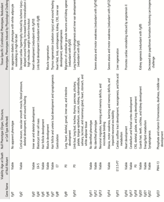
FGFs control a variety of physiological responses during embryonic development and in adult organisms. During development FGFs play key roles in patterning and morphogenesis by controlling cell proliferation, survival, migration and differentiation. In adult organisms, they are involved in tissue repair, response to injury, as well as energy, bile acid, mineral ion and metabolic homeostasis. The identification of the roles of most FGFs was made possible by the use of knock-out mouse models for one or more of these factors (Table 1).
Table 1. Phenotypes of germline and conditional loss-of-function Fgf mutations in mice. (Ornitz & Itoh 2015).
1.2 Fibroblast Growth Factor Receptors (FGFRs)
The FGFR family is composed by 5 proteins: FGFR1, 2, 3, 4 and L1. The FGFR1, 2, 3, 4 are single chain transmembrane tyrosine kinases that consist of a ligand binding extracellular domain, a single transmembrane domain (TM), and an intracellular tyrosine kinase domain that is separated into two parts by an insertion domain. FGFRL1 (also recently called FGFR5) is an exception to this scheme since it has a short intracellular tail with no tyrosine kinase domain (Figure 2). The FGFR extracellular domain contains two or three immunoglobulin (Ig)-like loops (D1, D2, D3). D1 loop and the D1-D2 link, also known as acidic box (AB) due to the presence of acidic residues, are involved in receptor autoinhibition (5) and they’re not necessary for FGF binding (6). FGFR portion responsible for the ligand binding is composed by D2 and D3, with the first loop presenting the heparin binding site (HBS). While FGFR4 and L1 present only one splice isoform, the other three FGFRs have been found to naturally encode different variants. In fact in FGFR1–FGFR3, two alternative exons (IIIb and IIIc) code for the second half of D3 and are spliced to the common exon IIIa (encoding the first half of D3) (Figure 2). These alternative splicing events occur in a tissue specific fashion, resulting in mesenchymal “c” and epithelial “b” isoforms and this variation defines ligand-binding affinity and specificity of FGFR1-3.
Figure 2. FGFRs structures and isoforms. (N: N-terminus; D1-3: immunoglobulin (Ig)-like loops; AB: Acidic Box; HBS: Heparin Binding Site; TM: Transmembrane domain; TK1-2: Tyrosine kinase domain; C: C-terminus; a,b,c: exons participating to splicing events) (1, 6).
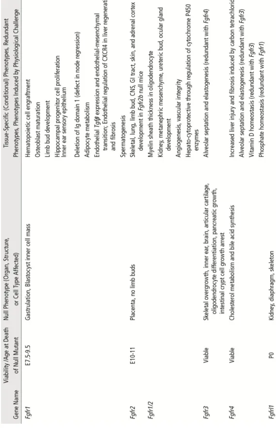
FGFRs role has been evaluated through the use of null or conditional loss-of- function transgenic mice (Table 2). Fgfr1-/- mice do not survive early embryonic phases while, using conditional inactivation of Fgfr1 in osteo-chondrocyte progenitor cells and in differentiated osteoblasts, it has been shown that FGFR1 signalling is important in limb establishment and bone formation (7, 8). Mice lacking Fgfr2 survive until embryonic day 10–11. These embryos fail to form a functional placenta and do not form limb buds (9). The conditional inactivation of Fgfr2 showed osteoprogenitors proliferation defects and mice presented dwarfism (10). Fgfr3-null mice are viable and present a skeletal overgrowth and other defects. Mice lacking Fgfr4 are viable, and they present minor liver-related defects (11). Finally Fgfrl1-/- mice present diaphragm hypoplasia, heart and skeletal anomalies (12, 13). The involvement and role of FGFRs in skeletal development will be later discussed.
Table 2. Phenotypes of germline and conditional loss-of-function Fgfr mutations in mice. (1)
Ligand binding specificity of the 18 secreted (canonical and endocrine) FGFs have been compared using various mitogenic assays and by directly measuring affinity for FGFRs (Zhang 2006). Each FGFR isoform react to specific FGFs (Table 3). These FGFs bind FGFRs in dimers in concert with heparan sulfate proteoglycan (HS). Complex FGF-HS-FGFR dimers stimulate the tyrosine kinase activity through a process of trans-autophosphorylation that triggers the transduction signalling that will end in the activation of the targeted genes. In fact the activated receptor is able to bind and phosphorylate other proteins that will lead to the transduction of the signalling through different pathways.
FGFR dimerization is needed to transduce biochemical signalling, but the widely believed role of FGFs in producing FGFR dimerization (14) is a current object of debate since a recent study shows the presence of FGFR dimers in FGFs absence (15). The same study indicates the presence of FGFR phosphorylation in unlinganded dimers, however the FGF binding still determines a more consistent phosphorylation of the FGFR dimers.
Table 3. Receptor specificity of canonical and endocrine FGFs. (1)
2. FGFR-related diseases
Mutations in FGFRs have been found responsible for several pathological syndromes. Here we will detail the FGFR-related diseases with a special focus on pathologies du to FGFR3 mutations.
2.1 FGFR1 and FGFR2-related diseases
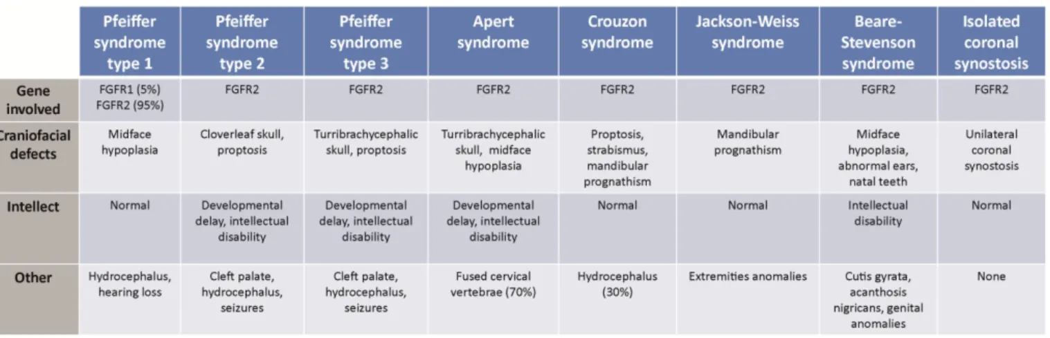
Fgfr1 is located on 8p11.23 and spans 58 kb whereas Fgfr2 is located on 10q26.13 spanning 120 kb. Both Fgfr1 and Fgfr2 transcripts are composed of 18 exons (17 coding exons), their expression is limited to bone and cartilage (16). Most of the activating mutations in Fgfr1 or Fgfr2 have been detected in cranyosinostosis syndromes that are briefly describe in Table 4. Craniosynostosis occurs in approximately 1 in 2 100 to 1 in 2 500 live births (17, 18), and is characterized by the premature fusion of one or more cranial sutures resulting in malformation of the skull. Potential consequences of abnormal skull growth include increased intracranial pressure, problems with hearing and vision, impaired blood flow in the cerebrum, and developmental delay (19).
Table 4. FGFR1 and FGFR2 related craniosynostoses.
2.2 FGFR3-related diseases
Fgfr3 is located on 4p16.3 and spans 16.5 kb. Its transcript is composed of 19 exons which only 2 to 18 are coding exons. The gene expression has been demonstrated in brain (astrocytes and glial cells), growth plate cartilage, intestine, pancreas, testis, cochlea and crystalline lens (20). Mutations in FGR3 are responsible for bone disorders (chondrodysplasias and craniosynostoses) and for various forms of cancer. Fgfr3 harbors almost exclusively dominant mutations leading to a gain of function in almost all cases. Only in the very rare case of Camptodactyly, tall stature, and hearing loss (CATSHL) syndrome it has been proposed a dominant negative mechanism. In any case Fgfr3 is not a haplo-insufficient gene: it means that removing one of the two copies of the gene doesn’t lead to mutant phenotype. In this chapter we explore the FGFR3-related pathologies highlighting their clinical features and the mutations underlying their etiology.
2.2.1 Osteochondral disorders
2.2.1.1 Chondrodysplasias
Chondrodysplasias is a family of genetic diseases characterized by a disturbance in the development of the cartilage of the long bones, especially of the epiphysial plates, resulting in arrested growth and dwarfism. Here we present chondrodysplasias due to point mutations in FGFR3 gene.
2.2.1.1.1 Achondroplasia (ACH)
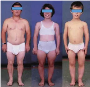
The term ‘achondroplasia’ was first used by Jules Parrot in 1878, and in 1900 Pierre Marie described the main features in children and adults. As ACH (OMIM#100800) is the most common disorder associated and with its disproportionate short stature is probably one of the best-known and -defined chondrodysplasias (Figure 3). This condition is estimated to occur in between 1 in 10 000 and 1 in 30 000 live births (21, 22). ACH patients present a rhizomelic dwarfism associated with macrocephaly, midface hypoplasia, prognatism (23), kyphoscoliosis (24), cervicomedullary compression, spinal stenosis and loss of synchondroses (25). ACH is an autosomal dominant disorder, frequently de novo and associated with
advanced paternal age due to an increased presence of FGFR3 mutations during spermatogenesis in men over 35 (26). The cause of this disease has been identified in a point gain-of-function heterozygous mutation in FGFR3 (27, 28). The most recurrent mutation (over 95% of cases (21)) is Gly380Arg localized in the TM domain of the receptor (Figure 9). In adulthood affected patients reach an average of 130 cm for males and 125 cm for females (29) due to an abnormal development that can be diagnosed in utero through medical ultrasound since the beginning of the third trimester (30, 31). ACH patients face higher rates of deaths compared to the unaffected population (basically due to sudden death, heart diseases end neurological diseases) (32) and present severe medical complications (spinal stenosis, tibial bowing, obstructive apnea).
Figure 3. ACH phenotype. In the left picture a couple of twins, the right one is affected by ACH. In the Right picture X-rays of ACH patient.
2.2.1.1.2 Hypochondroplasia (HCH)
HCH (OMIM#146000) (1 / 30 000 live births) is a skeletal dysplasia, characterized by short stature, disproportionately short arms and legs; broad, short hands and feet; mild joint laxity; and macrocephaly (33). The skeletal features are very similar to those seen in ACH but tend to be milder. Medical complications common to ACH (spinal stenosis, tibial bowing and obstructive apnea) occur less frequently in HCH, but intellectual disability and epilepsy may be more prevalent. The diagnosis is difficult to make in children under the age of three years, as skeletal disproportion tends to be mild and many of the radiographic features are subtle during infancy. DNA-based testing is possible and about 70% of affected individuals
are heterozygous for a mutation in FGFR3. However, there is evidence that locus heterogeneity exists, implying that mutations in other as-yet unidentified genes may result in similar, if not identical, phenotypes. The adult height for men with this condition ranges from 138 centimeters to 165 centimeters while the height range for adult women is 128 centimeters to 151 centimeters. Missense, gain-of-function, mutations responsible for this disease are localized in different FGFR3 domains: extracellular, transmembrane and tyrosine kinase with N540K as the most recurrent mutation.
Figure 4. Patients affected by HCH. These sufferers show a rhizomelic dwarfism less severe compared to ACH patients.
2.2.1.1.3 Thanatophoric Dysplasia (TD) and Severe Achondroplasia with Developmental Delay and Acanthosis Nigricans (SADDAN)
TD and SADDAN are the more severe diseases related to gain-of-function mutations in FGFR3. TD (1 / 20 000 to 1 / 50 000 live births) is a lethal skeletal dysplasia due to heterozygous mutations in FGFR3. This pathology was firstly described in 1967 (34). Two types of TD are clinically diagnosed based on ultrasound and radiographic findings. TD type 1 (OMIM#187600) patients have prominently curved femurs, while TD type 2 (OMIM#187601) patients typically have straight femurs, a severe form of craniosynostosis (often referred to as a cloverleaf skull) and a small chest (35). Several different gain-of-function mutations in FGFR3 cause TD type 1. Mutations R248C, S249C, S371C, and Y373C create novel cysteine residues in the extracellular and intramembranous domains, while other mutations causing TD
type 1, such as X807R, X807C, X807E, X807S, and X807W, obliterate the stop codon resulting in extension of the intracellular domain by an additional 141 amino acids. TD type 2 is caused by the FGFR3 mutation K650E. SADDAN Severe achondroplasia, developmental delay and acanthosis nigricans (OMIM#616482) was first discovered in 1999 (36) and it is caused by a K650M mutation in FGFR3. Most of the patients develops extensive areas of acanthosis nigricans in early childhood, suffers from severe neurological impairments, and survives without prolonged life-support measures (37). However the differences between TD and SADDAN phenotypes are not always evident (38). The substitution of a methionine residue at position 650 differentiates SADDAN from type 2 thanatophoric dysplasia, which arises from a glutamic acid substitution at the same position. Interestingly mutations at the same residue K650N, K650Q (39) and K650T (40) are responsible for the less severe hypochondroplasia. The SADDAN amino acid change induces a FGFR3 phosphorylation that is threefold greater than normal.
Figure 5. From left to right examples of TD type 1, TD type 2 and SADDAN. TD type 1 patients present femurs with a “telephone receiver" appearance. TD type 2 affected individuals are recognized by straight femurs and presence of cloverleaf craniosynostosis. SADDAN patients suffer of a severe dwarfism and feature the skin lesion acanthosis nigricans.
2.2.1.2 Craniosynostoses
As previously defined for FGFR1 and FGFR2 related craniosynostoses, these pathologies arise from the premature fusion of skull sutures. Also mutations in FGFR3 can lead to craniosynostosis: until now this gene contributes to craniosynostoses family with two syndromes.
2.2.1.2.1 Muenke Syndrome (MS)
MS (OMIM#602849) constitutes the most common syndromic form of craniosynostosis, with an incidence of 1 in 30 000 births. Of all patients with craniosynostosis, 8% have MS (17, 18). Both sporadic and familial cases have been reported. MS displays incomplete penetrance and a variable phenotype even within families (41). Characteristics include bi- or unicoronal synostosis, midfacial hypoplasia, macrocephaly, and downslanting palpebral fissures. Some affected individuals have additional features that may include sensorineural hearing loss, developmental delay, brachydactyly, and coned epiphyses in the hands and feet. MS craniosynostosis is a result of a specific heterozygous gain-of-function mutation, P250R, found in the linker region between domains D2 and D3 of FGFR3 (42). Increasing paternal age is a contributing factor for de novo mutations (43).
Figure 6. 4 month child affected by MS. The MR images in sagittal and axial views show the severity of the brachycephaly.
2.2.1.2.2 Crouzon with Acanthosis Nigricans syndrome (CAN)
Patients with CAN (OMIM#612247) have multiple sagittal or coronal fusions causing brachycephaly, trigonocephaly, and rare reports of cloverleaf skull malformation. Attributes typically include hypertelorism, a small midface, beaked nose and protrusion of the eyes. CAN has an estimated prevalence of 1 / 1 000 000 newborns and most cases are sporadic and associated with paternal aging, although familial cases consistent with autosomal dominant inheritance have been reported. Characteristic of this disease is the presence of hyperpigmentation of the skin (acanthosis nigricans), hyperkeratosis, and other skin findings. A specific FGFR3 heterozygous gain-of-function mutation A391E, responsible for the syndrome, is located in TM (44, 45).
Figure 7. Manifestation of CAN syndrome. Lateral close up and radiograph.
2.2.1.3 Other FGFR3-disorders
Camptodactyly, tall stature, and hearing loss (CATSHL) syndrome
Dominantly inherited, camptodactyly, tall stature, scoliosis, and hearing loss syndrome (CATSHL; OMIM#610474) is caused by a FGFR3 heterozygous missense mutation, R621H, residing within the tyrosine kinase domain generating a loss-of-function that promotes endochondral bone growth (46). For several reasons, it is unlikely that the loss of function caused by p.R621H results from haploinsufficiency. In fact, mice heterozygous for an Fgfr3 null allele are phenotypically normal and the
fibroblasts of individuals affected with CATSHL syndrome express both wild-type and mutant FGFR3 RNA in nearly equal proportions, and the expression levels of all five FGFRs in patients are similar to those of normal individuals. Furthermore, both mutant and wild-type FGFR3 localize to their normal position in the cell membrane (46). These observations suggest that p.R621H might, instead, cause loss of FGFR3 function by a dominant negative mechanism. Recently, a novel homozygous mutation T546K has been described as also causing skeletal overgrowth (47).
Figure 8. Patient affected by CATSHL syndrome. The disorder is characterized by tall stature and scoliosis.
Figure 9. FGFR3 mutations in osteochondro-related disorders. Most common mutations for each disease are underlined.
2.2.2 Cancer
As already shown, germline mutations in FGFR3 cause skeletal disorders. The same point mutations have been identified to cause cancer (48). In literature we can find various examples of FGFR3 constitutive activation in the cancer field. In fact somatic mutations in this gene can induce the cancer progenitors to fulfill their maturation, meaning that FGFR3 mutations are tumorigenic thus FGFR3 is an oncogene. FGFR3 have been reported to play a role in multiple myeloma (49) where a translocation t(4;14)(p16.3;q32) causes the overexpression of the gene. This translocation links FGFR3 to the immunoglobulin heavy chain ICH locus. These translocations are intergenic, with the breakpoints occurring ~70 kb upstream of FGFR3, and bring FGFR3 under the control of the highly active IGH promoter. It is important to note that the translocations involving FGFR3 in multiple myeloma also involve the adjacent multiple myeloma SET domain-containing (MMSET) gene, and the relative contributions of FGFR3 and MMSET to oncogenesis are subject to ongoing debate. The ultimate effect of the translocation is to overexpress FGFR3 out of context, which might result in aberrant dependent signalling or ligand-independent signalling. In a small proportion of t(4;14) multiple myeloma, FGFR3 is also mutated (~5% translocated cases), presumably further reinforcing FGFR3 signalling.
FGFR3 is also implied in bladder cancer, the fourth most common tumor among males. More than 90% of bladder cancers are urothelial cell carcinoma and about 5% are squamous cell carcinoma. The gender ratio of male to female is 3 to 1 and the best known environmental risk factor is smoking. Urothelial cell carcinoma patients are classified by pathologic stage. The stage classification differentiates between non-muscle invasive (NMI; Tis, Ta, and T1) and muscle-invasive (T2, T3, and T4) tumors according to the invasion depth. Ta tumors are restricted to the urothelium, T1 tumors present invasion of the lamina propria, T2 of the superficial muscle, T3 of the perivesical fat and T4 of surrounding organs. Tis is poorly understood and believed to be a precursor of muscle-invasive tumors. Most of the patients (70%) initially present NMI tumors and after treatment up to 70% of these patients will develop one or several local recurrences. About 25% of NMI patients progress to muscle-invasive tumors disease that can potentially lead to metastasis.
About 50% of all patients present an FGFR3 mutation and the most recurrent is S249C (50–52).
Somatic mutations were found also in other cancers: prostate cancer (53) with 2 missense mutations (S249C and A393E), spermatocytic cancer (54) with the K650E mutation and in cervix cancer as well (55, 56) with the only S249C missense mutation.
2.2.3 Skin lesions
FGFR3 mutations are also associated with particular skin lesions. In the case of SADDAN we have already discussed the appearance of the skin lesion acanthosis nigricans, a velvety hyperpigmentation of the skin. Somatic FGFR3 mutations were also identified in benign skin tumors. Analysis of human seborrheic keratoses revealed somatic FGFR3 mutations in 40% of the benign tumors. The prevalence of FGFR3 mutations was even higher (85 %) in adenoid seborrheic keratoses, a particular histological subtype of seborrheic keratosis (57). FGFR3 mutations in human skin can cause common non-organoid epidermal nevi (58). In that study normal skin adjacent to the epidermal nevus did not show the presence of FGFR3 mutations. Finally other studies showed the presence of FGFR3 mutations in solar lentigo, in 5 out 30 patients, (59) and in lichenoid keratosis, in 6 out of 52 patients (60).
2.2.4 Consequences of different mutations on receptor activity
FGFR3 mutations implied in chondrodysplasias are localised in different receptor domains (Figure 9). FGFR3 gain-of-function mutations are characterized by an abnormal phosphorylation of the receptor. However the phosphorylation level varies depending on the FGFR3 harboured mutation. FGFR3 studies in vitro have highlighted higher phosphorylation levels for the mutation K650M (SADDAN) and K650E (TD type 2) (61), with the K650M mutation presenting the higher phosphorylation level (62). This elevated FGFR3 phosphorylation will be transduced to the downstream signalling pathway activating its function disproportionately. We can imagine that mutations in TK1 and TK2 determine protein conformational
modification that increase the transphosphorylation between the receptors that make up the dimer (39). Even if other mutations are not localised in TK1 and TK2 as G380R (ACH), Y373C (TD Type 1) or A391E (CAN) the higher phosphorylation level compared to FGFR3 wild-type could be explained. In fact these mutations localised in TM domain could lead to an over-stabilization of the dimer causing an increase of the transmitted signals (63, 64). Other studies have also suggested that these mutations disrupt c-Cbl-mediated ubiquitination that serves as a targeting signal for lysosomal degradation and termination of receptor signalling. This defect would distance actively signalling receptors from lysosomes prolonging their survival and signalling capacity (65).
3. Skeletogenesis
Skeletogenesis it is a hard task that consists in elaborating an edifice of more than 200 pieces of bone and cartilage. Each skeletal piece is modeled at a distinct location in the body, is articulated with others, and reaches specific sizes, shapes, and tissue compositions according to both species instructions and role. Here we will explore the main stages of this process with a special focus on FGFR role in the phenomenon.
3.1 Early embryo development
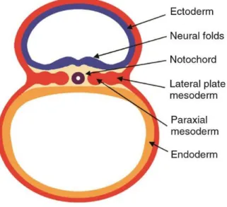
The first step in skeletogenesis consists in generating osteochondral progenitor cells. The origin of these cells is linked with the early stages of embryo development, when the vertebrate embryo is composed of three germ layers: ectoderm, mesoderm, and endoderm (Figure 10). From the neural crest several throat and craniofacial skeletal elements arise; the lateral plate mesoderm gives rise to other craniofacial skeletal structures, the limb skeletal elements (appendicular skeleton), the sternum (part of the axial skeleton); finally the paraxial mesoderm gives rise to somites, which develop into sclerotomes (ribs and vertebrae). The vertebrae, developing around the notochord, will force the notochord cells to change phenotype, migrate to the intervertebral spaces, and develop the nuclei pulposi (NP) of Intervertebral Discs (IVDs) (Lefebvre and Bhattaram, 2010).
Figure 10. Origin of osteochondro-progenitor cells in the vertebrate embryo. Scheme of a cross-section of mouse embryo after gastrulation at day 8 of development (equivalent to day 17 in humans). The three germ layers are shown: ectoderm, endoderm, and mesoderm (66)
These neural crest and mesoderm cells will reach their skeletal sites through the expression of specific factors as the Homeobox (Hox) genes (67). Before reaching their skeletal sites, neural crest- and mesoderm-derived cells produce a matrix rich in collagen-1, fibronectin, and hyaluronan, and they proliferate or die in a tightly controlled spatial and temporal manner (68). They thereby establish mesenchymal structures that prefigure the future skeletal elements. These cells are called osteochondro-progenitors because most of them give rise to osteoblasts and chondrocytes.
Development of the vertebrate skeleton occurs through the processes of endochondral and intramembranous bone formation. Endochondral-derived bones comprise the appendicular skeleton, facial bones, vertebrae, and medial clavicles, while intramembranous bones comprise the cranium and lateral clavicles. Endochondral ossification initiates with the condensation of mesenchyme, followed by the formation of a cartilaginous template that patterns the developing skeleton and the segmentation of this template into cartilaginous joints. Intramembranous bone forms from a mesenchymal condensation that directly gives rise to bone.
3.2 Chondrogenesis
Mesenchymal condensation is characterized by the aggregation of loose mesenchymal cells and the expression of extracellular matrix proteins and cell adhesion molecules (type I collagen (Col I), hyaluronan, N-cadherin, tenascin-C). The transcription factor Sry box containing 9 (Sox9), that acts in concert with Sox5 and Sox6, is necessary for the beginning of the chondrogenesis as well as the bone morphogenetic protein (BMP) receptors 1a and 1b (69). Proliferating cells within the mesenchymal condensation begin to express type II collagen (Col II) (chondroprogenitors) while cells on the borders of the structure express Col I (osteoprogenitors) (70). Proliferating chondrocytes form columns that are oriented along the longitudinal axis of the developing bone to define the future growth plate (Figure 11).
At the center of the developing bone chondrocytes expressing specific markers (type X collagen (Col X), Indian Hedgehog (IHH), ParaThyroid Hormone (PTH) 1 Receptor (PTH1R)) differentiate into prehypertrophic chondrocytes increasing their volume and arresting their proliferation. The down-regulation of Sox9 is necessary to allow this chondrocyte maturation, however a level of expression of this transcriptor factor is a requirement for the chondrocyte identity (71, 72). Runt- related protein 2 (Runx2) and Runx3 are required for chondrocyte differentiation, with Runx2 determining chondrocyte proliferation through Ihh expression (73). Several signalling pathways (IHH, PTH, FGF, BMP and Wnt) regulate the equilibrium between proliferating and hypertrophic chondrocytes (74). Wnt and PTH-related peptide (PTHrP) signals modulates the calcium/calmodulin-dependent protein kinase II (CAMK2) level, which induces chondrocyte hypertrophy increasing RUNX2 and β-catenin activity (75). Hypertrophic chondrocytes, pushed by the proliferative chondrocytes along the axis, in their late phase secrete vesicles containing enzymes that degrade the cartilage matrix and mineralize their surroundings (76). Finally those chondrocytes commit apoptosis or differentiate into osteoblasts (77, 78).
3.3 Endochondral ossification
The formation of a primary ossification center and bone collar that starts prenatally (79) is characterized by the differentiation of Col I-producing osteoblasts on the border of the hypertrophic chondrocyte zone and determines the origin of mineralized bone. Osteoprogenitor cells (belonging to the Osterix lineage) localized in the perichondrium and in the periosteum differentiate into osteoblasts responsible of the trabecuale formation. The osteoblasts derived from the late hypertrophic cells (belonging to the Col I lineage) will be responsible of the cortical bone (80). The expression of Vascular Endothelial Growth Factor (VEGF-A) in hypertrophic chondrocytes determines the beginning of the vascularization leading to the recruitment of endothelial cells, osteoprogenitor cells, and osteoclasts that will later participate in the bone remodeling process (81). The cartilaginous matrix left behind by the hypertrophic chondrocytes will work like a mold for the osteoblastic progenitors that they will fulfill with mineralized matrix giving rise to the primary ossification center. The primary ossification center will progressively elongate following the same longitudinal axis primarily determined by the columnar chondrocytes. The late hypertrophic chondrocytes, osteoblasts and osteoclasts will take the charge of dismantling the remains of the cartilage matrix through Matrix metalloproteinases (MMP) expression (82). With the postnatal formation of a secondary ossification center at the epiphysis we observe the classic growth plate with well-demarcated zones of cells presenting vascular elements in the primary bone (Figure 12). Finally osteoclasts bone remodeling abilities will be applied in the definition of the bone marrow cavity in the diaphysis that presents an intricate network of vessels compared to the growth plate.
Figure 11. Endochondral ossification. Long bones are formed by endochondral ossification (83).
Figure 12. Long bone and growth plate structure. Primary Ossification Center (POC), Growth Plate (GP) and Secondary Ossification Center (SOC).
3.4 Intramembranous ossification
In the structure that will give rise to the skull, at the beginning of intramembranous ossification (Figure 13) mesenchymal cells condensate and differentiate primarily into osteoprogenitor cells and secondly into osteoblasts, in total absence of cartilage and chondrocytes. These osteoblasts directly derived from osteoprogenitor give rise to ossification centers where they will produce mineralized (84). These ossification centers are responsible for the development of the skull bone, but their osteogenic
fronts will never merge determining the appearance of the sutures (85). An abnormal fusion of the sutures (synostosys) would impair the skull growth. Mesenchymal cells surround the suture and part of them differentiate into osteoprogenitors and then into osteoblasts lining the developing bone. When the bone is formed osteoblasts finally die by apoptosis or become embedded in the bone matrix as osteocytes, which then also eventually undergo apoptosis (86). The expression of Runx2 is responsible for the differentiation of mesenchymal stem cells into osteoblasts (opposed to Sox9 responsible for the differentiation of the same cells into chondrocytes). Runx2 is a fundamental transcription factor that regulates several genes in osteoblasts, such as Col I, bone sialoprotein (BSP), osteopontin (OP), alkaline phosphatase (ALP), transforming growth factor (TGFβ), and osteocalcin (OC) (87, 88). Transcription factors, such as Msh homeobox 2 (MSX2) and Distal-less homeobox 5 (DLX5), play important roles in osteoblast differentiation during cranial bone formation in part by interacting with RUNX2 (89). Several factors (as TGFβs, BMPs, FGFs, and Wnt signalling) are responsible for the regulation of differentiation and survival of the cells participating to the bone formation process (85, 89).
Figure 13. Intramembranous ossification. Craniofacial bones are formed directly from condensations of mesenchymal cells without the formation of a cartilage intermediate. (83).
3.5 FGFR signalling and early skeletal development
FGFs and FGFRs will participate in all the skeletal development stages. We can observe the first FGFR expression even before the mesenchymal cells condensation. The origin of the appendicular skeletal is recognizable in the embryonic mesenchymal limb bud formation (Figure 14). Different mechanisms have been proposed to determine the limb bud development process (90) and in all of them FGFRs and FGFs play an essential role. In fact the distal limb bud mesenchyme expresses both FGFR1 and FGFR2 (91, 92). FGFR3 and FGFR4 are not expressed, so they do not take part to the process (92). At the bottom of the limb bud the apical ectodermal ridge (AER) produces FGFs called AER-FGFs. AER-FGFs (FGF4 and FGF8) bind FGFR1 and FGFR2 stimulating the limb growth (90) (Figure 14). FGFR1 and FGFR2 are redundant in this tissue (93). Conditional inactivation mice have been used to determine the function of FGFR1 and FGFR2 in the mesenchymal limb bud. The conditional knockout in mesenchymal cells of both Fgfr1 and Fgfr2 or the knockout of Fgfr1 lead to severe skeletal hypoplasia highlighting the major role of the receptors in mesenchymal tissue (8, 94). On the other hand the knockout Fgfr1 or Fgfr2 in distal limb bud mesenchyme resulted in milder skeletal phenotypes (95, 96), thus FGFR1 and FGFR2 are redundant in distal limb bud.
At the beginning of endochondral ossification an increase of FGFR2 and Sox 9 expression is observed in condensing mesenchymal cells compared to the surrounding cells of the mesenchyme (92, 97) (Figure 15). At the borders of the mesenchymal condensation both FGFR1 and FGFR2 expressing-cells are present and they will constitute the perichondrium and periosteum (97) (Figure 15). With the differentiation of mesenchymal cells into chondrocytes there is an expression of FGFR3, Sox9 and Col II while FGFR2 expression is decreased (98). The expression of these markers indicate the appearance of proliferative chondrocytes. In hypertrophic chondrocytes FGFR3 expression is decreased whereas FGFR1 expression is increased (7, 99) (Figure 15). The mesenchymal condensation and the differentiation of its cells in chondrocytes is modulated by the FGFRs (94, 100). In fact in chondroprogenitor cells and in primary chondrocytes the FGF signalling is responsible for an increase of Sox9 expression and Extracellular Signal-Regulated Kinase 1/2 (ERK1/2) activation (101, 102). FGFR3 expression in proliferating
chondrocytes requires Sox9 expression, and Sox9 regulatory binding sites have been found in the Fgfr3 gene (103). FGFR3 is activated by FGF9 and FGF18 expressed in mesenchyme stimulating chondrocyte proliferation (104, 105). In the forming perichondrium, bonecollar, and trabecular bone the expression of FGFR1 in mesenchymal progenitors and FGFR2 in differentiating osteoblasts are detected (7, 106). Moreover FGFR1 and FGFR3 are co-expressed in mouse and human articular chondrocytes (107, 108). Difference in FGFRs expression has been observed between immature cultured osteoblasts and mature osteoblasts in vitro: the first express relatively higher levels of FGFR1, the second of FGFR2 (109)
FGFs (FGF2, FGF4, FGF9, FGF18) and FGFRs (FGFR1, FGFR2, and FGFR3) are also determinant for the early stages of intramembranous ossification (110, 111). FGFR2 is expressed in early mesenchymal condensations and then in sites of intramembranous ossification, where it interacts with FGF18 (97). At later stages during cranial bone development, FGF8 is expressed in mesenchymal cells and differentiating osteoblasts (110, 112). FGF9 is expressed throughout calvarial mesenchyme during mid to late stages of embryonic development (113). Mice lacking both FGF9 and FGF18 have severe defects in skull bone formation (114). FGFR1 and FGFR2, which are expressed in preosteoblasts and osteoblasts (109), are likely receptors for FGF9 and FGF18 in developing membranous bone (115).
Figure 14. Proximodistal limb bud axis development. The limb skeletal elements (stylopod, zeugopod, autopod) are determined early in embryo development. The origin of limb development is in the limb bud, however different models of this mechanism have been formulated. In all of them the role of AER and AER-FGFs is essential. Here it is presented the progress zone (PZ) model: the mesenchyme that underlies the AER contains mesenchymal cells regulated by AER signals. As limb bud growth progresses distally, proximal cells are too far to be influenced by AER-FGFs. The first cells to lack their influence will give rise to stylopod, the second the zeugopod and finally the autopod.(90).
Figure 15. FGF and FGFR expression during endochondral and intramembranous ossifications. (A–D) From chondrogenesis to the primary ossification center. (E) Embryonic growth plate. (F) Postnatal growth plate after formation of the secondary ossification center. (G) Intramembranous ossification. Cells and tissues are color-coded for expression domains of FGFs and FGFRs. (BM) Bone marrow. (93).
3.6 FGFR signalling in growth plate
Chondrocytes FGFRs expression is different within the growth plate (Figure 15). Briefly, there are low levels of FGFR2 in the resting zone, high levels of FGFR3 in the proliferating and prehypertrophic zone, and high levels of FGFR1 in hypertrophic chondrocytes (99, 116). We will now investigate the role of the FGFRs and their signaling pathways in this tissue.
Conditional inactivation experiments in mice shed light on FGFR2 function in resting chondrocytes. The conditional knockout in limb bud mesenchyme (10) leads to a reduced growth in mice. This data could suggest a role in skeletal development, but at the analysis of the growth plate no anomalies were detected in proliferative chondrocytes. The phenotype is probably linked to an effect on osteoblasts rather than on chondrocytes, or on an indirect increase of osteoclastic activity (10). Thus FGFR2 has more a redundant function in resting or proliferating chondrocytes.
In early embryonic stages of skeletal development, during the growth plate formation, FGFR3 stimulates the chondrocyte proliferative activity (62, 104). By cons, during postnatal skeletal growth, FGFR3 signalling inhibits chondrogenesis, leading the proliferating chondrocytes to their differentiation into prehypertrophic and hypertrophic chondrocytes (117, 118). This paradoxical activity of FGFR3 is the reason why chondrodysplastic disorders due to gain-of-function mutations in FGFR3 (that we have already discussed in chapter 2) have a decreased proliferation and differentiation of proliferating chondrocytes during pre-pubertal skeletal growth (119, 120).
FGFR3 downstream signalling pathways in growth plate chondrocytes involve several factors: Signal Transducer and Activator of Transcription 1 (STAT1), ERK1/2, p38, Snail1 (Zinc finger protein SNAI1), AKT, protein phosphatase 2a (PP2a), p107 and p130 (Figure 16) (121, 122). The chondrocyte proliferation arrest resulted from FGFR3 activation is effected through increased expression of the cell cycle inhibitor p21Waf1/Cip1 and activation of p107 and p130 (121, 123). Snail1 expression is induced by FGFR3 and is expressed at high levels in human thanatophoric dysplasia bone tissue. Ectopic activation of SNAIL1 in mice resulted in an ACH phenotype with decreased chondrocyte proliferation and shortened bones at late embryonic stages (124). Interestingly SNAIL1 acts activating both STAT1 and MAPK branches of the
FGFR3 signalling pathway, as the ectopic activation of the protein resulted in nuclear translocation of STAT1 and increased phosphorylation of ERK1/2 (124). The mechanism behind SNAIL1 regulation of STAT1 and MAPK pathways is still poorly understood. However it has been proposed, analyzing breast cancer cells, that SNAIL1 may regulate the nuclear localization of p-ERK (125). In the same cells it has also been shown that activated ERK2 directly phosphorylates SNAIL1, leading to its nuclear accumulation (126). In support of a requirement for SNAIL proteins in regulating chondrogenesis, conditional inactivation of both Snail1 and Snail2 resulted in increased p21Waf1/Cip1 and decreased chondrocyte proliferation (127). The bone growth anomalies lead by activating mutations in FGFR3 are due to decreased chondrocyte proliferation and differentiation. Experiments have led us to consider that these chondrocyte defects are diverse and regulated by different members of the FGFR3 signalling pathway. It has been proven that STAT1 regulates chondrocyte proliferation through in vivo experiments when proliferative defects in growth plate of mice with activating mutations in FGFR3 were rescued by the full inactivation of Stat1 (128). However, in this model, inactivation of Stat1 still does not rescue the overall ACH phenotype considering that chondrocyte differentiation to hypertrophy was not corrected: in FGFR3-related disorders impaired chondrocyte hypertrophy, and not only the defective proliferation, is responsible for the decreased bone growth. The same studies proved also that the activation of MAPK signalling suppressed chondrocyte hypertrophy, resulting in skeletal dwarfism (128).The mechanism may involve the suppression of SOX9 down-regulation in prehypertrophic chondrocytes by activated FGFR3 signalling (102, 129). Additionally, activation of the PP2A–B55α holoenzyme by FGFR3, which dephosphorylates and activates p107, could contribute to decreased chondrocyte hypertrophy (121, 122). In support of the model, previously presented, according to which STAT1 activation arrests chondrocyte proliferation and MAPK signalling is responsible for chondrocyte differentiation, there are the studies performed on the C-type natriuretic peptide (CNP) signalling pathway and its interaction with the FGFR3 pathway (130). In fact CNP expression was found to attenuate the phenotype of ACH mice through inhibition of MAPK signalling, which restored matrix production and hypertrophic differentiation. The activity of CNP was independent of the STAT1 one and did not modify chondrocyte proliferation. However that the functions of the STAT1 and MAPK pathways are completely distinct is still unsure. In fact it has been shown that in cultured chondrocyte the suppression
of proliferation required MAPK signalling and was independent of STAT signalling (131, 132). In support of these studies, mice lacking ERK1 and conditionally lacking ERK2 in chondrocytes showed an increase in chondrocyte proliferation at late embryonic stages (133, 134). Still we have to consider some variables that could have produced these results: the variability in the experiments may be due to in vitro culture conditions or by the developmental stage being examined. In fact it is really common to observe differences between the embryonic and postnatal signallings.
Figure 16. FGF/FGFR signalling pathways involved in proliferating chondrocytes. (93)
Other mechanisms by which FGFR3 signalling may indirectly regulate growth plate development include the regulation of BMP, Wnt, IHH, and PTHrP/PTH1R expression and activity (Figure 17). Comparisons of activating mutations in Fgfr3 and mice lacking Fgfr3 show that FGFR3 signalling suppresses expression of Bmp4, BMPR1a (BMP-receptor type 1A), Ihh, and PTH1R in postnatal growth plate chondrocytes (120, 135, 136). Additionally, in a chondrocyte cell line, overexpression of FGFR3 suppresses expression of both PTHrP and its receptor, PTH1R (137), and, in chondrocyte cell lines and micromass cultures, FGF signalling activates Wnt/ β-catenin signalling through phosphorylation of LRP6 (Low-density lipoprotein receptor-related protein 6) and functions to suppress hypertrophic differentiation (138, 139).
PTHrP may also regulate FGFR3 expression. Analyses of the Fgfr3 promoter identified a transcriptional regulatory element, CSRh, which was repressed by PTH through a cAMP and protein kinase A (PKA)-dependent mechanism (140).
Figure 17. Alternative pathways regulated by FGFR3.
FGFR1 is prominently expressed in prehypertrophic and hypertrophic chondrocytes and overlaps with FGFR3 in the latters (Figure 15). To understand the role of FGFR1 signalling experiments have been performed in mice through the targeted inactivation of FGFR1. At early stages of development, Fgfr1 conditional knockout mouse shows impaired chondrocyte hypertrophy (105) on the other hand the Fgfr1 conditional knock-out mouse for later stages presents an increased hypertrophic zone (7).
FGF9 and FGF18, FGFR3 ligands, are expressed in the perichondrium and are fundamental for growth plate organization (105, 112) (Figure 15). Mice lacking Fgf9, present rhizomelic shortening of the limbs. At early embryonic stages chondrocyte proliferation and differentiation are impaired in Fgf9 null mice (105). Mice lacking Fgf18 also showed decreased chondrocyte proliferation, but in the more distal part of the limb (141). These defects are related with FGF9 and FGF18 signalling to FGFR3 and the proliferative-inducing activity FGFR3 in embryonic chondrocytes (62, 105). In fact the inactivation of FGF9 and FGF18 at later stages leads to an expansion of the hypertrophic chondrocyte zone, thus confirming that in mature growth plate FGFR3 suppresses chondrocyte proliferation and differentiation (105, 110).
3.7 Bone modeling
Bone remodeling is required to repair old the bone and to prevent the aging effects and its consequences. This process requires balance between bone formation and bone resorption and direct communication among different bone cells. Cells of the osteoblast lineage (osteoblasts, osteocytes) and bone-resorbing cells (osteoclasts), together with their precursor cells, are organized in specialized units called bone/basic multicellular units (BMU) (142). We have already detailed the origin of osteoblasts, responsible for bone matrix synthesis and its subsequent mineralization, and osteoblasts-derived osteocytes. On the other hand osteoclasts are large, multinucleated giant cells formed from the fusion of mononuclear progenitors of the monocyte/macrophage in a process termed osteoclastogenesis (143). These cells are the main characters of the remodeling cycle. This process can be analyzed in seven sequential phases: quiescence, activation, resorption, reversal, formation, mineralization, and termination (Figure 18). The bone remodeling process is controlled by various local and systemic factors (Figure 19). The main factors regulating this system are Calcitonin, PTH, vitamin D3 and estrogen. PTH stimulates the proliferation and differentiation of osteoprogenitors to mature osteoblasts via IGF-1 (IGF-144), induces RUNX2 expression in osteoblasts (IGF-145), increases osteoblast numbers and extends their survival (146). Along with IGF-I, PTH induces Receptor Activator of Nuclear factor κB Ligand (RANKL) and Macrophage Colony-Stimulating Factor (MCSF) from mature osteoblasts to promote osteoclastogenesis (147). PTH elevates cAMP levels and inhibits Mef2-stimulated Sost promoter activity in osteocytes, leading to decreased expression of sclerostin and an elevated bone formation rate (148). Vitamin D3 stimulates osteoblastogenesis via differentiation of mesenchymal stem cells to osteoblasts (149). Calcitonin suppresses bone resorption by inhibiting the activity of osteoclasts (150). Estrogen inhibits bone resorption by directly inducing apoptosis of the bone-resorbing osteoclasts (151). Androgens can also indirectly inhibit osteoclast activity and bone resorption via effects on osteoblasts/osteocytes and the RANKL/RANK/OPG (OPG: Osteoprotegerin) system (152, 153). In addition to systemic hormonal regulation, it is known that growth factors such as Insuline Growth Factors (IGFs), TGF-β, FGFs, Epidermal growth factor (EGF), WNTs, and BMPs play significant roles in regulation of physiological bone remodeling (143).
Figure 18. Bone remodeling sequencial phases. The first stage of bone remodeling involves detection of an initiating remodeling signal, which has usually been described as resorption by osteoclasts. In the resorption phase, osteoblasts respond to signals generated by osteocytes or direct endocrine activation signals, recruiting osteoclast precursors to the remodeling site. It is followed by the reversal phase that is characterized by disappearance of almost all osteoclasts. The formation phase is distinct by complete replacement of osteoclastic cells with osteoblastic cells. The termination signals of bone remodeling include the terminal differentiation of the osteoblast. The resting bone surface environment is maintained until the next wave of remodeling is initiated. (143).
Figure 19. Regulation of bone remodeling. (143).
4. Therapeutic strategies
Skeletal anomalies due to aberrant FGFR signalling require several surgical treatments to extend and improve the life of affected patients. For ACH, life threatening features as cervicomedullary compression, hydrocephalus and lumbar spinal stenosis are solved by surgical interventions (154, 155). Moreover affected individuals attain adult heights that are well below the fifth percentile and undergo long, painful and costly limb lengthening surgical treatments (156).Other surgical interventions are required for other FGFR-related pathologies, notably for craniosynostoses where they are necessary to allow a correct brain development (157).
All treatments routinely administrated today to the affected population are surgical. There have been several trials of human growth hormone treatment in children with ACH: although there has been some increase in growth rate reported, especially early in the trials, no clear long-term benefit has been established and most experts do not recommend such treatment for ACH (158). However nowadays several alternative approaches have been proposed to enhance skeletal growth suppressing or counteracting the FGFR signalling pathway. We will now explore pharmacological approaches that have been tested or proposed to treat FGFR-related diseases, focusing on the FGFR3 signalling pathway and ACH treatments.
4.1 FGFR3 signalling targeting strategies
Figure 20. Strategies for therapeutic intervention. (1) chemical inhibitors to reduce fibroblast growth factor receptor 3 (FGFR3) tyrosine kinase activity; (2) antibodies to block receptor activation; (3) exogenous C-type natiuretic peptide (CNP) to enhance CNP-mediated antagonism of downstream signals; (4) RNAi to reduce FGFR3 production; (5) Hsp90 inhibitors to induce degradation of activated receptor; (6) agents to disrupt direct nuclear signalling of FGFR3. (7) Inhibition of FGF ligands that activate FGFR3; (8) chemical inhibitors to reduce SNAIL activity; (9) chemical inhibitors reducing MEK activity (modified from (159)).
4.1.1 FGFR3 tyrosine kinase inhibitors (TKIs) (1)
The rationale for this strategy is that activation and signalling of FGFR3 depend on its kinase activity; consequently, chemical inhibition of its kinase activity should block its inhibitory output and restore bone growth. Several FGFR3 inhibitors have been discovered. CHIR-258, a small molecule inhibitor of receptor tyrosine kinase, inhibits FGFR3. Therapeutic efficacy of CHIR-258 was demonstrated in a xenograft mouse model of FGFR3 multiple myeloma (160). Two others inhibitors of FGFR3, PD173074 and SU5402, inhibit the growth and induce apoptosis of multiple
myeloma cells (161). PD173074 in mice can effectively block angiogenesis induced by either FGF or VEGF with no apparent toxicity and block small cell lung cancer growth (162, 163). A large number of these FGFR tyrosine kinase inhibitors reached the clinical trial phase (164).
However only two FGFR3 selective inhibitors have been reported to have an effect on bone growth. Aviezer and colleagues described synthesis of an oxindole-based TKI selective for FGFR3 (165). The compound stimulated cartilage growth in cultured limb bones from a knock-in Fgfr3G369C/+ mouse model of ACH (166). Another effective TKI is A31 (167) that leads to the FGFR3 phosphorylation inhibition in cells and restores growth in limb explants of Fgfr3Y367C/+ mouse embryo, an ACH mouse model (168). However, because the structure of the tyrosine kinases domain is highly conserved among tyrosine kinase receptor subfamilies, kinase inhibitors frequently cross-react with other tyrosine kinase receptors. Such cross-reactivity is not problematic in cancer therapy, because cancer cells frequently depend on multiple tyrosine kinase receptors, but it could be more of a problem in the treatment of children with ACH, which involves only the single receptor. Moreover no TKI has been reported to be able to ameliorate growth in vivo (93). Gudernova and colleagues even assert that the use of TKIs in vivo show no positive effect on skeletal growth, but rather a toxic effect testing the compound AZD4547 (169). However such statements should be taken with a grain of salt since specificity and properties of chemical compounds as the TKIs vary widely depending on their molecular structure.
4.1.2 FGFR3 antibodies (2)
This strategy involves the use of humanized monoclonal antibodies to the extracellular domain of FGFR3. The binding between the antibody and the receptor constitutes a hindrance to the FGF ligand binding so that the activation of the receptor is no more inducible. These inhibitory antibodies have been developed and successfully applied as potential cancer therapeutics (170, 171). On the other hand a positive effect of these antibodies in an animal model of ACH has never been reported (93). Lack of efficacy for this approach in ACH studies could be linked to the nature of the receptor activation. If the gain of function mutation in the receptor leads
to its activation in absence of ligands, as it has recently been reported (15), the obstructive role of the antibody towards the FGF ligand would loose of importance.
4.1.3 CNP antagonism of FGFR3 signalling (3)
The C-type Natriuretic Peptide (CNP) approach is rooted in the observation that mice lacking CNP presented a severe dwarfism that was rescued when the strain was crossed with mice overexpressing CNP under the promoter Col2A1 (172). Considering the phenotypic similarity of CNP knock-out mice to mouse models of ACH, there was the chance that CNP could overcome the bone growth deficit caused by overactive FGFR3 in ACH. To explore this possibility, mice overexpressing CNP in cartilage were crossed with mice Fgfr3ach displaying an ACH phenotype resulting from overexpression of FGFR3 bearing the G380R ACH mutation in cartilage (120, 130). The same Col2A1 promoter was used to drive the expression of both mutant FGFR3 and CNP. The growth deficiency of the ACH mice was corrected by the local overexpression of CNP and a dose effect was demonstrated since bone growth was greater in mice homozygous compared with heterozygous for the CNP transgene insertion. The results suggested that the antagonism was primarily directed at MAPK-mediated FGFR3 signals.
These promising studies lead to the idea that administrating exogenous CNP could ameliorate, if not reverse, the dwarf phenotype. The first experiments exploring this strategy were performed through continuous exogenous CNP intravenous infusion and a significant rescue of the bone growth deficiency was observed after 3 weeks of treatment for the Fgfr3ach mice (128). However the study highlighted the limits of using exogenous CNP: since CNP half-life in plasma is estimated to be less than 2 minutes (173, 174) the continuous intravenous infusion was necessary to reach the positive effects on bone growth. It was clear that a similar treatment was not suitable for a pediatric population.
To overcome the obstacle the company BioMarin (Navato, California) designed and produced BMN 111, a 39-amino-acid pharmacological variant of CNP, with the same pharmacological properties and an increased half-life. BMN 111 harbors 17-amino-acid sequence necessary for CNP pharmacological activity and is resistant to neutral endopeptidase that normally digests the native CNP (175).The