HAL Id: tel-00838931
https://tel.archives-ouvertes.fr/tel-00838931
Submitted on 26 Jun 2013HAL is a multi-disciplinary open access
archive for the deposit and dissemination of sci-entific research documents, whether they are pub-lished or not. The documents may come from teaching and research institutions in France or abroad, or from public or private research centers.
L’archive ouverte pluridisciplinaire HAL, est destinée au dépôt et à la diffusion de documents scientifiques de niveau recherche, publiés ou non, émanant des établissements d’enseignement et de recherche français ou étrangers, des laboratoires publics ou privés.
Identification and characterization of molecular genetic
bases responsible for the predisposition to chronic
mucocutaneous candidiasis in humans
Luyan Liu
To cite this version:
Luyan Liu. Identification and characterization of molecular genetic bases responsible for the predis-position to chronic mucocutaneous candidiasis in humans. Human health and pathology. Université René Descartes - Paris V, 2013. English. �NNT : 2013PA05T009�. �tel-00838931�
1
Université Paris Descartes-Paris V
École Doctorale B3MI
Thèse
Présentée par : Luyan LIU
Soutenue le : 12 juin 2013
Pour obtenir le grade de : Docteur de l’Université Paris Descartes, Paris V
Discipline / Spécialité : Immunologie
Identification et caractérisation des bases génétiques
moléculaires responsables de la prédisposition
à la candidose cutanéo-muqueuse chronique chez l’homme
Laboratoire de Génétique Humaine des Maladies Infectieuses, INSERM U980 JuryDr Frédéric RIEUX-LAUCAT Président
Pr Guy GOROCHOV Rapporteur
Dr Vassili SOUMELIS Rapporteur
Dr Guillaume Laval Examinateur
Pr Jean-Laurent Casanova Examinateur
2
Remerciement
Cette thèse doit beaucoup aux nombreuses personnes qui m’ont encouragées, soutenues et confortées au long de toutes ces années. Qu’elles trouvent dans ce travail l’expression de mes plus sincères remerciements.
D’abord, Je tenais à remercier tous les patients et les médecins qui ont participé à cette recherche puisque sans eux ce travail n’aurait pas pu être réalisé. Un grand merci !
Je souhaite remercier le Pr. Jean-Laurent Casanova et le Dr. Laurent Abel, directeurs du laboratoire de Génétique Humaine des Maladies Infectieuse pour m'avoir accueillie au sein de leur laboratoire. Je suis également reconnaissante à Laurent pour ses qualités scientifiques, sa franchise et sa sympathie. J’aimerais également remercier Jean-laurent pour avoir suivi mes travaux pendant la durée de ma thèse. Ses conseils et remarques, toujours pertinents, m’ont permis de confronter et ajuster mes travaux à des problématiques pratiques et réelles.
Je souhaite renouveler mes remerciements à Anne pour avoir proposé, accepté et encadré cette thèse. J’aimerai aussi la remercier pour la confiance qu’elle m’a témoignée du début à la fin de ce travail, pour sa disponibilité, sa comprenhension et sa communication et sans oublier ses précieuses intuitions. Sincèrement, grâce à elle, j’ai pu apprendre beaucoup de choses dont certaines extrêmement utiles pour mes travaux de recherches bien sûr, mais aussi pour mon développement personnel. Enfin, je n’oublie pas son aide précieuse dans la relecture et la correction de ma thèse. La justesse de ses critiques a été très constructive et utile. Mises à part ses contributions de fond, elle a eu la pénible tâche de corriger mon anglais et mon français maladroits! Je lui exprime ma très profonde gratitude. Anne, parmi les milliards de choses (les manips, la rigueur, la gentillesse… …) que tu m’as apprises, je n’oublierais jamais cette phrase de ta part: « Petit à petit, on peut arriver ! » Tu vas me manquer !
Un immense merci à Fanny (ma binôme de nettoyage des incubateurs) et Laura (ma editrice du remerciement), mes copines très spéciales ! Les « trois boulets » d’Anne, qui se serrent les coudes tout en manipant, discutant et rigolant ensemble avec leur chef « superwomen ». Merci de m’avoir toujours remonté le moral, m’avoir soutenu dès le début et m’avoir aidé dans les périodes difficiles.
J’adresse une pensée particulière Najla. Ta gentillesse et ta douceur étaient si agréables. Nous avons tous apprécié ton séjour au labo. Merci également à Sophie, c’était un grand plaisir de travailler avec toi et te voir par écran interposé. Je suis chanceuse d’avoir été accompagnée par
3 les chansons enfantines de Maya et la musique plutôt « techno » de Malik. Ce travail n’aurait pas pu aboutir sans votre aide pour les ficolls, les fibroblastes, les B-EBV et les bases de données ! J’ai pu travailler dans un cadre particulièrement agréable, grâce à l’ensemble des membres du laboratoire GHMI. Que me pardonnent ceux et celles que j’oublie ici, mais j’aimerais adresser un remerciement particulier à Emmanuelle (toujours souriante et toujours sympathique), à Capucine (merci pour les prélèvements et les informations sur les patients), à Jacinta (notre bébé séquenceur tourne toujours !), à Mélanie (ta fille est trop belle et mignonne comme sa maman !), à Lazaro (tes blagues sorties toutes les 10 minutes sont trop marrantes !), à Francesca et Erica (fini les vrais tiramisus italiens, dommage...), à Marjorie (courage pour tes études en médecine!), à Yi et Ling (ma sœur et mon frère, solides chinois), à Julien (ton sourire et ta bonne humeur, un besoin toujours nécessaire!), à Jean-David et Benjamin (le cerveaux des normaliens, c’est quelque chose !) , à Jérôme (un vrai « pro » en informatique!), à Lahouari (un manager très compétent au labo, bravo !), à Quentin, Vincent, Jean et les autres épidémiologistes (votre intelligence m’impressionnera toujours !). Merci à tous pour votre bonne humeur, pour toutes ces séances de rires et de sourires, et pour toutes ces discussions autour d’un café où, comme il se doit, nous avons refait le monde...
J’adresse mes profonds remerciements au Professeur Guy GOROCHOV de l’Université Marie-Curie et Docteur Vassili Soumelis, directeur de recherche IRD à Institut Marie-Curie, d’avoir accepté d’être les rapporteurs de ma thèse et d’avoir bien voulu évaluer ce travail dans de très courts délais. Merci également à Guillaume Laval de l’Institut Pasteur Paris d’avoir accepté d’évaluer mon travail en tant qu’examinateur. Je remercie le Directeur de Recherche Frédéric RIEUX-LAUCAT, Responsable d'équipe Inserm U768, de présider ce jury.
La thèse a parfois été un moment difficile pour mes proches. Je n’ai pas pu passé avec eux le temps qu’ils méritaient. Arnaud, je te remercie pour tes encouragements et ton soutien. Un remerciement spécial pour mes parents, sans eux, je n’aurai jamais eu l’opportunité de continuer mes études en France et d’effectuer cette thèse. Je remercie aussi tous mes amis, d’ici et de là-bas. Merci pour m’avoir accompagné et aidé pendant ces dernières années.
4
Résumé
Mon projet de thèse a consisté en l’identification et la caractérisation moléculaire et immunologique de patients présentant une susceptibilité accrue aux infections fongiques par Candida sp. dans le syndrome Mendélien de candidose cutanéo-muqueuse chronique (CCMC).
La CCMC est caractérisée par des infections persistantes ou récurrentes de la peau, des ongles et des muqueuses par les champignons Candida, principalement C. albicans. Elle est fréquemment associée à d’autres infections opportunistes dans certaines immunodéficiences primaires ou acquises, ou bien elle peut être associée à un tableau auto-immun. La CCMC peut finalement être isolée (CCMCi) sans autre tableau clinique sévère: la plupart des cas rapportés sont sporadiques, mais il existe également des cas familiaux avec une hérédité mendélienne autosomique principalement dominante (AD) ou plus rarement récessive (AR).
Basés sur les données de la littérature, qui démontrent un rôle majeur de l’immunité dépendante des IL-17s dans la résistance aux infections mucocutanées vis-à-vis de C. albicans et nos résultats récents, qui démontrent un défaut de cette immunité dans certaines immunodéficiences primaires associées à une CCMC [les syndromes AD-HIES et AR APS-1, ainsi que chez les patients déficients en CARD9, nous avons émis l’hypothèse que parmi les patients atteints de CCMCi, certains pourraient présenter un défaut génétique affectant spécifiquement l’immunité IL-17-dépendante.
Au début de ma thèse, j’ai participé à l’identification des deux premières étiologies génétiques de la CCMCi : le défaut autosomique récessif (AR) complet en 17RA et autosomique dominant (AD) en IL-17F. Plus récemment, j’ai identifié la troisième et la plus fréquente étiologie génétique de la CCMC par l’identification de mutations gain de fonction dans le gène STAT1 suite à une approche explorant l’ensemble du génome (séquençage de l’ensemble des exons). Ces mutations engendrent une « hyper-réponse » aux interférons de type I et II et à l’IL-27 qui inhibent la différentiation des lymphocytes T sécréteurs d’IL-17, impliqués dans l’immunité mucocutanée vis-à-vis de C. albicans chez l’homme. En conclusion, nous avons identifié, en 2011, des trois premières étiologies génétiques de la CCMCi, avec les défauts AR en IL-17RA, AD en IL-17F et des mutations gain-de-fonction de STAT1, toutes associées à un défaut de l’immunité dépendante de l’IL-17. Des mutations gain-de-fonction de STAT1 représentent à ce jour la cause génétique la plus fréquente de la CCMCi avec au total 94 patients rapportés dans la littérature depuis 2011. Nous avons ainsi démontré que la CCMCi est une immunodéficience primaire, associée à un défaut de l’immunité réalisée par les IL-17s. Ces travaux ont des implications majeures dans le domaine immunologique avec la description et la caractérisation des mécanismes biologiques impliqués dans l’immunité protectrice spécifique de C. albicans et une meilleure compréhension des mécanismes physiopathologiques associés à une susceptibilité accrue aux infections fongiques, dans des conditions naturelles d’infection ; et dans le domaine médical, avec la possibilité de diagnostics moléculaires, un conseil génétique en cas de diagnostic positif, une meilleure prise en charge des patients.
5
Summary
My project consists in the molecular and immunological identification and characterization of patients with increased susceptibility to fungal infections with Candida sp. suffering from the Mendelian syndrome of chronic mucocutaneous candidiasis (CMC).
CMC is characterized by persistent or recurrent infections of the skin, nails and mucosae by Candida fungi, especially C. albicans. CMC is frequently associated with other opportunistic infections in some acquired or primary immunodeficiencies, or can be associated with autoimmune disorders. Finally, CMC may be present as an isolated form (chronic mucocutaneous candidiasis disease or CMCD) without any other severe infectious or autoimmune clinical manifestation: most reported cases are sporadic, but there are also familial cases with autosomal dominant (AD) or recessive (AR) Mendelian inheritance. Based the literature, which demonstrated a major role of IL-17 cytokines in mucocutaneous immunity with C. albicans, and our recent results, which show an impairment of IL-17 immunity in some primary immunodeficiencies associated with CMC (AD-HIES syndrome, AR APS-1, and CARD9-deficient patients), we hypothesized that among CMCD patients, some might have a genetic defect affecting specifically the IL-17-dependent immunity.
At the beginning of my PhD, I participated in the identification of the first two genetic etiologies of CMCD: complete AR IL-17RA and partial AD IL-17F deficiencies. More recently, I identified the third and most common genetic etiology of CMCD by identifying gain of function mutations in the STAT1 gene following an approach exploring the whole genome (sequencing of all exons). These mutations are responsible for a "hyper-response" to type I and II interferons and IL-27, which inhibit the differentiation of IL-17-producing T cells. Impaired IL-17 immunity results in reduced mucocutaneous defenses against
C. albicans in humans.
In conclusion, we have identified in 2011, the first three genetic etiologies of CMCD with AR IL-17RA and AD IL-17F deficiencies and gain-of-function STAT1 mutations, all associated with an impaired IL-17-dependent immunity. Gain-of-function STAT1 mutations represent the most frequent genetic cause of CMCD with a total of 94 patients reported in the literature since 2011. We have shown that CMCD is a primary immunodeficiency associated with inborn errors of IL-17 immunity. This work has important implications in the field of immunology with the description and characterization of the biological mechanisms involved in protective immunity specific to C. albicans and a better understanding of the pathophysiological mechanisms associated with increased susceptibility to fungal infections in natural conditions of infection, and in the medical field, with the possibility of molecular diagnostics, genetic counseling for a positive diagnosis, and a better follow-up of the patients.
6
Contents
List of publication
Abbreviation
1. Introduction
………..……….…….p92. Chronic mucocutaneous candidiasis (CMC)
……….………p10 2.1 Epidemiological and clinical data……….……….p10 2.2 Syndromic CMC: a role of IL-17 immunity?...p132.2.1 IL-17 immunity……….………..….p13 2.2.1.1 Pattern recognition receptor (PRR)-mediated fungal recognition……..…p15 2.2.1.2 Th17 differentiation………....….p16 2.2.1.3 Th17-dependant skin and mucosal immunity to Candida………..……p19 2.2.2 Impaired IL-17 immunity in syndromic CMC……….…….p21
3. Results: CMCD and specific genetic defects of IL-17 immunity
………...p22 3.1 Complete AR IL-17RA and partial AD IL-17F deficiency……….p22 3.2 Heterozygous gain-of-function STAT1 mutations……….…p233.2.1 Heterozygous STAT1 mutations are a major genetic etiology of CMCD……….…p23 3.2.2 Heterozygous STAT1 variations identified in CMCD mutations are
gain-of-function mutations……….….p26
3.2.3 GOF STAT1 mutations lead to impaired IL-17 immunity in CMCD………..…..p28 3.2.3.1 Hypothesis I: Major STAT1 activators (IFNs and IL-27) have a stronger
inhibitory effect on IL-17 T cell differentiation in CMCD patients…………..…..p29 3.2.3.2 Hypothesis II: higher STAT1 activity down regulates STAT3 activity and
results in impaired Th17 development in CMCD patients……...……….…p32
4. Conclusions
……….………..……..…p33Reference
Résumé en français
……….………..……..…p45Annex: publications
7
List of publications
1. Puel A*, Cypowyj S*, Bustamante J, Wright JF, Liu L, Lim HK, Migaud M, Israel L, Chrabieh M, Audry M, Gumbleton M, Toulon A, Bodemer C, El-Baghdadi J, Whitters M, Paradis T, Brooks J, Collins M, Wolfman NM, Al-Muhsen S, Galicchio M, Abel L, Picard C, Casanova JL. Chronic mucocutaneous candidiasis in humans with inborn errors of interleukin-17 immunity.
Science. 2011 Fev 24 332:65–68.
2. Liu L*, Okada S*, Kong XF*, Kreins AY*, Cypowyj S*, Abhyankar A*, Toubiana J, Itan Y, Audry M, Nitschke P, Masson C, Toth B, Flatot J, Migaud M, Chrabieh M, Kochetkov T, Bolze A, Borghesi A, Toulon A, Hiller J, Eyerich S, Eyerich K, Gulácsy V, Chernyshova L, Chernyshov V, Bondarenko A, Grimaldo RM, Blancas-Galicia L, Beas IM, Roesler J, Magdorf K, Engelhard D, Thumerelle C, Burgel PR, Hoernes M, Drexel B, Seger R, Kusuma T, Jansson AF, Sawalle-Belohradsky J, Sawalle-Belohradsky B, Jouanguy E, Bustamante J, Bué M, Karin N, Wildbaum G, Bodemer C, Lortholary O, Fischer A, Blanche S, Al-Muhsen S, Reichenbach J, Kobayashi M, Rosales FE, Lozano CT, Kilic SS, Oleastro M, Etzioni A, Traidl-Hoffmann C, Renner ED, Abel L, Picard C, Maródi L, Boisson-Dupuis S, Puel A, Casanova JL. Gain-of-function human STAT1 mutations impair IL-17 immunity and underlie chronic mucocutaneous candidiasis. J Exp Med. 2011 Aug 1;208(8):1635-48.
3. Okada S*,Cypowyj S*,Liu L*, Kong XF*, Kreins AY, Mekki N,Toubiana J,Hiller J,Okada C, Boisson B, Morel JD, Soltész B, Tóth B, Bensifi M, Toulon A, Gulácsy V, Schnopp C, Schaller M, Wijaya-Kusuma T, Jansson A, Sawalle J, Marie-Cardine A, Bue M, Drexel B, Hoernes M, Bustamante J, Firinu D, Meuwissen H, Grimprel E, Karmochkine M, Weiss L, Becerra J, Lagos M, Fouyssac F, Wallace MR, Lortholary O, Tucker M, Willis M, Leonard S, Ferdman R, Church J, Fieschi C, Suarez F, Hermine O, Kerns L, Yuenyongviwat A, Polak M, Bodemer C, Reich K, Debre M, Belohradsky B, Dupont B, Roesler J, Bousfiha A, Sanal O, Fischer A, Blanche S, Muhsen S, Kobayashi M, Reichenbach J, Seger R, Klein C, Renner E, Abel L, Traidl-Hoffmann C, Picard C, Maródi L, Boisson-Dupuis S, Puel A and Casanova JL. Gain-of-function STAT1 mutations underlying CMC: enhanced responses to IFN-α/β, IFN-γ, and IL-27 impair IL-17 T-cell immunity. Submitted.
4. Lanternier F*, Pathan S*, Vincent Q, Liu L, Cypowij S, Prando C, Migaud M, Taibi L, Ammar-Khodja A, Stambouli OB, Guellil B, Merad-Boudia A, Jacobs F, Goffard JC, Shepers K, Marmol V, Bachelez H, Michel L, Lefranc G, Fraitag S, Bougnoux ME, Lortholary O, Abel L, Jean-Laurent Casanova JL*, Picard C*, Grimbacher B* and Pue1 A*. Homozygous CARD9 mutations in patients with invasive dermatophytic disease. N Engl J Med, in press.
8
Abbreviation
AD Autosomal dominant
AD-HIES Autosomal dominant hyper-immunoglobulin E syndrome AMP Antimicrobial peptide
APECED Autoimmune polyendocrinopathy-candidiasis-ectodermal dystrophy APS-1 Autoimmune polyendocrine syndrome type 1
AR Autosomal recessive CCD Coiled-coil domain
CEPH Centre d’étude du polymorphisme humain (Center for the Study of Human Polymorphisms) CLR C-type lectin receptor
CMC Chronic mucocutaneous candidiasis
CMCD Chronic mucocutaneous candidiasis disease
DC-SIGN Dendritic cell specific ICAM-3-grabbing non-integrin DBD DNA-binding domain
GAS Gamma interferon activated Sequence GOF Gain-of-function
ISRE Interferon Stimuated Responses Element LOF Lost-of-function
MR Mannose receptor
PBMC Peripheral blood mononuclear cell PID Primary immunodeficiency PRR Pattern recognition receptors
SCID Severe combined immunodeficiency
STAT Signal transducer and activator of transcription TAD Transactivation domain
9
1. Introduction
The presence of a pathogen in the environment is an indispensable but not always a sufficient factor for the development of an infectious disease. A complex interplay between environmental (microbial and non-microbial) and human (non-genetic and genetic) factors determines immunity to infection and the resulting clinical outcome of infection. Accumulating evidences suggest that human genetic factors play a particularly important role in immunodeficiencies and susceptibility to infectious diseases [1-3]. According to the dominant paradigm, genetic predisposition to infectious diseases segregates in either a Mendelian or polygenic pattern of inheritance [2]. Following a Mendelian predisposition model, as of November 2011, more than 180 molecularly defined syndromes associated with abnormal immune function have been identified in more than 200 primary immunodeficiency diseases (PIDs) and new entities are reported almost every month [4]. Typically, these disorders are individually rare and confer predisposition to multiple infectious diseases (one gene, multiple infections). They affect immune responses in various ways, typically, but not exclusively, involving hematopoietic cells such as neutropenia, a lack of B or T cells. It gradually became clear that not all PIDs confer predisposition to multiple infections, or even recurrent infections. An increasing number of disorders are known to confer Mendelian predisposition to a single type of infection (one gene, single infection) [5]. We focus on the genetic determinism of such infectious diseases, and test the hypothesis that severe infections arising in otherwise healthy children may result from Mendelian (monogenic) genetic errors. We adopted candidate gene and hypothesis generating approaches to reveal Mendelian genetic etiologies for a fungal infectious disease: chronic mucocutenous candidiasis (CMC).
10
2. Chronic mucocutaneous candidiasis (CMC)
2.1 Epidemiological and clinical data
Candidiasis is a fungal infection caused by Candida species and more commonly by Candida albicans [6].
C. albicans is a commensal organism of the mucous membranes of the oral cavity, gastrointestinal tract
and vulvovaginal walls, and the prevalence of asymptomatic is approximately 50% in the general population. However, in some individuals (immunocompromised individuals, newborns, the elderly), C.
albicans becomes a major opportunistic pathogen causing two diseases generally exclusive: invasive
candidiasis (systemic candidiasis, which is often acute) or superficial candidiasis of the skin, nails and mucous membranes (mucocutaneous candidiasis, which is usually chronic, therefore it is called chronic mucocutaneous candidiasis or CMC) [7, 8]. Innate or acquired granulocyte defects are often responsible for systemic candidiasis, while innate or acquired T cell deficiencies are often responsible for mucocutaneous candidiasis [7-9]. My thesis focused on CMC.
CMC is a clinically highly heterogeneous disease, mainly characterized by severe, chronic or recurrent infections of the skin, nails and mucosae by the yeast Candida, mainly C. albicans. [8, 10, 11]. This disease might be severely disabling in particular due to debilitating esophageal stricture with potential mal-digestion/-absorption. In addition, it can be life threatening as the chronic infections are associated with the development of squamous cell carcinomas of the oral cavity or of the esophagus or with intracranial aneurysms [12-17]. A number of CMC patients also develop accompanying autoimmune disorders, mainly of the thyroid, suggestive of an underlying deregulation of the immune system.
It has long been recognized that protection from mucocutaneous candidiasis relies on cell-mediated immunity [18, 19]. Indeed, severe oro-pharyngeal candidiasis was among the most common opportunistic infections in HIV-infected patients, and represented a marker of disease progression before the introduction of effective antiviral treatments [18, 20]. Patients with severe combined immunodeficiency (SCID) or combined immunodeficiency (CID) and impaired numbers and/or function of T lymphocytes,
11 often suffer from persistent mucocutaneous infections with C. albicans [4, 5, 21]. CMC is also common in patients receiving immunosuppressors, steroids or antibiotics [8]. However, CMC is one among many other opportunistic infections occurring in these individuals who are vulnerable to a large spectrum of pathogens.
In contrast, CMC is one of the major infections found in autosomal dominant hyper-immunoglobulin E syndrome (AD-HIES). It is a complex PID characterized by high serum IgE levels, severe Staphyloccocus
aureus pulmonary infections, recurrent staphylococcal skin abscesses and impaired inflammatory
responses, referred to as cold abscesses, and skeletal abnormalities [22-24]. Classic AD-HIES is caused by dominant-negative mutations in STAT3 [25]. The transcription factor STAT3 (Signal Transducer and Activator of Transcription 3) is implicated in signaling pathway of a large number of cytokines, mainly those belonging to the gp130 family (IL-6, IL-11, OSM, LIF, CNTF, NNT-1, G-CSF, CT-1) and the IFN family (IL-10, IL-20, IL-22). It can also be activated by IFN-α/β, common gamma-chain family cytokines (IL-2, IL-7, IL-9, IL-15, IL-21), single chain family cytokine (GH) and receptor tyrosine kinases (EGF, PDGF, CSF-1, HGF) [25, 26]. An autosomal recessive (AR) form of susceptibility to CMC has been associated with a homozygous premature stop codon mutation in CARD9 found in a large consanguineous Iranian family. Patients of this family had recurrent superficial fungal infections with Candida sp. and dermatophytes, and/or invasive infection of the brain with Candida sp. [27]. The protein CARD9 is a key adapter in the signaling pathway of C-type lectin receptors (CLRs) such as Dectin-1, Dectin-2 and MINCLE [28-30], which are pattern recognition receptors (PRRs) critical for immune responses to fungi, including C. albicans [31-35].
CMC is one of the hallmarks of the autoimmune polyendocrine syndrome type 1 (APS-1), also known as autoimmune polyendocrinopathy-candidiasis-ectodermal dystrophy (APECED) syndrome [36]. Autosomal recessive APS-1 (AR APS-1) is due to biallelic mutations in the autoimmune regulator (AIRE) gene which result in T cell self-tolerance impairment [37, 38]. Present in over 90% of APS-1 patients, CMC is usually the earliest and only infectious clinical manifestation, but the underlying
12 immunodeficiency remained puzzling for a long time [21, 24, 39-43]. CMC was also described in combination with thyroid disease, segregating as an AD trait [21, 42-46]. From now on, throughout the text, I will reference the CMC associated with these conditions (AD-HIES, AR APS-1, AD CMC with thyroiditis, or AR CARD9 deficiency) as syndromic CMC.
Finally, CMC can exist as an isolated form (CMC disease or CMCD), arising in otherwise healthy individuals without any other overt infectious or autoimmune clinical manifestation [8, 10, 11, 21]. However, invasive candidiasis, dermatophytosis, bacterial infections of the lungs and skin (mainly by S.
aureus) [47-49], or severe viral disease [50] have been reported in some patients [21]. In addition, an
increasing number of CMCD patients with autoimmune, especially thyroid, diseases are reported [21]. Finally, CMCD is associated with an increased risk of developing oral or esophageal squamous cell carcinoma and cerebral aneurysms, which origin remains unknown [21]. It is a rare disease, which most often appears in early childhood, with a prevalence estimated at 1 out of 100,000 individuals.
The first CMCD cases were described during the 1960s, familial forms with mainly AD segregation, or more rarely AR inheritance in some consanguineous families, have been reported [8], suggesting a genetic predisposition to CMCD [21]. As many sporadic and familial cases have been described [21], it was suggested that CMCD could result from Mendelian genetic defects, at least for some patients [21]. Despite a large number of CMCD patients studied during the 1980s, no genetic etiology or robust immunological phenotype could be identified. Then, with the development of antifungal therapies, research on CMCD gradually declined. Recently, studies have strongly progressed in the identification and the characterization of genetic and physio-pathological mechanisms of CMC either syndromic or isolated.
13 2.2 Syndromic CMC: a role of IL-17 immunity?
In recent years, the search for the genetic predisposition to CMC was boosted by the development of oropharyngeal candidiasis in mouse models [51], the characterization of “Th17 cytokines” (17A, IL-17F and IL-22) [52, 53] and finally the characterization of the pathogenesis of primary immunodeficiencies associated with CMC [21, 23, 24, 41-43, 46, 54, 55].
2.2.1 IL-17 immunity
IL-17 and Th17 CD4 T cells are believed to confer protection against fungal pathogens including C.
albicans in mice [9, 42, 51] and humans [52, 56]. In addition, some studies in mice and humans have
demonstrated that IL-17 and Th17 also play a protective role against intracellular bacteria like Listeria
14
Table 1: IL-17 immunity in immunity against bacterial, mycobacterial or fungal pathogens [50]
Class Pathogen Protection Effects of IL-17 Host studied
Bacteria Bacteroides fragilis No Contributes to intra-abdominal
abscess formation Mice [58]
Bordetella pertussis Yes Required for vaccine-primed protection Mice [59]
Borrelia sp. No Contributes to the development of arthritis Mice [60]
Citrobacter
rodentium Yes Increases survival Mice [61]
Escherichia coli Yes Reduces bacterial burden Mice [62]
Helicobacter pylori No Associated with chronic
gastric inflammation Human [63, 64]
Klebsiella
pneumoniae Yes
Reduces bacterial burden, increases survival
Human [65], mice [66]
Listeria
monocytogenes Yes
Reduces bacterial burden in liver,
contributes to granuloma formation Mice [67, 68]
Mycoplasma
pneumoniae Yes
Enhances the kinetics of
bacterial clearance Mice [69]
Porphyromonas
gingivalis Yes
Prevents periodontal
bone destruction Mice [70]
Pseudomonas
aeruginosa No
Associated with pulmonary exacerbations in patients with
cystic fibrosis
Human [71], mice [72]
Streptococcus
pneumoniae Yes Prevents colonization Mice [73]
Salmonella enterica Yes Reduces bacterial burden Mice [74]
Mycobacteria Mycobacterium
tuberculosis Yes
Enhances T helper type 1 memory response, reduces mycobacterial
burden after vaccination
Mice [75]
M. bovis, bacille de Calmette–Guerin
(BCG)
Yes
Contributes to acute neutrophil-mediated inflammation and
granuloma formation
Mice [75]
Fungi Aspergillus
fumigatus No
Increases fungal burden
(intranasal inoculation) Mice [76]
C. albicans Yes
Reduces fungal burden, increases survival (intravenous inoculation or
natural cutaneous infection)
Mice [77], human [78]
C. albicans No Increases fungal burden
(intragastric inoculation) Mice [76]
Cryptococcus
neoformans Yes
Reduces fungal burden,
increases survival Mice [79]
Pneumocystis
15
2.2.1.1 Pattern recognition receptor (PRR)-mediated fungal recognition
C. albicans cell wall is a complex array of layered proteins and carbohydrates. Mannan and manoproteins
compose the outer portion, and β-(1,3)-glucan and chitin moieties compose the inner layer. C. albicans is a dimorphic fungus switching between yeast and hyphal forms. Expression of cell wall proteins and carbohydrates is significantly altered during transition of these two forms. The innate immune system recognizes components of the C. albicans cell wall, distinguishes fungal forms and directs skewing of Th cell responses by pattern recognition receptors (PRRs). PRRs capable of recognizing fungal components are identified in several PRR families: Toll-like receptors (TLRs), C-type lectin receptors (CLRs) and Complement receptor 3 (CR3) [81]. Of the TLRs, TLR2 and TLR4 are the major participants in C.
albicans recognition. TLR2 binds to phospholipomannans and β-glucan (the major component of yeast
zymosan) and acts in combination with Dectin-1 (a CLR receptor) to induce proinflammatory responses in a variety of Candida infection settings [82]. TLR4 recognizes C. albicans O-linked mannan and stimulates production of the inflammatory cytokine TNF-α in human mononuclear cells and murine macrophages [83]. CLRs appear to be more critical than TLRs in C. albicans recognition. Several extracellular and transmembrane CLRs, including the mannose receptor (MR), Dectin-1, Dectin-2, dendritic cell specific ICAM-3-grabbing non-integrin (DC-SIGN) and the collectins, are involved in antifungal immunity [84]. Although their roles need to be further elucidated, Dectin-1, dectin-2 and MINCLE are the best-characterized CLRs with respect to Candida. Those receptors recognize different antigens presented dynamically by either the yeast or hyphal form. Dectin-1 recognizes the β-(1,3)-glucan, which is usually buried underneath a layer of cell wall proteins and mannan moieties, posing an issue of accessibility for innate immune cells. Nevertheless, β-glucan is exposed in bud scars that are revealed during the process of hyphal transition, which facilitates its recognition and may be the essential signal that alerts the host of a transition from fungal colonization to infection [28]. Dectin-2 recognizes N-linked mannan sugars, which are localized in the exterior layer of the yeast cell wall [29], and appears to be especially important in recognition of hyphae [30]. In addition to Dectins, MINCLE and the mannose receptor (MR) recognize
16 mannan carbohydrates from Candida. In human peripheral blood mononuclear cells (PBMCs), mannans were found to induce more IL-17 than other fungal components such as β-glucan and chitin [85]. Although in vivo studies have not yet fully clarified which CLR is more important, details of their respective signaling pathways are progressively elucidated. They appear to mediate signaling through the Syk kinase, the adaptors CARD9/Bcl-10/MALT1, and the NF-κB and Ras/Raf-1 pathways [86, 87]. Ultimately, it is likely that signaling through a multiplicity of PRRs that recognize different components of C. albicans is needed to develop an optimal immune response. This combined pathways trigger expression and secretion of IL-6, IL-23, and IL-1β, which together with TGF-β induce naïve T cell differentiation towards Th17 cells via the activation of the transcription factors STAT3 downstream of IL-6 and IL-23 and ROR-γt [88].
2.2.1.2 Th17 differentiation
The T helper cell (Th) paradigm, introduced by Mosmann and Coffman more than two decades ago, has been used to explain how different adaptive immune responses are elicited in the host organism for the purpose of eradicating infections with diverse microbial pathogens [89]. Th1 and Th2 cells described in the original studies, have now been joined by Th17 cells who produce high levels of IL-17A, IL-17F, and IL-22 [90-92]. Later on, some studies distinguished high IL-22-producing T cells from Th17 and named them Th22 [93, 94]. These cells are involved in clearance of extracellular bacteria and fungi, particularly at mucosal surfaces [61, 95, 96]. Another subset of CD4+ T cells, the regulatory T cells (Tregs), has also emerged suppressing effector T cell responses thereby preventing their potentially pathogenic effects [97]. The T helper cell differentiation program is largely controlled by cytokines produced in response to microbial products by innate immune cells. These include IL-12/IFN-γ for Th1 differentiation, IL-4 for Th2 differentiation and TGF-β to induce Tregs (iTregs). Th17 differentiation program is more complex, and there are some discrepancies between human and mouse Th17 cell differentiation. The first one is the role of TGF-β. Indeed, TGF-β has been shown to be essential for Th17 cell differentiation in mice. In human, it shows a dose-dependent effect: an inhibitory effect at high doses and an inducing effect at low
17 doses. However, the presence of TGF-β is necessary but not sufficient for Th17 cell differentiation initiation. This process requires at the same time the presence of IL-6 in mice and pro-inflammatory cytokines such as IL-1, and IL-21 in human. Then, IL-23 functions both in mice and human, at a late stage of Th17 cell differentiation, for their expansion and maintenance [98-100].
The differentiation of each effector T cell subset requires the induction and/or function of a series of transcriptional regulators that interact with each other in complex networks and thus orchestrate the functional program of the cells [101]. Each T helper cell differentiation program is pivoted by their “master regulators”: T-bet for Th1 cells, GATA3 for Th2 cells [102], Foxp3 for Tregs, and Retinoid-related orphan receptor (ROR)-γt (ROR-γt for mice and its homologue RORC for human) for Th17 [103]. Th17 shares with iTreg cells an early common transcriptional programming which requires TGF-β. TGF-β signaling leads to an early transient co-expression of the lineage-defining transcription factors Foxp3 and ROR-γt. Foxp3 represses ROR-γt in a TGFβ dose dependent manner: high doses of TGF-β repressed ROR-γt via increased Foxp3 and drive iTreg differentiation, whereas low doses of TGF-β, in cooperation with Th17-promoting cytokines including IL-6, IL-21 and IL-23 that activate STAT3, override the Foxp3-mediated repression of ROR-γt, and favor Th17 differentiation [100]. In contrast to Th1 and Th2 differentiation, during late lineage specification, Th17 present an alternative developmental program than the one used during early differentiation. Functional maturation and maintenance of Th17 require exposure to IL-23, which receptor IL23R is expressed only in activated ROR-γt+ Th17 cells but not in naïve CD4 T cells. IL-23 is indispensable for maintaining and stabilizing the expression of Th17 cell signature genes including Ror-γt (mouse) or RORC (human), IL17, and IL23R, while repressing genes that destabilize Th17 cells such as IL2 and IL27R [99].
Since tissue damage can be impacted due to over-induction of inflammatory pathways by Th17 [92], their generation is strictly regulated. The earliest documented biological activity of IL-17 was its effects on synoviocytes from patients with rheumatoid arthritis [104]. IL-17 activity was immediately linked to inflammation by inducing the production of IL-6 and IL-8, as they lead to fever, acute phase responses
18 (caused by IL-6) and the accumulation of neutrophils in blood and tissue (caused by IL-8) [104]. IL-17 activity also contributes to chronic inflammation [105]. For example, it causes matrix destruction by inhibiting matrix production of chondrocytes and osteoblasts. IL-17 and Th17 have been associated with an increasing number of chronic inflammatory diseases including rheumatoid arthritis, psoriasis and psoriatic arthritis, ankylosing spondylitis, Crohn’s disease, multiple sclerosis, vasculitis and atherosclerosis, lung disorders, asthma and chronic obstructive pulmonary disease and chronic obstructive pulmonary disease [106].
As Th17 perform both protection and inflammatory effect, their generation is strictly regulated [90]. The negative regulatory mechanisms act at three levels: 1) STATs level: reciprocal down-regulated STAT activation came from studies on genetically modified STAT-deficient cells [107]. The essential Th17 transcription activator STAT3 has been shown to be inhibited by STAT1, STAT2, STAT4 and STAT6 [107]; 2) cytokine level: signaling with the help of different STATs and other transcription factors, some cytokines inhibit Th17 development [108]. For example, IL-27, type I and II Interferons (IFN-α/β and IFN-γ respectively) tightly negatively regulate Th17 cell initiation and maintenance, as inferred from studies in mice [109-116] and humans [45, 117-121], mainly in a STAT1-dependant way. Th17 negative regulators also include IL-2 (via STAT-5) [122], IL-4 (via STAT6), IL-10 (via STAT3) and high dose TGF-β (via FOXP3) [100]; 3) Th cell level: Th1, Th2 and Treg show an antagonist effect intermediated by their respective signature cytokines mentioned earlier [108].
Different from Th1 and Th2, Th17 show a higher flexibility and heterogeneity concerning their initiation, maintenance and function. For example, Th17 differentiation could occur in the absence of TGF-β in mice, and only Th17 generated in this condition were shown to be pathogenic in an EAE model [123]. In some studies, Th17 proinflammatory effect could not be limited by blocking IL-17A or IL-17F [106]. These findings suggest that it may not be sufficient to define T-cell lineage based on a single cytokine and heterogeneity of effector T cells could be more complex than initially thought.
19
2.2.1.3 Th17-dependant skin and mucosal immunity to Candida
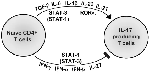
To date, there are six 17 family members [17A, 17B, 17C, 17D, 17E (25) and IL-17F], and five receptors (IL-17RA, IL-17RB/IL-25R, IL-17RC, IL-17RD/SEF and IL-17RE) (Table 2) [124]. Interleukin-17A and IL-17F are the most extensively studied. IL-17A/F producing CD4 T cells (Th17) specific for C. albicans, express on their surface the CCR4 and CCR6 receptors, which target them mainly to the skin and mucous membranes [125]. Epithelial cells express both IL-17RA, IL-17RC, IL-22R and 10R2 and thus can responses to Th17 cytokines mainly 17A, 17F (17A/17A and IL-17F/IL-17F homodimers or IL-17A/IL-17F heterodimers) and IL-22 [71, 96, 126]. Treatment of human bronchial lung epithelial (HBE) cells with IL-17 induces CXC chemokines such as IL-8 [71, 127], G-CSF[71], and antimicrobial proteins such as human β-defensin 2 (HBD2) [128]. IL-17 cytokines have also been shown to be important in regulating Th2 [129] and IgA [130] responses in the mucosa. IL-22 can activate STAT3 [131] and synergizes with IL-17 to increases the expression of human antimicrobial genes such as HBD2. A unique activity of IL-22, not shared with IL-17, is to increase the clonogenic potential of epithelial cells and accelerate wound repair [96]. More directly, treatment of skin keratinocytes with Th17 cytokines markedly increases anti-candidial activity in vitro, and this activity is lost when keratinocytes are tested in the presence of activated T cells from AD-HIES patients due to the absence of Th17 in these patients [132, 133]. Thus, Th17 cytokines are involved in mucocutaneous protection againt C. albicans (Figure 1) [56, 134-136]. Although Th17 have been suggested to be the main IL-17 cytokine producers, cellular sources of IL-17 include γδ T cells, innate NKT (iNKT) cells and innate lymphoid cells (iLCs) [137, 138]. Particularly in mucosal infections, the γδ T cells response can be the predominant source and their IL-17 production is critically regulated by both IL-23 and IL-1β [139, 140].
20
Table 2 IL-17 receptor and ligand families (adapted from [124])
Receptor complex Ligand(s)
IL-17RA/RC IL-17A, IL-17F, IL-17A/F, viral IL-17a
IL-17RA/RB IL-17E (a.k.a., IL-25)
IL-17RD (SEF) Unknown
IL-17RA/RD Unknown
IL-17RE IL-17C
Unknown IL-17D
Figure 1: PRR and Th17-based immunity to Candida albicans (adapted from [88])
(A) PRRs including CLRs (Dectin-1, Dectin-2, MINCLE) and TLRs (TLR2, TLR4) respond to Candida PAMPs by inducing through Syk/CARD9/BcL10/MALT1 or MyD88, respectively, the NF-kB and MAPK pathways, leading to the secretion of pro-inflammatory cytokines such as IL-6, IL-23, and IL-1β. (B) These cytokines bind to their receptors expressed on T cells, thereby inducing the differentiation of naïve T cells toward IL-17 producing T cells, in particular via the transcription factor STAT3, activated by IL-6, IL-23 (and IL-21) that in turn induces ROR-γt (ROR-γt for mice, its homologue RORC for human), leading to the transcription and the secretion of the IL-17 cytokines.
(C) IL-17A and IL-17F produced by Th17 cells bind to their receptors (IL-17RA/IL-17RC) expressed on various cells such as epithelial or mesenchymal cells to induce the expression of neutrophil attracting chemokines (IL-8, CXCL1, CXCL5) and activating/growth factors (G-CSF), as well as antimicrobial peptides (AMPs) such as defensins and S100 proteins [88]
21
2.2.2 Impaired IL-17 immunity in syndromic CMC
Considering that (i) IL-17 T cell plays an important role in immunity against C. albicans (based on studies in humans and mice), (ii) they are involved in the skin and mucous membranes protection, (iii) 6, 21, and 23 in particular, act via STAT3 to induce differentiation, proliferation and maintenance of IL-17 T cells, de Beaucoudrey et al. evaluated the presence of these cells in STAT3+/- patients with AD-HIES syndrome and syndromic CMC [23]. They have demonstrated in these patients, a significant decrease in the percentage of IL-17 T lymphocytes [55]. They have also observed a significant reduction in the proportion of these lymphocytes in patients deficient in IL-12p40 or IL-12Rβ1 with a complete lack of production or response to IL-12 and IL-23, respectively [55]. Although susceptible to mycobacterial infections or salmonella, approximately 25% of these patients develop moderate CMC [141]. These results therefore suggested that impaired IL-17 immunity could be at the origin of the syndromic CMC in these patients [21, 42, 43].
To further study the relation between impaired IL-17 immunity and CMC, our laboratory hypothesized that in APS-1 patient, CMC might result from autoimmunity against IL-17 cytokines. Indeed, these patients have numerous autoantibodies (auto-Abs), some directed against interferons IFN-α/-ω. However, these auto-Abs are probably not the cause of the associated CMC, as 65% of patients with thymomas have anti-IFN autoantibodies at diagnosis, and rare cases develop CMC [142. In addition, no CMC was reported in patients with various forms of STAT1 and TYK2 deficiency (unpublished data) and impaired responses to type I IFNs, or in patients with various forms of NEMO, UNC-93B, and TLR3 deficiencies and impaired production of type I IFNs {Al-Owain, 2010 #207]. In our laboratory, Puel et al. investigated the presence of auto-Abs against the IL-17s. In fact, along with another team, they found high titers of neutralizing auto-Abs against IL-17A, IL-17F, and/or IL-22, probably at the origin of the CMC occurrence in these patients [39-41]. These studies have contributed to the identification and characterization of the pathological mechanisms likely responsible for the CMC observed in these three syndromes, strongly suggesting that IL-17 T cells play a major role in mucocutaneous immunity against C. albicans in humans.
22 3. Results: CMCD and specific genetic defects of IL-17 immunity
3.1 Complete AR IL-17RA and partial AD IL-17F deficiency
According to literature, earlier studies had demonstrated the importance of IL-17 responses in host defense against mucosal candidiasis, both in mouse and human studies [88] and our laboratory results showed low IL-17 T cell proportions in PIDs with syndromic CMC [21, 42, 43], including AD-HIES syndrome [55] and AR APS-1 [41], as well as CARD9 deficient patients [27]. We therefore hypothesized that among CMCD patients, some may have a genetic defect affecting specifically IL-17-dependent immunity [21, 42, 43]. Puel et al. have undertaken the sequencing of candidate genes encoding 17s (including 17A,
IL-17F, IL-21, IL-22, and IL-26) and their receptors in a cohort of CMCD patients recruited over the last
years. Thus, in a child with CMCD (and skin infections with S. aureus) from a consanguineous family of Moroccan origin, we identified the first complete AR IL-17RA deficiency. The patient is carrying a homozygous mutation causing a premature stop codon located in the extracellular domain of IL-17RA (Q284X). This mutation was not found in any of public databases (1000 genomes, NCBI, Ensembl), nor in our own database of exomes (sequencing of the entire coding regions of the genome) sequenced in the laboratory (> 1000 today), nor among the 1052 control individuals from the Human Genome Diversity Project (HGDP) panel/ Center for the Study of Human Polymorphism (CEPH) panel, nor in 100 Moroccan controls sequenced, which excluded the variation being a polymorphism (i.e. frequent allele). I showed that this mutation abolished the expression of the IL-17RA protein at the surface of the patient’s cells (fibroblasts and PBMCs), as well as their response to the homo- or hetero-dimers of 17A and IL-17F. Parents and siblings heterozygous for the mutation do not suffer from CMCD, demonstrating the recessive nature of the deficiency. Transfection of the patient’s fibroblasts with a plasmid encoding the wild-type (WT), but not the mutant (Q284X) IL17RA or an empty plasmid, restored IL-17RA expression and the response to IL-17 cytokines in terms of IL-6 and Gro-α production. Thus, AR complete IL-17RA deficiency results in an abolished response to IL-17 cytokines and CMCD. At the same time, in a multiplex family with 5 CMCD individuals among 3 generations, we have identified the first partial AD
23 IL-17F deficiency, caused by a heterozygous missense mutation (S65L) in IL-17F. Again, it was not found in any of public or laboratory databases or CEPH control individuals, excluding a polymorphism. This mutation, located in the binding domain of the IL-17F to its receptor, is strongly hypomorphic (almost total loss of function) and dominant because it impairs the functionality of homo- (IL-17F/IL-17F) and hetero- (IL-17A/IL-17F) dimers containing the mutated protein, by blocking the binding of these complexes to their receptors [Publication #1]. This study has validated our hypothesis. It led to the discovery of the first two genetic etiologies of CMCD, showing that it is indeed a primary immunodeficiency. It confirmed the major role of IL-17s in mucocutaneous immunity against C. albicans in humans. However, unlike the situation observed in mice, these cytokines seemed redundant in the protection against the most common pathogens, since none of the reported patients had developed any severe infectious diseases besides CMCD. However, it is absolutely necessary to identify other patients with these deficiencies to draw definitive conclusion.
3.2 Heterozygous gain-of-function (GOF) STAT1 mutations
3.2.1 Heterozygous STAT1 GOF mutations are a major genetic etiology of CMCD
After the identification of the first two genetic etiologies of CMCD with AR IL-17RA and AD IL-17F deficiencies in one family each, using a candidate gene approach, mainly focusing on IL-17 immunity [Publication #1], we performed a “genome-wide” approach, by using whole-exome sequencing (WES), to identify novel morbid genes. We first performed WES for 6 CMCD patients. In order to analyze the WES data of the 6 CMCD patients, I first decided to focus on genes related to IL-17 immunity, as it is essential in mucocutaneous defense against Candida, even though the candidate gene strategy focused on IL-17 immunity (sequencing of IL17A, IL17F, IL17RA IL17RC, IL22, IL22RA1, IL10RB,) we used, let us to identify only 1 patient with AR IL-17RA deficiency in one family and five patients with AD IL-17F deficiency in another family. This suggested either that the gene (or genes) involved in IL-17 immunity and mutated in the rest of the CMCD cohort was (or were) not included in the candidate gene
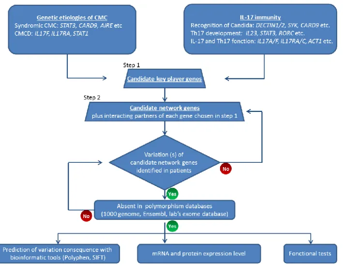
24 investigation; or that there was (or were) other pathogenesis mechanism(s) underlying CMC than impaired IL-17 immunity. Considering the first hypothesis probable, I focused on IL-17 immunity and enlarged the candidate gene list. I did this in three steps: 1) I chose all the genes found mutated in PIDs with syndromic or isolated CMC: STAT3, AIRE, CARD9, IL17F, IL17RA, IL12B, IL12RB1; 2) I also included essential genes involved in IL-17 immunity according to the literature, such as IL17A, RORC, SYK etc. The candidate genes chosen in these first two steps are named as “IL-17 immunity key genes”; 3) I then added 20 interacting partners to each of these “IL-17 immunity key genes”. The choice of these partners was done based on an online program “STRING”, which collects known and predicted protein interactions including direct (physical) and indirect (functional) associations [143]. My idea was therefore to first check for these “IL-17 immunity key genes” in the 6 patients’ exome data; then to check their interacting partners, and if no mutation could be found in any patient for this gene-list, then to increase the “candidate network genes”. I called this strategy “candidate network” (Figure n° 2). The strategy was therefore to combine the online gene network database (STRING) and the whole-exome sequencing data. Besides the “candidate network” strategy, I also performed studies by following “hypothesis-generating” strategy, which means trying to identify variations in one gene common in two or more patients without proposing pathogenesis hypothesis. Combining these different approaches, I identified in four out of the six patients, three different STAT1 heterozygous missense variations. STAT1 was the only “candidate network gene” which displayed variations in more than four patients. These variations were not reported in any public dababase (1,000-genome, National Center for Biotechnology Information NCBI, Ensembl, and dbSNP) or in our own database (250 exomes at that time), excluding the variations being polymorphisms but instead rare events (mutations). We confirmed these mutations by Sanger sequencing and also excluded their presence in the 1,052 controls from 52 ethnic groups from the HGD/CEPH panels. Thus, these mutations were suggested to be probably CMCD-causing variants rather than irrelevant polymorphisms.
25
Figure 2 “Candidate gene network” strategy
1) the “candidate key player genes” were chosen according to both experimental data from mouse infection models and epidemiological studies in humans concerning IL-17 immunity and/or CMC pathogenesis; 2) “candidate gene network” list covered not only “key player genes” but also their interacting partners with the help of protein network database STRING; 3) verification of variations of candidate genes in patients and elimination of polymorphism. If no “non-polymorphsim” variation is identified in patient(s), the “candidate gene network” list is enlarged by adding more “key players” partners; 4) bioinformatics prediction and in vitro functional test of variation(s)’ consequence.
26
3.2.2 Heterozygous STAT1 variations identified in CMCD patients are gain-of-function mutations
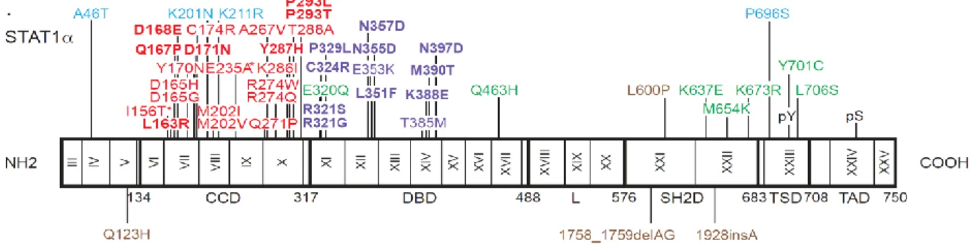
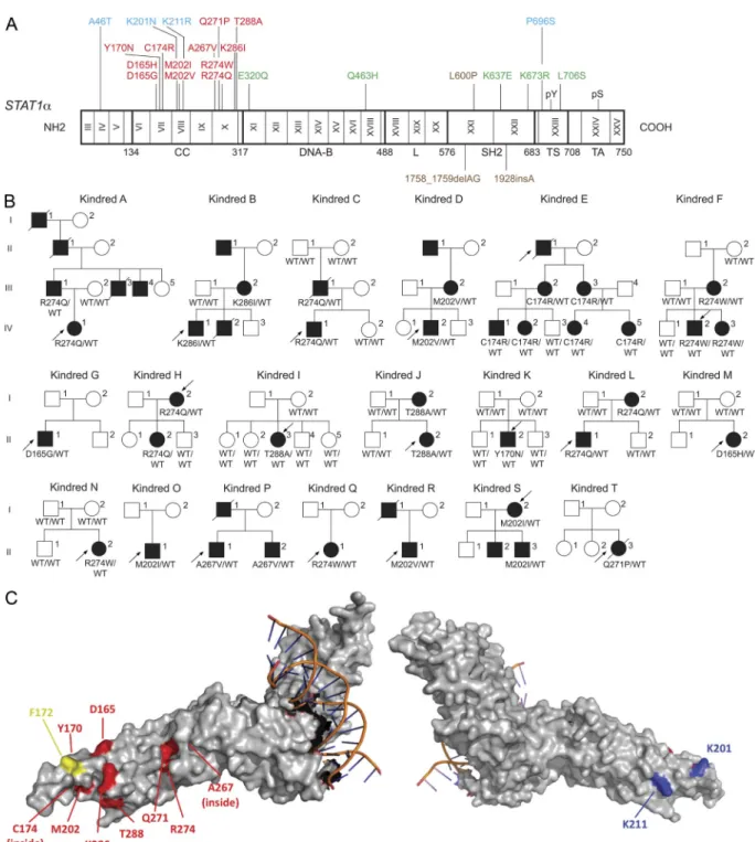
We identified 139 CMCD patients from 74 families with 32 heterozygous missense mutations affecting the coiled-coil domain (CCD) (106 patients from 53 kindreds with 12 mutations) or the DNA binding domain (DBD) (33 patients from 21 kindreds with 20 mutations) of STAT1 (Figure 3) [Publication #3], when we submitted the publication #3. To date, there are in total 55 heterozygous missense STAT1 CMC mutations identified by us or other teams in 196 patients from 121 families (unpublished data). Their clinical penetrance appeared to be complete, as all CMCD patients from the kindreds tested were heterozygous, whereas none of these mutations was found at the heterozygous state in any of the healthy relatives sequenced. The mutations were not found in any public or in-house databases.
Figure 3 Heterozygous missense mutations affecting the CCD and DBD of STAT1 in kindreds with AD CMCD. The human STAT1 alpha isoform is shown, with its known pathogenic mutations. Coding
exons are numbered with Roman numerals and delimited by a vertical bar. Regions corresponding to the coiled-coil domain (CCD), DNA-binding domain (DBD), linker domain (L), SH2 domain (SH2D), tail segment domain (TSD), and transactivator domain (TAD) are indicated, together with their amino-acid boundaries, and are delimited by bold lines. Tyr701 (pY) and Ser727 (pS) are indicated. Mutations in green are dominant and associated with partial STAT1 deficiency and Mendelian susceptibility to mycobacterial disease (MSMD). Mutations in brown are recessive and associated with complete STAT1 deficiency and intracellular bacterial and viral disease. Mutations in blue are recessive and associated with partial STAT1 deficiency and intracellular bacterial and/or viral disease. Mutations in red are dominant, located in the region encoding the CCD and associated with a gain of function of STAT1 and CMCD. Mutations in violet are dominant, located in the region encoding the DBD and associated with gain of function of STAT1 and CMCD..
27 The fact that heterozygous STAT1 mutations were identified in more than half of the CMCD patients from our cohort (almost 300 patients) and absent in healthy controls, already gave a strong argument for the causality of these STAT1 mutations in CMCD. However, the next question was what was the molecular impact of these STAT1 alleles and how could they lead to the CMCD clinical phenotype?
Signal transducer and activator of transcription 1 (STAT1) belongs to a family of transcription factors comprising STAT1, STAT2, STAT3, STAT4, STAT5A, STAT5B and STAT6. STAT1 exists as two isoforms: STAT1α (91kDa) and STAT1β (84kDa) resulting from alternative splicing of the transcript. Both isoforms contain an N-terminal domain, a coiled-coil domain (CCD), a DNA-binding domain (DBD), and a SH2 domain. Only the STAT1α isoform possesses a transactivation domain (TAD) including two phosphorylated sites: tyrosine 701 and serine 727. Phosphorylated tyrosine 701 is required for STAT1 dimer formation and its transcription activity. STAT1 is an essential effector of IFNs including type I (IFN-α/β) and type II (IFN-γ), but also IFN-λ and IL-27. Following activation of the IFN-γ or IL-27 receptor, STAT1 is phosphorylated on tyrosine 701 and forms a homodimer. In response to IFN-α/β, phosphorylated STAT1 forms a heterotrimer with phosphorylated STAT2 and p48/IRF-9. Activated STAT1 homodimers or heterotrimers translocate into the nucleus, where they bind to specific consensus sequences: the GAS (gamma interferon activated sequence) or the ISRE (interferon stimulated response element) sequences, respectively. STAT1 homodimers (GAF: gamma interferon activating factor) bind to GAS DNA sequences via their N-terminal domain and stimulate the transcription of genes mainly involved in antibacterial immunity. STAT1/STAT2/p48 heterotrimers (ISGF3 complex) induce the transcription of genes involved in antiviral immunity via their binding to ISRE DNA sequences. STAT1 is also activated in response to other cytokines, including IL-6, IL-21 and IL-23; and in response to growth factors including EGF and PDGF [144]. STAT1 knockout mice respond very poorly to IFN-α/β, IFN-γ and IL-27. These mice are susceptible to viruses, bacteria and parasites [46]. However, when they are challenged with C. albicans, their response is similar to that of wild-type mice[145].
28 In human, biallelic or monoallelic mutations in STAT1 had already been identified [46]. Human AR complete STAT1 deficiency leads to life-threatening intra-macrophagic bacterial and viral disease, and partial AR STAT1 deficiency presents a milder susceptibility to bacterial and viral diseases. AD STAT1 deficiency results in a rare syndrome characterized by infections with weakly pathogenic/virulent mycobacteria (Mendelian susceptibility to mycobacterial disease: MSMD syndrome). None of these STAT1 deficient patients had to our knowledge a susceptibility to fungal infection. Therefore, we made the hypothesis that the STAT1 mutations identified in CMCD patients could be gain-of-function (GOF) instead of loss-of-function (LOF) as those previously identified, at least in response to IFNs and IL-27. To test this hypothesis, we functionally characterized the CMCD-causing STAT1 allele, R274Q, found in several kindreds. We compared it with a WT and an MSMD-causing LOF STAT1 allele (L706S). A higher STAT1 activity in response to IFNs and IL-27 was observed in STAT1-deficient U3C cells transfected with R274Q compared with WT or LOF alleles. Similarly, we also observed a higher STAT1 activity in Epstein-Barr (EBV) - transformed B cells from a patient heterozygous for the STAT1 R274Q allele compared with those from controls or LOF patients. These experiments demonstrated the GOF and the dominant nature of the CMCD-causing STAT1 mutation [Publication #2].
3.2.3 GOF STAT1 mutations lead to impaired IL-17 immunity in CMCD patients
The susceptibility to intracellular bacterial and/or viral infections in STAT1 deficient patients is explained by impaired STAT1-dependent IFN immunity. In order to understand the pathological mechanism of
STAT1 GOF mutations in CMCD we asked two questions: 1) whether IL-17 immunity was impaired in
these CMCD patients with STAT1 GOF alleles similarly to patients with syndromic CMC; 2) if so, whether GOF STAT1 mutations were the cause of the impaired IL-17 immunity.
Indeed, we highlighted an impaired development of IL-17-producing T cells, both ex vivo and after in
vitro differentiation starting from CMCD patients’ bulk leukocytes (Figure 4) [Publication #2], We also
demonstrated that CMCD patients heterozygous for STAT1 gain-of function alleles displayed poor IL-17-producing T-cell development from naïve CD4+ T cells after in vitro differentiation [Publication #3],
29
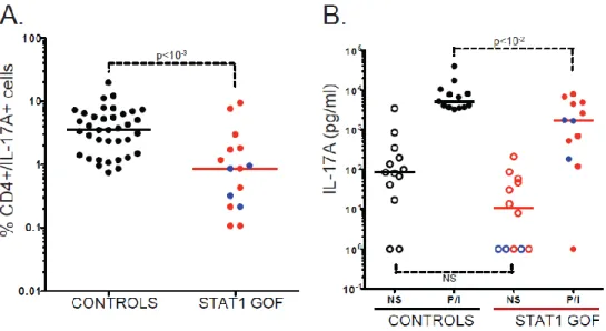
Figure 4 The differentiation of naïve CD45RA+ CD4+ T cells into IL-17- producing T cells in vitro is impaired in patients with AD CMCD and STAT1 GOF mutations. (A-B) Proportion of
IL-17A-producing T cells (A) and IL-17A secretion (B), after 12 days of naïve CD4+ T cell differentiation in the presence of anti-CD3 and anti-CD28 antibodies, IL-1β and IL-23, as determined by flow cytometry and ELISA, respectively, after stimulation with PMA and ionomycin for 12 hours for flow cytometry analysis, and in the absence of stimulation (open symbols) or after 48 hours of stimulation (closed symbols) for ELISA analysis. Each symbol represents a value from a healthy control individual (black circles), a patient bearing a STAT1 gain-of-function (GOF) mutation affecting the CCD (red circles) or a patient bearing a STAT1 GOF mutation affecting the DBD (violet circles). Horizontal bars represent medians. The p-values for the nonparametric Wilcoxon tests are shown for comparisons of patients with STAT1 GOF mutations (STAT1 CCD: n = 11 and STAT1 DDB: n = 4) and controls (n = 37) (A) patients with STAT1 GOF mutations (STAT1 CCD: n = 9 and STAT1 DDB: n = 3) and controls (n = 13) (B).
3.2.3.1 Hypothesis I: major STAT1 activators (IFNs and IL-27) have a stronger inhibitory effect on IL-17 T cell differentiation in CMCD patients.
Both mouse [109-116] and human [45, 117-121] studies have shown that IFNs and IL-27 inhibit Th17 cell development in a STAT1 dependent manner in mice and humans. Thus, we hypothesized that CMCD in patients with GOF STAT1 mutations could result from stronger STAT1 dependent inhibitory effect, downstream of IFNs and IL-27, on IL-17 T cell differentiation. To test this hypothesis, I first investigated IL-17 T cell differentiation starting from naïve CD4 T cells as an in vitro model to investigate whether the CCD and DBD STAT1 mutations impaired the development of IL-17 T cells. A combination of TGF-β
30 and IL-6 had been shown to be essential for the initial differentiation of IL-17 T cells in mice [146, 147], but the key cytokines required in humans remained less clearly defined (Sallusto et al., 2012). Various combinations of cytokines had been used for the differentiation of human 17 T cells: (1) TGF-β and IL-21; (2) IL-1β and IL-6; (3) IL-1β and IL-23 and (4) TGF-β and IL-23 [148-153]. I thus purified CD45RA+ CD4+ T cells by magnetic beads and cultured them in the presence of coated antibody (Ab) against CD3 and soluble Ab against CD28, together with individual cytokines or all possible combinations of TGF-β, IL-1β, IL-6, IL-21 and IL-23, in the presence of IL-2. I measured the proportion of IL-17A-expressing T cells and the secretion of IL-17A from days 5 to 12, by flow cytometry and ELISA, respectively. I obtained the most reproducible results within and between controls with a combination of IL-1β and IL-23 for 12 days (data not shown). IL-6 was not retained, as it increased inter-individual variability. In these conditions, I showed that patients heterozygous for CCD or DBD STAT1 mutations had lower (p < 10-3) proportions of IL-17A T cells and secreted smaller amounts of IL-17A (p < 10-2) [Publication #3], Impaired IL-17 T-cell development in these patients was similar to that seen in patients with other conditions conferring CMC, including AD-HIES patients with heterozygous LOF STAT3 mutations and AR MSMD patients with biallelic IL12RB1 LOF mutations [Publication #3]. Collectively, these data demonstrate that the GOF STAT1 mutations caused CMCD by impairing IL-17 T-cell immunity.
To further investigate the mechanisms by which CMCD-causing GOF STAT1 alleles prevent the development of IL-17 T cells, in the culture conditions defined above, I added a combination of suboptimal doses of Th17 cell differentiation inhibitors (IFN-α2a, IFN-β1a, IFN-γ and IL-27). I observed a large decrease in the proportion of IL-17A T cells and in the secretion of IL-17A, in both the healthy controls and the STAT1 patients tested [Publication #3]. The effect was statistically significant when measured both by flow cytometry and ELISA, for CMCD patients (p < 10-3), and for controls (p < 5 x 10⁻³ and p < 10-2 respectively). Moreover, in these inhibitory conditions, the difference in terms of IL-17A T cell proportion and IL-17A production between controls and patients was more significant than in the absence of IFNs and IL-27 (p < 10-3) [Publication #3]. At higher concentrations of IFN and IL-27, a
31 stronger inhibition was observed in the cells of controls and patients, with similar levels of inhibition in both [Publication #3]. These data suggested that the poor development of IL-17 T cells in patients heterozygous for STAT1 alleles might result, at least in part, from enhanced IFN-α/β, IFN-γ and IL-27 responses via STAT1. I tested this hypothesis, by treating the cells with a combination of neutralizing Abs against IFN-α/βR2, IFN-γ and IL-27. These Abs rescued the development of IL-17 T cells carrying GOF
STAT1 mutations, whereas this effect was not detectable in healthy control IL-17 T cells [Publication #3].
Indeed, the effect of these Abs reached significance only in the patients’ cells (p < 10-3 by flow cytometry and p < 5. x 10-4 by ELISA). Moreover, in these conditions, the difference between the cells of the controls and those of the patients was abolished [Publication #3]. In these conditions, the proportion of CD4+ IFN-γ+ was slightly lower in the patients’ cells, whereas the amounts of IFN-γ and IL-27 secreted were similar for controls and patients [Publication #3]. Overall, these experiments established that the poor development of IL-17 T cells in CMCD patients carrying GOF mutations affecting the CCD or DBD of STAT1 involves STAT1-dependent inhibition via IFN-α/β, IFN-γ and/or IL-27.
Figure 5: hypothesis I: major STAT1 activators (IFNs and IL-27) have a stronger inhibitory effect on IL-17 T cell differentiation in CMCD patients. Activating molecules, such as IL-23 and IL-21
(acting mostly through STAT3, and to a lesser extent, STAT1), IL-6, IL-1β, and TGF-β ,and inhibiting molecules, such as IFN-α/β, IFN-γ, and IL-27 (acting mostly through STAT1 and, to a lesser extent, STAT3) are represented.
![Table 1: IL-17 immunity in immunity against bacterial, mycobacterial or fungal pathogens [50]](https://thumb-eu.123doks.com/thumbv2/123doknet/2273620.21527/15.918.126.799.134.1028/table-il-immunity-immunity-bacterial-mycobacterial-fungal-pathogens.webp)
![Table 2 IL-17 receptor and ligand families (adapted from [124])](https://thumb-eu.123doks.com/thumbv2/123doknet/2273620.21527/21.918.117.777.256.812/table-il-receptor-ligand-families-adapted.webp)