THÈSE
En vue de l’obtention du
DOCTORAT DE L’UNIVERSITÉ DE TOULOUSE
Délivré par l'Université Toulouse 3 - Paul Sabatier
Présentée et soutenue par
XIAOHUI TONG
Le 4 septembre 2018
Rôle de la synthèse des miARN par le tissu adipeux dans
les pathologies de vieillissement.
Ecole doctorale : BSB - Biologie, Santé, Biotechnologies Spécialité : SCIENCES DU VIEILLISSEMENT
Unité de recherche :
I2MC - Institut des Maladies Métaboliques et Cardiovasculaires Thèse dirigée par
Philippe VALET Jury
Mme Soazig LE LAY, Rapporteur
M. Miguel GODINHO FERREIRA, Rapporteur M. Ez-zoubir AMRI, Rapporteur
Mme Laurence NIETO, Examinateur M. Philippe VALET, Directeur de thèse M. Cedric DRAY, Co-directeur de thèse
RESUME
D'une part, le dicer, l'endoribonucléase RNase III responsable de la maturation des microARN, aurait été réduit chez les adipocytes au cours du vieillissement. Avec l'utilisation de souris dicerlox / lox / adiponectine-CreERT2 inductibles par le tamoxifène, nous avons constaté que le déficit en adipocytes favorisait l'apparition de certaines complications liées à l'âge, telles que la réduction de la taille des adipocytes et le dysfonctionnement du métabolisme. L'abrogation des marqueurs adipocytaires blancs, tels que Pparγ, Glut4 ou Hsl, indique que le dicer est indispensable au maintien de l'identité des adipocytes blancs. De plus, les résultats montrant une accumulation de lipides et une fibrose hépatique chez des souris traitées par le damerlox / lox / adiponectine-CreERT2 + traitées au tamoxifène ont indiqué que la carence en adicocyte pourrait contribuer au vieillissement du foie.
Mécaniquement, la fonction mitochondriale semblait être régulée à la hausse en raison d'une déficience en dicer adipocytaire, indiquée par une augmentation des taux de protéines des composants d'OXPHOS et de PGC1α. En ligne, les répresseurs des mitochondries FOXO1 et FOXO3 ont été phosphorylés et inactivés, dont les cibles antioxydantes en aval, la Catalase et le Sod, ont également diminué. En outre, P16, un marqueur de la sénescence, a montré une tendance à augmenter en raison de la carence en dicer. Comme le surplus de ROS mitochondrial peut entraîner des dommages à l'ADN et la sénescence, nous avons assuré que la déficience en dicer adipocytaire pourrait induire une combinaison d'activation mitochondriale et une réduction de la réduction de la détoxification possiblement induite par l'inactivation de FOXO1 et FOXO3a. Enfin, le résultat selon lequel la restriction en éléments nutritifs a régulé positivement le taux de dicer dans les adipocytes a également confirmé qu'il existe une voie de vieillissement conservée dans les adipocytes impliquant le dicer.
Par ailleurs, grâce à la sélection de microréseaux et à la validation RT-qPCR, nous avons rapporté que le vieillissement augmentait le mir-1949 dans les adipocytes chez les souris de type sauvage et favorisait probablement sa sécrétion à partir du tissu adipeux perigonadal in vitro. De manière correspondante, des résultats in vitro ont également suggéré que la sénescence augmentait la production de mir-1949 et la sécrétion d'adipocytes. Sur le plan fonctionnel, la régulation positive du mir-1949 dans les adipocytes
3T3-F442A régulait négativement le niveau du complexe mitochondrial II et la capacité de consommation d'oxygène, associées à l'accumulation de lipides. Fait intéressant, la régulation à la hausse soutenue du mir-1949 au cours de l'adipogenèse des préadipocytes 3T3-F442A avait tendance à augmenter les marqueurs adipocytaires blancs, tels que la leptine, Glut4 ou Hsl (N = 2). Enfin, combiné aux résultats indiquant que le jeûne de 24 heures a significativement augmenté le mir-1949 dans les tissus adipeux péri-radicaux et que la régulation positive du mir-1949 tendait à augmenter l'accumulation de lipides dans les adipocytes séniles 3T3-F442A (N = 1) augmente l'expression de mir-1949 dans les adipocytes, ce qui pourrait tenter de sauver des dysfonctionnements liés à l'âge dans les adipocytes, tels qu'une altération du stockage des lipides. Néanmoins, les actions précises de mir-1949 doivent être validées in vivo par injection de mimétique d'AAV-aP2-mir-1949 chez des souris âgées.
Parallèlement à mon projet de thèse, nous avons dû moduler les expressions géniques dans des adipocytes différenciés à partir de lignées cellulaires immortalisées avec des techniques de routine telles que les transfections d'ARNsi. Nous avons donc cherché à développer une technique permettant de conserver des adipocytes isolés pour des expériences. À l'aide d'inserts de culture cellulaire, nous avons réussi à séquencer des adipocytes matures flottants et à les exposer à l'air. En outre, en réduisant la tension de surface avec la supplémentation en co-solvants, le taux de survie des cellules adipeuses en culture a apparemment augmenté. Ensuite, les résultats de la qPCR ont démontré que les taux d'ARNm des marqueurs des adipocytes augmentaient après presque une semaine de culture, ce qui correspondait à la taille accrue des cellules. En outre, les adipocytes cultivés dans notre système de culture se sont avérés fonctionnels, pouvant répondre respectivement à la stimulation catécholamine / insuline pour la lipolyse et le transport du glucose. Par conséquent, la nouvelle façon dont nous avons invité est capable de rester des adipocytes matures pour une étude in vitro sans perdre ses caractéristiques de morphologie et de fonction.
Ensemble, ces deux aspects de ma thèse nous amèneraient à mieux comprendre le domaine des adipocytes et du vieillissement.
ABSTRACT
On the one part, dicer, the RNase III endoribonuclease responsible for microRNAs maturation, has been reported to be decreased in adipocytes during ageing. With the use of tamoxifen inducible dicerlox/lox/ adiponectin-CreERT2 mice, we found that adipocyte dicer deficiency promoted the onset of some of the age-related complications, such as reduced adipocyte sizes and dysfunctions in systemic metabolism. The abrogation of white adipocyte markers such as Pparγ, Glut4 or Hsl, indicated that dicer is indispensible for the maintainance of white adipocyte identity. In addition, the results that there were lipid accumulation and fibrosis in liver in tamoxifen treated dicerlox/lox/ adiponectin-CreERT2+ mice, indicated that adipocyte dicer deficiency might contribute to liver aging.
Mechanistically, mitochondrial function seemed to be upregulated due to adipocyte dicer deficiency, indicated by increased protein levels of OXPHOS components and PGC1α. In line, mitochondria repressors FOXO1 and FOXO3 were phosphorylated and inactivated, whose downstream antioxidant targets Catalase and Sod were also decreased. Moreover, P16, a marker of senescence, exhibited a trend to be increased due to adipocyte dicer deficiency. Since mitochondrial ROS surplus can lead to DNA damage and senescence, we assured that adipocyte dicer deficiency might induce a combination of mitochondrial activation and reduction in detoxification reduction possibly mediated by the inactivation of FOXO1 and FOXO3a. Finally, the result that nutrient restriction positively regulated dicer level in adipocytes further supported that there is a conserved aging pathway in adipocytes involving dicer.
On the other part, through microarray screen and RT-qPCR validation, we reported that aging increased mir-1949 in adipocytes in wild type mice and possibly promoted its secretion from perigonadal adipose tissue in vitro. Correspondingly, in vitro results also suggested that senescence increased mir-1949 production and secretion from adipocytes. Functionally, upregulation of mir-1949 in 3T3-F442A adipocytes negatively regulated mitochondrial complex II protein level and oxygen consumption capability, associated with lipid accumulation. Interestingly, sustained upregulation of mir-1949 during adipogenesis of 3T3-F442A preadipocytes, tended to increase white adipocyte markers, such as Leptin, Glut4 or Hsl (N=2). Finally, combined with the results that 24h fasting significantly increased
mir-1949 in perigonadal adipose tissue as well as that upregulation of mir-mir-1949 exhibited a trend to increase lipid accumulation in senescent 3T3-F442A adipocytes (N=1), we assured that aging increases mir-1949 expression in adipocytes which might try to rescue age-related dysfunctions in adipocytes, such as impaired lipid storage. Nevertheless, the precise actions of mir-1949 need to be validated in vivo by injection of AAV-aP2-mir-1949 mimic into aged mice.
In parallel of my PhD project, we faced the matter of modulating gene expressions in differentiated adipocytes from immortalized cell lines with routine techniques like siRNA transfection. Therefore, we aimed to develop a technique enabling us to conserve isolated adipocytes for experiments. With the help of cell culture inserts, we managed to sequence buoyant mature adipocytes from floating and exposing into air. Moreover, by reducing surface tension with supplementation of co-solvents, the survival rate of cultured fat cells was apparently increased. Then, RT-qPCR results demonstrated that mRNAs levels of adipocytes markers increased after nearly one week of culture, in consistent with the increased cell sizes. Besides, cultured adipocytes in our culture system were proved to be functional, being able to response to catecholamine/ insulin stimulation for lipolysis and glucose transport, respectively. Therefore, the new way that we invited is capable of remaining mature adipocytes for in vitro study without losing its characteristics of morphology and function.
Taken together, these two aspects of my PhD would lead us to understand more in the field of adipocytes and aging.
1
LISTS OF FIGURES AND TABLES
FIGURES
Introduction part
Figure 1: Growing interests in adipose biology over the last century. ... 10
Figure 2: Adipose tissue distribution associates with the incidence of diabetes development. ... 11
Figure 3: Representation and transmission electron microscopy of the morphology of white adipocytes. ... 14
Figure 4: Extracellular support of adipocytes in vivo. ... 15
Figure 5: Schematic representation of FFA uptake mediated by CD36 in caveolin-1-FABP rich microdomain. ... 18
Figure 6: Insulin signaling regulates GLUT4 exocytosis by engaging the trafficking machinery. ... 20
Figure 7: Tight control of lipolysis process by IIS, β-adrenergic signaling and NP receptor A signaling pathways. ... 22
Figure 8 : Adipocytes crosstalk with various organs through adipokines. ... 26
Figure 9 : Simplified scheme of white adipocyte origins. ... 28
Figure 10: The classical pathway of microRNA biogenesis... 43
Figure 11: Hallmarks of senescent cells. ... 63
Figure 12 : The p53 and RB tumor-suppresor pathways. ... 64
Figure 13: Simple illustrations of microRNAs’ participations in calorie restriction process in different species. ... 70
Figure 14: Examples of effects of microRNAs in sirtuins during aging or senescence. ... 73
Figure 15 : 3T3-F442A adipocytes differentiated from 3T3-F442A fibroblasts. ... 75
Figure 16: Isolation of SVF from adipose tissue with collagenase digestion and centrigugation for in vitro adipogenesis. ... 75
Figure 17: 3D adipogenesis of 3T3-L1 cells inelectrospun polycaprolactone scaffolds. ... 77
Figure 18: Formation of spheroids of 3T3-L1 adipocyte aggregates on surface tethered with ELP-PEI. ... 78
Figure 19: Ceiling culture of isolated adipocytes. ... 79
Results part Result 1: Role and regulation of microRNAs in adipose tissue during aging Figure 1: Inducible reduction of dicer in adipocytes by tamoxifen in dicerLox/Lox/ adiponectin-CreERT2+ mice. ... 85
Figure 2: Reduction in the average sizes of perigonadal adipocytes due to dicer deficiency. ... 87
Figure 3: Possible involved mechanisms underlying the effects of adipocyte dicer deficiency. ... 89
Figure 4: Adipocyte dicer deficiency promoted lipid accumulation and fibrosis in liver. ... 93
Figure 5: Regulation of dicer level in adipose tissue and in in vitro adipocytes. ... 96
Figure 6: Age-associated increase of mir-1949. ... 99
2
Figure 8: Proposed molecular mechanisms underlying adipocyte dicer in aging. ... 105
Figure 9: GFP expression in adipocytes from ZsGreenlx/lx/adiponectin-CreERT2+ mice by 4-hydrotamoxifen. ... 108
Figure 10: Possible role of mir-1949 in the prevention of age-related dysfunctions. ... 110
Result 2: A novel method of culturing isolated adipocytes in vitro Fig 1: Illustration of the working scheme. ... 119
Fig 2: Image of adipocytes in culture under different condtions. ... 122
Fig 3: Functional investigation of adipocyte in vitro. ... 125
Fig 4: Characterizations of adipocytes during chronic culture in vitro. ... 127
TABLES
Introduction part Table 1: The average sizes of adipocytesin developing and adult human ... 16Table 2: List of miRNAs involved in adipogenesis ... 46
Table 3 : MiRNAs involved in age-associated cardiovascular diseases ... 59
Results part Result 1: Role and regulation of microRNAs in adipose tissue during aging Table 1: Plasma parameters (fed) ... 91
3
ABBREVIATION
ACC Acetyl CoA carboxylase
ACSL Adipose acyl-CoA synthetase
ADSCs Adipose tissue-derived stem cells
ADEV Adipose tissue derived extracellular vesicles ADRP Adipose differentiation-related protein
ADRB3 Β3-adrenergic receptor
AGO Argonaute
AGRP Agouti-related protein
ATGL Adipose tissue lipase
ALDH3A2 Aldehyde dehydrogenase 3 family member A2
AMPK AMP-activated protein kinase
ATF6 Activating transcription factor-6
ATMs Adipose tissue macrophages
ATSC Adipose tissue stromal cell
APAF1 Apoptotic protease-activating factor 1
APCs Adipoycte progenitor cells
ARF6 ADP-ribosylation factor 6
BAT Brown adipose tissue
BCL2 BMP
B-cell lymphoma 2
Bone morphogenetic protein
BMI Body mass index
BMSCs Bone marrow stromal cells
BSCL Berardinelli-seipcongential generalized lipodystrophy
BNIP3 BCL2/adenovirus E1B 19kDa interacting protein 3 CART Cocaine and amphetamine regulated transcript
C/EBPα CCAAT/enhancer binding protein α C/EBPβ CCAAT/enhancer binding protein β
ChREBP Carbohydrate response element binding protein
CR Calorie restriction
CPT Carnitine palmitoyl transferase
CLAs Conjugated linoleic acids
DGAT1 Diacylglycerol acyltransferase 1
DDRs DNA damage responses
DNL De novo lipogenesis
DGCR8 DiGeorge critical region 8
ECM Extracellular matrix
EPCs Endothelial progenitor cells
4
eNOS Endothelial NO systhesis
EVs Extracellular vesicles
FABP4 Fatty acid binding protein 4
FABPpm Plasma membrane fatty acid binding protein FAT10 HLA-F adjacent transcript 10
FAT/CD36 Fatty acid translocase FATP Fatty acid transport protein
FAS Fatty acid synthesis
FFAs Free fatty acids
FPLD Familial partial lipodystrophic syndromes
FOXO Forkhead box O
FGF21 Fibroblast growth factor 21
GCs Glucocorticoids
GH Growth hormone
GLUT4 Glucose transporter 4
GLP-1 Glucagon-like peptide-1
GO Gene ontology
HDAC9 Histone deacetylase 9
HEK 293T Human embryonic kidney cell line 293T
HK Hexokinase
HDL High density lipoprotein
HMGA2 High mobility group AT-hook 2
HOMA-IR Homeostatic model assessment for insulin resistance
HSC Hematopoietic stem cells
HSL Hormone-sensitive lipase
IBMX 3-isobutyl-1-methylxantine
IGF Insulin like growth factor
IRS Insulin receptor substrate
JAK Janus Kinase
JNK Jun-N-terminal kinase
KSRP KH-type splicing regulatory protein
LCFA Long chain fatty acid
LD Lipid droplet
MGL Monoglyceride Lipase
MiRNAs MicroRNAs
MiT/TFF Microphthalmia/ Transcription Factor E
5
mTOR Mammalian target of rapamycin
MUFA Mono-unsaturated fatty acid
NAMPT Nicotinamide phosphoribosyltransferase
NF-κB Nuclear factor-κb
NGS Next-generation sequencing
NPY Neuropeptide-Y
NLRP3 Nod-like receptor 3
OPN Osteopontin
PAI Plasminogen activator inhibitor PDK Phosphoinositide-dependent kinase
PDE3b Phosphodiesterase 3b
PEPCL Phosphoenolpyruvate carboxykinase
PGC-1 Peroxisome proliferator-activated receptor γ coactivator 1
PKA Protein kinase A
PLIN1 Perilipin 1
POMC Proopiomelanocortin
PPARγ Peroxisome proliferator-activated receptor-γ
PRDM16 PR domain containing 16
Pri-miRNAs Primary microRNAs Pre-miRNAs Precursor microRNAs
PTP1B Protein tyrosine phosphatase
PTEN Phosphatase and tension homologue
RB Retinoblastoma
RGS Rosiglitazone
RICTOR RPTOR-independent companion of mtor complex 2 RIP140 Receptor-interacting protein 140
RISC RNA-induced silencing complex
ROS Reactive oxygen species
SA-β-gal Senescence-associated β-galactosidase SCD1 Stearoyl coenzyme A desaturase-1
S6k1 Ribosomal S6 protein kinase
SIRT1 Sirtuin 1
SOD Superoxide dismutase
SREBP-1c Sterol regulatory element binding transcription factor 1c STAT Signal transducers and activators of transription
SVF Stromal vascular fraction
TG Triglyceride
TGFβ Transforming growth factor β
6
TIP47 Tail interacting protein of 47 kda family TRAF6 TNF receptor-associated factor 6
TNFα Tumor necrosis factor Α
TXNRD2 Thioredoxin reductase 2
UCP1 Uncouping protein 1
UCP9 Ubiquitin carrier protein 9 3’-UTRs 3’-Untranslated regions
UV Ultraviolet
7
Introduction ______________________________________________________________ 10 PART I: White adipose tissue and aging ________________________________________ 10
1. The adipose tissue ______________________________________________________ 10
1.1. Subcutaneous adipose tissue (sWAT) ______________________________________________ 11 1.2. Visveral adipose tissue (vWAT) ____________________________________________________ 12
2. White Adipocytes ______________________________________________________ 13
2.1. Architecture __________________________________________________________________________ 13 2.2. Homeostasis of lipid droplets in adipocytes _______________________________________ 15 2.2.1. Synthesis of lipid droplets (LDs) ___________________________________________________ 16 2.2.2. Hydrolysis of lipid droplets _________________________________________________________ 20 2.2.3. Lipid driplets “burning” _____________________________________________________________ 22 2.3. Endocrinal functions _________________________________________________________________ 23 2.4. Origins of white adipocytes _________________________________________________________ 27 2.4.1. Adipocyte progenitor cells (APCs) __________________________________________________ 28 2.4.2. Adipogenesis _________________________________________________________________________ 29
3. Alterations of WAT by aging ______________________________________________ 32
3.1. Senescence of adipocyte progenitors_______________________________________________ 33 3.2. Altered lipid turnover _______________________________________________________________ 34
4. Contribution of WAT to aging _____________________________________________ 36
4.1. Nutrition/age-associated obesity attenuates longevity___________________________ 36 4.2. Removal or mobilization of vWAT increases longevity ___________________________ 38 4.3. Novel insights from microRNAs ____________________________________________________ 40 PART II: MicroRNAs in white adipose tissue _____________________________________ 42
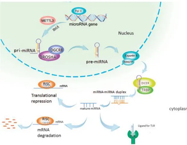
1. Biogenesis and regulation of microRNAs ____________________________________ 42
1.1. Biogenesis of microRNAs ____________________________________________________________ 42 1.2. Regulation of miRNAs expression in adipose tissue ______________________________ 44
2. MiRNAs in the formation of adipose tissue __________________________________ 45
2.1. MiRNAs in differentiation of adipocytes progenitors _____________________________ 45 2.2. Determination of adipocytes identities ____________________________________________ 48 2.3. Involvement in adipose tissue functions ___________________________________________ 50
8
PART III: MiRNAs in aging ___________________________________________________ 56
1. Dysregulations of miRNAs in aging ________________________________________ 56
1.1. MiRNAs involved in overall longevity ______________________________________________ 56 1.2.1. Age-associated cardiovascular diseases ___________________________________________ 57 1.2.2. Age-associated sarcopenia and osteoporosis ______________________________________ 60 1.2.3. Age-related liver dysfunctions ______________________________________________________ 61 1.2.4. Age-related immune defects ________________________________________________________ 61
2. MiRNAs to senescence __________________________________________________ 62
2.1. Cellular senescence __________________________________________________________________ 62 2.2. miRNAs in p53/RB tumor-supressor pathways ___________________________________ 64 2.3. miRNAs in DNA damage pathways _________________________________________________ 66
3. MiRNAs in conserved aging ______________________________________________ 67
3.1. miRNAs affecting caloric restriction process ______________________________________ 68 3.2. miRNAs affecting sirtuin families ___________________________________________________ 71 PART IV: A novel method of culturing isolated adipocytes __________________________ 74
1. Current in vitro models of adipocytes_______________________________________ 74
1.1. Two-dimensional (2D) adipogenesis of preadipocytes ___________________________ 74 1.2. Three-dimensional (3D) adipogenesis of preadipocytes _________________________ 76
2. First attempt to culture isolated adipocytes _________________________________ 78
Result 1: Role and regulation of microRNAs in adipose tissue during aging ____________ 84 Result 2: A novel method of culturing isolated adipocytes in vitro __________________ 117 Bibliography _____________________________________________________________ 136
9
10
Introduction
PART I: White adipose tissue and aging
1. The adipose tissue
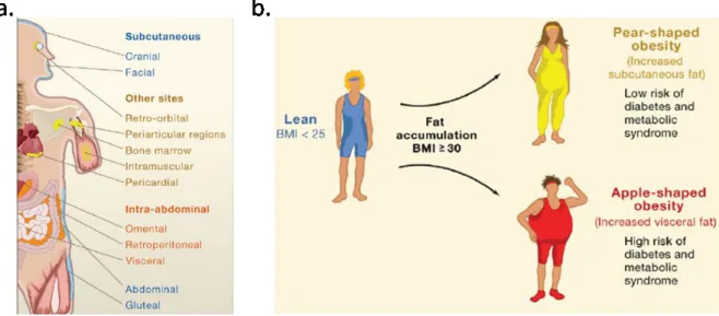
The adipose tissue is a master regulator of energy balance and nutritional homeostasis. Viewed as an inert energy storage site and a form of connective tissue not long time ago, surging evidence now have revealed that adipose tissue is a remarkably complex organ with important endocrinal functions which enable its crosstalk with diverse organs. Moreover, with the very recent discovery of brown and beige adipocytes, interests have been fueled in the study of adipose biology (Figure 1).
Figure 1: Growing interests in adipose biology over the last century.
Adopted from2.
Adipose tissue is a dynamic organ which can range from 4% to over 40% of total body composition in adult humans and is dispersed throughout human body. Founctionally,
11
adipose tissue could be categorized into white adipose tissue (WAT) and brown adipose tissue (BAT). BAT generates heat during cold exposure by adaptive thermogenesis to maintain normal body temperature and is more abundant in neonates and young children, and gradually declined in human adults3. Here in our topic, we are going to focus mainly on WAT.
Geographically, WAT is categorized into subcutaneous and visceral adipose tissue (sWAT and vWAT, respectively) (Figure 2a). Surprisingly, a pioneering clinical study in the 1950s firstly reported that location of fat depots has a great influence on the likelihood of an individual to develop many of the complications of obesity4 (Figure 2b). The data indicate that there is heterogeneity between sWAT and vWAT.
Figure 2: Adipose tissue distribution associates with the incidence of diabetes development.
(a) Distributions of WAT in human. (b) Incidence of development of diabetes associated with the fat distributions. Modified from5.
1.1. Subcutaneous adipose tissue (sWAT)
In human, sWAT forms just below the dermis, and includes periscapular and inguinal depots. Increased amounts of subcutaneous fat, especially in the gluteofemoral regions, are associated with improved insulin sensitivity and a lower risk of developing metabolic
12
syndromes6. In mice, transplantation of subucutaneous fat into the cavity of visceral fat significantly decreases body weight and total fat mass as well as improves systemic glucose tolerance, whereas transplantation of vWAT into the depot of sWAT has little effects7.
The metabolic benefits of sWAT might be associated with its unique property: sWAT is capable of ‘browning’ in responsive to cold exposure, β-agonists or other hormone-like stimuli8, which includes the induction of uncoupling protein 1 (UCP1) expression, a gene giving rise to uncoupled respiration and heat production. The UCP positive cells would emerge in white fat depots under certain stimuli and are termed beige or brite cells. Adipocyte-specific mutant of PR domain containing 16 (PRDM16) markedly inhibites beige adipocyte function in sWAT following cold exposure or β3-agonist treatment, acquiring numerous properties of vWAT including decreased thermogenic and increased inflammatory gene expression as well as macrophage infiltration. Moreover, transplantation of the sWAT derived from PRDM16 mutants into obese mice shows a loss of metabolic benefit9.
Recently, it has been reported that inhibitors of the Janus kinase/ signal transducers and activators of transcription 1/3 (JAK/ STAT1/3) signaling, like tofacitinib and R406 are strongly inducers of UCP1 exressionin sWAT but not in vWAT10, indicating that inhibitions of JAK/ STAT1/3 signaling might mediate the browning process in sWAT, whereas it is not employed in the futile energy cycle in vWAT.
1.2. Visveral adipose tissue (vWAT)
In human, there are 6 visceral fat depots: perirenal, gonadal, epicardial, retroperitoneal, omental and mesenteric depots. In contrast to sWAT, it is revealed that the size of abdominal adipocyte is a significant risk predictor for the development of type 2 diabetes, as revealed by a follow-up study of around 245 women from 1974 to 200111. Several plausible mechanisms may be associated with the properties of vWAT.
Firstly, Aliki Kosteli et al, demonstrat that vWAT has a higher rate of basal lipolysis and therefore releases more free fatty acids (FFAs) compared to sWAT12. Since vWAT is anatomically drained by the portal vein, the increased levels of portal FFAs might induce metabolic complications in liver, such as hepatic steatosis or hepatic insulin resistance in
13
obesity13. What’s more, FFAs are also strong inducers of adipose tissue’s recruitment of macrophages, whose chronic accumulation would lead to elevated local inflammatory state12, which is negatively associated with insulin resistance. In addition, with the use of whole-body fluorodeoxyglucose positron emission tomography (FDG-PET) scans, it is found that vWAT has higher rates of glucose uptake compared to sWAT in both lean and obese humans. The isolated stromal vascular fractions (SVFs) from vWAT exhibites significant increase in both basal and cytokine stimulated glucose consumptions associated with a higher expression of hexokinase (HK) compared to that from sWAT14. These results indicate that vWAT has higher metabolic rates in comparision to sWAT.
Interestingly, although to a lesser extent, visceral fat is capable of responding to chronic β3-adrenergic receptor stimulation demonstrated by the appearance of lipid droplet mutilocularity and robust mitochondrial biogenesis, which might be explained by a prominent increase in futile creatine cycling in visceral fat under stimulation15. However, the physiological role of futile cycling of vWAT remains to be investigated.
Secondly, vWAT is also an endocrine organ that actively secretes lots of cytokines and bioactive mediators, which influence systemic inflammation, lipid metabolism and insulin sensitivity16. For example, B cells isolated from vWAT expressed higher levels of inflammatory immune activation markers and significantly higher nuclear factor-κB (NF-κB) activation and phospho-STAT317. Expression of proinflammatory adipokines in vWAT could be elevated by obesity, which might contribute to their circulating elevation during obesity18.
2. White Adipocytes
Adipose tissue is composed of mature adipocytes and various other cell types in stromal vascular factions (SVFs) that include leukocytes (e.g., macrophages and lymphocytes) and adipose tissue stromal cells (ATSCs) that include preadipocytes and fibroblasts. Adipocytes contribute mainly to metabolism and functions of adipose tissue19.
2.1. Architecture
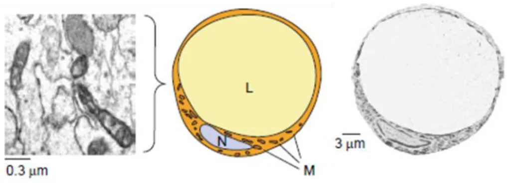
White adipocytes contain a single large lipid droplet (LD) occupying approximately 90% of the cytoplasm, with the nucleus being squeezed to the cell periphery and the cytoplasm
14
forming a very thin rim. Mitochondria are scarce and internal cristae are poorly developed (Figure 3). Due to these characteristics, these cells are also termed unilocular adipocytes.
Figure 3: Representation and transmission electron microscopy of the morphology of white adipocytes.
Abbreviations: L, lipid; M, mitochondria; N, nucleus. Modified from20.
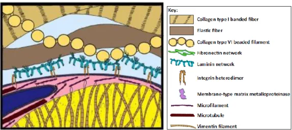
At the tissue level, spherical adipocytes are typically arranged in an imperfect hexagonal packing architecture resembling honeycomb, with smaller preadipocytes and interweaving capillaries and nerves filling the interstitial space21,22. Adipocytes themselves are interconnected via gap junctions and thereby share cytoplasm and respond in concert to electrical stimuli23. Additionally, adipocytes are intensively interacted with the extracellular matrix (ECM), which provides both the flexibility needed for cell migration and hypertrophy and the stability needed for the structural and functional integrity of the tissue as a whole (Figure 4). For example, adipocytes are interacted with an isotropic network of collagen and elastic fibers, extracellular fibronectin and laminin24, via attachment points for integrins anchored in the adipocyte membrane25.
15
Figure 4: Extracellular support of adipocytes in vivo.
Structural support is provided by type I (100 nm to 100 µm diameter) and type VI (50 nm diameter) collagen networks. The LD is caged in vimentin (10 nm, brown filament). The nucleus (blue) moves to the cell periphery and is deformed by the LD. Modified from26.
The rounded LDs are surrounded by a single lipid layer (of endoplasmic reticulum membrane origin), which is in turn ensheathed by endoplasmic reticulum (ER). LDs are very ofter surrounded by one or more proteins of the PAT (perilipin, adipose differentiation-related protein (ADRP), and TIP47 (tail interacting protein of 47 kDa family)27. The LDs of adipocytes are encased in a coating of perilipins, which have an important role in regulating triglyceride hydrolysis (breakdown of triglyceride into glycerol and free fatty acids), with perilipin A being the most abundant isoform in adipocytes. Perilipin null adipocytes exhibites elevated basal lipolysis but attenuated sitmulated lipolytic activity, indicating that perilipin is required for maximal lipolytic activity28.
2.2. Homeostasis of lipid droplets in adipocytes
Adipocytes are central in the regulation of triglyceride (TG) and FFAs homeostasis during energy expenditure and deprivation. The storage and removal of TG in adipocytes is essential to determine the sizes of adipocytes. In vivo, the sizes of adipocytes vary greatly according to the depots and the nutritional states (Table 1). The sizes of adipocytes correlate with the functions of adipocytes. It has been demonstrated that small white adipocytes have lower expression of enzymes involved in fatty acids use (β-oxidation) including acyl-CoA
16
dehydrogenase and carnitine palmitoyl transferase 2 (CPT2), as well as in fatty acid synthesis (FAS) compared to large white adipocytes, coinciding with that fact that small adipocytes have low storage of TG29.
Table 1: The average sizes of adipocytesin developing and adult human a
25-30 weeks gestation,b1-3 months postpartum. Adopted from30.
Several major functions of adipocytes determine the size of LDs in adipocytes: fatty acid uptake, de novo lipogenesis (DNL), lipolysis and possible burning of fat by beiging, which we will discuss in details in the following parts.
2.2.1. Synthesis of lipid droplets (LDs) Fatty acid uptake
Most of the lipids stored in adipocytes are transferred directly from diets. Several proteins are involved in fatty acid uptake in adipocytes: caveolin-1, fatty acid translocase (FAT/CD36), the 43-kDa plasma membrane fatty acid binding protein (FABPpm), and fatty acid transport protein (FATP), adipose acyl CoA synthetase (ACSL).
Caveolin-1
Caveolae are a subset of so-called membrane lipid raft domains that are often experimentally defined by their resistance to solubilization by nonionic detergents.
The surface of adipocytes is abundantly covered by caveolae (nearly 30%)31. Caveolin-1 is the primary protein driving the formation of caveolae in adipocytes32. Caveolin-1 is speculated to bind the fatty acids accumulated with the caveolar membrane, leading to membrane asymmetry and budding of caveolae from plasma membrane to form vesicles. These fatty
17
acid loaded caveolae vesicles could deliver fatty acids to subcellular compartments for further metabolism (Figure 5)33.
FABPpm
Expression of FABPpm in 3T3-L1 preadipocytes that normally do not express FABPpm induces a significant increase in fatty acid uptake. Anti-FABPpm antibodies selectively inhibit uptake of oleate in 3T3-L1 adipocyte monolayer without affecting 2-deoxyglucose or octanoate (medium-chain fatty acid) uptake34. Currently, the mechanism by which FABPpm used in LCFA uptake is unknown.
CD36
CD36 is an 88-kDa single chain membrane glycoprotein expressed in many tissues and cell types, including adipocytes, heart, skeletal muscle and macrophages. C/EBPα and C/EBPβ would increase CD36 expression in different types of cells, with C/EBPα exhibits a significantly higher potency than C/EBPβ. A C/EBP responsive element is identified in the CD36 promoter35. It has been speculated that CD36 may bind and internalize long chain fatty acid (LCFA) by endocytosis or may bind LCFA and work in concert with FABP to provide a high concentration gradient of LCFA across the plasma membrane facilitating its uptake by other fatty acid transporters (Figure 5)33.
FATP1 and ACSL
FATP1 and ACSL are two different classes of membrane bound enzymes catalyzing the ATP-dependent esterification of long-chain and very long-chain fatty acids into acyl-CoA derivatives36,37. FATP1 and ACSL1 are enriched in adipose tissue.
FATP1 is induced during the differentiation of 3T3-L1 adipocytes, and increased LCFA uptake. Insulin could trigger FATP1 translocation from a perinuclear compartment to the plasma membrane, indicating that FATP1 is facilitated by insulin38. Since it is reported that the conversion of incoming LCFA to TG occurs on/or around the plasma membrane in rat adipocytes39, FATP1 might facilitate LCFA uptake by tightly coupling it to metabolism: by converting it into acyl-CoAs which is preferentially shunted into TG synthesis (Figure 5)33.
18
ACSL1 is found to be involved in the reacylation of LCFA released from the LD during basal and hormone-stimulated lipolysis. Acylation of LCFA leads to reincorporation into the TG thereby preventing its cellular efflux40.
Figure 5: Schematic representation of FFA uptake mediated by CD36 in caveolin-1-FABP rich microdomain.
Adopted from33.
De novo lipogenesis (DNL)
On the other hand, adipocytes are capable of synthesizing new lipids from carbohydrates through DNL. The two major enzymes of DNL, fatty acid synthase (FAS) and acetyl CoA carboxylase (ACC), which are under the control of carbohydrate-response-element-binding protein (ChREBP) or sterol-response-carbohydrate-response-element-binding protein 1c (SREBP1c), are abundantly expressed in fat cells41. Surprisingly, DNL contributes only a few percent of fatty acids to the TG that accumulates in the lipid droplets of adipocytes42. In addition, it was found that in vitro 3T3-L1 adipocytes preferentially use glycolysis to depose glucose into lactate even with the abundant oxygen availability, while only a small fraction of carbon from glucose would be converted into TG43. However, DNL contributes to a number of
19
adipose-derived putative bioactive lipids, such as palmitoleate44, the novel fatty acid esters of hydroxy-fatty acids45 and PPARγ ligand alkyl ether-lipid46. In addition, studies have shown that up-regulation of DNL in adipose tissue mediated the improved insulin sensitivity and glycemic control of GLUT4 overexpression in adipocytes, whereas loss of ChREBP has the opposite effects41,47. Besides, FAS was dramatically upregualted in WAT by caloric restriction (CR) process which is associated with increased insulin sensitivity and longevity, indicating that DNL might be important metabolic adaptations of caloric restriction benefits48. Conversely, impairment of DNL, which is under rictor/mTORC2 signaling in adipose tissue, was found to be necessary for the loss of insulin sensitivity induced by high fat diet in wild type mice49.
Glucose transport:
Insulin signaling plays an important role in lipid storage and the process of adipogenesis for adipocytes (Figure 6). The phosphatidylinositol-3-OH kinase (PI(3)K)-dependent signaling is initiated by binding of insulin onto its tyrosine kinase receptor on the cell surface, leading to the recruitment and tyrosine phosphorylation of the insulin receptor substrate (IRS). The phosphorylated IRS then serves as docking sites for the SH2 domain of the p85 regulatory subunit of class I PI3K, resulting the activation of PI3K, which promotes the synthesis of phosphatidylinositol-3,4,5-trisphosphate PtdIns(3,4,5) P3 from PtdIns (4,5) P2 at the plasma membrane. PtdIns(3,4,5) P3 then serves as a docking site for several PH
domain-containing Ser/Thr kinases that are implicated in glucose uptake, such as phosphoinositide-dependent kinase 1 (PDK 1) and AKT (also known as PKB) 50. AKT was then activated by PDK1 (phosphoinositide-dependent kinase 1) and mTORC2 through dual Ser/Thr phosphorylation. Downstream, AKT kinase affects the exocytic arm of the GLUT4 (glucose transporter 4) trafficking itinerary, probably by regulating the translocation, targeting and fusion of GLUT4-containing vesicles51.
On the other hand, insulin could also initiate a PI3K-independent signaling by recruiting the adaptor protein APS, which has high affinity to the activated insulin receptor52. Following insulin stimulation, APS recruits a complex that comprises the proto-oncogene c-CBL and c-c-CBL-associated protein (CAP), triggering tyrosine phosphorylation of c-CBL by insulin receptor53. Upon phosphorylation, CBL is then translocated to lipid rafts, recruiting the
CrkII-20
C3G complex to lipid rafts, where C3G specifically activates the small GTP-binding protein TC10, which interacts with effector proteins that regulate GLUT4 vesicle exocytosis54.
Adipocyte GLUT4 ablation resulted in systemic insulin resistance in liver and muscle55, indicating that GLUT4 mediated glucose transport in adipocytes is indispensable for whole body glucose homeostasis.
Figure 6: Insulin signaling regulates GLUT4 exocytosis by engaging the trafficking machinery.
Modified from56.
2.2.2. Hydrolysis of lipid droplets
Lipolysis represents the process for FFAs to be liberated from TG in LDs. Total lipolysis is the sum of basal lipolysis and stimulated lipolysis. Basal lipolysis is the relase of FFAs from adipocytes occurring in the absence of negative energy balance, which correlates positively with adipocyte size and is increased by obesity57. Stimulated lipolysis refers to the hormonally regulated release of FFAs in response to nutritional demands. The lipolysis of TG in adipocytes is under tight control of various pathways in vivo (Figure 7).
Typically, the main regulating signaling pathway of stimulated lipolysis in adipocytes is β-adrenergic signaling. At least three major enzymes are responsible for the lipolytic process. The primary cleavage of TG to diacylglycerols is performed by the adipose tissue lipase (ATGL), which is predominantly expressed in adipose tissue. In ATGL-/-mice, absence of
21
ATGL induced severe lipid accumulation in adipose tissues and heart, with dramatic inhibition of basal and isoproterenol stimulated lipolysis as well as premature death. On the contrary, the inability of fat catabolism due to ATGL deficiency led to increased glucose use, indicating that ATGL is rate limiting in the catabolism of cellular fat58. Administration of Atglistain, a small molecule inhibitor of ATGL effectively improves high fat diet-induced obesity associated insulin resistance and liver steatosis in mice. Moreover, the drug predominantly targeted adipose tissue and the liver, and therefore did not induce cardiac lipid accumulation or cardiomyopathy even after long-term treatment59. ATGL is highly regulated at both the transcriptional and post-transcriptional levels, including multiple phosphorylation events and translocation to the surface of LDs. It could be activated by its coactivator comparative gene identification-58 (CGI-58), which is normally bound in an inactive state by the LD protein perilipin 1 (PLIN1). And upon activation of β-adrenergic signaling, PKA induced phosphorylation of PLIN1 leading to the release of CGI-58, allowing its binding and activation of ATGL60.
Subsequently, hormone-sensitive lipase (HSL) is dispensable for lipolysis. HSL is the major diglyceride lipase in adipocytes, with monoglyceride lipase (MGL) completes the process by generating glycerol and FFAs. Together, these three enzymes account for over 90% of the lipolytic activity in the adipocytes61.
Physiologically, insulin is the major suppressor of lipolysis, which functions through several different ways to block lipolysis: 1) It activates phosphodiesterase 3b (PDE3b) in a AKT dependent manner, which antagonize the intracellular levels of cAMP and thereby blocking protein kinase A (PKA) activation62. 2) Recently, a non-canonical pathway has been proposed in which insulin blocks activation of PKA selectively on PLIN1 through a PI3K-mediated, AKT-independent pathway63. 3) Insulin could also repress lipolysis by transcriptionally silencing lipase genes via repressions of transcription factors forkhead box protein O1 (FOXO1) and interferon regulatory factor 4 (IRF4)64 (Figure 7).
The size of fat cells per se also have an impact on both the spontaneous and hormone-stimulated rates of lipolysis, with big adipocytes exhibited higher rates of basal and hormone-stimulated hydrolysis, probably owing to the fact that hypertrophic adipocytes are enriched in the expressions of ATGL, HSL and PLIN1. However, the receptor sensitivities for
22
hormones like insulin, natriuretic peptides, dobutamine, terbutaline and clonidine, are comparable between small and large fat cells65.
Figure 7: Tight control of lipolysis process by IIS, β-adrenergic signaling and NP receptor A signaling pathways.
Adopted from62.
2.2.3. Lipid driplets “burning”
Over the last three decades, it has been realized that mature adipocytes displayed high plasticity, especially those in the subcutaneous depots. In response to some external stimuli, including chronic cold exposure, exercise, long-term treatment with PPARγ agonists or β3-adrenergic receptor agonists, cancer cachexia, and tissue injury, a group of beige (also called brite) adipocytes are induced within WAT which are characterized by possessions of abundant cristae-dense mitochondria that express UCP1 and mutilocular LDs66. Fatty acids are rapidly consumed and deprived by mitochondria β-oxidationa and UCP-mediated heat production in beige adipocytes, resulting in a smaller appearance of beige adipocytes compared to normal white adipocytes67. It also has been demonstrated that beige-like adipocytes also exist in human adults68.
23
Although some of beige adipocytes might directly come from adipogenesis of distinctive populations of progenitors within WAT69,70, it has been reported that stimulation of β3-adrenergic receptor (Adrb3) in mature adipocytes could directly lead the transformation into mitochondrial abundant beige adipocytes via activation of PRDM71.
Conversely, after withdrawal of external stimuli such as cold acclimation or β3- adrenergic agonists, the produced beige adipocytes then possibly transform into normal white adipocytes, by upregulation of mitophagy (mitochondrial clearance via autophagy) activated by which is initiated by the activation of MiT/TFF (microphthalmia/ transcription factor E) transcription factor-mediated lysosome biogenesis67.
2.3. Endocrinal functions
Adipocytes have been well acknowledged as a bona fide endocrine cell type that regulates systemic metabolic homeostasis in accordance to nutrient flux by equally matching the metabolic demands of positive or negative energy balance, via secreting a variety of bioactive peptides, known as adipokines (Figure 8). Secretion of adipokines, including leptin, adiponectin, bone morphogenetic protein (BMP)-4, dipeptidyl peptidase 4 (DPP-4), etc, is altered by adipose dysfunction and may contribute to obesity-associated diseases. Therefore, adipokines are promising candidates for pharmacological treatment and as diagnostic tools.
Leptin
Leptin was discovered more than 20 years ago72. Circulating levels of leptin are proportional to fat mass, and leptin receptors are abundantly expressed in various tissues, including on the adipocyte. Leptin negatively regulate adiposity. Indirectly, leptin negatively regulate satiety via inhibiting the orexigenic pathway comprising neuropeptide-Y (NPY) and agouti-related protein (AGRP)-containing neurons, while upreguation of anorexigenic pathway consisting of proopiomelanocortin (POMC) and cocaine- and amphetamine-regulated transcript (CART)-containing neurons in hypothalamus73. Moreover, leptin would drive adiposity depletion by stimulation of lipolysis through activating sympathetic neurons innervating adipocytes in WAT in vivo74. Also, in vitro, leptin was reported to decrease
24
lipogenesis, increase TG hydrolysis and oxidation of fatty acids in isolated rat white adipocytes75.
Adiponectin
Adiponectin was firstly reported in 199576, which is almost exclusively synthesized by adipocytes. Adiponectin concentrations inversely correlate with fat mass and are down-regulated in obesity and type 2 diabetes77.Overexpression of adiponectin in 3T3-L1 cells enhanced adipogenesis and lipid storage78. Also, adiponectin has been shown to increase insulin sensitivity and maintains healthy adipose tissue expansion while rescuing ectopic lipid accumulation in mice79.
The beneficial effects of adiponectin on insulin sensitivity seem to be mediated in part by its ability to activate AMP-activated protein kinase (AMPK) in skeletal muscle and liver80.For example, adiponectin decreases hepatic lipogenesis and increases β-oxidation through activation of AMPK, a cellular energy sensor, inhibits lipogenesis by phosphorylating the rate-limiting enzyme of DNL, acetyl CoA carboxylase-1 (ACC-1). This decreases in ACC-1 activity which leads to decrease in malonyl CoA production, thereby relieves inhibition of carnitine palmitoul transferase-1 (CPT-1) activity and enhances fatty acid transport into the mitochondria to undergo β-oxidation.
Adiponectin is also associated with cardio-protection. Administration of adenovirus-mediated adiponectin reduces atheroscleric lesion size in apolipoprotein E knockout mice, accompanied by reduction in the expression of inflammatory cytokines81. Moreover, adiponectin is also reported to promote angiogenesis and inhibits endothelial apopotosis82.
Fatty acid binding protein 4 (FABP4/ aP2)
aP2 was elevated in circulation in obese humans and mice83. Furthermore, aP2 was demonstrated to be an adipokine released from adipocytes under obesogenic conditions, such as hypoxia, to augment insulin secretion from pancreatic β-cells in vivo, while insulin could in turn inhibit aP2 secretion84.
25
Dipeptidyl peptidase 4 (DPP4) is a 110 kDa glycoprotein, which is ubiquitously expressed on different cell types as well as in circulation. By proteomic profiling of the adipocyte secretome, soluble DPP4 is identified as a novel adipokine, for which mature adipocytes constitute are a major source85. DPP-4 may not only reflect, but also contribute to adipose tissue dysfunction. Compared with healthy lean individuals, patients with obesity and insulin resistance have higher DPP-4 expression in vWAT, which may contribute to the higher circulating DPP-4 levels86. In obesity, hypoxia-mediated upregulation of DPP-4 shedding from adipocytes by metalloprotease-9 might contribute to induced DPP-4 activity87
. Also, in mice, production of DPP-4 in adipocytes is shown to be negatively regulated by high glucose concentration under physiological conditions, such an effect is abolished in mouse models of diabetes88.
Under physiological conditions, DPP-4 rapidly degrades the incretin hormones including glucose-dependent insulinotropic polypeptide (GIP) and glucagon-like peptide-1 (GLP-1)89
, which have been shown to enhance glucose-dependent insulin secretion and inhibit glucagon secretion, which together may reduce hepatic gluconeogesis90. Moreover, under physiologic concentrations, DPP-4 directly induced insulin resistance in adipocytes and skeletal muscle85
Nicotinamide phosphoribosyltransferase (NAMPT)
Very recently, NAMPT, a key NAD+ biosynthetic enzyme, which has two different forms, intra- and extracellular (iNAMPT and eNAMPT) in mammals, was found predisposed to secretion from adipocytes via deacetylation by the mammalian NAD+-dependent deacetylase SIRT1. Adipose tissue-specific NAMPT knockout and knockin mice show reciprocal changes in circulating eNAMPT, affecting the hypothalamic NAD+ production, SIRT activity and neural activation accordingly91. The story provides an elegant example in which adipocyte-derived molecules could be transferred and exert their function in other organs.
BMP-4
BMP-4 is secreted by differentiated preadipocytes and adipocytes, with higher expression in larger adipocytes92. It has been recently shown that differentiated human adipocytes can promote adipogenesis via endogenous BMP-4 activation, a process that may
26
be counter-balanced by the expression of the BMP-4 (and -7) inhibitor gremlin-192. In hypertrophic obesity, adipose precursor cells are resistant to BMP-4, which may contribute to the limited expandability of adipose tissue and, subsequently, to obesity-related metabolic diseases92
.
Interestingly, adipose tissue has been very recently demonstrated as a major source of circulating microRNAs, and adipocyte-derived microRNAs could regulate gene expressions in liver in mice93, indicating that, aside the classical view that most of the adipokines are proteins, other factors like microRNAs might be considered as novel adipokines.
Figure 8 : Adipocytes crosstalk with various organs through adipokines.
27 2.4. Origins of white adipocytes
Although adipose tissue development is poorly understood, the phenomenon that, in lipodystrophies, regionalized depot atrophy coexists with preservation and often expansion of other depots suggests that adipose tissue is an organ of multiple lineages95. In mice, it has been demonstrated that sWAT and vWAT form in an ordered and timed manner throughout embryogenesis and within the first few weeks of birth. sWAT begins to develop during embryogenesis and the compartment of progenitors is established for all the depots of sWAT before the first few days of life. Whereas, vWAT principally form postnatally: the perigonadal lineage forms approximately between postnatal day 3 and the second week of life. The mesenteric WAT adipose compartment completes its establishment of lineage between the second and third weeks of life. The retroperitoneal WAT is formed in-between these pre- and postnatal stages, and has a morphogenesis, texture, and histology that also seem intermediate96. Transcriptomic analysis of preadipocytes isolated from the SVF of various fat depots have revealed consistent differences in developmental gene profiles between vWAT and sWAT, indicating the possibility that sWAT and vWAT are of distinct origins 97.
Three critical events are necessary for the establishment and maintenance of functional adipocytes (Figure 9):
1) Determination of preadipocyte, commitment of multipotent progenitors into the adipocyte lineage.
2) Adipocyte differentiation, in which committed preadipocytes undergo a morphological and biochemical transition into mature adipocytes in response to appropriate stumuli.
3) Adipocyte maintenance, in which the cellular identity and functional properties if the terminally differentiated cells are maintained.
28
Figure 9 : Simplified scheme of white adipocyte origins.
2.4.1. Adipocyte progenitor cells (APCs)
Pluripotent stem cells, residing in the vascular stroma of adipose tissue, are able to be recruited into preadipocytes, which when appropriately induced, undergo mitotic clonal expansion and then differentiate into adipocytes (Figure 9). Adipocyte progenitor cells (APCs) reside in the SVF and could be separated from non-adipose precursors (e.g., endothelial progenitors, immune cell populations) using early adipocyte progenitor cells markers (Lin-: CD29+;CD34+;SCA1+:CD24+) with the technology of fluorescence-activated cell sorting (FACS), which could successfully proliferate and differentiate into WAT in A-Zip lipodystrophic transgenic mice98.
To investigate the architecture of the stromal vascular (SV) compartment, Wei T., et al, developed an SV particulate (SVP) isolation procedure designed to partially maintain the native SV structure while removing adipocytes that obscure visualization of the precursor location. In the SVPs, the majority of peroxisome proliferator-activated receptor γ (PPARγ)/GFP cells were arrayed in tubelike structures, which immunohistochemically
29
expressed platelet endothelial cell adhesion molecule (PECAM) and mural cell markers: SMA (endogenous smooth muscle actin), PDGFRβ (platelet-derived growth factor receptor β), indicating that these adipocyte progenitors reside in the mural cell compartment of the adipose vasculature99.
The large C2H2 zinc-finger transcription factor, Zfp423, is a factor associated with preadipocyte commitment. Zfp423GFP reporter mice show that Zfp423 distinguished adipogenic from inflammatory-like mural cells. Zfp423GFP expression was found not only in mature adipocytes but also in a distinct subset of perivascular cells expressing mural cell marker PDGFRβ, supporting the hypothesis that preadipocytes resemble pericyte-like cells. Upon high fat diet feeding, the frequency and absolute number of cells positive for GFP and Pdgfrβ significantly increase in gonadal WAT by comparision to inguinal WAT of male mice, consistent with the fact that gonadal WAT of adult mice expands by both adipocyte hypertrophy and adipocyte phyerplasia, while inguinal WAT expands predominantly through cellular hypertrophy70.
Besides, adipocytes within different depots might have distinct progenitors. For example, the majority of the precursors and mature sWAT adipocytes in mice are labeled by paired related homeobox transcription factor 1 Cre (Prx1-Cre), while in contrast, few visceral adipocytes are marked by Prx1-cre100. Conversely, the wilms tumor gene (Wt1) expressing cells give rise to adipocytes in vWAT but not adipocytes in sWAT, and are enriched in the mesothelium, supporting that the lateral plate mesoderm as a major source of vWAT101.
Multiple protein factors could increase the commitment of stem cells into preadipocytes. For example, following treatment of the C3H10T1/2 stem cells with bone morphogenic protein 4 (BMP4) during proliferation followed by differentiation inducers at growth arrest, the cells synchronously enter S phase and undergo mitotic clonal expansion, which is a hallmark of preadipocyte differentiation102.
2.4.2. Adipogenesis
During conversion of preadipocytes to adipocytes, preadipocytes employ a sequential induction of a series of transcription factors, including C/EBP families and PPARγ to express genes necessary for adipocyte function. They also undergo change in morphology to become
30
rounded lipid-laden adipocytes. In the 1990s, the nuclear hormone receptor PPARγ was identified as a master regulator of adipocyte differentiation. PPAR has three isoformes: PPARα, PPARδ, and PPARγ. PPARγ predominately cooperate with C/EBPα in the promotion of adipognesis103.C/EBPδ and C/EBPβ are induced early during adipognesis, peaking at day 2 of adipogenesis and after decreased104. The early upregulation of C/EBPδ and C/EBPβ facilitate the expression of PPARγ, and then PPARγ along with C/EBPβ and C/EBPδ activates C/EBPα expression. Chromatin immunoprecipitation (ChIP) study revealed that most induced genes in adipogenesis were bound by both PPARγ and C/EBPα. Thus, PPARγ and C/EBP factors cooperatively orchestrate adipocyte biology by adjacent binding, which activate hundreds of genes responsible for terminal adipocyte differentiation105. PPARγ can promote adipogenesis in C/EBPα-deficient cells. However, conversely, ectopic expression of C/EBPα could not stimulate adipognesis in PPARγ-deficient fibroblast, demonstrating that C/EBPα and PPARγ participate in a single pathway of fat cell development with PPARγ being the proximal effect of adipogenesis106. C/EBPα-deficient fibroblast underwent adipogenesis via activation of PPARγ with several apparent defects, including impaired insulin stimulated glucose uptake secondary to reduced gene expression and tyrosine phosphorylation for the insulin receptorand IRS-1. Later it was demonstrated that insulin receptor promoter or enhancer was preferentially transactivated by C/EBPα107.
Signal transducer and activator of transcription 3 (STAT3) was reported to be activated by Janus kinase 2 (JAK2) within 2h of adipogenesis induciotn on 3T3-L1 adipocytes, translocating from cytoplasm to the nucleus and regulating the transcription of C/EBPβ by binding the distal region of its promoter108 (Figure 9).
PRDM16 is more enriched in sWAT compared to that in vWAT in mice, associated with its role in the development and function of beige cells. Ablation of PRDM16 caused a switch of sWAT to vWAT like phenotype, and impairment of thermogenic gene expression in response to cold exposure, indicating that PRDM16 pathway is necessary for the differentiation of brite adipocytes9. In human, the elevated levels of BMP4 in WAT correlated with a lean phenotype. Forced expression of BMP4 in mice induced a brown fat-like alterations in WAT through upregulation of mitochondrial biogenesis in association with increased energy expenditure and insulin sensitivity, which is mediated by the PGC1α/
31
p38/MAPK/ATF2 pathway109, indicating a potential role of BMP4 in the biogenesis of brite adipocytes (Figure 9).
Wnts are a family of paracrine and autocrine factors that regulate cell growth and cell fate. Signaling is initiated when Wnt ligands bind to transmembrane receptors of the Frizzled family, which signals through Dishevelled to inhibit the kinase activity of a complex containing glycogen synthase kinase 3 (GSK3), Axin, catenin, etc. This complex targets β-catenin for rapid degradation through phosphorylation. Thus, once hypophosphorylated due to Wnt signaling, β-catenin is stabilized and translocate to the nucleus where it binds the TCF/LEF family of transcription factors to regulate the expression of Wnt target genes. When Wnt signaling was activated in 3T3-L1 or 3T3-F442A, adipogenesis was significantly impaired, with no alterations in the expressions of C/EBPδ and C/EBPβ, but decreased levels of C/EBPα and PPARγ. Consistently, over-expression of C/EBPα and PPARγ could partly rescue the impaired adipogenesis due to Wnt activation110 (Figure 9).
Pref-1 is a transmembrane protein encoded by the pref-1/dlk1 gene and belongs to a family of EGF-like repeat-containing proteins that include Notch/Delta/Serrate, which are involved in cell fate determination.Interestingly, Pref-1 is highly expressed in 3T3-L1 preadipocytes, but almost abolished in adipocytes111. The extracellular domain of Pref-1 undergoes two proteolytic cleavage events that generate 50 and 25 kDa soluble products. By fusing Pref-1 to human immunoglobulin-γ constant region (Pref-1/hFc), transgenic mice expressing the Pref-1/hFc transgene under the aP2 promoter, showed reduced expression of adipocyte markers and adipokines, whereas the preadipocyte markers Pref-1 was increased. Moreover, mice expressing the Pref-1/hFc transgene exclusively in liver under the control of the albumin promoter also showed a decrease in adipose mass and markers, suggesting an endocrine role of Pref-1 on adipogenesis112 (Figure 9).
The seven Sirtuins in mammals (Sirt1-Sirt7), which are involved in the regulation of several essential cellular processes, also have important roles in adipogenesis. Sirtuin 1 (Sirt 1) is the ortholog of the yeast protein silent information regulator 2 (Sir2) and a nuclear NAD+-dependent class III histone deacetylase class III, and is a key component activated by CR. Sirt 1 partly mediated the inhibitory effects of CR on adiposity by attenuating adipogenesis through suppressing PPARγ by docking with its cofactors NCoR (nuclear
32
receptor corepressor) and SMART (silencing mediator of retinoid and thyroid hormone receptors). Conversely, Sirt7 which is known to specifically deacetylate H3K18, is reported to promote adipogenesis by binding and repressing Sirt1 activity113. The forkhead transcription factor FOXO1 is an important downstream effector of insulin/PI3K/AKT signaling, which lead to its phosphorylation and thereby nuclear exclusion. Constitutively active FOXO1 attenuated adipogenesis, while dominant-negative FOXO1 restored adipogenesis of fibroblasts from insulin receptor deficient mice114. Interestingly, mammalian Sirt2 is localized mainly in the cytoplasm and is reported to be down regulated during adipogenesis, and its over-expression inhibits differentiation via enhancing insulin-stimulated phosphorylation of FOXO1115.
3. Alterations of WAT by aging
Over the last decades, the growing number of elderlies (aged 65 years and over) has become a considerable challenge to health and social care service provision and funding. Moreover, the numbers of the very old, those aged 85 years and over, are set to double over the next 20 years116. Aging is defined as a decline in survival and fecundity with advancing age, which is caused by damage to macromolecules and tissues.
What are the consequences of aging on WAT?
In human, age per se is a strong risk factor for fat accumulation117. Morphologically, computational tomography scans in men and women revealed that, as age increases, sWAT decreases while vWAT increases118. Rodents also demonstrate the same trend to develop age-associated central obesity119. Consequently, the dramatic alterations in fat distributions and functions occurring during life, are linked with the increasing risks of the developments of various aging-associated diseases, such as diabetes, cognitive dysfunction and atherosclerosis120. Multi-isotope imaging mass spectrometry (MIMS) is an imaging method that enables multiplexed measurement of stable isotope tracers with subcellullar resolution, and utility of stable isotope tracers of DNA synthesis and DNL to measure cell birth and lipid turnover. It was demonstrated that there’s an age-dependent decline in the plasticity of adipose tissue whih is characterized by impaired adipogenesis and adipocyte lipid turnover in subcutaneous fat, which might be mediated by a decline in insulin like growth factor-1 (IGF-1) in humans121. In addition, by measuring the 2nd World Warnuclear bomb test-derived
33
14
C in lipids accumulated in adipocytes (in Hiroshima), it was reported that during the average ten-year-life span of human adipocytes, TG were renewed six times. Adipocyte lipid turnover is strongly related to conditions with disturbed lipid metabolism122.
Therefore, in the following part, we will detail effects of aging on adipogenesis and lipid turnover, respectively.
3.1. Senescence of adipocyte progenitors
Compromised adipogenesis during aging contributed to the impaired ability of adipose tissue to store lipids, leading to FFAs spillover and ectopic lipid accumulation in liver or muscle123. Senescence of adipocyte progenitors mediates partly the impaired adipogenesis during aging.
Senescence of adipocytes progenitors is increased in aged adipose tissue, demonstratd with upregulation of senescence-associated markers: SA-β-galactosidase and p16/p53 expressions124.
The senescence burden could further accelerate age-related dysfucntions in fat via both intrinsic and autocrine manners125. For instance, senescent adipocyte progenitors would impede the adipogenesis of non-senescent cells, via the activation of the JAK/STAT and over production of the activin A protein126.
Growth hormone (GH) plays a central role in regulating mammalian growth, metabolic homeostasis and adiposity. Both mice and human who have excessive GH production have increased mortality rates compared to age-matched counterparts 127. Ames and Snell dwarf mutant mice (Prop1df and Pit1dw, respectively) are the first GH-deficient genetic mutants to display extension of lifespan. Interestingly, deficiency of GH in Ames and Snell mice attenuated the age-related central accumulation of vWAT. Besides, the levels of senescent fat progenitors measured by p16 expressions or SA-β-galactosidase, were negatively associated with the levels of GH. Thus, attenuation of senescent burden in vWAT might mediate the life-extending effects of GH deficiency128.
Clearance of senescent cells confers counteractions to age-related irregulations in adipocytes. For example, genetic clearance of p16 positive cells delayed or attenuated the