Contents lists available atScienceDirect
Bone Reports
journal homepage:www.elsevier.com/locate/bonr
Investigating the incidence and magnitude of heterotopic ossi
fication with
and without joints involvement in patients with a limb fracture and mild
traumatic brain injury
Marianne Jodoin
a,b, Dominique M. Rouleau
a,c, Erik Therrien
a,c, Jean-Marc Chauny
a,
Emilie Sandman
a,c, Camille Larson-Dupuis
a,b, Stephane Leduc
a,c, Nadia Gosselin
a,b,
Louis De Beaumont
a,c,⁎aCentre de Recherche de l'Hôpital Sacré-Coeur de Montréal, Montréal, Québec, Canada bDépartment de Psychologie, Université de Montréal, Montréal, Québec, Canada cDépartment de Chirurgie, Université de Montréal, Montréal, Québec, Canada
A R T I C L E I N F O
Keywords:
Mild traumatic brain injury Isolated limb fracture Heterotopic ossification Orthopedic complications Return to work
A B S T R A C T
Objectives: This study seeks to evaluate the incidence rate of heterotopic ossification (HO) formation in patients afflicted by an isolated limb fracture (ILF) and a concomitant mild traumatic brain injury (mTBI).
Methods: The current study is an observational study including ILF patients with or without a concomitant mTBI recruited from an orthopedic clinic of a Level 1 Trauma Hospital. Patients were diagnosed with a mTBI according to the American Congress of Rehabilitation Medicine (ACRM) criteria. Radiographs taken on average 3 months post-trauma were analyzed separately by two distinct specialists for the presence of HO proximally to the fracture site (joints or extra joints). Both raters referred to Brooker's and Della's Valle's classification to establish signs of HO. First, analyses were conducted for the full sample. Secondly, a matched cohort was used in order to control for specific factors, namely age, sex, type of injury, and time elapsed between the accident and the analyzed radiograph.
Results: The full sample included a total of 183 patients with an ILF (94 females; 47.5 years old), of which 50 had a concomitant mTBI and 133 without. Radiographic evidence of HO was significantly higher in patients with an ILF and a mTBI compared to ILF patients (X2= 6.50; p = 0.01). The matched cohort consisted of 94 participants
(i.e.; 47 patients from the ILF + mTBI group and 47 patients from the ILF group). Again, ILF + mTBI patients presented significantly higher rates of HO signs in comparison to ILF patients (X2= 3.69; p = 0.04). Presence of
HO was associated with prolonged delays to return to work (RTW) only in ILF + mTBI patients (F = 4.055; p = 0.05) but not in ILF patients (F = 0.823; p = 0.37).
Conclusions: Studyfindings suggest that rates of HO are significantly higher proximally to fracture sites when ILF patients sustain a concomitant mTBI, even after controlling for factors known to influence HO. Moreover, results show that HO is associated with a prolonged RTW only in ILF patients with a concomitant mTBI but not in ILF-only patients. The impact of mTBI on HO formation warrants further attention to detect early signs of HO, to identify shared physiopathological mechanisms and, ultimately, to design targeted therapies.
1. Introduction
Heterotopic ossification (HO), defined as an abnormal bone for-mation occurring in extra-skeletal tissues, is a possible complication following fractures (Kaplan et al., 2004). The risk of developing HO varies depending on the type of fracture, with incidence of HO reaching
nearly 40% in patients with elbow fractures (Eisenstein et al., 2018; Foruria et al., 2013, 2014). HO develops around the fracture site, more typically near a joint, making certain fractures, such as elbow and hip fractures, more prone to HO formation (Pape et al., 2004). As a result, most studies have investigated HO in this context and the impact of fractures occurring away from joints on HO remains less known.
https://doi.org/10.1016/j.bonr.2019.100222
Received 13 February 2019; Received in revised form 25 July 2019; Accepted 12 August 2019
⁎Corresponding author at: 5400 Gouin Ouest, Suite E-1340, Montreal, Quebec, Canada.
E-mail addresses:marianne.jodoin@umontreal.ca(M. Jodoin),erik.therrien@umontreal.ca(E. Therrien),jean-marc.chauny@umontreal.ca(J.-M. Chauny),
emilie.sandman@umontreal.ca(E. Sandman),camille.larson-dupuis@umontreal.ca(C. Larson-Dupuis),stephaneleduc@hotmail.com(S. Leduc),
nadia.gosselin@umontreal.ca(N. Gosselin),louis.de.beaumont@umontreal.ca(L. De Beaumont).
Available online 13 August 2019
2352-1872/ © 2019 The Authors. Published by Elsevier Inc. This is an open access article under the CC BY-NC-ND license (http://creativecommons.org/licenses/BY-NC-ND/4.0/).
Clinical manifestations of HO, including soft-tissue loss, joint con-tractures, motion deficits, stiffness, and chronic pain, can become a debilitating condition for the affected patients (Vanden Bossche and Vanderstraeten, 2005). HO has been associated with reduced quality of life mainly due to extended medical treatment and higher probability of undergoing additional surgical procedures to remove heterotopic bone (Winkler et al., 2015). It is therefore not surprising that HO has been identified as a major obstacle to rehabilitation (Nauth et al., 2012).
HO initially follows similar physiological patterns as the natural fracture healing process (Nauth et al., 2012). However, HO's patholo-gical mechanisms are thought to originate from the convergence of multiple factors including prolonged nervous system and immune system responses to injury (Forsberg et al., 2014;Convente et al., 2015; Kraft et al., 2016;Sullivan et al., 2013). More precisely, recent studies suggest that HO results from exaggerated inflammatory cytokine re-lease, osteoprogenitor cell proliferation due to inflammation, increased leptin levels, vascularization of injured tissues, and the activation of bone morphogenic protein (BMP) signaling, all known to promote bone formation in extra-skeletal locations (Eisenstein et al., 2018; Nauth et al., 2012;Firoozabadi et al., 2017).
Traumatic brain injury (TBI) is a known risk factor for the devel-opment of HO in polytrauma patients (Dizdar et al., 2013;Coelho and Beraldo, 2009;Ranganathan et al., 2015;Bajwa et al., 2018). Recent estimates suggest that nearly 20% of patients who suffer from TBI or spinal cord injuries will develop HO (Cipriano et al., 2009). Moreover, concomitant limb fracture and TBI is associated with a twofold increase risk of HO occurrence (Foruria et al., 2014;Dizdar et al., 2013). A possible explanation for the high occurrence of HO in orthopedic pa-tients with a TBI is the overlapping physiopathological mechanisms involved in both injuries, namely dysfunctions in the blood-brain bar-rier permeability, substance P increase, and prolonged pro-in-flammatory cytokine release, making the physiological environment more prone to HO formation (Huang et al., 2018;Evans et al., 2012). These pathological mechanisms are also observed after the mildest form of TBI, the mild TBI (mTBI).
MTBIs account for approximately 70–90% of all TBIs sustained and are frequent among patients who suffered from fractures, with an in-cidence rate estimated at 23% (Cassidy et al., 2004; Jodoin et al., 2016). Although considered the mildest form of TBIs, a growing body of evidence shows that concomitant mTBI can have a significant impact on recovery in patients with fractures, highlighting the importance of considering the interaction between these two injuries (Jodoin et al., 2017a,b). To our knowledge, the association between mTBI and HO has not been investigated. Lack of medical follow-ups after mTBIs, sub-clinical HO signs associated with less severe accidents as well as un-derdiagnosed mTBI in trauma patients presenting with fractures could partly underlie this lack of scientific interest (Jodoin et al., 2016). Here, we tested whether isolated limb fracture patients (ILF) presenting with a concomitant mTBI have a higher incidence rate of HO when com-pared to ILF patients without a mTBI.
2. Methods
2.1. Participants selection
All participants included in this study were selected from a previous sample recruited consecutively from a single orthopedic clinic of a Level 1 Trauma Hospital to evaluate the incidence rate of mTBI among ILF patients (For more details;Jodoin et al., 2016). Each participant has consented to grant access to their research data for future studies. This sample consisted of 251 participants with an ILF of which 58 partici-pants had suffered from a mTBI based on the American Congress of Rehabilitation Medicine (ACRM) clinical criteria (loss of consciousness, loss of memory for the events immediately before or after the accident, and alteration of mental state at the time of the accident) (Carroll et al., 2004). A mTBI diagnosis was given when a patient reported at least
three of the four abovementioned criteria. Moreover, patients' medical files were also screened to gather more information related to the ac-cident and to the injuries. Patients were eligible to take part in this study if they had suffered from an ILF and did not meet any of the exclusion criteria, namely being under 18 years old, substance-related intoxication at the emergency room, Glasgow Coma Scale under 13 at emergency admission, health-related complications other than mTBI in the acute and post-acute injury phases, and non-extremity fractures (hip, pelvis, ribs, neck, spinal cord, and skull). Moreover, patients were excluded from the analyses if they presented with signs of HO prior to the accident and if raters were unable to distinguish between bone fragment and HO. The study was approved by a local ethics committee. 2.2. Characterization of HO
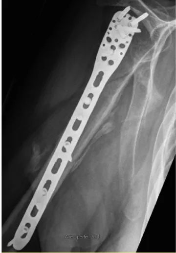
Participants were included from the initial sample only if radio-graphs were taken at least 45 days post-trauma. This cut-off was set as signs of HO can be adequately detected at that time (Cipriano et al., 2009). Moreover, in cases of multiple radiograph availabilities for a single patient, the radiograph conducted the closest to three months post-trauma was selected considering that medical check-ups are fre-quent at this time and that it falls within the range when HO formation is typically best detected (Cipriano et al., 2009). Radiographs of all patients were analyzed separately by a trained senior orthopaedist re-sident and a senior orthopedic surgeon both blind to the subjects' group classification. To evaluate signs of HO, both raters used a specialised radiology display system (NEC Display Solutions; MultiSync Monitor LCD 2090UXi-20.1; Made in China) to detect the presence of abnormal bone formation located in extra-skeletal soft tissues. More specifically, signs of HO were sought for near the fracture location, independently of joints involvement (seeFig. 1for a representative case of HO among the current sample). Hypertrophic callus was excluded from HO cases as the ossification identified needed to be at the heterotopic site and not at the fracture callus itself. In addition, Brooker's and Della Valle's clas-sifications were used conjointly as suggested by Toom et al. (2005) aiming to improve inter-observer reliability in the assessment of HO. Inter-rater reliability was verified and reached an almost perfect agreement according to Cohen's kappa coefficient (k = 0.93). In case of disagreements among raters, both raters reviewed together the radio-graph to reach an agreement concerning the presence of HO formation. 2.3. Matched sample procedure
Further steps were taken to control for potential factors known to affect the risk of HO formation. Patients from the ILF + mTBI group were matched with ILF patients according to age, sex, type of injury (area of fracture), and time elapsed between the accident and the radiograph. The importance of matching for the delay between the accident and the analyzed radiograph is to control for the risk of HO signs developing after the analyzed radiograph (Cipriano et al., 2009). To do so, we proceeded by using a one-on-one matching approach based on the following criteria: 1) age ( ± 5 years); 2) sex; 3) type of injury (area of fracture); 4) time elapsed between the accident and the radiograph ( ± 14 days). A match was made when all four criteria corresponded for two participants from each experimental group (ILF + mTBI group and ILF group). When more than one participant from the control group matched with a ILF + mTBI patient based on the aforementioned criteria, we selected the control participant who cor-responded most closely to the ILF + mTBI patient. This matching pro-cess allowed to form 47 near-identical pairs. The remaining participants who were not matched according to the criteria were excluded from these analyses.
2.4. Analyses
groups from our study (ILF + mTBI group and ILF group). Results from descriptive analyses are expressed as means, SD (standard deviation), and percentages (refer toTables 1–2). We used Pearson chi-square tests to compare the incidence rate of HO between the two experimental groups (ILF + mTBI group and ILF group). Additional chi-square ana-lyses were conducted to evaluate the possible impact of sex, age group (18–24; 25–44; 45–64; 65+ years old), joint involvement (periarticular fracture versus diaphyseal fracture) and surgical procedures on HO formation. A linear regression analysis was computed to give an esti-mate on which independent variable, mTBI or joint involvement, best predicted HO development. Statistical tests were carried out with a α-levelfixed at 0.05. The same pattern of analyses was used to test the study hypothesis among the matched sample. Moreover, a 2 × 2 ANOVA was used to assess the impact of HO and mTBI on return to work (RTW) among the matched sample. RTW was used in this study to reflect potential impact of HO development on functional outcome as it is known to be a good marker of recovery (Clay et al., 2010).
Information on RTW was collected in the context of a previous study conducted by our group using the same sample (see Jodoin et al. (2017b) for more details). Statistical analyses were performed using IBM SPSS software version 24 (Armonk, NY, United States).
3. Results
3.1. Results of full sample analysis
A total of 183 participants were selected, from a study cohort of 251 individuals recruited by our group (see participantflowchart inFig. 2). The remaining participants were excluded from the current study due to the inability to access their radiograph. Among thefinal sample, 50 patients were in the ILF + mTBI group (females = 19; mean age = 43.8) and 133 patients were in the ILF group (females = 75; mean age = 48.9). On average, radiographs were analyzed 86.8 days post-trauma (range: 45 days–201 days), a delay that was similar be-tween groups (F = 0.01; p = 0.92) (see Table 1). There was a sig-nificantly higher rate of periarticular fractures, as opposed to diaphy-seal fractures, in the ILF group compared to the ILF + mTBI group (X2= 16.69; p = 0.01) (see Table 2). This difference can be mainly attributed to the low rate of ILF + mTBI patients with ankle and distal radius fractures, compared to the ILF patients. Given the higher in-cidence of mTBI in fractures occurring proximally to the head (Jodoin et al., 2016), risks of suffering from a mTBI are rare in individuals treated for ankle and distal radius fractures.
Patients in the ILF + mTBI group showed significantly more signs of HO compared to patients with an ILF alone (X = 6.50; p = 0.01), with the majority of patients presenting with low grade HO according to Brooker's and Della's Valle's classification (see Tables 3–4). The in-cidence rates of HO signs were 46.0% in ILF + mTBI patients (23/50) as opposed to 26.3% in patients with an ILF alone (35/133). Of note, sex (X2= 2.32; p = 0.10), age group (X2= 2.08; p = 0.56), and sur-gical procedures (X2= 1.71; p = 0.13) were unrelated to the detection of signs of HO. Furthermore, rates of HO signs were found to be similar whether the fracture occurred proximally (periarticular fracture) or distally (diaphyseal fracture) to a joint (X2= 1.68; p = 0.24). See Table 5for more details. Lastly, results from the computed linear re-gression analysis show that sustaining a concomitant mTBI significantly predicted risks of HO development (β-coefficient = 0.18; t = 2.29; p = 0.02), whereas joint involvement was unrelated to HO develop-ment (β-coefficient = −0.05; t = −0.56; p = 0.58).
Fig. 1. Representative case of HO among sample.
Table 1
Descriptive characteristics of full study cohort by group.
Total mTBI No TBI P-value N (subjects) 183 50 133 – Age (years [SD]) 47.5 (15.5) 43.8 (15.3) 48.9 (15.3) 0.75 Sex (Female [%]) 94 (51.4) 19 (38.0) 75 (56.4) 0.02* Surgical procedures (% of sample) 32.7 26.8 34.6 0.23 Delay between trauma and
analyzed radiograph (days)
86.8 87.2 86.7 0.92
*=Level of significance was set at p < 0.05
Table 2
Distribution of fracture characteristics.
Body distribution of fractures [Number of patients] Total mTBI No TBI
- Metacarpal 2 1 1 - Metatarsal 11 3 8 - Proximal humerus 21 7 14 - Humerus diaphysis 5 1 4 - Distal humerus 6 0 6 - Scapula 3 2 1 - Clavicle 18 11 7 - Proximal ulna 5 2 3 - Ulna diaphyseal 3 2 1 - Distal radius 45 8 37 - Femur 2 1 1 - Patella 2 1 1 - Proximal tibia 4 2 2 - Diaphyseal tibia 7 3 4 - Distal tibia 9 2 7 - Ankle 40 4 36
Joint involvement Total mTBI No TBI P-value - Yes (periarticular fracture) 146 30 116 0.01 - No (diaphyseal fracture) 37 20 17
3.2. Results of analyses from the matched sample
A total of 94 participants were matched (i.e.; 47 patients from the ILF + mTBI group and 47 patients from the ILF group). Participants from both groups were equivalent according to the following criteria:
age (t = 0.00; p = 1.00), sex (X2= 0.00; p = 1.00), area of injury (X2= 0.00; p = 1.00), and delay between the accident and the ana-lyzed radiograph (t = 1.08; p = 0.30). Groups did not differ based on rates of surgical procedures (X2= 1.73; p = 0.25). Refer toTable 6to obtain detailed descriptive characteristics regarding the matched sample.
Similar to results obtained with the full sample, HO incidence was significantly higher in ILF + mTBI patients in comparison to ILF pa-tients (X2= 3.69; p = 0.04) (seeTable 7). This result further supports the notion that concomitant mTBI puts ILF patients at greater risk of developing HO. More specifically, 46.8% of ILF + mTBI patients (22/ 47) from the matched sample presented signs of HO compared to only 27.7% in ILF patients without a mTBI (13/47). Presence of HO nega-tively impacted RTW delays in patients with ILF + mTBI (F = 4.055;
251 participants
1) 58 ILF+mTBI patients
2) 193 ILF patients
Initial sample
183 participants
3) 50 ILF+mTBI patients
4) 133 ILF patients
Patients excluded due to inability
to access follow-up radiographs
or because follow-ups ended
within 45 days post-trauma
1) 8 ILF+mTBI patients
2) 60 ILF patients
Full sample
First round of analyses
1) Age
2) Sex
3) Type of injury
4) Delay between accident and analyzed
radiograph
94 participants
1) 47 ILF+mTBI patients
2) 47 ILF patients
Patients excluded following the
matching process
1) 3 ILF+mTBI patients
2) 86 ILF patients
Matching process
Matched sample
Second round of analyses
Fig. 2. Participant selectionflowchart. Table 3
HO signs among full sample.
mTBI No TBI X2 P-Value
HO signs (Number of patients [%]) 23/50 (46.0) 35/133 (26.3) 6.50 0.01*
p = 0.05). Return to work delays did not statistically differ according to the presence of HO in ILF patients without a comorbid mTBI (F = 0.823; p = 0.37). More specifically, ILF + mTBI patients with HO took, on average, 379 days to RTW compared to 106 days for ILF pa-tients with HO but without a mTBI. As for ILF + mTBI papa-tients without HO, it took, on average, 214 days to RTW as opposed to 168 days for ILF patients without HO and mTBI.
4. Discussion
This study investigated the incidence rate of HO among ILF patients with or without a concomitant mTBI. Results from the present study suggest that presence of HO is significantly higher in patients with both trauma injuries (mTBI and ILF) compared to ILF patients, even after controlling for factors known to influence HO, such as age, sex, area of injury, and time elapsed between the accident and the analyzed radiograph. Moreover, results from linear regressions show that sus-taining a concomitant mTBI significantly predicts risks for HO devel-opment whereas suffering from a fracture near a joint was unrelated. These findings are of particular interest, considering the high pre-valence of both injuries, namely ILF and mTBI, and the possible dele-terious consequences of HO on recovery and quality of life. In addition, the clinical symptoms linked to HO combined with possible additional surgical procedures to remove the heterotopic bone represent stag-gering financial burdens (health care expenditures and loss of pro-ductivity) (Eisenstein et al., 2018).
Another strikingfinding from this study is that the combination of HO formation and mTBI was associated with significantly longer RTW delays after an isolated limb fracture. Of note, mTBI without HO also negatively impacted RTW in ILF patients, but to a lesser extent than in the presence of HO. Indeed, results show a near 45% increase in delays to RTW when HO signs were detected in ILF + mTBI patients compared
to ILF + mTBI patients without HO. This is particularly alarming con-sidering that almost half of the assessed patients with an ILF and a comorbid mTBI presented signs of HO. Thisfinding points to the clin-ical relevance of systematclin-ically implementing a follow-up visit at least with ILF + mTBI patients if we are to investigate the impact of mTBI on clinical outcomes associated with HO such as pain, stiffness, and ar-ticular amplitude.
Most studies interested in the impact of concomitant TBI on the risk for HO formation focused on polytrauma patients or severely injured patients who suffered from a moderate to severe TBI and mostly focused on HO occurring near a joint (Bajwa et al., 2018; Garland, 1991a,b; Boes et al., 2006). The present study, however, shows that patients with an injury considered less severe, such as an ILF, are significantly more vulnerable to HO formation, regardless of joint involvement, when also afflicted by a comorbid mTBI. To the best of our knowledge, this is the first study specifically investigating the impact of mTBI on HO forma-tion among an orthopedic populaforma-tion. The fact that mTBI typically re-ceives limited medical attention beyond the acute post-accident phase can serve as a possible explanation. Another possibility could be that mTBI patients is not a clinical condition that justifies exposing unin-jured bones to X-ray radiation, thus preventing the detection of HO formation in mTBI-alone patients.
From a clinical standpoint, these results shed light on the im-portance of accounting for the presence of mTBI when treating ILF patients considering that over 44% of patients presenting with both injuries will develop HO. HO presence is classically studied in a context of hip and elbow secondary ankylosis and severe neurological con-comitant injury. Although conjectural, this study provides preliminary evidence of the significant impact of mild HO on patient outcome and extends HO screening beyond joints. Importantly, the addition of dia-physeal HO screening provides new information on whole-bone in-cidence rates of HO following a single fracture. Multiple factors may be at stake with regard to the higher incidence of HO among ILF + mTBI patients. For example, HO is believed to originate from the convergence of multiple mechanisms that closely involve the interaction of the
Table 4
Identification of HO according to Brooker's and Della Valle's classifications.
Total mTBI No TBI Number of subjects per classification
- A0
Absence of ossification
124 24 100 - A1
Isolated ossifications less than 1 cm in length
46 20 26 - B1
Isolated ossifications at least 1 cm in length – leaving MORE than 1 cm distance between pelvis and femur
2 2 0
- B2
Marginal ossifications – leaving MORE than 1 cm distance between pelvis and femur
3 0 3
- C1
Isolated ossifications at least 1 cm in length – leaving LESS than 1 cm distance between pelvis and femur or ankylosis
3 2 1
- C2
Marginal ossifications – leaving LESS than 1 cm distance between pelvis and femur or ankylosis
4 2 2
- C3
Ankylosis– leaving LESS than 1 cm distance between pelvis and femur or ankylosis
1 0 1
Table 5
Risks of HO in relation to joint involvement.
Periarticular fracture Diaphyseal fracture P-value mTBI
- Number of subjects with HO [%] 14/30 (46.7) 9/20 (45.0) 0.57 No mTBI
- Number of subjects with HO [%] 29/116 (25.0) 6/17 (35.3) 0.38
P-value 0.02* 0.40
*=Level of significance was set at p < 0.05
Table 6
Descriptive characteristics of matched sample by group.
Total mTBI No TBI P-value N (subjects) 94 47 47 – Age (years [SD]) 43.5 (15.1) 43.5 (15.5) 43.5 (14.7) 1.00 Sex (Female [%]) 34 (36.2) 17 (36.2) 17 (36.2) 1.00 Surgical procedures (% of sample) 33.7 26.3 40.0 0.25 Delay between trauma and
analyzed radiograph (days)
92.4 98.8 86.1 0.30
Table 7
HO signs among matched sample.
mTBI No TBI X2 P-Value
HO signs (Number of patients [%]) 22/47 (46.8) 13/47 (27.7) 3.69 0.04*
immune system and the central nervous system (Forsberg et al., 2014; Convente et al., 2015;Kraft et al., 2016;Sullivan et al., 2013). More specifically, a growing body of evidence highlights the involvement of the blood-brain-barrier (BBB) in HO formation (Huang et al., 2018). Interestingly, BBB permeability dysfunction is a well-known con-sequence of TBI and has been identified as a cause for high incidence rates of HO in patients with moderate to severe TBIs (Toffoli et al., 2008). Recent studies have shown that mTBI also leads to BBB dys-function which can act as a facilitator in the central nervous system invasion of peripheral immune response substances, such as in-flammatory cytokines, following a peripheral insult (Rowe et al., 2016). Additionally, neuroendocrine regulation, a system that is often deficient following mTBI, is closely involved in bone remodeling and HO for-mation (Undurti et al., 2018). Although speculative, it may be possible that the physiopathology of bone fracture and that of mTBI synergis-tically interact to promote HO formation. Shedding light on the possible involvement of physiopathological underpinnings of mTBI in HO could help identify new treatment targets and clinical management strategies aiming to minimize HO formation. In this study, HO was most fre-quently classified as low grade with small bone formation. This level of HO most likely does not cause decreased function by itself. We hy-pothesize that this low-grade HO is a sign of increased local soft tissue injury and increased neurological inflammation that is secondarily af-fecting outcome.
One limitation to the current study is that it uses data from parti-cipants recruited in the context of a previous study, which potentially restricts study findings generalization. Secondly, collection of pro-spective data should systematically control for the time elapsed since the injury at the time of radiographs (for example, all taken at three months post-accident) so as to reduce risks for missed HO diagnoses. One interesting avenue in further investigating the relation between RTW delays and HO formation would be to specify the type of work conducted (light versus heavy work) as well as the quality of the RTW (successful RTW versus work accommodations needed). Moreover, in-vestigating RTW delays in relation with both prospective functional recovery measures and low-grade HO could help us identify therapeutic targets for optimal orthopedic trauma recovery. Given that some frac-tures are more prone to HO formation, larger-scale replication studies should consider data stratification analyses according to injury types. Gained knowledge would allow us to further refine classification of at-risk patients. Finally, future studies should account for additional fac-tors, such as injury severity, duration of immobilization, and pre-injury conditions, such as, but not limited to, history of HO and genetic pre-disposition, as they are known to impact HO formation (Pape et al., 2004;Dizdar et al., 2013).
5. Conclusion
In conclusion, studyfindings highlight that sustaining a comorbid mTBI puts ILF patients at significantly higher risk of developing HO. Moreover, ILF patients with a mTBI are greatly impacted by HO in relation with RTW, a factor associated with high productivity costs and risks for chronic fracture injury symptoms. This is of significant clinical interest considering the high incidence of both injuries, the frequency at which mTBI goes undiagnosed, and the clinical impact of HO on re-covery. The impact of mTBI on HO formation warrants further attention to detect early signs of HO, to identify shared physiopathological me-chanisms and, ultimately, to design targeted therapies.
Conflict of competing interest
None of the authors report any conflict of interest in relation to this paper. The institution of one or more of the authors (HSCM) has re-ceived funding from: Arthrex, Conmed, Depuy, Smith and Nephew, Synthes, Tormier, Zimmer.
These companies were not involved in any aspect of the study
design, data collection, analysis, nor the manuscript preparation, and authors had complete control over all the study data that supports the publication.
Transparency document
The Transparency document associated with this article can be found, in online version.
References
Bajwa, N.M., Kesavan, C., Mohan, S., 2018. Long-term consequences of traumatic brain injury in bone metabolism. Front. Neurol. 9, 115.
Boes, M., Kain, M., Kakar, S., Nicholls, F., Cullinane, D., Gerstenfeld, L., Einhorn, T.A., Tornetta III, P., 2006. Osteogenic effects of traumatic brain injury on experimental fracture-healing. J. Bone Joint Surg. Am. 88 (4), 738–743.
Carroll, L.J., Cassidy, J.D., Holm, L., Kraus, J., Coronado, V.G., W.H.O.C.C.T.F.o.M.T.B. Injury, 2004. Methodological issues and research recommendations for mild trau-matic brain injury: the WHO Collaborating Centre Task Force on Mild Trautrau-matic Brain Injury. J. Rehabil. Med. (43 Suppl), 113–125.
Cassidy, J.D., Carroll, L.J., Peloso, P.M., Borg, J., von Holst, H., Holm, L., Kraus, J., Coronado, V.G., W.H.O.C.C.T.F.o.M.T.B. Injury, 2004. Incidence, risk factors and prevention of mild traumatic brain injury: results of the WHO Collaborating Centre Task Force on Mild Traumatic Brain Injury. J. Rehabil. Med. (43 Suppl), 28–60.
Cipriano, C.A., Pill, S.G., Keenan, M.A., 2009. Heterotopic ossification following trau-matic brain injury and spinal cord injury. J. Am. Acad. Orthop. Surg. 17 (11), 689–697.
Clay, F.J., Newstead, S.V., Watson, W.L., McClure, R.J., 2010. Determinants of return to work following non life threatening acute orthopaedic trauma: a prospective cohort study. J. Rehabil. Med. 42 (2), 162–169.
Coelho, C.V., Beraldo, P.S., 2009. Risk factors of heterotopic ossification in traumatic spinal cord injury. Arq. Neuropsiquiatr. 67 (2B), 382–387.
Convente, M.R., Wang, H., Pignolo, R.J., Kaplan, F.S., Shore, E.M., 2015. The im-munological contribution to heterotopic ossification disorders. Curr. Osteoporos. Rep. 13 (2), 116–124.
Dizdar, D., Tiftik, T., Kara, M., Tunc, H., Ersoz, M., Akkus, S., 2013. Risk factors for developing heterotopic ossification in patients with traumatic brain injury. Brain Inj. 27 (7–8), 807–811.
Eisenstein, N., Stapley, S., Grover, L., 2018. Post-traumatic heterotopic ossification: an old problem in need of new solutions. J. Orthop. Res. 36 (4), 1061–1068.
Evans, K.N., Forsberg, J.A., Potter, B.K., Hawksworth, J.S., Brown, T.S., Andersen, R., Dunne, J.R., Tadaki, D., Elster, E.A., 2012. Inflammatory cytokine and chemokine expression is associated with heterotopic ossification in high-energy penetrating war injuries. J. Orthop. Trauma 26 (11), e204–e213.
Firoozabadi, R., Alton, T., Sagi, H.C., 2017. Heterotopic ossification in acetabular fracture surgery. J. Am. Acad. Orthop. Surg. 25 (2), 117–124.
Forsberg, J.A., Potter, B.K., Polfer, E.M., Safford, S.D., Elster, E.A., 2014. Do inflammatory markers portend heterotopic ossification and wound failure in combat wounds? Clin. Orthop. Relat. Res. 472 (9), 2845–2854.
Foruria, A.M., Augustin, S., Morrey, B.F., Sanchez-Sotelo, J., 2013. Heterotopic ossifica-tion after surgery for fractures and fracture-dislocaossifica-tions involving the proximal as-pect of the radius or ulna. J. Bone Joint Surg. Am. 95 (10), e66.
Foruria, A.M., Lawrence, T.M., Augustin, S., Morrey, B.F., Sanchez-Sotelo, J., 2014. Heterotopic ossification after surgery for distal humeral fractures. Bone Joint J. 96-B (12), 1681–1687.
Garland, D.E., 1991a. Surgical approaches for resection of heterotopic ossification in traumatic brain-injured adults. Clin. Orthop. Relat. Res.(263), 59-70.
Garland, D.E., 1991b. A clinical perspective on common forms of acquired heterotopic ossification. Clin. Orthop. Relat. Res.(263), 13-29.
Huang, H., Cheng, W.X., Hu, Y.P., Chen, J.H., Zheng, Z.T., Zhang, P., 2018. Relationship between heterotopic ossification and traumatic brain injury: why severe traumatic brain injury increases the risk of heterotopic ossification. J. Orthop. Translat. 12, 16–25.
Jodoin, M., Rouleau, D., Charlebois-Plante, C., Benoit, B., Leduc, S., Laflamme, Y., Gosselin, N., Larson-Dupuis, C., De Beaumont, L., 2016. Incidence rate of mild traumatic brain injury among patients who have suffered from an isolated limb fracture: upper limb fracture patients are more at risk. Injury 47 (8), 1835–1840.
Jodoin, M., Rouleau, D.M., Gosselin, N., Benoit, B., Leduc, S., Laflamme, Y., Larson-Dupuis, C., De Beaumont, L., 2017a. Comorbid mild traumatic brain injury increases pain symptoms in patients suffering from an isolated limb fracture. Injury 48 (9), 1927–1931.
Jodoin, M., Rouleau, D.M., Larson-Dupuis, C., Benoit, B., Leduc, S., Laflamme, G.Y., Gosselin, N., Sabir, M., De Beaumont, L., 2017b. Effects of concomitant mild trau-matic brain injury on resuming work after suffering from an isolated limb fracture: a cohort study. Brain Inj. 31 (12), 1683–1688.
Kaplan, F.S., Glaser, D.L., Hebela, N., Shore, E.M., 2004. Heterotopic ossification. J. Am. Acad. Orthop. Surg. 12 (2), 116–125.
Kraft, C.T., Agarwal, S., Ranganathan, K., Wong, V.W., Loder, S., Li, J., Delano, M.J., Levi, B., 2016. Trauma-induced heterotopic bone formation and the role of the immune system: a review. J. Trauma Acute Care Surg. 80 (1), 156–165.
Nauth, A., Giles, E., Potter, B.K., Nesti, L.J., O'Brien, F.P., Bosse, M.J., Anglen, J.O., Mehta, S., Ahn, J., Miclau, T., Schemitsch, E.H., 2012. Heterotopic ossification in
orthopaedic trauma. J. Orthop. Trauma 26 (12), 684–688.
Pape, H.C., Marsh, S., Morley, J.R., Krettek, C., Giannoudis, P.V., 2004. Current concepts in the development of heterotopic ossification. J. Bone Joint Surg. Br. Vol. 86 (6), 783–787.
Ranganathan, K., Loder, S., Agarwal, S., Wong, V.W., Forsberg, J., Davis, T.A., Wang, S., James, A.W., Levi, B., 2015. Heterotopic ossification: basic-science principles and clinical correlates. J. Bone Joint Surg. Am. 97 (13), 1101–1111.
Rowe, R.K., Ellis, G.I., Harrison, J.L., Bachstetter, A.D., Corder, G.F., Van Eldik, L.J., Taylor, B.K., Marti, F., Lifshitz, J., 2016. Diffuse traumatic brain injury induces prolonged immune dysregulation and potentiates hyperalgesia following a peripheral immune challenge. Mol. Pain 12.
Sullivan, M.P., Torres, S.J., Mehta, S., Ahn, J., 2013. Heterotopic ossification after central nervous system trauma: a current review. Bone Joint Res. 2 (3), 51–57.
Toffoli, A.M., Gautschi, O.P., Frey, S.P., Filgueira, L., Zellweger, R., 2008. From brain to bone: evidence for the release of osteogenic humoral factors after traumatic brain
injury. Brain Inj. 22 (7–8), 511–518.
Toom, A., Fischer, K., Martson, A., Rips, L., Haviko, T., 2005. Inter-observer reliability in the assessment of heterotopic ossification: proposal of a combined classification. Int. Orthop. 29 (3), 156–159.
Undurti, A., Colasurdo, E.A., Sikkema, C.L., Schultz, J.S., Peskind, E.R., Pagulayan, K.F., Wilkinson, C.W., 2018. Chronic hypopituitarism associated with increased post-concussive symptoms is prevalent after blast-induced mild traumatic brain injury. Front. Neurol. 9, 72.
Vanden Bossche, L., Vanderstraeten, G., 2005. Heterotopic ossification: a review. J. Rehabil. Med. 37 (3), 129–136.
Winkler, S., Wagner, F., Weber, M., Matussek, J., Craiovan, B., Heers, G., Springorum, H.R., Grifka, J., Renkawitz, T., 2015. Current therapeutic strategies of heterotopic ossification—a survey amongst orthopaedic and trauma departments in Germany. BMC Musculoskelet. Disord. 16, 313.