Discriminant BOLD Activation Patterns during
Mental Imagery in Parkinson’s Disease
Jessica Schrouff1,2, Julien Cremers1,3, Kevin D’Ostilio1, Val´erie Delvaux3,
Ga¨etan Garraux1,3, and Christophe Phillips1,2
1
Cyclotron Research Centre, University of Li`ege, Belgium
2
Department of Electrical Engineering and Computer Science, University of Li`ege, Belgium
3
Department of Neurology, Li`ege University Hospital, Belgium
Abstract. Using machine learning based models in clinical applications has become current practice and can prove useful to provide information at the subject’s level, such as predicting an (early) diagnosis or monitor-ing the evolution of a disease. However, the performance of these models depends on the choice of a biomarker to detect the presence or absence of a disease. Choosing a biomarker is not straightforward, especially in the case of Parkinson’s disease when compared to healthy subjects. In the present work, we investigated the mental imagery of gait as a biomarker of Parkinson’s disease and showed that the signal in the mesencephalic locomotor region during the mental imagery of gait at a comfortable pace can discriminate significantly between idiopathic Parkinson’s disease pa-tients and healthy subjects. Although there is room for improvement, the results of this preliminary study are promising.
1
Introduction
During the past decades, advances in neuroimaging techniques allowed inves-tigating the brain structure and functioning in both health and disease. More particularly, Positron Emission Tomography (PET) and Magnetic Resonance Imaging (MRI) enabled the identification of biomarkers in dementias, such as Alzheimer’s disease [1] or in different states of consciousness [2]. They also al-lowed the characterisation of more complex paradigms such as the mental im-agery of gait in both healthy [3] and Parkinson’s diseased subjects [4].
Up to now, the methods used to analyse such data focused on characteriz-ing the individual relationship between a cognitive or perceptual state and each voxel, i.e. following a univariate approach. For functional images, a well-known univariate technique is Statistical Parametric Mapping [5], detecting which vox-els show a statistically significant response to the experimental conditions at the individual subject level but also within and between groups of subjects. For structural images, a common approach is Voxel-Based Morphometry, (VBM, [6]), which investigates focal differences in grey matter density between groups of sub-jects. However, these studies typically infer group differences while conclusions
at the subject’s level would be desirable for diagnosis, prognosis, treatment plan-ning or the monitoring of disease progression [7].
Recently, univariate analyses have been complemented by the use of machine learning based models [8]. These methods aim at predicting a variable of interest (e.g. mental state 1 vs. mental state 2, or patients vs. controls) from the pattern of brain activation/anatomy over a set of voxels. They have been particularly useful in clinical applications [7], e.g. to distinguish between healthy subjects and Alzheimer’s disease patients [9,10] or to assess the level of consciousness of vegetative states patients [11]. Machine learning based models have also been applied to Parkinson’s Disease (PD) when discriminating Idiopathic Parkinson’s Disease (IPD) from Parkinson Plus Syndromes (PPS) [12]. However, it seems that finding a biomarker distinguishing IPD from healthy controls remains an open issue [7,12].
In the present work, we investigated whether the mental imagery of both comfortable and brisk walking could predict the presence or absence of IPD, based on previous works [3,4].
2
Material and Methods
The material considered in this work is the same as in [4]. Therefore, only a brief description of the population and experimental design will be provided.
2.1 Population
In total, 29 subjects participated in the study: 14 patients (7 males; mean age: 65.1 ± 9.5 years) diagnosed with IPD with different degrees of severity of gait disturbances and 15 controls matched for age (63.8 6 ± 8.1 years) and gender (7 males). Written informed consents for this research protocol approved by the local ethics committee were obtained from all participants.
2.2 Design
Before fMRI, the subjects were asked to walk comfortably and then briskly on a 25m path. After gait evaluation, they were trained to mentally rehearse themselves walking on the path.
All subjects then underwent an fMRI session comprising three taks: mental imagery of standing on the path (STAND), walking at a comfortable pace along the path (COMF) and walking briskly along the path (BRISK). The COMF and BRISK conditions were self-paced, subjects indicating when they had completed each trial by a key press, while each trial of the STAND condition was constrained by the duration of the previous COMF trial. Eight trials of each condition (12 for BRISK to account for a shorter duration of the trials) were randomly presented to each subject.
In addition to BOLD fMRI, all participants underwent a high-resolution volumetric anatomical MRI of the brain.
2.3 Preprocessing
fMRI data preprocessing and univariate analysis were performed using SPM81
. Functional images were realigned and co-registered to the structural image before normalisation using DARTEL [13]. Finally, smoothing was applied using a 8mm FWHM Gaussian kernel.
A General Linear Model then summarized the time series from each subject by modelling each condition by a boxcar function convoluted with a canonical haemodynamic response function. In the end, three images per subject were considered for further analysis: the parametric maps of STAND, COMF and BRISK representing the BOLD signal activity associated with each condition.
2.4 Multivariate Modelling
The multivariate analysis was performed using PRoNTo2
. This Matlab based software (MathWorks, Natick, MA) provides a framework to perform pattern recognition based on machine learning models.
In its current version, PRoNTo does not provide wrapper or embedded feature selection. Three masks were therefore used as filters before building the linear kernel, based on [4]:
– A “whole brain” mask, selecting all voxels within the brain.
– A “motor mask”, built on the WFU-PickAtlas [14] and comprising the areas involved in gait (both in healthy subjects and patients), as described in Table 1 of [15].
– A mask comprising the Mesencephalic Locomotor Region (MLR) and pe-dunculopontine nucleus, further referred to as “MLR mask”.
Classification was performed using binary SVM [16], as in [12] and [7]. For the between groups classification, the three tasks will be combined in every pos-sible way (e.g. BRISK, BRISK+COMF, . . . ) and tested for each mask, leading to 7 combinations times 3 masks, equals 21 models. In addition to the IPD vs. CTRL comparison, the discrimination between the three tasks was also assessed by pooling together the data of both groups. This multiclass classification re-quired the use of multiclass Gaussian Processes [17]3
. For both classification problems, leave-one-subject-out cross-validation was performed to compute the balanced and class accuracies, as well as Positive Predictive Values (PPV). The significance of accuracy measures was assessed by non-parametric testing using 1000 random permutations of the training labels.
1
www.fil.ion.ucl.ac.uk/spm
2
Application for a demonstration of PRoNTo at NIPS has been submitted,
www.mlnl.cs.ucl.ac.uk/pronto
3
Binary Gaussian Processes could also have been used on the between groups com-parison but [18] has shown that SVM and GP had similar performances on balanced datasets with reduced computational expenses for SVM.
3
Results
3.1 Between groups comparison
Results are presented for each (combination of) condition(s) in Table1in terms of balanced accuracy while weights of the COMF model are represented in Fig.1
for each mask.
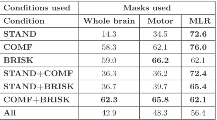
Table 1. Balanced accuracy (in %) for the IPD vs. CTRL classification for each com-bination of the three tasks (rows) and for each mask (columns). “All” represents the combination of the three tasks. Significant results are displayed in bold.
Conditions used Masks used
Condition Whole brain Motor MLR
STAND 14.3 34.5 72.6 COMF 58.3 62.1 76.0 BRISK 59.0 66.2 62.1 STAND+COMF 36.3 36.2 72.4 STAND+BRISK 36.7 39.7 65.4 COMF+BRISK 62.3 65.8 62.1 All 42.9 48.3 56.4
Overall, the whole brain mask led to a poor discrimination of IPD vs. CTRL, with the accuracy reaching 62.3% when considering both the COMF and BRISK conditions together (only significant result). Slightly better results were obtained from the features in the motor mask, as shown by a higher balanced accuracy for the BRISK-COMF combination, as well as for the BRISK condition (both significant at p < 0.05). The signal comprised in the MLR mask led to the best results, the highest performance being reached when considering the COMF condition. For this model, the balanced accuracy had a value of 76% (p=0.01), with the class accuracies reaching 78.6% for IPD and 73.3% for CTRL (both significant at p < 0.05). PPV are in the same range, with a PPV of 78.6% for CTRL and 73.3% for IPD.
3.2 Between tasks comparison
In terms of balanced accuracy, significant results could be obtained from the discrimination between the three tasks across the two groups of subjects when considering the whole brain and motor masks (Table 3.2). This result is con-firmed by the PPV for each class, which are quite high. However, the signal in the MLR doesn’t lead to a significant classification of the three tasks, which is further confirmed by the PPV (almost all samples are classified as STAND).
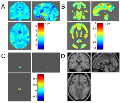
Fig. 1. Weights of the SVM model discriminating between CTRL (label +1) and IPD (label -1) based on the combination of the BRISK and COMF conditions, for A the whole brain, B, motor and C MLR masks (Note that the cross-hair position has been centred on the MLR for this panel). D displays the SPM single subject canonical structural image for better representation.
Table 2. Balanced accband class accuracies (in %, PPV in brackets) of the multiclass
GP model discriminating between the three tasks (STAND, COMF and BRISK) when considering both groups jointly. Significant results are displayed in bold.
Mask accb STAND COMF BRISK
Whole brain 65.5 58.6 (65.4) 41.4 (75.0) 96.6 (62.2) Motor areas 66.7 62.1 (64.3) 41.4 (70.6) 96.6 (66.7) MLR area 32.2 89.7 (32.1) 0.0 (0.0) 6.9 (33.3)
4
Discussion
In the present work, we classified the parametric maps of three tasks (STAND, COMF and BRISK) to identify idiopathic Parkinson’s disease patients from healthy controls. The best model discriminated significantly between IPD and
controls with a balanced accuracy of 76%, when using the signal comprised in the mesencephalic locomotor region. Furthermore, combining the mental imagery of gait at comfortable and brisk paces led to consistent and significant results across feature sets.
Although these results are not overwhelming, it is (to the best of our knowl-edge) one of the first significant classification between IPD and controls [7,12]. In view of the simplicity of the model (SVM with a filter feature selection), this re-sult is promising and suggests that mental imagery of gait could be a biomarker of Parkinson’s disease although improvements need to be performed before being able to deal with more complex issues, such as diagnosing Parkinson’s disease in its early stage, or distinguishing between IPD and Parkinson Plus Syndroms.
However, distinguishing between the three tasks using both groups led to significant results when considering the whole brain and motor masks, suggest-ing that the between-subject variability within one group is large compared to the between-groups variability for those features. This result highlights the importance of feature selection in the present case and favours the use of wrap-per or embedded feature selection techniques to increase the wrap-performance of the machine learning based models. Regarding the Parkinsonian group, the between-subjects variability might further be explained by the heterogeneity of the gait disorders in patients. Distinguishing between patients with light or sever gait disorders, for example by considering the Freezing of Gait (FoG, [19]), might in-crease the ratio of between versus within group variability and thereby improve the classification.
Finally, although a large overlap has been observed between mental imagery of gait and actual gait in healthy subjects [20], our results question the overlap between mental imagery of disturbed gait and actual disturbed gait, especially in the STAND and BRISK conditions. Solving this issue is not straightforward but developments in ambulatory Electro-EncephaloGraphy (EEG) acquisition systems and in the decoding of this type of signal might bring a solution by directly acquiring the brain activity under actual gait.
5
Conclusion
Finding a biomarker of Parkinson’s Disease allowing for the detection of IPD remains an open issue, but we have shown here that BOLD signal recorded during controlled mental imagery is a good candidate. Indeed the activity in the mesencephalic locomotor region induced by mental imagery of gait at a comfortable pace leads to promising results.
Acknowledgements
This work was funded by the FNRS-FRS and FRIA, Belgium and by the Uni-versity of Li`ege.
References
1. Zakzanis, K.K., Graham, S.J., Campbell, Z.: A meta-analysis of structural and functional brain imaging in dementia of the Alzheimer’s type: A neuroimaging profile. Neuropsychology Review 13 (2003) 1–18
2. Monti, M.M., Vanhaudenhuyse, A., Coleman, M.R., Boly, M., Pickard, J.D., Tshibanda, L., Owen, A.M., Laureys, S.: Willful modulation of brain activity in disorders of consciousness. New England Journal of Medicine 362 (2010) 579–589 3. Cremers, J., Dessoullieres, A., Garraux, G.: Hemispheric specialization during
mental imagery of brisk walking. Human Brain Mapping 33 (2012) 873–882 4. Cremers, J., D’Ostilio, K., Stamatakis, J., Delvaux, V., Garraux, G.: Brain
ac-tivation pattern related to gait disturbances in Parkinson’s disease. Movement Disorders in press (2012)
5. Friston, K., Ashburner, J., Kiebel, S., Nichols, T., Penny, W.: Statistical Para-metric Mapping: the analysis of functional brain images. Elsevier Academic Press, London (2007)
6. Ashburner, J., Friston, K.: Voxel-based morphometry – the methods. NeuroImage 11 (2000) 805–821
7. Orr`u, G., Pettersson-Yeo, W., Marquand, A.F., Sartori, G., Mechelli, A.: Using Support Vector Machine to identify imaging biomarkers of neurological and psychi-atric disease: A critical review. Neuroscience & Biobehavioral Reviews 36 (2012) 1140 – 1152
8. Pereira, F., Mitchell, T., Botvinick, M.: Machine learning classifiers and fmri: a tutorial overview. NeuroImage 45 (2009) S199–S209
9. Vemuri, P., Gunter, J.L., Senjem, M.L., Whitwell, J.L., Kantarci, K., Knopman, D.S., Boeve, B.F., Petersen, R.C., Jack, C.R.: Alzheimer’s disease diagnosis in individual subjects using structural MR images: Validation studies. NeuroImage 39 (2008) 1186–1197
10. Kl¨oppel, S., Stonnington, C.M., Chu, C., Draganski, B., Scahill, R.I., Rohrer, J.D., Fox, N.C., Jack, C.R., Ashburner, J., Frackowiak, R.S.J.: Automatic classification of MR scans in Alzheimer’s disease. Brain 131 (2008) 681–689
11. Phillips, C., Bruno, M.A., Maquet, P., Boly, M., Noirhomme, Q., Schnakers, C., Vanhaudenhuyse, A., Bonjean, M., Hustinx, R., Moonen, G., Luxen, A., Lau-reys, S.: ’Relevance Vector Machine’ consciousness classifier applied to cerebral metabolism of vegetative and locked-in patients. NeuroImage 56(2) (2011) 797– 808
12. Focke, N.K., Helms, G., Scheewe, S., Pantel, P.M., Bachmann, C.G., Dechent, P., Ebentheuer, J., Mohr, A., Paulus, W., Trenkwalder, C.: Individual voxel-based subtype prediction can differentiate progressive supranuclear palsy from idiopathic Parkinson syndrome and healthy controls. Human Brain Mapping 32 (2011) 1905– 1915
13. Ashburner, J.: A fast diffeomorphic image registration algorithm. NeuroImage 38 (2007) 95 – 113
14. Maldjian, J.A., Laurienti, P.J., Burdette, J.B., A, K.R.: An automated method for neuroanatomic and cytoarchitectonic atlas-based interrogation of fMRI data sets. NeuroImage 19 (2003) 1233–1239
15. Maillet, A., Pollak, P., Deb˜u, B.: Imaging gait disorders in parkinsonism: a review. Journal of Neurology, Neurosurgery & Psychiatry 83(10) (2012) 986–993
16. Burges, C.: A tutorial on Support Vector Machines for pattern recognition. Data Mining and Knowledge Discovery 2 (1998) 121–167
17. Rasmussen, C.E., Williams, C.K.I.: Gaussian Processes for Machine Learning. Adaptive Computation and Machine Learning. the MIT Press (2006)
18. Schrouff, J., Kuss´e, C., Wehenkel, L., Maquet, P., Phillips, C.: Decoding semi-constrained brain activity from fMRI using Support Vector Machines and Gaussian Processes. PLoS One 7 (2012) e35860
19. Karachi, C., Grabli, D., Bernad, F.A., Tand´e, D., Wattiez, N., Belaid, H., Bardinet, E., Prigent, A., Nothacker, H.P., Hunot, S., Hartmann, A., Leh´ericy, S., Hirsch, E., Chantal, F.: Cholinergic mesencephalic neurons are involved in gait and postural disorders in Parkinson disease. Journal of Clinical Investigation 120 (2010) 2745– 2754
20. Dobkin, B.H., Firestine, A., West, M., Saremi, K., R, W.: Ankle dorsiflexion as an fMRI paradigm to assay motor control for walking during rehabilitation. NeuroImage 23 (2004) 370381