Phycologia (1990) Volume 29 (1), 19-26
Presence of CU-phycoerythrin in the marine benthic
blue-green alga Oscillatoria cf. corallinae
L. HOFFMANN', L. TALARIC02 AND A. WILMOTTE' , Department 0/ Botany, University 0/ Liege, Liege, Belgium 2 Department 0/ Biology, University o/Trieste, Trieste, Italy
L. HO FFMANN, L. TALARICO AND A. WILMOTTE. 1990. Presence of CU-phycoerythrin in the marine benthic blue-green alga Oscillatoria cf. corallinae. Phycologia 29: 19-26.
The presence of CU-phycoerythrin, a phycobiliprotein characterized by the presence of phycourobilin chromophores in addition to phycoerythrobilins, and so far found in only eight blue-green algae, is reported for the first time from a marine benthic blue-green alga, Oscillatoria cf. corallinae.
INTRODUCTION
Phycobiliproteins are the major accessory light harvesting pigments of blue-green algae and red algae. Their colour is due to the presence of co valently bound open-chain tetrapyrrole pros thetic groups, the phycobilins. Different ph yco biliproteins are distinguished in blue-green algae on the basis of their visible absorption spectra (Table 1).
The phycobiliproteins are organized into su pramolecular complexes, the phycobilisomes, which are found as regular arrays on the surface of the thylakoid membranes (Gantt 1980, 1981; Glazer 1984). Additional uncoloured polypep tides serve to link biliproteins in the phycobili somes and to attach them to the thylakoid mem branes (Wehrmeyer 1983). Phycobiliproteins constitute an energy-transfer chain through which the incident light energy passes from phycoery thrin or phycoerythrocyanin to chlorophyll a (Gray & Gantt 1975; Grabowsky & Gantt 1978; Searle et at. 1978; Lundell & Glazer 1981; Pel legrino et at. 1981) as shown below:
ph ycoerythrin
Phycocyanin and allophycocyanin seem to be universally present in blue-green algae. Allophy cocyanin B was identified in many but not all blue-green algae (Glazer & Bryant 1975; Ley et at. 1977).
Phycoerythrocyanin, characterized by the presence of the two chromophores phycocy anobilin and phycobiliviolin (Bishop et at. 1987), is mainly found in heterocystous blue-green algae. Phycoerythrocyanin and phycoerythrin are mutually exclusive (Bryant 1982).
The phycoerythrins are widely distributed among all the taxonomic groups and form the spectroscopically most variable class of phyco biliproteins. The classical phycoerythrin (CPE) has a single absorption maximum at 560 nm due to the presence of phycoerythrobilin (PEB) as a chromophore. In some blue-green algae, spectral forms with broadened absorption bands and maxima at 550-565 nm, and those possessing two absorption maxima in the 550-570 nm range are found. In many cases, these spectral forms are apparently denatured and dissociated forms of phycoerythrin (Mac Coli & Guard-Friar 1987),
0'
)---?>
phyooey,n;n - allophy""y,n;n - chlocophyll. phycoerythrocyaninCorrespondence: Dr L. Hoffmann, Department of Botany, University of Liege, Sart Tilman B22, B-4000 Liege, Belgium.
reflecting different types of protein-bilin inter action brought about by variation in pH, con centration of ions and biliproteins in the solution (Glazer 1984). Many, but not all, blue-green al gae containing phycoerythrin undergo chromatic adaptation. In fact, three types of response to 19
20 Phycologia, Vol. 29 (1), 1990
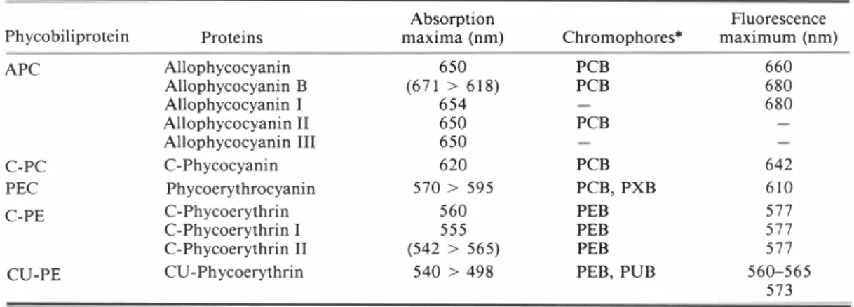
Table 1. Properties of phycobiliproteins present in blue-green algae. Modified from Gantt (1981) and Zilinskas & Greenwald (1986)
Absorption Fluorescence
Phycobiliprotein Proteins maxima (nm) Chromophores* maximum (nm)
APC Allophycocyanin 650 PCB 660 Allophycocyanin B (671 > 618) PCB 680 Allophycocyanin I 654 680 Allophycocyanin II 650 PCB Allophycocyanin III 650 C-Phycocyanin 620 PCB 642 C-PC PEC Phycoerythrocyanin 570 > 595 PCB, PXB 610 C-Phycoerythrin 560 PEB 577 C-Phycoerythrin I 555 PEB 577 C-PE C-Phycoerythrin II (542 > 565) PEB 577
CU-PE CU -Phycoerythrin 540> 498 PEB, PUB 560-565
573 * PCB: phycocyanobilin; PXB: phycobiliviolin; PEB: phycoerythrobilin; PUB: phycourobilin. red, green and white light conditions have been
observed in blue-green algae (Tandeau de Mar sac 1977). Strains designated as type I do not adapt chromatically and the ratio of PE to PC remains constant. Type II strains adapt by mod ulating PE synthesis alone; its synthesis is pro moted in green light and repressed in red light, whereas PC synthesis remains constant. Type III strains are able to alter both PC and PE synthesis. In green light PE synthesis is high and this bili protein becomes the dominant pigment; in red light PE synthesis is repressed, but the exposure of these strains to red light induces the de novo synthesis of a unique PC not present in green light-grown cells (Bryant 1981; Bryant & Cohen Bazire 1981).
Another type of phycoerythrin is characterized by the presence of a peak in the 500 nm range, due to the presence of phycourobilin (PUB) chro mophore in addition to PEB. This phycoery thrin, called CU-phycoerythrin by MacColl & Guard-Friar (1987), appears to be similar to B and R-phycoerythrins from red algae in that it contains the same chromophores (PUB, PEB). This pigment class has so far only been dem onstrated in eight blue-green algal species be longing to the Chroococcales and the Oscillato riaceae (Table 2). The pigment was studied in detail for Synechococcus sp. DC-2 (Alberte et al. 1984; Kursar et al. 1981), Synechococcus sp. WH 8103 (Ong et al. 1984), and Gloeobacter viola ceus Rippka, Waterbury et Cohen-Bazire (Bryant et al. 1981).
The rare occurrence of this pigment in blue green algae is of interest and adds a marine ben thic Oscillatoria species to the list.
MATERIALS AND METHODS
Oscillatoria cf. corallinae (Kiitzing) Gomont ex Gomont growing on a calcareous worm tube on the stem of Posidonia oceanica Delile was col lected by V. Demoulin at a depth of 5 m in July 1984 in the harbour of the oceanographical sta tion ST ARESO (Calvi, Corsica). The strain CJ 1 is maintained in the culture collection of the De partment of Botany (University of Liege). The morphological variability of the strain and its growth limits for temperature and irradiance were determined as described by Wilmotte (1988). To determine the growth limits for salinity the strain was grown in MN medium (Rippka et al. 1979) with the salinity adjusted to 8, 50, 75, 100,200, and 500% of seawater at 22°C, and at a contin uous illumination of 20 �mol photons m-2 S-I. To determine the presence of a chromatic adaptation, cultures were grown for 3 weeks under red and green filters (Lee filters 106 and 124) at 22°C at a continuous illumination of 17 �mol photons m-2 S-I. The change of colour was recorded visually.
For pigment analysis, the strain was grown in MN medium in 5 I flasks at 22°C and with a continuous lateral illumination of 30 �mol pho tons m-2 S-I provided by Phytor LF40W cool white fluorescent tubes. The culture was agitated continuously by magnetic stirring and aerated with 99.5% N2 and 0.5% CO2. After 3 weeks, plants were harvested by centrifugation and the pellet was stored at -70°C until use.
Frozen Oscillatoria cells were homogenized by a liquid nitrogen-cooled electric grinder. The homogenate was suspended in a 50 mM Na-K
Table 2. Properties of CU-phycoerythrin from blue-green algae
Habitat
Gloeobacter violaceus Rippka Terrestrial
et al.
Synechococcus sp. DC-2 Marine
pico-(= WH7803) plankton
Synechococcus sp. WH8103 Marine pico-plankton Synechocystis cf. trididemni Marine
sym-Lafargue et Duclaux biont Oscillatoria irrigua Gomont Freshwater
Oscillatoria sp. Thermal Oscillatoria cf. corallinae Marine benthic
Gomont
Oscillatoria spongeliae Marine
sym-(Schulze) Hauck biont
Trichodesmium cf. thiebautii Marine
plank-Gomont ton
* PEB: phycoerythrobilin; PUB: phycourobilin. t sh: shoulder.
* In vivo spectrum.
§ Three bands are observed in the 29 kD region.
Absorption maxima (nm) 501,564 500, 542 492,543 495, 540 496, 540 495, 565 498, 567 494, 540 498, 542 500, 547, 565 (sh)t 493, 567* 495, 550* 505, 542, 557 496, 542 495, 547, 565 F1uores-cence maximum (nm) 574, 577 560 565 570 569 573 574 573 Subunits
molecular PEB : Subunit composition* weight PUB
(D) ratio a {3
a-20 500 6 : I {3-21 700
a-17000 4: 1 2 PEB 2 PEB + I PUB {3-19 500
a-19500
{3-20 000 0.6: I 1 PEB + I PUB 'Y-29000§
a-18700 5: I 2 PEB 3 PEB + 1 PUB {3-19 800 a-18 000 {3-19 500 1.76 : I 'Y-29 000 a-18 000 {3-20 000 References Rippka et al. (1974) Bryant et al. (1981) Alberte et al. (1984) Kursar et al. (1981) Ong et al. (1984) Parry (1984) Cox et al. (1985) Neveux et al. (1988) Hirose et al. (1969) Stadnichuk et al. (1985) Present study Larkum et al. (1987) Fujita & Shimura (1974) Shimura & Fujita (1975)
Lewis et al. (1988) McCarthy & Carpenter
(1979) Haxo et al. (1987) :r:
�
2i � ;::s ;::s ('I) ... e:. (') c::: I "0 ::r '< (") o ('I) � ... ::r :J. ::l 5'2
'"�
<:) .... is' IV22 Phyc% gia, Vol. 29 ( I ), 1990
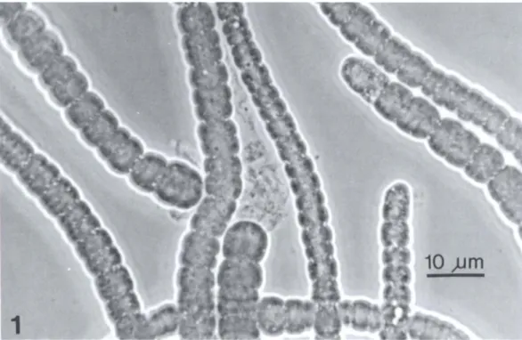
Fig. 1. Oscil/atoria cf. corallinae from culture (phase-contrast microscopy). phosphate buffer (pH 6.8) in a 1 : 5 (w: v) ratio
and kept in the dark at 4°C for 12 h. After cen trifugation at 27 000 g for 20 min at 4°C to re move cellular debris, the aqueous extract was
monitored with a Perkin-Elmer 554
spectrophotometer. The extract was then frac tionated successively with 30, 40, 50, and 60% ammonium sulphate. At each fractionation step, the extract was left overnight in the dark at 4°C and then centrifuged at 27 000 g; the absorption spectra of the precipitate resuspended in I mM phosphate buffer and of the supernatant were taken to check the composition of the fractions. The purest fraction was dialysed against 1 mM phosphate buffer (pH 6.8). Fluorescence emis sion was monitored with a Perkin-Elmer LS-5 luminescence spectrometer. A pure fraction was denatured with 20% acetic acid. Optical densities at 494 nm (PUB) and 540 nm (PEB) were mea sured and considered in a linear system of der ivation permissible as the absorption spectra of protein bound bilins, when denatured, closely resemble those of free bilins (Glazer et at. 1982). The PUB: PEB ratio was calculated with the bil in molar extinction coefficients given by Klotz & Glazer (1985).
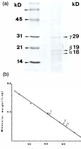
SDS-urea gradient gels were used to determine the molecular masses of the subunits. Subunits
were prepared by adding 1% dithiothreitol and 0.1 % !J-mercaptoethanol to the samples and heating them at 100°C for 10 min. Polyacryl amide gel gradients from 10 to 20% in Tris-HCI buffer (pH 8.9) with 4 M urea were used. Lyso zyme (14 400 Daltons), soybean trypsin inhibitor (21 500 D), bovine carbonic anhydrase (31 000 D), ovalbumin (45 000 D), bovine serum albu min (66 200 D), and rabbit muscle phosphory lase (97 400 D) were used as markers. Gel slabs were run at 6 mA for 12 h in a Protean II Cell (Bio-Rad) with a circulating water-cooling sys tem. They were stained with Coomassie blue and destained in a mixture of acetic acid (40%) and methanol (10%) in water.
RESULTS
Strain characteristics
The following description is based on a culture grown at 2YC, at a continuous illumination of 7 j.tmol photons m-2 S-l, under which conditions variations in cell length are smallest. Dimensions are expressed as: minimum-maximum (mean) determined from 50 measurements. The tri chomes form a red ( lOD7, Kornerup & Wan scher 1978) cushion-like, loose aggregate in liquid
Hoffmann et al.: CU-phycoerythrin in Oscillatoria 23
(a)
kD
'\kD
.. .<> � 0.5 ° 400 5 600 7 0 Wavelength (nm )Fig. 2. Absorption spectra of the biliprotein crude ex tract (- - -) and of the purified CU -phycoerythrin ( . . . .. ) from Oscil/atoria cf. corallinae. Note the pres ence of APC (Am.x at 646 nm) and PC (Am.x at 608 nm) in the crude extract.
culture. The trichomes are 4.8-6.4 (5.5) /Lm wide, without any visible sheath, and are flexuous and constricted. The cells are wider than long, 1.9-4.0 (2.8) /Lm long, with a length: width ratio of 0.3-0.8 (0.5). Newly formed hormogonia are not attenuated at the apex and have rounded apical cells, whereas fully developed trichomes are at tenuated towards the apex with a conical, pale apical cell (Fig. I). The trichomes break by the formation of necridia and move by gliding. In old cultures, dark granules and refringent gran ules, possibly gas vesicles, are present along the cross-walls.
Culture conditions mainly influence cell width. At 12°C, cell width is narrower (4.1-6.1 /Lm). At 25°C and at high irradiances (40-74 /Lmol pho tons m-2 S-I) cell width is slightly larger (5-S /Lm). Maximum cell width (10.7 /Lm) was observed in a 100% salinity medium.
Cell length is less variable and maximum vari ation is found at 25°C at higher irradiances (1.8-5.S /Lm) and at 100% salinity (1.6-5.9 /Lm).
The strain grows between 7 and 74 /Lmol pho tons m-2 S-I at 2Ye. At 12°C, some filaments survive between 7 and 40 /Lmol photons m-2 S-I and at 35°C complete lysis is observed. Highest yields in MN medium are observed at 25°C and at 17 and 40 /Lmol photons m-2 S-I. At the higher irradiance (74 /Lmol photons m-2 S-I) the colour of the cells turns more orange (SB4, Kornerup & Wan scher 1978). The salinity range tolerated by
(b)
5.0 � '" ° ...J � .., '" � � = " u " '0 ::E 45 -31 - 21 -14 _ 30 6 0 90 Relative mobility(mm)y29
�19
a18
Fig. 3. (a) SDS-urea gradient gel (10-20%) of the puri fied CU-phycoerythrin after staining with Coomassie blue. Three bands corresponding to the a-, {3-, and 'Y-subunits are visible. (b) Molecular weight determi nation of the sub-units of CU-phycoerythrin. Esti mated values are 18 kD (a), 19 kD ({3), and 29 kD (-y). the alga lies between 50 and 100% of seawater salinity, and best growth is obtained at 100%. Pigments
Three different phycobiliproteins have been found in this species of Oscillatoria (Fig. 2). The phy cocyanins are only present in small amounts un der the culture conditions used; the absorption maxima at 60S and 646 nm most probably in dicate the presence of C-phycocyanin and allophycocyanin respectively. The absorption spectrum of the isolated phycoerythrin, the ma jor phycobiliprotein of strain CJ 1, possesses two maxima at 494 nm and 540 nm. The pigment has a fluorescent emission maximum at 573 nm.
24 Phycologia, Vol. 29 (1), 1990 .. .., 0 .5 400 Wavelength Cnm) ---... ,... 700
Fig. 4. Absorption spectra of purified ( . . ... ) and de natured (- - -) CU-phycoerythrin. PUB: PEB ratio was found to be 0.56.
The alga shows no chromatic adaptation under red light. The molecular weights of the subunits are 18 000 D (a), 19 500 D «(3) and 29 000 D (y) (Fig. 3). After denaturation (Fig. 4) the PEB: PUB ratio was found to be 1.76: I , indicating that there are about two phycoerythrobilins for each phycourobilin in the subunits.
DISCUSSION
The marine strain CJ 1 most closely resembles descriptions of Oscillatoria corallinae and O. ni groviridis Thwaites ex Gomont. These two species differ mainly by the presence or absence of gran ules at the cross-walls, a variable character which depends on the physiological state of the organ isms (Anagnostidis & Komarek 1988). Gomont (1890) and Lindstedt (1943) proposed uniting the two species, but Gomont (1893) subsequent ly treated them as two separate species.
The strain CJ I differs from these two Osci/ latoria species by its dimensions, its colour, and the morphology of the apical cell. However, the cell dimensions in the original descriptions of these two species are generally greater than those observed for our strain, but they lie within the possible variation range. Also, the colour of the strain CJ l is red (even under red light), whereas Gomont (1893) and later authors (e.g. Setchell & Gardner 1919; Lindstedt 1943; U mezaki 1961) mention colours varying from blue-green, olive green, eruginous to pale brown. According to
Anagnostidis & Golubic (1968), O. corallinae ex
hibits chromatic adaptation. No thickening of the cell wall of the apical cell as mentioned by Gomont (1893) is found in strain CJ 1. However, Gomont's illustration (1893, pI. 6, fig. 21) shows no such thickening, as has been pointed out by Lindstedt (1943). The conical apical cell of strain C] I appears only in fully developed trichomes . Its frequency may thus be variable in the field, depending on the sampling conditions, and it may have been overlooked in the original descrip tions.
The morphology of the strain CJ 1 is also close to that of O. boryana (Bory) Gomont ex Gomont. It differs from this species in having shorter cells and mainly by its habitat, as O. boryana is a species found in thermal areas (Geitler 1932). As uncertainties exist about the correct name to apply to this strain, we prefer to refer to it as Oscillatoria cf. corallinae.
The presence of two peaks at 494 and 540 nm is typical of phycoerythrins carrying phycouro bilin and phycoerythrobilin chromophores. This group of pigments, generally simply called phy coerythrin in the literature, was named CU-phy coerythrin (CU-PE) by MacColl & Guard-Friar (1987). The properties of this type of phycoer ythrin are still not very well known. It is rather heterogeneous with respect to its spectroscopic and subunit properties. Table 2 summarizes the properties of CU-phycoerythrin. In general, there are two absorbance peaks, one in the 490-505 region and one in the 540-567 region; a supple mentary peak or shoulder may be present in
Trichodesmium sp. (Fujita & Shimura 1974; Haxo et at. 1987). Isolation of a phycourobilin containing phycoerythrin from a species of Os cillatoria expands our knowledge about the dis tribution of similar pigments among blue-green algae. Strain CJ I is indeed only the ninth entity for which the pigment has been demonstrated. Among the unicellular blue-green algae, CU phycoerythrin has been identified in four taxa belonging to the genera Synechococcus (marine picoplankton), Synechocystis (a marine sym biotic species) and Gloeobacter (a terrestrial species). For the filamentous blue-green algae, the presence of CU-phycoerythrin has been de tected in the genera Oscillatoria (symbiont of sponges and ascidians; freshwater and thermal species) and Trichodesmium (marine plankton). So far it has not been found in nanocyte- and heterocyst-forming blue-green algae. This is the first report of the presence of CU-phycoerythrin
Hoffmann et al.: CU-phycoerythrin in Oscillatoria 25
in a free-living marine Oscillatoria species. Ow ing to the rareness of the pigment among blue green algae, its presence may be a taxonomic character of some importance.
It is not clear why this pigment replaces phy coerythrin in these blue-green algae, as its pres ence has been reported from widely different habitats. Wyman et at. (1985) suggested that for marine Synechococcus species phycoerythrin might function as a nitrogen reserve. CU-phy coerythrin seems especially important for pico planktonic marine Synechococcus species. Al berte et at. (1984) showed that when marine Synechococcus strains were grown under low ir radiances, the phycoerythrin-containing clones showed higher photosynthetic performance than strains lacking phycoerythrin. Furthermore, Glover et at. (1986) showed that a Synechococ cus clone containing the PUB chromophore was able to photosynthesize more efficiently at low fluxes of blue light than a Synechococcus clone lacking this chromophore. These results indicate the high photosynthetic efficiency of phycoery thrin-containing organisms in low-light environ ments common to mid-depth neritic and oceanic habitats. Strain CJ 1 was isolated from the in fralittoral zone; such environments characteris tically have primarily blue and green wave lengths available for photosynthesis because of the preferential absorption of red to yellow wave lengths by the water column. The presence of the phycourobilin chromophore, with an absorbance in the 450-500 nm region, should widen the ab sorption cross-section in the blue part of the spectrum and would thus be advantageous for organisms inhabiting the infralittoral zone. ACKNOWLEDGMENTS
We thank A. Rossi and P. Ferrari for technical assistance. This study was performed in the framework of the FRFC contract 24550-80. LH
and A W were research assistants at the Belgian National Fund for Scientific Research (FNRS) during this study. Financial support of the FNRS and the Italian Consiglio Nazionale delle Ricerche (CNR) to LH is also acknowledged. We thank Dr V. Demoulin for reading the manuscript.
REFERENCES
ALBERTE R.S., WOOD A.M., KURSAR T.A. & GUILLARD
R.R.L. 1984. Novel phycoerythrins in marine
Synechococcus spp. Characterization and evolution ary and ecological implications. Plant Physiol. 75: 732-739.
ANAGNOSTIDIS K. & GOLUBIC S. 1968. Uber die Oko logie einiger Spiru/ina-Arten. Nova Hedwigia 11: 309-335.
ANAGNOSTIDIS K. & Ko MAREK J. 1988. Modern ap proach to the classification system of cyanophytes. 3-0scillatoriales. Arch. Hydrobiol. Suppl. 80 (AI gological Studies 50-53): 327-472.
BISHOP J.E., RApOPORT H., KOLTZ A.V., CHAN c.F.,
GLAZER A.N., FUGLISTALLER P. & ZUBER H. 1987.
Chromopeptides from phycoerythrocyanin. Struc ture and linkage of the three bilin groups. J. Am. Chem. Soc. 109: 875-881.
BRYANT D.A. 1981. The photoregulated expression of multiple phycocyanin species. A general mecha nism for the control of phycocyanin synthesis in chromatically adapting cyanobacteria. Eur. J. Bio chem. 119: 425-429.
BRYANT D.A. 1982. Phycoerythrocyanin and phy coerythrin: properties and occurrence in cyanobac teria. J. Gen. Microbiol. 128: 835-844.
BRYANT D.A. & COHEN-BAZIRE G. 1981. Effects of
chromatic illumination on cyanobacterial phycobil isomes. Evidence for the specific induction of a sec ond pair of phycocyanin subunits in Pseudanabaena
7409 grown in red light. Eur. J. Biochem. 119: 415-424.
BRYANTD.A., COHEN-BAZIREG. & G LAZERA.N. 1981. Characterization of the biliproteins of Gloeobacter violaceus. Chromophore content of a cyanobacterial phycoerythrin carrying phycourobilin chromophore.
Arch. Microbiol. 129: 190-198.
Cox G.c., HILLER R.G. & LARKUM A.W.D. 1985. An unusual cyanophyte, containing phycourobilin and symbiotic with ascidians and sponges. Mar. Bioi. 89: 149-163.
FUJITA Y. & SHiMURA S. 1974. Phycoerythrin of the
marine blue-green alga Trichodesmium thiebautii. Plant Cell Physiol. 15: 939-942.
GANTT E. 1980. Structure and function of phycobil isomes: light harvesting pigment complexes in red and blue-green algae. Int. Rev. Cytol. 66: 45-80. GANTT E. 1981. Phycobilisomes. Ann. Rev. PI. Phys
iol. 32: 327-347.
GEITLER L. 1932. Cyanophyceae. In: Kryptogamen Flora von Deutschland, Osterreich und der Schweiz
(Ed. by L. Rabenhorst) 14: 1-1196. Akademische Verlagsgesellschaft, Leipzig.
GLAZER A.N. 1984. Phycobilisome. A macromolecu lar complex optimised for light energy transfer. Biochim. Biophys. Acta 768: 29-51.
GLAZER A.N. & BRYANT D.A. 1975. Allophycocy anin B (Amax 671, 618 nm): a new cyanobacterial phycobiliprotein. Arch. Microbiol. 104: 15-22. GLAZER A.N., WEST J.A. & CHAN C. 1982. Phycoer
ythrins as chemotaxonomic markers in red algae: a survey. Biochem. System. Ecol. 10: 203-215. GLOVER H.E., KELLER M.D. & GUIL LARD R.R.L. 1986.
Light quality and oceanic ultraphytoplankters. Na ture 319: 142-143.
GOMONT M. 1890. Essai de classification des Nos tocacees homocystees. J. Bot. 4: 349-357.
26 Phyc% gia, Vol. 29 (1), 1990
(Nostocaceae homocystees). Ann. Sci. Nat., Bot. ser 7, 16: 91 -264, 7 pis.
GRABOWSKY J. & GANTT E. 1 978. Photo physical
properties of phycobiliproteins from phycobili somes: fluorescence life times, quantum yields and polarization spectra. Photochern. Photobiol. 28: 39-45.
GRAY B.H. & GANTT E. 1975. Spectral properties of
phycobilisomes and phycobiliproteins from the blue green alga Nostoc sp. Photochern. Photobiol. 21: 1 2 1 -1 28.
HAXO F.T., LEWIN R.A., LEE K.W. & LI M.-R. 1 987.
Fine structure and pigments of Oscillatoria (Trich odesrniurn) aff. thiebautii (Cyanophyta) in culture.
Phycologia 26: 443-456.
HIROSE H., KUMANO S. & MADOKO K. 1969. Spec troscopic studies on phycoerythrins from cyanophy cean and rhodophycean algae with special reference to their phylogenetical relations. Bot. Mag. Tokyo 82: 197-203.
KLOTZ A.V. & GLAZER A.N. 1 985. Characterization
of the bilin attachment sites in R-phycoerythrin. J.
BioI. Chern. 260: 4856-4863.
KORNERUP A & W ANSCHER J.H. 1 978. Methuen
Handbook of Colour. Eyre Methuen Ltd., London,
252 pp.
KURSAR T.A, SWIFT H. & ALBERTE R.S. 1 981 . Mor phology of a novel cyanobacterium and character ization of light-harvesting complexes from it: im plications for phycobiliprotein evolution. Proc. Natl. Acad. Sci. USA 78: 6888-6892.
LARKUM A.W.D., Cox G.c., HILLER R.G., PARRY D.L. & DIBBAYAWAN T.P. 1987. Filamentous cyano phytes containing phycourobilin and in symbiosis with sponges and an ascidian of coral reefs. Mar. Bio!. 95: 1 -13.
LEWIS M.R., ULLOA O. & PLATT T. 1 988. Photosyn thetic action, absorption, and quantum yield spectra for a natural population of Oscillatoria in the North Atlantic. Lirnnol. Oceanogr. 33: 92-98.
LEY AC., BUTLER W.L., BRYANTD.A & GLAZER AN.
1 977. Isolation and function of allophycocyanin B
of Porphyridiurn cruenturn. Plant Physio!. 59:
974-980.
LINDSTEDT A 1 943. Die Flora der rnarinen Cyano phyceen der schwedischen Westkiiste. Hakan Ohls sons Buchdruckerei, Lund, 121 pp., I I pis. LUNDELL D.J. & GLAZER A.N. 1 981 . Allophycocy
anin B. A common (3 sub-unit in Synechococcus al lophycocyanins (Amax 670 nm) and allophycocyanin (Amax 650 nm). J. BioI. Chern. 256: 1 2600-1 2606. MACCOLL R. & GUARD-FRIAR D. 1 987. Phycobili
proteins. CRC Press, Boca Raton, Florida, 218 pp. McCARTHY J.J. & CARPENTER E.J. 1 979. Oscillatoria
(Trichodesrniurn) thiebautii (Cyanophyta) in the cen tral north Atlantic Ocean. J. Phycol. 15: 75-82. NEVEUX J., DUCLAUX G., LAFARGUE F., WAHL M. &
DEVOS L. 1988. Pigments of some symbiotic cy anobacteria. Vie & Milieu, ser. A, BioI. Mar. 38: 251-258.
ONG L.J., G LAZER A.N. & WATERBURY J.B. 1 984.
An unusual phycoerythrin from a marine cyanobac terium. Science 224: 80-83.
PARRY D.L. 1984. Cyanophytes with R-phycoery thrins in association with seven species of ascidians from the Great Barrier Reef. Phycologia 23: 503-505.
PELLEGRINO F., WONG D., ALFANO R.R. & ZILINSKAS B.A 1981 . Fluorescence relaxation kinetics and quantum yield from the phycobilisomes of the blue green alga Nostoc sp. measured as a function of single picosecond pulse intensity. Photochern. Photobiol. 34: 691 -696.
RIPPKA R., DERUELLES J., WATERBURY J.B., HERDMAN M. & STANIER R.Y. 1979. Generic assignments, strain histories and properties of pure cultures of cyanobacteria. J. Gen. Microbiol. 111: 1 -61. RIPPKA R., WATERBURY J. & COHEN-BAZIRE G. 1 974.
A cyanobacterium which lacks thylakoids. Arch. Mi crobio!' 100: 419-436.
SEARLE G.F.W., BARBER J., PORTER G. & TREDWELL
C.J. 1 978. Picosecond time-resolved energy trans fer in Porphyridiurn cruenturn. Part II. In the isolated light harvesting complex (phycobilisomes). Biochirn.
Biophys. Acta 501: 246-256.
SETCHELL W.A. & GARDNER N.L. 1 91 9. The marine
algae of the Pacific coast of North America. Part I. Myxophyceae. Univ. Calif. Publ. Bot. 8: 1 -1 39.
SHIMURA S. & FUJITA Y. 1 975. Phycoerythrin and photosynthesis of the pelagic blue-green alga Trich
odesrniurn thiebautii in waters of Kuroshio, Japan.
Mar. BioI. 31: 1 21 -128.
STADNICHUK LN., ROMANOVA N.l. & SELYAKH 1.0. 1 985. A phycourobilin-containing phycoerythrin from the cyanobacterium Oscillatoria sp. Arch. Mi crobio!. 143: 20-25.
TANDEAU DE MARSAC N. 1 977. Occurrence and na ture of chromatic adaptation in cyanobacteria. J. Bact. 130: 82-91 .
UMEZAKl I. 1 961. The marine blue-green algae of
Japan. Mern. Coli. Agric. Kyoto Univ. 83: 1-149. WEHRMEYER W. 1 983. Organization and composi
tion of cyanobacterial and rhodophycean phycobil isomes. In: Photosynthetic Prokaryotes: Cell Differ entiation and Function (Ed. by G.c. Papageourgiou & L. Packer) pp. 1 -22. Elsevier Science Publ., Am sterdam.
WILMOTTE A. 1 988. Growth and morphological vari ability of six strains of Phorrnidiurn cf. ectocarpi Go mont (Cyanophyceae) cultivated under different temperatures and light intensities. Arch. Hydrobiol. Suppl. 80 (Algological Studies 50-53): 35-46.
W YMAN M., GREGORY R.P.F. & CARR N.G. 1 985. Novel role for phycoerythrin in a marine cyanobac terium, Synechococcus strain DC2. Science 230: 818-820.
ZILINSKAS B.A. & GREENWALD L.S. 1 986. Phycobil isome structure and function. Photosyn. Res. 10: 7-35.