Biochem. J. (1981) 193, 83-86 83 Printed in Great Britain
The
penicillin-binding site
in
the exoceliular
DD-carboxypeptidase-transpeptidase ofActinomadura R39
Colette DUEZ, Bernard JORIS, Jean-Marie FRERE and Jean-Marie GHUYSEN
ServicedeMicrobiologie, Faculte deMedecine,Institutde Botanique, UniversitedeLiege,Sart Tilman, B-4000 Liege,Belgium
and Jos VAN BEEUMEN
LaboratoriumvoorMicrobiologie, Rijksuniversiteit Gent,K.L.Ledeganckstraat 35,B-9000Gent, Belgium (Received2 May1980/Accepted6 August1980)
Heatdenaturation and Pronasedegradation ofthecomplex previously formed between benzylpenicillin and the exocellular DD-carboxypeptidase-transpeptidase of Actino-madura R39 yields a heptapeptide H-Leu-Pro-Ala-Ser-Asn-Gly-Val-OH, where the benzylpenicilloyl group is ester-linked tothe serineresidue. This
linkage
is verylabile and its hydrolysis causes the release of benzylpenicilloate. In contrast, the native benzylpenicilloyl-enzyme complex is very stable (half-life 70h at 370C) and its breakdown proceeds via fragmentation ofthe bound benzylpenicilloyl group [Fuad, Frere,Ghuysen, Duez &Iwatsubo(1976) Biochem.J. 155,623-6291.The complexes formed by interaction between penicillin and the
DD-carboxypeptidases
havere-ceived much attention. With the exocellular DD-carboxypeptidase-transpeptidase of Streptomyces R61 (Frere et al., 1976a; Degelaen et
al., 1979)
and the membrane-boundDD-carboxypeptidases
of Bacillus subtilis and B.stearothermophilus
(Georgopapadakou et al., 1977; Yocum et al.,1979),
it is known that:(1)
penicillin acylates the enzymes and forms rather stablepenicilloyl-enzyme
complexes,(2)
thepenicilloylgroupis ester-linkedto an enzyme serine residue (as least as found after denaturation ofthecomplexes)
and(3)
breakdown of the native complexes leads to enzyme regeneration and proceeds viafragmentation ofthe boundpenicilloyl moiety.
As shown with the Streptomyces R61 enzyme(Frere
etal., 1976b),
this processimplies splitting
ofthe C-5-C-6 bond and protonation of C-6, causing the release of N-acylglycine andN-formyl-D-penicillamine.
The exocellular
DD-carboxypeptidase-trans-peptidase of Actinomadura R39(in short,
the R39enzyme)
resembles the R6 1 and Bacillus enzymes at least in two respects.First,
the inter-action between the R39 enzyme and fi-lactam antibiotics leads to the formation of stablecom-plexes in which the bound metabolite probably occursin the formofapenicilloylorcephalosporoyl derivative. Thus,in such
complexes, cephaloglycine,
cephalexin and cephalosporin C have their621,
decreased to the same extent as thatobtained after the action of
fi-lactamase (Fuad
etal.,
1976),
and nitrocefin has a64.7/e355
ratioof2.40, which is that NVol. 193obtainedafter theaction of ,8-lactamase(Frereetal., 1974). Secondly, spontaneous breakdown of the native penicilloyl-R39 enzyme complex also pro-ceeds (slowly) via fragmentation of the bound metabolite (Frere et al., 1975). The present paper describes experiments which were carried out to characterizetheamino acid residue whichmay serve asthepenicillinattachmentsite in the R39 enzyme. Materials and methods
The R39 enzyme, the
['4C]benzylpenicillin
(57
mCi/mmol)
and the[14Clbenzylpenicilloyl-R39
enzyme complex were those previously used (Fuad etal., 1976). Trypsintreated with 1-chloro-4-phenyl-3-L-toluene-p-sulphonamidobutan-2-one (TPCK-trypsin)was purchased from Worthington. Pronase was from Sigma. The following chromatography solvents were used: I, butan-l-ol/acetic
acid/
pyridine/water
(15:3:10:12, by vol.); II,chloro-form/methanol/acetic
acid (44:5:1, by vol.); III, butan-l-ol/water/acetic acid/ethanol
(10:4:3:3,by vol.). T.l.c. wasperformed on silica gel G-60 plates. Highvoltage electrophoreses were carried out with a Gilson electrophorator modelDW at60V-cm-',
incollidine/acetic acid/water
(33:13:5000, by vol.) (pH6.5), using Whatman 3MM papers andre-frigerated tanks. Peptides were stained with fluorescamine
(F.
Hoffmann-La Roche Inc., Basel, Switzerland) as described by Vandekerckhove & Van Montagu(1974).
A Packard Tri-Carb 2425 liquid-scintillation spectrometer and a Packard radiochromatogram scanner model 7201 were used 0306-3275/81/010083-04$01.50/1 © 1981The BiochemicalSocietyC. Duez, B. Joris, J.-M.Frere,J.-M. Ghuysen and J. VanBeeumen for the radioactivity measurements. Amino acid
analyses (after hydrolysis of the samples in azeo-tropicHCIfor24hat1050C)wereperformed witha Beckman Multichrom 4255 automatic amino acid analyser. Amino acid sequencing was carried out according tothe micromethod of Bruton & Hartley (1970). The dansyl amino acids were characterized as described by Weiner et aL (1972)on 5 cm x 5cm polyamide plates. Benzylpenicilloate was charac-terizedby paper electrophoresis atpH6.5 (mobility 25cm*h- at
60V.cm-1)
andby
t.l.c. in both solventII(RF
0.03)andsolventIII(RF
0.56). ResultsEffectsof heat denaturationand trypsindegradation on the stability of the
[14Clbenzylpenicilloyl-R39
enzymecomplex
Heat denaturation ofthe
[14C]benzylpenicilloyl-R39 enzyme complex (by boiling a solution ofthe complex for 1min in 10mM-sodium phosphate, pH7.0) had two effects. (1) As determined
by
the rate of release of radioactivity, the denatured complexhadadecreasedhalf-life of 10h(instead
of 70h for the native complex). (2) Spontaneous breakdown of the heat-denatured complex resulted in the releaseof[14C]benzylpenicilloate
(and
didnot proceed through fragmentation of the benzyl-penicilloylmoiety
as observed with the native complex).Note thatdenaturationby sodiumdodecyl sulphate(instead
ofheat)
also caused a decreased stability of the complex (half-life 150min at 37°C; Frere etal., 1974).Degradation of the heat-denatured
[14C]benzyl-penicilloyl-R39 enzyme with TPCK-trypsin further destabilized the bond between the peptide and the benzylpenicilloyl group. The heat-denatured com-plex(2nmol in30,1of
lOmM-Tris/HCl,
pH8.0)was supplemented with1.5,ug
of trypsin. After 2h at 370C, afreshsample of1.5,ug.of
trypsin
wasadded and the mixture was further incubated for 2h. Analysis of the degradation products by paper electrophoresis at pH 6.5 showed that all the radioactivity was present in -the form of['4C]-benzylpenicilloate. However, if a higher amount of trypsin
(15,ug)
was used, so that theincubation at 370C could be decreased to 30min (insteadof4h), electrophoresis ofthe degradation products permit-ted detection, in addition to[14C]benzylpenicilloate,
of a radioactively labelled, negatively charged peptide that had a mobility of 7cm*h-l at60V.cm-'.
Isolationofa
[14Clbenzylpenicilloyl-heptapeptide
Essentially, the same procedure as above was used except that Pronase was used instead of trypsin. Moreover, the proteolytic degradation at
370Cwasmadeasshortaspossibleand all the other operationswerecarriedout at40C. Theheat-treated complex (57nmolin 50,u1 of10mM-sodium phosph-ate, pH7.0) was incubated with 240,g ofPronase
(two
successive additions of120,ug)
for 30min at 370C. Filtration of the sample in water through a column (60cmx1.0cm) of Biogel P-2 at 40C yielded tworadioactive fractions. The fraction with KD 1.0 contained[14C]benzylpenicilloate.
The frac-tion withKD 0.62 was lyophilized and submittedto paper electrophoresis for 2 h at pH 6.5 and 60V *cm-'.
Half of theradioactivityinitially present in the fraction with KD 0.62 was recovered as[14C]benzylpenicilloate
and the other half was found to be associated with a negatively chargedpeptide (mobility 7.5cm.h-1 at 60V.cm-1). In turn, this radioactive peptide was submitted to descending paper chromatography in solvent I at 40C. Again, half of the radioactivity was recovered as [14C]-benzylpenicilloate (which migrated close to the solvent front) and the other half as found to be associated with a peptide ofRF
0.64. This radio-active peptide was eluted with water at 40C. In termsofradioactivity, the final yield was 7% of that present intheoriginalcomplex. Chromatographyof the radioactive peptide on Biogel P-2 (in water at 40C) indicatedamol.wt. ofapprox. 1100. Itsamino acid composition (Asx, 1.2; Ser,0.6; Pro, 1.0; Gly, 1.0; Ala, 0.8; Val, 1.1; Leu, 0.6) suggested that it was aheptapeptideconsisting of one residue each of Asx, Ser, Pro, Gly, Ala, Val and Leu. On the basis of this composition, the corresponding penicilloyl-heptapeptide should have a theoretical mol.wt. of 970.Evidence for the presence of an esterlinkagein the
[4
4Clbenzylpenicilloyl-heptapeptidefragment
The heat-denatured
[14lbenzylpenicilloyl-R39
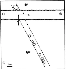
en-zyme complex (45nmol) was treated with Pronase, thedegradation productswerefilteredonBiogelP-2 (at 4°C) and the radioactive fraction with KD 0.62 was lyophilized, as described in the preceding section. Thefraction with KD0.62 wasanalysed by two-dimensional paperelectrophoresis atpH 6.5 and 60V-cm-1 under the following conditions. A first electrophoresiswascarriedout on aWhatman 3MM paper strip (5cm x150cm) for 30 min. The paper strip (from which that part containing free [14C]-benzylpenicilloate was eliminated) was exposed to NH3 vapourfor 16h at room temperature, dried at 800C and sewn to a Whatman 3MM paper sheet (27cmx 150cm). A secondelectrophoresiswas then carriedoutfor 60 min inadirectionperpendicular to that used for the first one. Staining of the electro-phoretogram withfluorescamine showedthat all the peptides were positioned along one oblique line except oneof them (Fig. 1). Obviously,thispeptide (spot 1) had migrated as anacidic compound with a 1981 84Active site ofActinomadura R39 DD-carboxypeptidase
1~~~.
e A B~ %I~~~~~~~~~~
"''\\
' '\
'\\
*2 \%\ 2cm ' 'Fig. 1. Purification of the penicillin-binding peptide by two-dimensional electrophoresis
The first electrophoresis took place in direction A, the second in direction B. Fordetails, see the text.
Spot 1 was fluorescent and non-radioactive (the Heptapeptide). Spot 2 was radioactive and
non-fluorescent(a-amideof[14C]benzylpenicilloicacid).
mobility of 7.5cm-h-l during the first electro-phoresis, but had been transformed during treat-ment with NH3 so that it behaved as a neutral peptide during the second electrophoresis (its slight migration towards the cathode was due to electro-osmosis). In turn, scanning of the electrophoreto-gram permitted detection ofone single radioactive compound (spot
2).
This compound which, during the first electrophoresis, had migrated towards the anode with the same mobility (7.5cm.h-') as that observed for peptide 1 before NH3 treatment, migrated, during the second electrophoresis, with a mobility of l5cmh'1,
i.e. most likely that of the a-amide of benzylpenicilloic acid. Peptide 1, as it occurred in the fraction with KD 0.62, thus behaved as a negatively charged benzylpenicilloyl-peptide. After NH3 treatment, the same peptide 1 then behaved as a neutral, benzylpenicilloyl-free com-pound. The benzylpenicilloyl-free peptide 1 was eluted from the electrophoretogram; 7nmol were obtained (yield 16%). Amino acid composition and sequencing studies gave the following primary structure: H-Leu-Pro-Ala-Ser-Asn-Gly-Val-OH. Residue 5 was assumed to be amidated, on the basis of the neutral character of the peptide at pH6.5. Attachment of a penicillin molecule to this neutral heptapeptide, so that the derivative thus formed acquires one negative charge, exhibits low stability and breaks down in water with release ofbenzyl-penicilloate, necessarily involves penicilloylation of theserine residue via formation ofan esterlinkage. Discussion
Heat treatment andproteolytic degradation (with either trypsin or Pronase) of the benzylpenicilloyl-R39 enzyme complex considerably labilizes the linkage between the bound metabolite and its attachment site on the enzyme. In spite of thishigh lability, a benzylpenicilloyl-heptapeptide complex (generated by Pronase treatment) has beenisolated and the penicilloyl group has been shown to be ester-linked to its serine residue. Like the R61 and theBacillus DD-carboxypeptidases,the R39 enzyme thusappears to be aserineDD-carboxypeptidase.
Spontaneous hydrolysis ofthe ester bond which links thebenzylpenicilloylgroup to theheptapeptide is rapidandleadstothe releaseofbenzylpenicilloate. In contrast, the native benzylpenicilloyl-enzyme is very stable (half-life 70h at 370C) and its spon-taneous
(and
slow) breakdown proceeds via frag-mentationof the penicilloyl group. It should be noted that heat denaturation of the complex, without any furtherproteolytictreatment,alreadydestabilizes the complex, prevents the fragmentation reaction from occurring and, consequently, causes the release of benzylpenicilloate. With the R61 DD-carboxy, peptidase, denaturation andproteolytic degradation of the benzylpenicilloyl-enzyme complex have also drastic effectsonthe stability ofthecomplexand the natureofthe released products (Frere etal., 1976a; C. Duez, J. M. Frere, J. M. Ghuysen, J. Van Beeumen &J. Vandekerckhove, unpublished work). Theseobservationscan be bestinterpreted(Ghuysen etal., 1979) by assumingthatformationofan ester bondbetween C-7 ofthe penicillinmolecule and the active serine residue on the enzyme (binding site 1) involves other interactions between both the acyl side chain and the monocyclic thiazolidine ring of the bound metabolite, and two specific amino acid groupings(enzyme binding
sites 2 and 3). The relative disposition of these three enzyme sites (which depends on the conformation of the penicilloyl-enzymecomplex)
and, consequently,the distortionthattheyconferonthe penicilloyl moiety, appear to be an important parameter that governs thestabilityof the complex and the fate of the bound metabolite.A serine residue (70) is also involved in the interaction between penicillin and the,-lactamase I ofB. cereus and that of Escherichia coli RTEM (Knott-Hunziker et al., 1979; Fisher et al., 1980; J. R. Knowles, personal communication), and this serineresidue is conservedintwo other
JJ-lactamases
of known sequences, i.e. those of Staphylococcus aureusandB. licheniformis (Ambler, 1979). It thus follows that the amino acid sequences around the Vol. 193
86 C.Duez,B.Joris,J.-M.Frere, J.-M.Ghuysenand J. Van Beeumen
Table 1. Sequencesaround theactive serine residue infourDD-carboxypeptidases andfourfi-lactamases TheactiveserineintheB.subtilis DD-carboxypeptidaseoccursatposition36.
DD-CarboxypeptidasesorDD-carboxypeptidases-transpeptidases
Streptomyces R61 Val-Gly- Ser Frereetal., 1976a
ActinomaduraR39 Leu-Pro-Ala- Ser -Asn-Gly-Val Thepresentpaper B.stearothermophilus Gly-Ile-Ala- Ser -Met
. Yocum etal., 1979
B.subtills Pro-Ile-Ala- Ser -Met-Thr-Lys)
,-Lactamases
Staphylococus aureus Ala-Tyr-Ala- Ser
-Thr-Ser-Lys-B.cereus569/H I Ala-Phe-Ala- Ser -Thr-Tyr-Lys
Ambler,1979 B.licheniformis 749/C Ala-Phe-Ala- Ser -Thr-Ile-Lys
E.coli RTEM Pro-Met-Met- Ser -Thr-Phe-LysJ
presumed active serine residue are known for four different DD-carboxypeptidases and four different f-lactamases. Alignment of the active serine residues in these eight different enzymes (Table 1)shows an obvious homology only between the four a-lactamases, as well as between the two Bacillus DD-carboxypeptidases.
The work has been supported in part by theNational Institutesof Health,U.S.A.(contractno. 2RO1 Al13364-04), the Fonds de la Recherche Scientifique Medicale, Bruxelles (contract no. 3.4501.79) and the Actions Concert6es,Bruxelles(conventionno.79/84-I1). References
Ambler, R. P. (1979) in ,-Lactamases(Hamilton-Miller,
J. M. T. & Smith, J. T.,eds,),pp. 99-125, Academic Press, NewYork
Bruton, C.J. & Hartley, B. S. (1970)J. Mol. Biol. 52,
165-173
Degelaen, J., Feeney, J., Roberts, G. C. K., Burgen,
A. S.V.,Frere,J. M. &Ghuysen,J. M.(1979)FEBS Lett. 98,53-57
Frere, J. M.,Ghuysen, J. M.,Reynolds, P. E., Moreno,
R. & Perkins, H. R. (1974) Biochem. J. 143,
241-249
Frere, J. M., Ghuysen, J. M., Degelaen, J., Loffet, A.
&Perkins, H. R. (1975)Nature (London) 258, 168-170
Fisher,J., Belasco, J. G., Charnas, C. L., Khosla, S. & Knowles, J. R. (1980) Philos. Trans. R. Soc. London Ser. B289, 309-319
Frere, J. M., Duez, C., Ghuysen, J. M. &
Van-dekerckhove,J.(1976a)FEBS Lett. 70,257-260
Frere, J. M., Ghuysen, J. M., Vanderhaeghe, H.,
Adriaens, P., Degelaen, J. & De Graeve,J. (1976b)
Nature(London) 260,451-454
Fuad, N., Frere, J. M., Ghuysen, J. M., Duez, C. &
Iwatsubo,M.(1976)Biochem. J.155, 623-629 Georgopapadakou, N., Hammarstrom,S. &Strominger,
J. L. (1977) Proc. Natl. Acad. Sci. U.S.A. 74, 1009-1012
Ghuysen, J. M., Frere,J. M.,Leyh-Bouille, M., Coyette,
J., Dusart, J. & Nguyen-Disteche, M. (1979)Annu. Rev.Biochem.48,73-101
Knott-Hunziker, V., Waley, S. G., Orlek, B. S. &
Sammes,P.G.(1979)FEBS Lett.99,59-61
Vandekerckhove, J. & Van Montagu, M. (1974)Eur. J.
Biochem.44,279-288
Weiner, A. M., Platt, T. & Weber, K. (1972)J. BioL Chem.247,3242-3251
Yocum, R. R., Waxman, D. J., Rasmussen, J. R. &
Strominger, J. L. (1979)Proc.Natl.Acad. Sci. U.S.A. 76,2730-2734