Primary
and predicted secondary
structures
of the Actinomadura
R39 extracellular
DD-peptidase,
a
penicillin-binding protein
(PBP)
related
to
the
Escherichia
coli
PBP4
Benoit
GRANIER,* Colette DUEZ,* Sophie LEPAGE,* Serge ENGLEBERT,*
JeanDUSART,*T
Otto
DIDEBERG,* Jozef VAN BEEUMEN,t Jean-Marie FRERE* and Jean-Marie GHUYSEN*
*Centred'Ingenierie des Proteines, Universite de Liege, InstitutdeChimie, B6, B-4000 Sart Tilman (Liege 1),and tLaboratorium
voor Microbiologie enMicrobiele Genetica, Rijksuniversiteit-Gent, K.L. Ledeganckstraat 35, B-9000 Gent, Belgium
As derived from gene cloning and sequencing, the 489-amino-acid DD-peptidase/penicillin-binding protein (PBP) produced by Actinomadura R39 has a primary structure very similar to that of the EscherichiacoliPBP4 [Mottl, Terpstra & Keck (1991) FEMS Microbiol. Lett. 78, 213-220]. Hydrophobic-cluster analysis of the two proteins shows that, providing that a large 174-amino-acid stretch is excluded from the analysis, the bulk of the two polypeptide chains possesses homologues of the active-site motifs andsecondary structures found in the class A/-lactamaseof Streptomyces albus Gof known three-dimensional structure. The 174-amino-acid insert occurs at equivalent places in the two PBPs, between helices a2 anda3, away from the active site. Such an insert is unique among the penicilloylserinetransferases. It is proposed that the Actinomadura R39 PBP and E. coli PBP4 form a special class, class C, of low-Mr PBPs/
DD-peptidases.A vectorhas been constructed and introducedby electrotransformation in the original Actinomadura R39
strain, allowing high-level expressionand secretionof theDD-peptidase/PBP (250 mg * 1-1). The gene encoding the desired protein is processed differently in Actinomadura R39 and Streptomyces lividans. Incorrect processing in Streptomyces
lividans leadsto a secreted protein whichis inert interms ofDD-peptidase activity and penicillin-binding capacity.
INTRODUCTION
The
fi-lactamases,
the low-Mr penicillin-binding proteins (PBPs)/DD-peptidases and the penicillin-binding domainsofthe bifunctionalhigh-M,
PBPs arebelieved to form asuperfamily ofevolutionarily related penicilloyl serine transferases [1]. How-ever,only a few
,-lactamases
of class A, one ,B-lactamase of class CandtheStreptomyces R61low-Mr
PBP/DD-peptidase of class B areofknown three-dimensional structures [1]. Hence the caseofadivergentevolution ismainly supportedby predictional studies. Hydrophobic-cluster analysis [2,3] is a powerful method for analysing proteins that areweakly related intheprimary struc--ture. Byusing thismethod, ithas been shown that, amongthe low-Mr PBPs/DD-peptidases, the Streptomyces K15 PBP, the
Escherichia coli PBP5 and the Bacillus subtilis PBP5 of class A have similarity in the polypeptide folding with the class A /1-lactamases[4]. Recently, Mottl et al.[5] reportedthat the E. coli PBP4has a typeofprimarystructurewhich isuniqueamongthe penicillin-interactive proteins. Gene cloningandsequencing has
shownthat the secretory PBPproducedbyaverytaxonomically
distant species, Actinomadura R39, has a primary structure which isextremely similar to thatofPBP4. Itis proposed that these two proteins form a distinct class, class C, of low-Mr
PBPs/DD-peptidases.
MATERIALS AND METHODS
Bacterial strains, plasmids and media
Actinomadura R39(fromthislaboratory)wasgrownat28'C
with vigorous orbital shaking intryptone soya broth (Oxoid).
PlasmidspBR322 andpBR325wereused forcloning experiments
in EscherichiacoliHB1O1. Growthwas at37
°C
inLuria-Bertanimedium. Streptomyces lividansTK24 andplasmids pIJ486 and pIJ702 were from the John InnesInstitute,
Norwich,
U.K.; S.lividans was grown in MYEME medium [6] and recombinant Actinomadura R39 was grown in TAU medium [7]. The R2YE medium [8] was also used.
Recombinant DNA techniques, radioactiveoligonucleotide probe and nucleotide sequence
The Actinomadura chromosomal DNA was prepared as
de-scribed in [8], and the recombinant DNA techniques were
performed essentiallyasdescribed in[9].
The sequence ofthe 58-amino-acid N-terminal region of the Actinomadura R39 PBP was determined on 3.2 nmolofprotein
(purified as described in [10]), using a 477-A pulsed liquid
sequenator with on-line analysis of the amino acid phenyl-thiohydantoin derivatives and a 120-A analyser (Applied
Bio-systems, Forster City, CA, U.S.A.). On the basis of these data and the known Actinomycetes codon usage, and by using the
PCR procedure[11],a 109 bp DNA segment was prepared and, from this, the non-degenerated 27-mer probe
3'-CGG-CAC-AGC-CCC-CAC-CAG-CAG-CAC-CTG-5' was synthesized.
This probe was complementary to the nucleotide sequence encoding the nonapeptide A21VSGVVVVD29 of the PBP. The
probe was labelled with [y-32P]ATP and served to screen gene libraries by hybridization [12,13].
DNA segments cloned into M13 vectors were sequenced by
the dideoxynucleotide-chain-termination method [14]. Zones of base compression due to highGC content wereresolved
using
dITP instead of dGTP (Sequenase kit; USB,Cleveland,
OH, U.S.A.). Codonusagewasanalysedwith Staden'sprogram [15]andFickett'stest [16]usingthe Actinomadura R39 8-lactamase [17] as reference.
Immunological screening
Rabbit anti-(Actinomadura R39
DD-peptidase/PBP)
anti-serumwasprepared byGamma S.A.(Tavier,
Belgium)
andwasAbbreviations used:PBP,penicillin-bindingprotein; ORF,openreadingframe. $ Towhomcorrespondenceshouldbeaddressed.
E
6805l
0
eRt
Fig. 1. Restriction maps of plasmids A(a), B(b),pDML1O (c)andpDML15(d)
Inserts areshown inheavy lines. The open boxes in plasmids A and B indicate thehybridizingregions. The position of the initiation codon ATG
andtheorientation of theORF inplasmidsAand B areindicated.Thehatchedline inpDMLIOandpDML15specifiesthe Actinomadura R39
DD-peptidase/PBP-encoding gene. Abbreviations: mel, tyrosinase; tsr,thiostrepton resistance.
used to screen PBP-secretory clones. Bio-Rad Immuno Blot Alkaline Phosphatase Assay Systemswere employed.
DD-Peptidase activity
Measurement of the DD-carboxypeptidase activity was per-formedonAcetyl2-L-Lys-D-Ala-D-Ala (releaseof the C-terminal D-Ala) as describedin [18].
Electrotransformation
Cells of Actinomadura R39
[a
48hculture made in 25ml of tryptone soya broth containing 0.5%(w/v)
glycine]
weredis-persedby gentlesonicationonice(5burstsof 30s
each;
20kHz),
washed bycentrifugation (4 °C; 4000 g; 15min)first with coldwater (three times) and then with a
100%
(v/v)
glycerol/water
solution(once)and resuspended in 1 ml ofglycerol/water.
Thecell suspension (100lO) and the plasmid (50 ng; see the Results section)weremixed ina 1.5 mlpolypropylene tube. The mixture
wassuccessively transferred to a 0.2 cm electroporation cuvette, pulsed once(Cellject, Eurogentec S.A., Liege, Belgium; 40 ,F; 2.5kV; 192Q), supplemented with 1 ml of SOC medium [19] and homogenized with a Pasteur pipette (all these operations
were carried out at 4 C). The suspension was shaken at 250 rev./min for 1 h at 37°C in a 17 mm x 100mm poly-propylene tube.The cells were plated on TAU agar medium and, after 10hat 28°C, overlaid with 3 ml of soft agar containing
50,tgofthiostrepton/ml.
Hydrophobic-cluster analysis
Thisprocedure [2,3]restsuponarepresentation of the amino
acid sequenceson ana-helical two-dimensional patternin which
,- 408 bp 527bp : I #t,I I~ .)i _at Io k
f)
Eco
(,E,)
co E II II II I I l ! 7 1990 bp +.0- 539bp --0) -0. 0 ( (I)Fig. 2. Strategyof nucleotide sequencing
The phage vectors M13mp18 tg 130 and tg 131 were used to clone the 1990 bp SphI, the 1926-bp PstI-KpnI and the 539 bp SphI-KpnI subfragments. The phagevectorM13mp 0(cutwith SamI and dephosphorylated)wasusedtoclone the 408 bp and 527 bp SmaI subfragments.
Nucleotide sequencesinitiated with the M13 universal primeraremarked by'O'.Those initiated with the DNA probearemarked 'Cl'.The
arrows indicate the orientation andlength of the sequencedsegments.
the hydrophobic residues tend to form clusters that usually correspond to the secondary structure elements. Clusters of similar shapes, sizesand relative positions express similarity in
the polypeptide folding of the proteins. When compared with
methods based only on single-amino-acid property/identity
(Goad& Kanehisa[20];BESTFIT[21]),thehydrophobic-cluster analysis allows distant information to becomemorevisible and
allows deletions or insertions to be introduced more easily
between the secondary structures.
RESULTS
Gene cloning
The genomic DNA of Actinomadura R39 was cleaved with
BamHl, BclI, BglII, NotI, Sall, SphI and NcoI, and the DNA fragments were cloned in pBR322 or pBR325 (for the NcoI
library). Among the 4500 ampicillin-resistant E. coli
trans-formants obtained, one clone A fromthe Bcll library and one
clone B from the NotI library gave a strong signal with the radioactive probe after washing the filtersat 70 °C (Tm-6 °C).
The restrictionmapsareshown inFigs. 1(a) and 1(b). TheDNA
segmentsresponsiblefor thehybridization signaloccurredatthe
extremity of each ofthe inserts(open boxesintheFigures). The
500bp SmaIsubfragment fromplasmid Aand the 800bp SphI subfragment from plasmid B were prepared, cloned into M13
and sequenced by usingthe M13 universal primer. Both inserts encoded the N-terminal region ofthe Actinomadura R39 PBP.
In order to allow the orientation of the gene, the 1.45 kb
SphI-KpnI subfragment from plasmid A and the 1 kb
SphI-BamHI subfragmentfromplasmid Bwerepreparedand cloned
into M13. Nucleotide sequencing, initiated with the M13 uni-versal primer from the SphI site, revealed that plasmid A contained thecomplete PBP-encodinggene(with4500bp
down-stream of the initiation codon ATG), but probably not the
completepromoter(with only370bpupstream ofATG).Plasmid B contained only part of thePBP-encoding gene (with 300bp
downstream ofATG), butalarge 1.25kbsegmentupstream of
ATG.Consequently,the BamHI375-BamHI 1354DNAsegment
was excised from plasmid B and replaced by the BamHI
1981-BamHl 5018 DNA segment of plasmid A, yielding
pDMLIO (Fig. lc), where the structural gene is very probably
preceded byitsownpromoter.
Genesequencingandprimarystructure
Establishment ofthe nucleotidesequenceof theActinomadura
R39 PBP-encoding gene (using the strategy shown in Fig. 2)
revealed a 1614-nucleotide open reading frame(ORF) (Fig. 3).
The ORF started with an ATG codon, presented the biased
pattern of codon usage typical of Actinomycetes genes and
terminated with an Amber codon TGA. This ORF translated
intoa538-amino-acidproteinprecursorwhose49-amino-acid
N-terminal region had the features ofa long signal peptide. It
containedonelysine residueatposition -48, six arginine residues
atpositions -40, -38, -37, -33, -29 and -28, and along
hydrophobic stretch from Ala-27 to Ala-6. The amino acid
sequenceof theprotein fromArg- toSer-38wasthat of the
N-terminal region of thematureActinomaduraR39DD-peptidase/
PBP.
Expression of the clonedgeneinS. lividansandActinomadura
R39
The BamHI 375-PstI 3896 DNA segment, containing the
complete structural gene and the 590bp upstream region was
excised from pDMLIO and ligated to the Streptomyces high-copy-number plasmid pIJ702 (previously cleaved with BglII
and PstI and treated with bacterial alkalinephosphatase). The
resulting plasmid pDML15 (Fig. Id) was used to transform S. lividansTK24 protoplasts. Among the transformants (selected
on R2YE agar plates containing
6,ug
ofthiostrepton/ml), S.lividansBG2 was the bestproducerof the expected protein, as
evidencedbytheimmunologicaltest. However, when S. lividans
BG2 was cultivated in MYEME liquid medium, the secreted
protein, though reacting with the anti-(Actinomadura R39
DD-peptidase/PBP) antiserum, lacked both DD-peptidase activity
andpenicillin-binding capacity. Moreover,itmigratedonSDS/
PAGE withanapparent molecularmassabout 3 kDalargerthan
thatof theoriginal DD-peptidase/PBP.
Inspiteof theseabnormalities,pDML15wasre-isolated from
S. lividans BG2 and introducedin theActinomadura R39strain Li3
Ii~~~~~~~~~~~~~E-II~~~~~~~~~~~~~~~~~~~~~. -Gi co cn-Z:.49 -40 44
Net Lys Gln Ser Ser ProGluPro LeukgProkg ArgTbrGly GlyArg Gly Gly OCCATGAAGChiTCCTCCOOCGUAOCC CTG OGCOCC CGC CGCACC GGA GGGCGCGGC GGC 57
-301? -20 -10 41
Ala kg ArgAlaAla Ala Leu ValThr lie Pro Leu Lou Pro NetThrLeuLou Gly Ala Ser Pro Ala Lou Ala Asp Ala SerGly Ala GCCCGG A6G GCC GOCCTCGTCACG ATC CCCCTG CTG CG ATG ACG CTC CTGGGAGCGTCCCGCGCTCGOCQCGOC TOCGGA GQC 147
1 10 20 30
Arg LeuThrGlu Leukg GluAspIle Asp AlaIle LeuGluAspProAlaLouGluGlyAlaVal SerGlyVal Val Val ValAspThr
CGC CTG OCGAAC OGCGAG GAC ACGACCATC CTG GAGGACCCGCACTGGAGGGCGOC GTTCGGGG GTGGTC GTCGTGGACACC 237
40 ' 50 60
Ala ThrGly Glu GluLeu Tyr SorArgAsp GlyGlyGluGlnLeu Leu ProAla SerAsnNet LysLeuPhe TbrAlaAla Ala Ala Leu GCGACC GGCGA GG CTGTACTCGOGCGAC GGCGGCGAG CAG CTG CTGCOC GOC TCCAACATGAAGCTG TTC ACCGOGGCCGCC GCCCTG 327
70 80 90
Glu Val LouGiy AlaAspHisSerPheGlyTbr Glu ValAla AlaGlu SerAla ProGlyArgArgGlyGluValGlnAspLouTyrLou GAGGTCCTGGGC GCCGACCCTCC TTC GGG ACC GAG GTCGCGGOCGAG TCC GCT OCC GG CGC CGGGGAGAG GTGCAGGAC CTC TAC CTG 417
100 110 120
ValGly ArgGly AspProThr LeU SerAla GluAspLeuAspAlalletklaklaGlu Val AlaAla SerGlyValArgThrValArgGly
GTGGGCCGGGGCGACCCG ACG CTCTCCGCCGA CCTGACGCC ATGGCGCCGAG GTCGCGGCC TCC GGG GTC CGC ACGGTCAGG GGC 507
130 140 150
AspLeuTyrAlaAspAspTnrTrpPheAspSerGluArgLeuValAspAsp TrpTrpProGluAspGlu ProTyrAlaTyrSerAlaGln
GACCTGTAC GCCGACGCACCGTGGTTC GAC TCC GG CGGCTCGTG GAC GC GG TGG CCCGAGGAC GAG CCC TAC GCC TAC TCG GC CAG 597
160 170 180
IleSerAla LeuThrValAlaHis GlyGlukgPheAsp Thr GlyValThrGluVal SerVal ThrPro AlaAla GluGlyGlu ProAla
ATCTOGGCCCTGACG GTCGCCCAC GGGGAGCOCTTCGAC ACCGGC GTGACG AGGTC TCG GTG ACCCCGCGGCGGAGGGCGA CCCGOC 687
190 200 210
AspValAsp LeuGlyAlaAlaGluGlyTyr AlaGluLou AspAsnArgAlaValThrGlyAla Ala GlySerAlaAn ThrLeuVal Ile GACGTGGACCTC GGCGOCGCGGAGGGCTACGCC GCTCGACAAC GGGCCGTCACC GGCGCCGCCGGC AGCGCC AAC ACC CTC GTC ATC 777
220 230 240
Asp Arg ProValGlyTbr Asu Tbr Ile AlaValThrGlySerLeuProAlaAsp Ala Ala Pro ValThrAla LeukgThrValAsp Glu GACCGCCCGGTGGGCACC AACACCATCGCGGTCAQCGGCTOGCTCCCC GCGGACGCC GCACQC GTG AOCGCGCTG CGGACGGTCGACGAG 867
250 260 270
Pro AlaAla LouAlaGlyHisLeuPhe Glu Glu AlaLeu GluSer AsnGlyValTh ValLysGlyAsp ValGlyLeu GlyGlyVal Pro CCCGCCGCGCTCGCGGGCCAC CTC TTCGAGGAG GCGCTGGAGAGCA1CGGCGTCACC GTG AAG GGCGACGTCGGCCGGGCGGT GTCCCC 957
280 290 300
AlaAspTrpGlnAspAla Glu Val Leu Ala AspHisTbrSerAla Glu LeuSerGluIleLeuValProPheNetLysPhe Ser Asn Asn
GCCGACTGGCAGGACGCC GAGGTG CTC GCC GC C&CACGTCG GCC GAGCTC CCGAG ATCCTC GTG CC TTCITG AG TTCAGC AACAAC 1047
310 320 330
GlyHisAlaGlu Net Leu Val LysSerIleGly Gln GluThrAla Gly Ala Gly Thr Trp Asp Ala GlyLouVal Gly ValGlu GluAla
GGGCAC GCCGAGATGCTGGTC AAGAGCLTCGGCC&GGAG ACCGCCGGCGCG GGC ACC TGGGACGC GGG CTC GTC GGC GTG GAGGA GCG 1137
340 350 360
Leu SerGlyLeuGlyValAspThrAlaGlyLeuVal LeuAsnAspGly SerGly Leu SerkgGlyAsnLeu ValThrAlaAspThrVal
CTG TCCGGCCTGGGCGTGGCACGQCGC CTG
GGTC
AACGACGGCTOC GGC CTG TO CGG GGC AiCCTG GTC ACC GCGGACACCGTC 1227370 380 390
Val AspLuLouGlyGlnAla Gly SerAlaProTrp AlaGlnTbrTrp SerAlaSerLouProValAlaGlyGluSer Asp ProPbeVal GTCGACCTG CC GGGCAG GCG GGT TCC GCC COC TGGGCGCAGAOC TGTMCGCCTCGCTGCGGTC GOG GGCGAGAGCGAC CG TTCG 1317
400 410 420
Gly Gly ThrLeuAlaAsnkg NetArgGlyThr AlaAlaGluGlyVal ValGiuAlaLysThrGly Thr Net Ser Gly Val SerAlaLeu
GGCGGCACC CTCGCC AAC CGG ATG GCGGTACCGCC GCCGAGGGCG
GTC
GAGGCCAGACC GGACGATGAGCGGG GTC AGCGOCCTC 1107430 440 450
SerGlyTyr Val ProGlyProGluGlyGlu LeuAlaPheSerIle ValAsnAsnGlyHisSer Gly Pro Ala ProLeu
Ala
Val GlnAsp TOC GGG TAC GTG CCC GGG CCG GG GGC GG CTG GCG TTC AGC ATC GTG AAC AAC GGC CACTQC GGT OCCGCGCCCCTC GCG GTG CAGQC 1497460 470 480
Ala Ile AlaVal Arg LeuAlaGlu TyrAla GlyHisGln Ala ProGlu Gly Ala Arg NetNetArg Gly Pro ValGlnGlySerGlyGlu
GCG ATC GCGGTGCGCCTGGCGAGTAC GCG GGC CAC CAGGCGCCG GAG GGC GCC AGG ATG ATG CGC GGCCCGGTCCAG GGCAGCGGC GAG 1587
489
LouGlu Cys Ser TrpValGln AlaCys **
CTGGAGTGCTCCTGGGTG CAG GCC TGC TGA
CCGGGAGGAGTACCTGCTCCGGGGCGGG
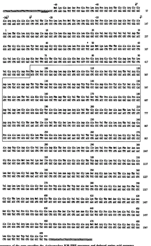
Fig. 3. Nucleotide sequence of the geneencodingtheActinomaduraR39 PBP precursor and deduced amino acid sequence
Thesiteofcleavagebythe leaderpeptidaseisindicatedbytheverticalarrow1.Otherpotential
cleavage
sites2,3and 4arealso indicated(seethe text). Ser*, active-siteserine. TheT97-P270insert(seethetext)isboxed.Theputative ribosome-bindingsiteGAGGAG is underlined.
R39 mature protein pDML10 (
-5 -1 +1
Asp Ala Ser Gly Ala Arg Leu Thr Glu Leu
5'... GAC GCC TCC GGA GCC CGC CTG ACC GAA CTG ... ... 3'
Hill
Streptomyces
(c) signal peptide Polylinker
*
~
~~~~
II
l
l
5' GGG TCCGGA TG CTGCAG CA TCCGGA CCC 3'
L I I I L I BspEI Pstl BspEl
41
5'... GACG CCT CCG GAT BspEI Synthetic oligonucleotide -1 +1 Ala ArgGCT GCA GCA TCC GGA GCC CGC CTG ACC GAA CTG ... ... 3'
Pstl BspElH1dI
Pstl BspEI Hindlill
39 new mature protein
+1
Ser Gly Ala Arg Leu Thr
TCC GGA GCCCGC CTG ACC ... 3'
!-BspEl
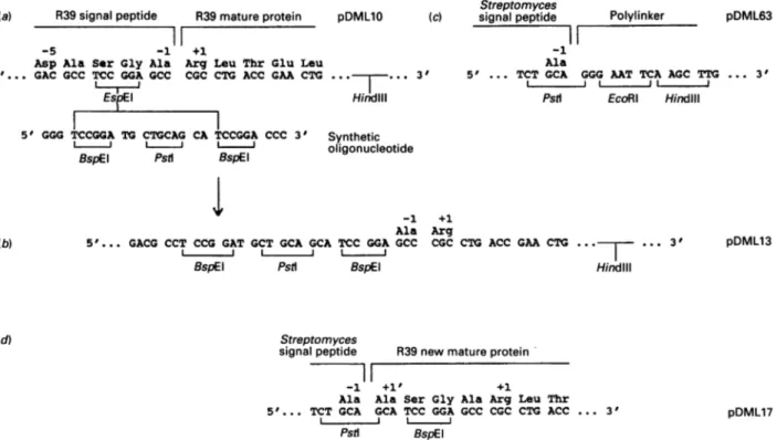
Fig. 4. Introduction of the DNAsequenceencoding thematureActinomaduraR39DD-peptidase/PBP in theStreptomyces secretion vectorpDML 63: construction ofpDML17
(a)Aperfectpalindromic oligonucleotidewassynthesized and allowedtoself-hybridize, givingaDNAsegmentwithaPstIsiteflankedoneach
side withaBspEI site. The hybridwasdigested with BspEI and inserted in the unique BspEI 3166 site ofpDML10 (seeFig. lc),yieldingpDML13
(b), in which the natural signal peptidase cleavage site (Ala-'-Ala"') ofthe Actinomadura R39 DD-peptidase/PBP is thus preceded bya
17-nucleotidesequencecontainingaPstI andaBspEIsiteseparated byadinucleotide CA. Digestion ofpDML13 withPstI andHindIIIliberated
a3100-bpsegmentthatcontainedtheregion encoding thematurepartof thePBP-encodinggenedownstreamofa12-nucleotidesequencecoding
forAla,Ser, Gly and Ala. This 3100-bpsegmentwasintroduced in the polylinker of pDML63 (c),aStreptomyces high-copy-numbersecretion
vector(see the text), giving risetopDML17(d) in which the Streptomyces signal sequenceis in phasewith thegeneencoding the Actinomadura
R39matureprotein (withafour-amino-acid N-terminalextension).
by electrotransformation, yielding Actinomadura BG3 (note that transformation of the Actinomadura strain by the usual
PEG-assisted procedure failed and that electrotransformation gave
onlyonetransformant in severalassays). Underoptimal growth
conditions in TAU medium, Actinomadura BG3 produced 250mg of active DD-peptidase/PBP/litre of culture, instead of 15mg/litre for the original strain (grown under identical
con-ditions). Thesecretedproteinhad the'correct' molecularmass.
Alikely hypothesisderived fromthe abovestudieswasthat,in S. lividans, incorrect processing of the DD-peptidase/PBP
pre-cursoroccurred,producinganinactiveproteinwithanextended
N-terminal region. Examination of the ORF shows that the
signal peptide possesses, upstream of the site cleaved in Acti-nomadura R39 (marked 1 in Fig. 3), other potential cleavage
sites(marked 2,3and4) [22].Inparticular,site 2isidentical with the S. lividans ,-galactosidase signal-peptide-cleavage site [23]. Cleavageof theDD-peptidaseprecursoratthissitewouldgenerate
aproteinwith amolecularmassof 52339 (instead of50053 for
the protein processedatsite 1).
In order to test the hypothesis, the mature-protein-encoding
DNA wasintroduced into the Streptomyces high-copy-number
secretion vector pDML63 (a derivative of pIJ702; A.Brans,
M. V.Lenzini, C. Fraipont-Piron & J. Dusart, unpublished work). Thisvector possesses Streptomyces transcription,
trans-lation and secretion signals, followed by a polylinker (PstI,
EcoRI,HindIlI, SmaI,XbaI)whosePstI site is inphasewith the
translation signal. Therefore a PstI site was introduced in the
unique BspEI site of the cloned gene, close to the junction
betweencleavage site 1 and the mature-protein-encoding DNA
(Fig. I c), the strategyshown in Fig.4beingfollowed. The final
construction was called pDML17. S. lividans transformed with pDML17 andgrowninMYEMEmedium secretedatleast 18mg
ofthe activeDD-peptidase/PBP per litre.
Hydrophobic-cluster analysis: similarityto the E. coliPBP4
andstructural relatedness withStreptomyces albus G fI-lactamase of classA
Both the Goad & Kanehisa algorithm [20] and BESTFIT
program revealed high similarity, in the primary structure,
between theActinomaduraR39 PBP and the E.coliPBP4(results
notshown). By using thesameprocedure,no, oronly marginal,
similarity was observed with the other groups and classes of
penicilloyl serine transferases. Hydrophobic-cluster analysis (Fig. 5) confirmed that the Actinomadura R39 PBP and the E. coli PBP4 were indeedremarkably similar. Providing that a
largedeletion wasmade inthe PBPs, theanalysis also revealed
similarity,inthepolypeptide folding,between thetwoPBPsand the class A Streptomyces albus G ,-lactamase of known three-dimensional structure [24,25]. Fig. 6 shows the amino acid
alignments as derived from thisanalysis.
DISCUSSION
As a first attempt to obtain the desired Actinomadura R39
gene, aS. lividansTK24-pIJ702 cloningsystem andan
immuno-logical screeningtestwereused(B. Granier, unpublished work).
Clones were isolated which produced aprotein that effectively reacted with the anti-(Actinomadura R39 DD-peptidase/PBP)
(a) R39signal peptide pDML63
-1
Ala
5' ... TCT GCA GGG AAT TCA AGC TTG ... 3'
Pstl EcoRl Hindlil (b) (d) pDML13 Streptomyces signal peptide R
7i17
-1 +1' Ala Ala E 5'... TCT GCA GCA I l I Pst pDML17 -JLIcn *E a) C) 10
S:
a) U, e, IN CL CLn In )-N CLO _:,n Ln LnL 0 01 QC r-tn EL C) LLi 1992 rs. :L I Cul N 21 r-2 CN Ln N JN es X w OL. ta I-*s C .e low q Ct Ca co co la . .0I
Uto Ut .=Ut Ca cot h. Ca Ca0 CA CI C .vicc
-o(DCu
rz .9 49 U) 13~ Q E 0 0). (if) N z .-T cl. 9 9& t 9 0 0 0 cc ...a4 M41 co 04 .... 0 13 .C7x CD t(70,ABL)
ALBUS .GSGSVSDA ERRLAGL.ER AS..GARLGV YAYDTGSGRT 56 VAYRA...DE LFPMCSVFKT LSSAAVLRDL DRNGEFLSRR ILYTQDDVEQ 104
PBP4 MRFSRFIIGL TSCIAFSVQA ANVDEYITQL PA..GAN..L ALMVQKVGAS 26 APAIDYHSQQ MALPASTQRV ITALAALIQL GPDFRFTTTL ETKGNVENGV 76
R39 .... .. RLTELR EDIDAILED. PALEGAVSGV VVVDTATGEE 35 LYSRDGG..E QLLPASNMRL FTAAAALEVL GADHSFGTEV AAESAPGRRG 83
ALBUS ADGAPETGKP ... ... ... 1... 12 ... 112
PBP4 LKGDLVARFG ADPTLKRQDI RNMVATLKKS GVNQIDGNVL IDTSIFASHD 126 KAPGWPWNDM TQCFSAPPAA AIVDRNCFSV SLYSAPKPG. ..DMAFIRVA 173 R39 EVQDLYLVGR GDPTLSAEDL DAMAAEVAAS GVRTVRGDLY ADDTWFDSER 133 LVDDWWPEDE PYAYSAQISA LTVAHGERFD TGVTEVSVTP AAEGEPADVD 183
ALBUS ... ... ... ... ... 112 ... . ... ... ... ...QN 114
PBP4 SYYPVTIPSQ VRTLPRGSAE AQYCELDVVP GDLNRFTLTG CLPQRSEPLP 223 LAFAVQDGAS YAGAILKDEL KQAGITWSGT LLRQTQVNEP G...TVVA 268
R39 LGAAEGYAEL DNRAVTGAAG SANTLVIDRP VGTNTIAVTG SLPADAAPVT 233 ALRTVDEPAA LAGHLFEEAL ESNGVTVKGD VG...LGGVP ADWQDAEVLA 280
(130,ABL) (166,ABL)
ALBUS LANGMTVEEL CEVSITASDN CAANLMLREL G... ...GPAAVTR 150 FVRSL.GDRVTRLDRWEPEL NSAEPGRGVT DTTSPRAITR TYGRLVLGDA 198
PBP4 SKQSAPLHDL LKIMLKKSDN MIADTVFRMI GHARFNVPGT WRAGSDAVRQ 318 ILRQQAGVDI GNTIIADGSG LSRHNLI... ...APATMMQ VLQYIAQH.. 360
R39 DHTSAELSEI LVPFMKFSNN GHAEMLVKSI GQETA.GAGT WDAGLVGVEE 329 ALSGL.GVDT AGLVLNDGSG LSRGNLV... ...TADTVVD LLGQAGSA.. 370
(234,ABL)
ALBUS LNPRDRRLLT SWLLANTTSG DRFRAGL... ....PDDWTLGDRTGAGRYG 242 TNNDAGVTWP ...PGRAPIV LTVLTAKTEQ DAA...RDDG LVADAARVLA 287
PBP4 .DNELNFISMLPLAGY.DGS LQYRAGLHQA ....GVDGKV SAIKTGSLQGV 404 YNL...AGFI TTASGQRMAF VQYLSGYAVE PADQRNRRIP LVRFESRLYK 451
R39 ...PWAQTWS ASLPVAGESD PFVGGTLANR MRGTAAEGVV EAKTGTMSGV 417 SAL...SGYV. .PGPEGELA FSIVNNGHSG P..APLAVQD AIAVRLAEYA 460
ALBUS ETLG... ... ... 291
PBP4 DIYQNN.... 457
R39 GHQAPEGARMMRGPVQGSGE LECSWVQAC. 489
Fig. 6. Amino acid sequencealignmentoftheActinomadura R39PBP, the E. coliPBP4andtheStreptomycesalbus G
fI-lactamase
of class A Theproposed alignment derives fromthatofFig.5.ALBUS, Streptomycesalbus G BLA; PBP4, E. coli PBP4; R39, Actinomadura R39 PBP. Themotifsthatformthe,-lactamase active site areindicated(ABL numbering).
antiserum. This protein had an apparent molecular mass
larger than that ofthe original DD-peptidase/PBP and it was inert in terms of DD-peptidase and penicillin-binding activity, suggestingthat theuseofapolyclonal antibody
might
have beenmisleading. Subsequently,andasdescribedabove, the genewas
clonedusingE. coliashost and anucleotide probeasscreening tool, butintroduction of the gene in S. lividans, viapDML15, againresulted in thesynthesis(inMYEMEmedium)ofaprotein whichwasdevoidofenzymic activityandwasalso about 3 kDa
larger than the original DD-peptidase/PBP. However, and at
variance with this observation, S. lividans TK24 transformed
with a secretion vector in which the signal sequence of the Actinomadura gene was replaced by a Streptomyces signal
sequence,producedand secretedanactiveDD-peptidase/PBPof normal size.These studiesthus supporttheview that theprotein
precursor isprocesseddifferentlyinActinomadura and in
Strep-tomyces and that the N-terminal extension resulting from the incorrect processing in S.lividansissufficient topreventcorrect
folding of theprotein.
Hydrophobic-cluster analysis ofthe Actinomadura R39 PBP and theE. coliPBP4and
pairwise comparison
ofthe PBPs with theclass A Streptomycesalbus G,3-lactamase (of
known three-dimensional structure) leads to thefollowing
conclusions. (1)Thetwo PBPshave, alongthe amino acidsequences, the same
typical pattern of distribution of hydrophobic clusters and of hydrophilic residues between the conserved
hydrophobic
clusters. (2)Thepeptide stretchesT97-P270 in theActinomaduraPBPandT90-P263in theE. coliPBPhave noequivalentsin the
/3-lactamase.
(3) When these adducts are eliminated from the amino acid sequences, the bulk of thepolypeptide
chainof thetwo PBPs exhibits
similarity
with that of the/,-lactamase. (4)
Secondary structures
equivalent
to the,3-lactamase
strands/31,
,82,
/83,
/34
and,35
and helicesal, a2,a4,
a5, a6, a8, a9,
alOandal1 are easily identified in the PBPs.
(5)
Identification in thePBPsof the S*XXK motif
(where
S* is theessentialserine)
ona2,the SXN motif betweena4anda5and the KTG motifon/3
is not a matterof controversy. (6) D345 in the Actinomadura PBP
andD335intheE. coli PBP occur at a position comparable with that of E166 of the EPELNmotif, between a6 and a8, in the
/3-lactamase. E166 functions as proton abstractor in the attack of thecarbonylcarbonofthescissileamidebond of penicillin by the y-OH of the ,-lactamase active-site serine residue [25]. The
homologuesD345andD335might play a similar role in the PBPs. Note, however, that another aspartic acid, D337 orD327,occurs
immediately upstream from D34s and D335.
Site-directed-muta-genesisexperiments will give answers tothe question regarding
theessentiality of these acidic sidechains in the PBPs. (7)The inserts T97-P270 in the Actinomadura R39PBPand p90_P263 in the
E. coli PBP4 (whose position is slightly different from that
proposedbyMottletal.[5])occurbetweena2and a4, i.e.onthe
surface oftheproteins.Thestructureandpossiblefunction of the inserts areunknown.
TheActinomadura R39 PBP isasecretoryprotein.Themajority
of theE. coli PBP4 is exportedin the periplasm, atleast in the
overproducing strains [26]. Byanalogytothe ,-lactamases, the
polypeptide chain ofthese two water-soluble PBPs terminates
immediately or almost immediately after al 1. In contrast, the membrane-boundE.coli PBP5 andB.subtilis PBP5areanchored inthe membranebyashortpeptidestretch locatedatthe end of
along (> 100aminoacids) additionalC-terminal extension
[27].
The Actinomadura R39 PBPand the E. coliPBP4are func-tionally homologous with respect to the reactions that they catalyse onD-alanyl-D-amino acid-terminatedpeptides [26-28].
The Actinomadura PBP, however,performs transpeptidation
reactions in vitro whenincubated in the presence of structured amino acids and peptides [28] and it isextremely susceptible
toinactivation by benzylpenicillin
(second-order
rate constant ofacylation of the essential serine:
300,000
M-1 5s- at 37°C,
ascompared withabout 7000M-1-s- for the E.coli
PBP4)
[29].
Alastcomment mustbe made. The immediate environmentofthe active-site serineinthe Actinomadura R39PBPis
LPASNMK,
last tworesidues wereimpurities andtheisolatedpeptidewasin fact thepentapeptideLPASN.
We thank Mr. D.Klein for technical assistance in DD-peptidase purification. This work was supported, in part, by the Belgian programme on Interuniversity Poles of Attraction initiated by the
Belgian State, PrimeMinister'sOffice, Science Policy Programming(PAI n° 19), Actions concerteeswith the Belgian Government (conventions
86/91-90 and 89/94-130), the Fonds de la Recherche Scientifique
Medicale (contract n°3.4537.88), aConvention tripartite between the Region wallonne, SmithKline Beecham, U.K., and the University of Liege,andthe Belgian IncentiveProgram onFundamentalResearchin LifeSciences (contract BIO 22). C.D.andJ. D. areChercheursqualifies
of the FondsNationalde la Recherche Scientifique,Brussels.
REFERENCES
1. Ghuysen,J. M. (1991) Annu. Rev. Microbiol. 45,37-67
2. Gaboriaud, C., Bissery, V., Benchetrit,T. & Mornon, J. P. (1987)
FEBS Lett.224,149-155
3. Henrissat, B., Saloheimo, M., Lavaitte, S. & Knowles, J. K. C.
(1990)ProteinsStruct. Funct. Genet. 8, 251-257
4. Palomeque-Messia, P., Englebert, S., Leyh-Bouille, M., Nguyen-Disteche, M.,Duez,C.,Houba, S., Dideberg, O.,VanBeeuman,J. &Ghuysen,J. M. (1991) Biochem.J. 279, 223-230
5. Mottl,H.,Terpstra,P. &Keck,W.(1991)FEMS Microbiol. Lett.
78, 213-220
6. Erpicum, T., Granier, B., Delcour, M., Lenzini, V.M., Nguyen-Disteche,M.,Dusart J.& Frere, J. M.(1990) Biotechnol. Bioeng. 35,
719-726
7. Granier, B.(1990)Ph.D.Thesis, UniversityofLi&ge
8. Hopwood, D.A., Bibb, M.J., Chater, K.F., Kieser, T., Bruton,
C.J., Kieser, H.M., Lydiate, D.J., Smith, C. P., Ward, J. M. & Schrempf, H. (1985) Genetic Manipulation of Streptomyces: A LaboratoryManual,TheJohnInnesFoundation, Norwich
9. Maniatis, T., Fritsch, E. F. & Sambrook, J. (1982) Molecular
Cloning. A LaboratoryManual, ColdSpring HarborLaboratory,
ColdSpringHarbor, New York
10. Frere, J. M.,Ghuysen,J. M.,Reynolds,P. E., Moreno,R.,Perkins,
H.R.,Dierickx,L.&Delcambe,L.(1974) Biochem.J.143, 241-249
11. Saiki, R. K.,Gelfand, D.H.,Stoffel, S.,Scharf, S.J.,Higuchi, R.,
Horn, G. T., Mullis, K.B. & Ehrlich, H. A. (1988) Science 239,
487-491
12. Wallace, R. B.,Johnson,N.J.,Hirose,T.,Miyake,M.,Kawashima,
E. H. &Itakura,K.(1981) Nucleic AcidsRes. 9,879-894
13. Woods,D.(1984)Focus6, 1-3
14. Sanger, F.,Nicklen, S. &Coulson, A. R. (1977)Proc.Natl. Acad. Sci. U.S.A. 74, 5463-5467
15. Staden, R.(1984) Nucleic Acids Res. 12, 551-567 16. Fickett,J. W. (1982)Nucleic AcidsRes. 10, 5303-5318
17. Houba, S., Willem, S.,Duez,C.,Molitor,C., Dusart, J., Frere, J.M.
&Ghuysen,J. M.(1989) FEMS Microbiol. Lett. 65,241-246
18. Frere,J. M.,Leyh-Bouille,M.,Ghuysen,J. M.,Nieto,M. &Perkins,
H. R.(1976)MethodsEnzymol.45B,610-636 19. Hanahan, D. (1983)J. Mol. Biol. 166, 557-580
20. Goad, W. B. & Kanehisa, M.I. (1982) Nucleic Acids Res. 10, 247-263
21. Devereux, J.,Haeberli,P.&Smithies,0. (1984)NucleicAcidsRes. 12,387-395
22. vonHeijne, G. (1984)J.Mol. Biol. 173, 243-251
23. Eckhardt,T.,Strickler,J.,Gorniak,L., Burnett, W. V. & Fare, L. R.
(1987)J.Bacteriol. 169, 4249-4256
24. Dideberg, O., Charlier, P., W6ry, J. P., Dehottay, P., Dusart, J.,
Erpicum,T., Frere, J. M. &Ghuysen,J. M.(1987) Biochem. J. 245, 911-919
25. Lamotte-Brasseur, J., Dive, G., Dideberg,O., Charlier, P., Frere, J. M. & Ghuysen,J. M.(1991)Biochem. J. 279, 213-221
26. Korat,B.,Mottl,H. &Keck,W.(1991) Mol. Microbiol.5,675-684
27. Pratt, J. M.,Jackson, M. E. & Holland, I. B. (1986) EMBO J. 5, 2399-2405
28. Ghuysen, J.M., Reynolds, P. E., Perkins, H. R., Frere, J. M. & Moreno, R. (1974) Biochemistry13, 2539-2547
29. Frere, J. M. & Joris, B. (1985) CRC Crit. Rev. Microbiol. 11, 299-396
30. Duez, C.,Joris, B., Frere,J.M.,Ghuysen,J. M. & Van Beeumen, J.
(1981) Biochem.J. 193, 83-86
Received 12August 1991; accepted 25 September 1991