Acknowledgements
I would like to thank all the members of my jury: Prof. Corinne Chanéac, Dr. Rabah Boukherroub, Dr. Fabienne Gauffre and Prof. Romuald Poteau for accepting to read the manuscript and examine my Ph.D work.
I would like to give my deep appreciation to my supervisors Dr. Myrtil Kahn and Dr. Christophe Mingotaud for giving me the opportunity to study in Toulouse, and for their endless guidance, patience, support and encouragement. Their rigorous academic attitude, innovative thinking, and profound knowledge of chemistry have left me a deep impression and will benefit me in my whole lifetime. I also would like to thank their kind-hearted help when I face some difficulties in my life beyond the research. And, I would like to thank Dr. Jean-Daniel Marty and Dr. Clément Roux, who also participate in this work and help me a lot during all these three years.
I would like to thank Dr. Pierre Fau who helped me a lot to measure the electronic properties. I would also like to thank Dr. Yannick Coppel for NMR experiments and fruitful discussions, many thanks for your patience to teach me even I had never used NMR before I came here. I also would like to thank Dr. Juliette Fitremann for her help to analyze the rheological and mechanical properties; Dr. Christine Lepetit for the molecular simulations and valuable discussions; Pierre Lecante for the WAXS experiments; Pierre Alphonse for the BET measurements; Jérome Esvan for the XPS measurements; Laure Vendier for the PXRD measurements; Vincent Collière for the high resolution electronic microscopy characterization; Stéphanie Seyrac for TGA characterization; Stéphane Gineste for the help to synthesize the ligand with azobenzene group; and all the others who indeed help me finish my PhD work.
All current and former Equipe T members in LCC and Equipe IDeAS members in IMRCP, Yohan Champouret, Pauline Loxq, Emillie Tailhades, Justyna Jonca, Jérémy Cure, Glenna Drisko, Zhiqin Zheng, Hala Assi, Guillaume Garnide, Ségolene
Palussière, Maxime Puyo, Xuming Zhang, Florian Wodlei, Baptiste Amouroux, Cherles-Louis Serpentini, Dr. Anne-Francoise Mingotaud, Dr. Christophe Coudret, and Dr. Katia Fajerwerg for their fruitful discussions and friendship.
Thanks to the help from some of my friends in Toulouse: Jinhui Wang, Chongwei Zhu, Longai Lang, Qian Liao, Lin Yang, Changlong Wang, Chen Zhang, Jieru Qiu, Liang Chen, Fan Li and Yin Zhang who have brought me a lot of fun during the 3 years.
Many thanks to all LCC and IMRCP employees for their kind-hearted help.
China Scholarship Council (CSC) and CNRS are also acknowledged for financial support.
The last but not the least, my greatest appreciation are dedicated to my parents Quangen Zhao and Hong Yue, my sister Xinmei, and my lovely daughter Sixuan and my wife Xiaoqi, who sacrificed much during these 3 years.
_________________
Systémes hybrides : de la
nanoparticule aux gels
_________________
Zhihua Zhao
15/09/2017
Sommaire
Chapitre I - Revue bibliographique et objectifs de la thèse
Introduction ... 11
I. 1. Les nanoparticules ... 11
I. 1. 1 Introduction général aux nanosciences ... 11
I. 1. 2 Les nanoparticules d’oxydes métalliques ... 12
I. 2. Les différentes approches de synthèse de NP d’oxydes métalliques... 14
I. 3. L’approche “sol-gel” pour la préparation de NP d’oxydes métalliques .... 15
I. 4. L’approche organométallique pour la préparation de NP d’oxydes métalliques ... 16
I. 5. Les travaux antérieurs du LCC ... 18
I. 5.1. Introduction ... 18
I. 5.2. Le rôle des ligands sur la synthèse des NP ... 20
I. 5.3. Les métairaux hybrides ... 23
I. 6. Objectifs de la thèse ... 25
Références ... 26
Chapitre II – Analyse statistique d’objets anisotropes
Introduction ... 43II.1. Principe de l’analyse multivariée ... 44
II.2. Applications de l’analysis par représentation 2D ... 49
Sommaire
Références ... 58
Chapitre III – Une vision nouvelle sur la croissance de
nanoparticules anisotropes de ZnO
Introduction ... 67III.I. Influence du temps de murissement et de la vitesse de réaction ... 67
III.2. Influence de la quantité d’eau ... 79
Conclusion ... 94
Références ... 95
Chapitre IV – Une vision nouvelle sur la croissance de
nanoparticules anisotropes de ZnO
Introduction ... 103IV. 1. Formation de gels ... 104
IV. 2. Etude rhéologique ... 105
IV. 3. Analyse structurale ... 109
IV. 3. 1 Etudes NMR ... 109
IV. 3. 2 Modélisation moléculaire ... 121
IV. 3. 3 Diffusion des rayons-X aux grand angles (WAXS) ... 125
IV. 4. Mise en forme ... 126
IV. 5. Généralisation de l’approche ... 134
Conclusion ... 140
Sommaire
Conclusion Générale
………147Partie expérimentale
Introduction ... 159 Méthodes de synthèse ... 159 Synthèses : Chapter II ... 159Synthèses : Chapter III ... 159
Synthèses : Chapter IV ... 161
Méthodes de caractérisation ... 163
Microscopie électronique à transmission ... 163
Microscopie électronique à balayage à émission de champ (FE-SEM)…….163
Analyse calorimétrique différentielle (DSC) ... 163
Microscopie optique polarisée (POM) ... 163
Spectroscopie de photoluminscence ... 163
Spectroscopie RMN ... 164
WAXS experiments ... 164
Diffraction des rayons-X ... 165
Modélisation moléculaire ... 165
Rhéologie ... 165
Tests de compression ... 166
Mesures de photocourant ... 166
Sommaire
Analysis statisitque ... 167
Programme R... 167
Produits chimiques ... 169
Chapitre I
_________________
Revue bibliographique et objectifs de
la thèse
Résumé
Dans ce chapitre introductif, nous nous attachons à présenter une introduction aux nanosciences en nous focalisant sur les nanoparticules d’oxydes métalliques qui sont au cœur de ce travail de thèse. Nous présentons les différentes méthodes de préparation de ces nanoparticules d’oxydes métallique en détaillant plus précisément l’approche « sol-gel » et l’approche organométallique. Cette dernière est en effet la méthode que nous avons utilisée tout au long de ce travail. Les travaux antérieurs à ce travail sont ensuite présentés afin de bien identifier les objectifs de cette thèse.
Contenu
Introduction ... 11 I. 1. Les nanoparticules ... 11 I. 1. 1 Introduction général aux nanosciences ... 11 I. 1. 2 Les nanoparticules d’oxydes métalliques ... 12 I. 2. Les différentes approches de synthèse de NP d’oxydes métalliques... 14 I. 3. L’approche “sol-gel” pour la préparation de NP d’oxydes métalliques .... 15 I. 4. L’approche organométallique pour la préparation de NP d’oxydes
métalliques ... 16 I. 5. Les travaux antérieurs du LCC ... 18 I. 5.1. Introduction ... 18 I. 5.2. Le rôle des ligands sur la synthèse des NP ... 20 I. 5.3. Les métairaux hybrides ... 23 I. 6. Objectifs de la thèse ... 25 Références ... 26
Chapter Ⅰ Literature Review and Project Objectives
11
Introduction
In this chapter, firstly I will discuss some basic concepts about the different kinds of nanoparticles (NPs). Then the discussion will be shifted to the metal oxide NPs and the versatile synthesis methods to get them, especially the organometallic chemistry strategy. The past published work in our group will then be shown in this chapter, and among them, the importance of the ligand effect on the different properties will be emphasized. At last, the project objectives of my thesis will be derivatized from those discussions.
I. 1. Nanoparticles
I. 1. 1 General Introduction of Nanoscience
Nanoscience involve studying and working with matter on an ultra-small scale.
One nanometer is one-millionth of a millimeter, and a single human hair is around 80000 nanometers in width. Nanoscience encompass a range of techniques rather than a single discipline, and stretch across the whole spectrum of science, touching medicine, physics, engineering and chemistry [1]. They have huge application potential in different
fileds such as new energy production, healthcare, communication and information technologies, environment protection and the preparation of new materials with better mechanical properties. Therefore, it becomes more and more important to develop in-depth study to better understand this discipline. In fact, the actual investment for nanomaterials and nanostructures occupies 49% in the investment for the study of nanoscience [2]. Now, in order to improve the national competitive ability in the
scientific and technological fields, the motivation for the study of nanomaterials and nanostructures becomes mainly National Strategy Requirements. Furthermore, the study of nanomaterials and nanostructures is of vital importance for establishing new
Chapter Ⅰ Literature Review and Project Objectives
12
methods, new technologies and new principles, thereby potentially leading to breakthroughs in great scientific problems [3]. At the same time, the nanomaterial
market is also a native power for the development of nanomaterials and nanostructures. Recently there has been substantial interest in the preparation and characterization of materials consisting of particles with dimensions in the metal oxide nanocrystalline materials [4]. One factor driving the current interest in nanoparticle research is the
perceived need for further miniaturization of both optical and electronic devices [5,6].
Figure I. 1. Diagram illustrating the scale of nanomaterials [7].
I. 1. 2 Meal Oxide Nanoparticles
Metal oxides play a significant role in the various areas of chemistry, physics, and materials science. Metallic elements can form a large diversity of oxide compounds, which can adopt a vast number of structural geometries with electronic structures that can exhibit metallic, semiconductor, or insulator character [8]. In technological
applications, metal oxide nanoparticles are used in the fabrication of microelectronic circuits, sensors, piezoelectric devices, fuel cells, coatings for the passivation of surfaces against corrosion, and as catalysts [8]. For example, among them, a lot of works
about zinc oxide, tin oxide and iron oxide nanoparticles have been done due to their interesting properties paving the ways for fascinating applications.
Zinc oxide is a material with great potential for a variety of practical applications,
Chapter Ⅰ Literature Review and Project Objectives
13
such as piezoelectric transducers, optical waveguides, surface acoustic wave devices, varistors, phosphors, transparent conductive oxides, chemical and gas sensors, spin functional devices, and UV-light emitters [9-12]. Its wide bandgap (≈3.37 eV at room
temperature) makes ZnO a promising material for photonic applications in the UV or blue spectral range, while the high exciton-binding energy (60 meV) allows efficient excitonic emission even at room temperature. In addition, ZnO doped with transition metals shows great promise for spintronic applications. It has also been suggested that ZnO exhibits sensitivity to various gas species, namely ethanol (C2H5OH), acetylene
(C2H2), and carbon monoxide (CO), which makes it suitable for sensing applications
[13-15]. Moreover, its piezoelectric property (originating from its non-centrosymmetric
structure) makes it suitable for electromechanical sensor or actuator applications [16-18].
Also, ZnO is biocompatible which makes it suitable for biomedical applications [19-21].
Consequently, there is considerable interest in studying ZnO in the form of powders, single crystals, thin films, or nanostructures [22].
Tin oxide is another important metal oxide semiconductor, which is considered to be technologically important materials and has been investigated for a wide range of applications such as high energy density rechargeable lithium batteries [23], storage of
solar energy [24], gas sensors [25], electrocatalysis [26] and photocatalysis [27]. This
diversity in the application is a function of the size, morphology, phase, and crystallinity of the nanocrystals. Various geometrical morphologies of tin oxide have been produced, for example, spherical particles [28], networks of ribbons [29], hollow microspheres [30],
sheets [31], flowers [32] and belts [33]. SnO2 sensor response is because of the physical and
chemical changes on its surface due to the adsorption of a chemical stimulant and the sensitivity can be fine-tuned by reducing the size up to nano dimensions, varying the crystal structure and morphology and/or adding dopants (typically noble metals and other metal oxides) to create nanocomposite materials [34].
Iron oxide is also a kind of important metal oxide material, which has served humans for centuries, for example, the application of small iron oxide nanoparticles as a contrast agent for in vivo diagnostics has been practiced for nearly half century [35]. In
Chapter Ⅰ Literature Review and Project Objectives
14
developed not only for its fundamental scientific interest but also for its many technological applications, such as targeted drug delivery, magnetic resonance imaging (MRI), magnetic hyperthermia and thermoablation, bioseparation, and biosensing [36].
Eight iron oxides are known [37], among these iron oxides, hematite (α-Fe2O3),
magnetite (Fe3O4) and maghemite (γ-Fe2O3) are very promising and popular candidates
due to their polymorphism involving temperature-induced phase transition. Each of these three iron oxides has unique biochemical, magnetic, catalytic, and other properties which provide suitability for specific technical and biomedical applications.
I. 2. Synthesis of Metal Oxide Nanoparticles
In the past decades, a number of specific methods have been developed to prepare metal oxide nanoparticles, some examples about the broadly used methods will be discussed as follow: (1) Coprecipitation methods: This involves dissolving a salt precursor (chloride, nitrate, etc.) in water (or other solvent) to precipitate the hydroxide form with the help of a base. Very often, control of size and chemical homogeneity in the case of mixed-metal oxides is difficult to achieve [38]. (2) Microemulsion technique:
Microemulsion or direct/inverse micelles represent an approach based on the formation of micro-/nano-reaction vessels under a ternary mixture containing water, a surfactant, and oil. Metal precursors on water precede precipitation as hydroxides within the aqueous droplets, typically leading to monodispersed materials with size limited by the surfactant-hydroxide contact [39]. (3) Solvothermal methods: In this case, metal
complexes are decomposed thermically either by boiling in an inert atmosphere or by using an autoclave with the help of pressure. A suitable surfactant agent is usually added to the reaction media to control particle size growth and limit agglomeration [40]. (4)
Template/surface derivatized methods: Template techniques are common to some of the previously mentioned methods and use two types of tools: soft templates (surfactants
[41]) and hard templates (porous solids as carbon [42] or silica [43]). Template- and surface-
mediated nanoparticle precursors have been used to synthesize self-assembly systems
Chapter Ⅰ Literature Review and Project Objectives
15
main advantages of those 2 methodologies are that they produce uniform and pure nanoparticles and films, however, the disadvantage of these methods is that they require a careful initial setting up of the experimental parameters [46]. Besides the above
mentioned methods, there are other two important methods, which are sol-gel route and organometallic method for which detailed introduction will be described in the following.
I. 3. Sol-gel route towards Metal Oxide Nanoparticles
The sol-gel method prepares metal oxides via hydrolysis of precursors, usually metal alcoxides in alcoholic solution, resulting in the corresponding hydroxides. Condensation of molecules by giving off water leads to the formation of a network of the metal hydroxide: hydroxyl species undergo polymerization by condensation and form a dense porous gel. Appropriate drying and calcination lead to ultrafine porous oxides [47].
The sol-gel route offers some particular advantages, centered on the ability to produce a solid-state material from a chemically homogeneous precursor. By ensuring atomic level mixing of reagents, one should be able to produce complex inorganic materials such as ternary and quaternary oxides. Furthermore, sol-gel chemistry should enable greater control over particle morphology and size. In reality, producing a homogeneous precursor at room temperature does not ensure homogeneity throughout a reaction and many sol-gel routes have therefore been designed to combat or control phase segregation during synthesis. In fact, some of the most interesting advances in the sol-gel field in recent years have come from gels that have some degree of ordering and structure [48].
However, the sol-gel process is mainly restricted in aqueous condition, which brought some major limitations forward when it came to the preparation of their nanoscale counterparts. Aqueous sol-gel chemistry is rather complex, mainly due to the high reactivity of the metal oxide precursors and the double role of water as ligand and solvent. In many cases, the three reaction types (hydrolysis, condensation, and
Chapter Ⅰ Literature Review and Project Objectives
16
aggregation) occur almost simultaneously (and are difficult to control individually), so slight changes in experimental conditions result in altered particle morphologies, a serious issue regarding the reproducibility of a synthesis protocol. Furthermore, the as-synthesized metal oxides are often amorphous, and it is difficult to retain full control over the crystallization process during any additional annealing step. All these parameters might be controlled well enough for the preparation of bulk metal oxides, however, they represent a big challenge in the case of nanoparticle synthesis [49].
Therefore, it is of importance to develop nonaqueous sol-gel processes in organic solvents under exclusion of water, which are able to overcome some of the limitations. The advantages of nonaqueous sol-gel processes are closely related to the manifold role of the organic components in the reaction mixture. They not only act as the oxygen-supplying agent for the metal oxide but also strongly influence particle size, shape, surface and assembly properties, and, in selected cases, even composition and crystal structure. Furthermore, in contrast to aqueous systems with nearly indefinable composition, the characterization of the organic compounds in organic media can easily be performed with standard techniques like nuclear magnetic resonance (NMR) spectroscopy or gas chromatography-mass spectrometry (GC-MS). By retro-synthetical analysis, it is possible to correlate the processes leading to these organic species to the growth mechanisms of the oxide nanoparticles, offering a powerful tool toward the development of a rational synthesis strategy for a broad family of inorganic nanomaterials [49].
I. 4. Organometallic Method towards Metal Oxide NPs
There are many precursors for making metal oxide nanoparticles, and all the compounds could be classified as two categories: (a) inorganic metal salts including metal nitrate, metal chloride, metal acetate and so on; (b) organic metal compound, such as metal isopropoxide, or some other organometallic complex like alkyl metal. As with all synthesis procedures, each of the precursors described above has its characteristic
Chapter Ⅰ Literature Review and Project Objectives
17
scope and limitations. Many rely on the reactions of low-energy reagents at high temperatures, especially for those about inorganic metal salts, and for the other extreme, so called organometallic method, is a pathway involving the reaction of intrinsically higher energy organometallic complex at lower temperature.
In the literature, some results about the metal oxide nanoparticles prepared by organometallic pathway have been reported. For instance, Steigerwald et al. reported a new synthesis of TiO2 nanocrystals that is based on the gentle oxidation of
Bis(cyclooctatetraene)titanium (Ti(COT)2), which is a very reactive organometallic
complex [50]. Lewiński and co-workers [51-58] developed an organometallic method to
prepare ZnO nanoparticles using diethylzinc (ZnEt2) and its derived organometallic
complex as precursor. For example, they used the ZnEt2 as the precursor and carboxyl
acid as stabilized ligand to obtain ZnO nanocrystals (Figure I. 2) and at last they successfully prepared photoactive Langmuir-Blodgett, freely suspended and free standing films of carboxylate ligand [54].
Figure I. 2. One-pot synthesis procedure for the preparation of ZnO nanocrystals
coated by the carboxylate ligands [55].
Sabastian Polarz and co-workers [59-83] developed another organometallic precursor
(alkylzincalkoxides with heterocubane architecture) to ZnO materials. A large variety of the tetrameric compounds [RZnORʹ]4 had been prepared from the reaction of ZnR2
with the respective alcohol in high yield, and then were reacted to ZnO either by thermal reaction or the reaction with water (Figure I. 3).
Chapter Ⅰ Literature Review and Project Objectives
18
Figure I. 3. The reaction of a Zn4O4 heterocubane with water to yield ZnO [68].
Another work reported by Charlotte K. Williams is about the ZnO nanocrystals prepared by hydrolysis of zinc clusters, which obtained by the reaction of ZnEt2 and
phosphinate ligands. They found that the ligand is sequestered to a stable zinc cluster during the majority of the synthesis and only becomes coordinated to the nanoparticle surface, this interesting finding provide an understanding of the role of well-defined molecular precursors during the synthesis of small nanoparticles [84].
In fact, a lot of organometallic precursors have been chosen as the precursor to prepare metal oxide nanoparticles. Compared to the other method such as aqueous sol-gel process, this process has some advantages, for instance, normally the organometallic species could be dissolved in organic solvent, and thus the kinetic of the chemical reaction could be monitored by the chemical characterization tools like NMR spectra, furthermore, many of them have high energy, which could make the release of the metal atom in a relatively mild condition, for example, when mixed with the stabilizing ligands, some organometallic complex is very easy to be hydrolyzed just by simply expose to the air, and finally well-defined metal oxide NPs were obtained. However, there are also some disadvantages like relatively high price, and sometimes
the organometallic complex is too reactive for the formation of the nanoparticles.
I. 5. The works of our group in LCC
I. 5.1. General introduction
In our group, we also use the highly energetic organometallic complexes, which are sensitive to both air and water, as the precursor, and the metal oxide nanoparticles could
Chapter Ⅰ Literature Review and Project Objectives
19
be obtained by direct hydrolysis or by another strategy, which involves two steps: the first step is hydrogenolysis of the organometallic complex in solution to get metal NPs, and then use some methods to oxidize the metal NPs to obtain metal oxide NPs. The shape and size of these nanoparticles can be controlled by the different parameters of the system (such as the nature of the organometallic precursor, the ligands or surfactants present, the solvent used and so on) [85].
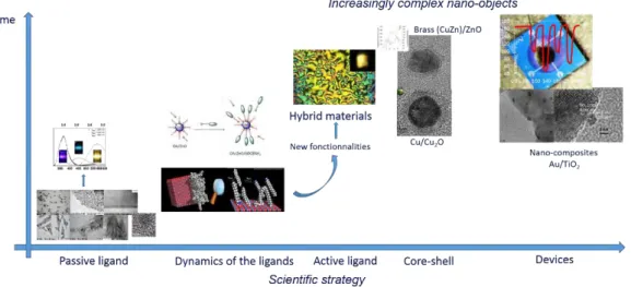
Figure I. 4. Schematic illustration for the scientific strategy in our group.
There is a strong correlation between the size and shape and properties of those NPs, and in our group, we use the NMR spectroscopy to monitor the dynamics of the stabilizing ligands like alkyl amine and (or) alkyl acids [94]. Some stabilizing ligands
with special functional groups were synthesized and used either by direct coordination on the surface of the NPs or by an interesting method called ligand exchange, then the organic-inorganic hybrid materials with new functionalities like liquid crystal property could be prepared [98]. We also prepared some more complex nano-objects which have
two or more metal species, and used them to make devices for the application of electronics like gas sensors [99]. In summary, the scientific strategy in our group could
be depicted in figure I. 4, and several kinds of metal oxide NPs prepared in our group will be shown in the following content.
Chapter Ⅰ Literature Review and Project Objectives
20
I. 5.2. The effect of stabilizing ligands on the prepared NPs
The first example is about the synthesis of ZnO NPs, which have been prepared by hydrolysis of [Zn(C6H11)2] ([ZnCy2]) in the presence of different kind of stabilizing
ligands (figure I.5) [86-101]. Interestingly, when the synthesis was performed in solvent
and the alkyl amine or (and) alkyl acid was (were) chosen as ligands, the isotropic ZnO nanoparticles could be obtained, however, when the synthesis was performed without any solvent, the anisotropic nanoparticles could be prepared just by exposure to the air (figure I.6). The size and shape of the ZnO NPs could strongly affect the properties like optical property [108] and gas sensitivities [110].
Figure I. 5. Schematic illustration for the preparation process of ZnO nanoparticles
using pioneered by Kahn et al. [87].
f
Figure I. 6. Representative TEM images of the ZnO nanoparticles prepared by the
process depicted in figure I. 5: the isotropic ZnO NPs stabilized by 1 eq. OA (a), 1 eq. DDA and 1 eq. OlAc (b), 1 eq. HDA and 1 eq. OlAc (c) in THF; the anisotropic ZnO NPs stabilized by 5 eq. OA (d), 2 eq. OA (e), and 1 eq. OA (f) in the absence of solvents.
a
b
c
Chapter Ⅰ Literature Review and Project Objectives
21
Furthermore, there is also correlation between the solubility and the stabilizing ligands. For example, if commercially available amino-PEG oligomers were used as the ligands, metal and metal oxide NPs including ZnO NPs could be obtained by hydrolysis of the organometallic complex, and those NPs could be solubilized in many non-polar and polar solvent including water [77, 89].
The stabilizing ligands are also very important for the self-organization property of the colloidal ZnO nanoparticles. For example, after gradually changing the concentration of the ZnO colloidal solution by evaporation of the solvent, no organization phenomenon could be monitored if long-alkyl-chain amines or carboxylic acids are used as stabilizing ligands, some superlattices of organized ZnO nanoparticles could be observed by TEM and SEM as well as by DLS when binary mixtures of long-alkyl-chain amines or carboxylic acids are used [114]. The coordination mode of the
ligands at the surface of ZnO nanoparticles was fully characterized by different NMR technologies, which suggest that at least three different modes of interaction of the amines at the surface of the NPs. The first mode corresponded to a strong interaction between a small amount of amine and the NPS. The second mode corresponded to a
weak interaction between the amines and the surface of the ZnO NPs. The third, and weakest mode of the interaction corresponded to the formation of a second ligand shell by the amine around the NPs that held together through van der Waals interactions [94, 115].
There are also some effects of the stabilizing ligands on the optical properties of the ZnO nanoparticles. For example, ZnO NPs were firstly prepared using alkyl-chain amine as the stabilizing ligand, after that, alkyl-chain thiol was added through a ligand exchange pathway. Interestingly, the emission wavelength of the ZnO NPs was not affected by the type of ligand at the surface of the NPs, however, the emission was reduced significantly after only 0.2 equiv. (with respect to the amine ligand) of thiol ligand was added [103]. Another work finished in our group is about the photocontrol of
luminescent of ZnO NPs through an organic molecular switch, dithienylethene (DTE). It is very interesting that both isomeric forms of the DTE switch could quench the emission of ZnO NPs, and the efficiency of the closed isomer was ten times higher than
Chapter Ⅰ Literature Review and Project Objectives
22
the open one [112]. The above works did in our group pave the way to the application for
optical sensors.
The second kind of metal oxide NPs prepared in our group is tin oxide NPs. We used the organometallic compound [Sn(NMe2)2]2 as the precursor because of the
relative thermodynamic weakness of the Sn-N bond, and tin/tin oxide nanoparticles could be obtained after a decomposition reaction by thermolysis in the organic solvent with water, and after an oxidation process, pure SnO2 nanoparticles could be obtained
[118-122]. Furthermore, when the alkyl amine like HDA (hexadecylamine,
CH3(CH2)15NH2) was chosen as the stabilizing ligand, the particles with special
morphology like octahedra could be obtained, PXRD confirmed the Sn3O2(OH)2
structure prepared at room temperature, and SnO2 structure could be prepared at last
after calcination at 450 °C [123]. The above prepared nano- or micro- particles are very
sensitive to reducing gases like CO, therefore those materials had been developed to high sensitive gas sensors [123].
The third example in our group is about the iron oxide NPs, organometallic compound [Fe{N(SiMe3)2}2]2 was chosen as the precursor and ultra-small iron oxide
NPs could be obtained by a hydrolysis reaction in the organic solvent. In fact, the crystalline phase of the NPs could be adjusted by different oxidation process, for instance, exposed to the air or pure oxygen. And, the size of the iron oxide NPs was different if the alkyl amine with different length of alkyl chain was used as the stabilizing ligand [124]. Furthermore, polyacrylic acid (PAA) was used to trigger the iron
oxide NPs to aggregate, which was water-biocompatible, and those prepared aggregates could be used in the field of magnetic resonance imaging[124-127].
In summary, the stabilizing ligands play a crucial role throughout the preparation of the material, and they can also have strong effects on the physical and chemical properties of the final nanoobjects. In fact, some stabilizing ligands can also play another role, for instance, as a functional part in the NPs hybrid materials.
Chapter Ⅰ Literature Review and Project Objectives
23
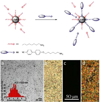
I. 5.3. Hybrid materials
In the past decades, the hybrid materials attract more and more attention since they could combine different properties from different compositions in the matrix. In our group, the main efforts had been made to design hybrid materials based on metal oxide nanoparticles. The organic part in those hybrid materials could be introduced either by direct coordination on the surface of the NPs as the stabilizing ligands or by a simple ligand exchange process. For example, long alkyl amine was evidenced to be a good stabilizing ligand for the ZnO NPs, so some other kinds of stabilizing ligands with amine group could be synthesized and used to play the same role, therefore interesting functional group with specific properties like liquid crystalline property could be introduced in those designed ligands and at last become a part of the well-tailored hybrid materials [97]. In our group, some works about ZnO NPs liquid crystalline hybrid
materials had been published [7, 95-101]. The first example [96] published is about
6OCBNH2/ZnO/OA hybrid material, which was prepared by a ligand exchange process
between an amine-containing mesogenic liquid crystalline, 6OCBNH2 and octylamine
(OA)-protected ZnO NPs (Figure I. 7).
Figure I. 7. Schematic diagram of the proposed ligand exchange mechanism occurring
in solution. TEM micrograph (a) and POM images of the hybrid (b) at 80 °C, (c) 140 °C and (d) cooling down to 84 °C [98].
Chapter Ⅰ Literature Review and Project Objectives
24
The prepared hybrid material present mesomorphic behavior even for a high content of inorganic material. In combination with promising luminescent properties, it may be exploited for electro-optical applications.
The liquid crystal macromolecules could also be directly used as the stabilizing agents for the synthesis of ZnO NPs in solution [100-101]. Interactions between [Zn(Cy)2]
and the branched structure (through amine groups) were evidenced both by experiments in solution and by solid state 13C-NMR studies in the LC state. Under isotropic
conditions, hydrolysis of the [Zn(Cy)2] precursor led to isotropic NPs. More precisely,
the pure branched LC and hyperbranched LC are isotropic at 30 °C and 45 °C, respectively. When the experiments were performed in the nematic phase state of the LC compound (i.e. at lower temperatures), anisotropic ZnO structures, were obtained. As shown in Figure I. 8, either nano-worm-like or nano-wires structures were grown in branched LC and hyperbranched LC. They have an average width of 2.5(0.2) nm and 2.7(0.4) nm, respectively. Polydispersed lengths vary from a few nanometers up to around 100 nm when the branched LC was used, while lengths from around 10 to 200 nm were obtained when the hyperbranched LC was used. Thus, a direct correlation between the structural characteristics of the LC and the morphology of the nanostructures, was demonstrated. The new LC/NPs composites present, at the same time, LC properties and optical properties originating from ZnO [98].
Figure I. 8. TEM micrographs of ZnO NPs synthesized under various conditions: (A)
in branched LC at 30°C (scale: 200 nm) or at 5°C (scale: 50 nm) (B) in hyperbranched LC at 45°C (scale: 50 nm) or at 5°C (scale: 50 nm) [98].
Chapter Ⅰ Literature Review and Project Objectives
25
All of the above synthesizes had been done in the presence of solvents, and we recently found that a kind of ZnO NPs/liquid crystal hybrid materials could also be prepared in the absence of solvents. A mixture of hexadecylamine (HDA) and [ZnCy2]
(in a ratio of 2 equivalents of amine per zinc precursor) was chosen without additional solvent, whereas both are solids at room temperature, admixture of [ZnCy2] and HDA
(in the 1:2 molar ratio) under an inert atmosphere rapidly leads to a liquid phase due to an acid-base reaction. After hydrolysis the mixture for 3 days, ZnO NPs hybrid materials with liquid crystal and luminescent properties could be obtained. More interestingly, the reaction temperature could strongly affect the anisotropy of the resulted NPs in the hybrid matrix [107].
I. 6. Project objectives
The initial objective of this project is to control and understand the growth process of ZnO NPs prepared through the simple organometallic pathway developed in our group, especially the anisotropic growth in the absence of any solvent. To reach this goal, an interesting 2-D plot analysis was developed. The formation of gels was evidenced and link between the gelification process and the size and shape of the NPs highligthened. Finally, new strategy toward processable and reshapable metal oxide hybrid materials was developped paving the way to a new generation of sensors.
Chapter Ⅰ Literature Review and Project Objectives
26
References
[1]. A. Henglein, Chem. Rev. 1989, 89, 1861–1873. [2]. S. Suresh, Nanosci. Nanotechno. 2013, 3, 62-74.
[3]. M. L. Steigerwald, L. E. Brus, Acc. Chem. Res. 1990, 23, 183-188.
[4]. M. G. Bawendi, M. L. Steigerwald, L. E. Brus, Annu. Rev. Phy. Chem. 1990, 41, 477-496.
[5]. A. P. Alivisatos, J. Phys. Chem. 1996, 100, 13226-13239. [6]. J. A. Stroscio, D. M. Eigler, Science 1991, 254, 1319-1326. [7]. S. Saliba, Université de Toulouse 2011.
[8]. M. Fernández-García, J. A. Rodriguez, Metal oxide nanoparticles,
Encyclopedia of Inorganic and Bioinorganic Chemistry 2011.
[9]. H. C. Ong, T. G. Du, J. Cryst. Growth 2004, 265, 471-175. [10]. R. B. Lauer, J. Phys. Chem. Solids 1973, 34, 249-253.
[11]. Z. Feng, R. Jia, B. Dou, H. Li, Z. Jin, X. Liu, F. Li, W. Zhang, C. Wu, Sol.
Energy 2015, 115, 770-776.
[12]. S. Pearton, F. Ren, Int. Mater. Rev. 2014, 59, 61-83.
[13]. M.C. Carotta, A. Cervi, V. di Natale, S. Gherardi, A. Giberti, V. Guidi, D. Puzzovio, B. Vendemiati, G. Martinelli, M. Sacerdoti, D. Calestani, A. Zappettini, M. Zha, L. Zanotti, Sens. Actuators B 2009, 137, 164-169.
[14]. Y. Zhang, J. Xu, Q. Xiang, H. Li, Q. Pan, P. Xu, J. Phys. Chem. C 2009, 113, 3430-3435.
[15]. J. Xu, Q. Pan, Y. Shun, Z. Tian, Sens. Actuators B 2000, 66, 277-279.
[16]. S. C. Minne, S. R. Manalis, and C. F. Quate, Appl. Phys. Lett. 1995, 67, 3918. [17]. X, Wen, Y. Fang, Q. Pang, C. Yang, J. Wang, W. Ge, K. S. Wong, S. Yang, J.
Phys. Chem. B 2005, 109, 15303–15308.
[18]. T. Shibata, K. Unno, E. Makino, Y. Ito, S. Shimada, Sens. Actuators A 2002, 102, 106-113.
Chapter Ⅰ Literature Review and Project Objectives
27 21, 433-447.
[20]. J. Zhou, N. S. Xu, Z. L. Wang, Adv. Mater. 2006, 18, 2432-2335.
[21]. H. Xiong, Y. Xu, Q. Ren, Y. Xia, J. Am. Chem. Soc. 2008, 130, 7522–7523. [22]. A. B. Djurisic, Y. H. Leung, Small 2006, 2, 944-961.
[23]. Y. Idota, T. Kubota, A. Matsufuji, Y. Maekawa, T. Miyasaka, Science 1997, 276, 1395-1397.
[24]. G. Hodes, L. Thompson, J. DuBow, K. Rajeshwar, J. Am. Chem. Soc. 1983, 105, 324–330.
[25]. A. Kolmakov, Y. Zhang, G. Cheng, M. Moskovts, Adv. Mater. 2003, 15, 997-1000.
[26]. W. Du, Q. Wang, D. Saxner, N. A. Deskins, D. Su, J. E. Krzanowski, A. I. Frenkel, X. Teng, J. Am. Chem. Soc. 2011, 133, 15172–15183.
[27]. G. Wang, W. Lu, J. Li, J. Choi, Y. Jeong, S.Y. Choi, J.B. Park, M. K. Ryu, K. Lee, Small 2006, 2, 1436-1439.
[28]. R. Demir-Cakan, Y. Hu, M. Antonietti, J. Maier, M. Titirici, Chem. Mater.
2008, 20, 1227.
[29]. Z. Wang, Z. Pan, Adv. Mater. 2002, 14, 1029.
[30]. H. Yang, J. Qian, Z. Chen, X. Ai, Y. Cao, J. Phys. Chem. C. 2007, 111, 14067. [31]. J. Liu, Y. Wan, F. Meng, X. Huang, J. Liu, J. Mater. Chem. 2012, 22, 2885. [32]. A. Chen, X. Penng, K. Koczkur, B. Miller, Chem. Commun. 2004, 1964. [33]. S. Sun, G. Meng, Y. Wang, T. Gao, M. Zhang, Y. Tian, X. Peng, L. Zhang,
Appl. Phys. A, 2003, 76, 287.
[34]. D. Mohanta, M. Ahmaruzzaman, RSC Adv. 2016, 6, 110996-111015. [35]. W. Wu, Q. He, C. Jiang, Nanoscale Res. Lett. 2008, 3, 397.
[36]. N. Lee, T. Hyeon, Chem. Soc. Rev. 2012, 41, 2575.
[37]. W. Wu, Z. Wu, T. Yu, C. Jiang, W. S. Kim, Sci. Technol. Adv. Mater. 2015, 16, 023501.
[38]. K. S. Suslick, S. B. Choe, A. A. Cichowlas, and M. W. Geenstaff, Nature, 1991, 353, 414.
Chapter Ⅰ Literature Review and Project Objectives
28
[40]. M. Rajamathi, R. Seshadri, Curr. Opin. Solid State Mater. Sci. 2002, 6, 337-345.
[41]. N. R. Jana, L. Gearheart, C. J. Murphy. Adv. Mater. 2001, 13, 1389.
[42]. M. A. Correa-Duarte, N. Sobal, L. M. Liz-Marzán, M. Giersig, Adv. Mater.
2004, 16, 2179-2184.
[43]. T. A. Crowley, K. J. Ziegler, D. M. Lyons, D. Erts, H. Olin, M. A. Morris, J. D. Holmes, Chem. Mater. 2003, 15, 3518-2522.
[44]. Y. Yin, Y. Lu, B. Gates, Y. Xia, J. Am. Chem. Soc. 2001, 123, 8718–8729. [45]. J. Ohring, ‘The Material Science of Thin Films’, Academic Press, San Diego,
1992.
[46]. M. Fernández-García, J. A. Rodriguez, 2011. Metal Oxide Nanoparticles.
Encyclopedia of Inorganic and Bioinorganic Chemistry.
[47]. L. V. Interrante and M. J. Hampen-Smith, ‘Chemistry of Advanced Materials: An Overview’, While-VCH, New York, 1998.
[48]. A. E. Danks, S. R. Hall, and Z. Schnepp, Mater. Horizon. 2016, 3, 91-112. [49]. M. Niederberger, Acc. Chem. Res. 2007, 40, 793-800.
[50]. J. Tang, F. Redl, Y. Zhu, T. Siegrist, L. E. Brus, and M. L. Steigerwald, Nano
Lett. 2005, 5(3), 543-548.
[51]. A. Grala, M. Wolska-Pietkiewicz, W. Danowski, Z. Wróbel, J. Grzonka, and J. Lewiński, Chem. Commun. 2016, 52, 7340.
[52]. M. V. Pavliuk, A. M. Cieślak, M. Abdellah, A. Budinská, S. Pullen, K. Sokolowski, D. L. A. Fernandes, J. Szlachetko, E. L. Bastos, S. Ott, L. Hammarström, T. Edvinsson, J. Lewiński, and J. Sá, Sustainable Energy Fuels
2017, 1, 69-73.
[53]. P. Krupiński, A. Kornowicz, K. Sokolowski, A. M. Cieślak, and J. Lewiński,
Chem. Eur. J. 2016, 22, 7817-7823.
[54]. J. Paczesny, M. Wolska-Pietkiewicz, I. Binkiewicz, Z. Wróbel, M. Wadowska, K. Matuła, I. Dzięcielewski, D. Pociecha, J. S. Koziorowska, J. Lewiński, and R. Hołyst, Chem. Eur. J. 2015, 21, 16941-16947.
Chapter Ⅰ Literature Review and Project Objectives
29
K. Matuła, W. Nogala, J. Lewiński, and R. Hołyst, ACS Appl. Mater. Interfaces
2016, 8, 13532-13541.
[56]. K. Sokołowski, I. Justyniak, W. Bury, J. Grzonka, Z. Kaszkur, Ł. Mąkolski, M. Dutkiewicz, A. Lewalska, E. Krajewska, D. Kubicki, K. Wójcik, K. J. Kurzydłowski, and J. Lewiński, Chem. Eur. J. 2015, 21, 5488-5495.
[57]. A. M. Cieślak, M.V. Pavliuk, L. D'Amario, M. Abdellah, K. Sokołowski, U. Rybinska, D.L.A. Fernandes, M.K. Leszczyński, F. Mamedov, A.M. El-Zhory, J. Föhlinger, A. Budinská, M. Wolska-Pietkiewicz, L. Hammarström, J. Lewiński, J. Sá, Nano Energy 2016, 30, 187.
[58]. D. Prochowicz, K. Sokołowski, J. Lewiński, Coordin. Chem. Rev. 2014, 270-271, 112-126.
[59]. S. Polarz, A. Orlov, A. Hoffmann, M. R. Wagner, C. Rauch, R. Kirste, W. Gehlhoff, Y. Aksu, M. Driess, M. W. E. van den Berg, M. Lehmann, Chem. Mater.
2009, 21, 3889.
[60]. D. Lehr, M. Luka, M. R. Wagner, M. Buelger, A. Hoffmann, S. Polarz, Chem.
Mater. 2012, 24, 1771– 1778.
[61]. C. Lizandara-Pueyo, M. C. Morant-Minana, M. Wessig, M. Krumm, S. Mecking and S. Polarz, RSC Adv. 2012, 2, 5298–5306.
[62]. M. A. Dreher, M. Krumm, C. Lizandara-Pueyo, and S. Polarz, Dalton Trans.
2010, 39, 2232-2238.
[63]. S. Dilger, C. Lizandara-Pueyo, M. Krumm, and S. Polarz, Adv. Mater. 2012, 24, 543–548.
[64]. M. Gerigk, J. Bahner, T. Kollek, S. Helfrich, R. Rosenberg, H. Cölfen, S. Polarz, Part. Part. Syst. Charact. 2017, 34, 1600215.
[65]. K. Hagedorn, W. Li, Q. Liang, S. Dilger, M. Noebels, M. R. Wagner, J. S. Reparaz, A. Dollinger, J. Schmedt auf der Günne, T. Dekorsy, L. Schmidt-Mende and S. Polarz, Adv. Funct. Mater. 2016, 26, 3424–3437.
[66]. V. Ischenko1, S. Polarz1, D. Grote, V. Stavarache, K. Fink, and M. Driess, Adv.
Funct. Mater. 2015, 15, 1945–1954.
Chapter Ⅰ Literature Review and Project Objectives
30
S. Polarz, CrystEngComm, 2014, 16, 1525-1531.
[68]. C. Lizandara-Pueyo, S. Siroky, M. R. Wagner, A. Hoffmann, J. S. Reparaz, M. Lehmann, S. Polarz1, Adv. Funct. Mater. 2011, 21, 295–304.
[69]. S. Polarz , F. Neues , M. W. E. van den Berg , W. Grünert , and L. Khodeir, J.
Am. Chem. Soc. 2005, 127, 12028–12034.
[70]. C. L. Pueyo, S. Siroky, S. Landsmann, M. W. E. van den Berg, M. R. Wagner, J. S. Reparaz, A. Hoffmann, and S. Polarz, Chem. Mater. 2010, 22, 4263–4270. [71]. M. Krumm, C. L. Pueyo, and S. Polarz, Chem. Mater., 2010, 22, 5129–5136. [72]. S. Dilger, M. Wessig, M. R. Wagner, J. S. Reparaz, C. M. Sotomayor Torres,
L. Qijun, T. Dekorsy, and S. Polarz, Cryst. Growth Des. 2014, 14, 4593–4601. [73]. M. Gerigk, P. Ehrenreich, M. R. Wagner, I. Wimmer, J. S. Reparaz, C. M.
Sotomayor Torres, L. Schmidt-Mende, and S. Polarz, Nanoscale, 2015, 7, 16969-16982.
[74]. C. Lizandara-Pueyo, M. W. E. van den Berg, A. De Toni, T. Goes, S. Polarz, J.
Am. Chem. Soc. 2008, 130, 16601–16610.
[75]. S. Polarz, A. V. Orlov, M. W. E. van den Berg, and M. Driess, Angew. Chem.
Int. Ed. 2005, 44, 7892–7896.
[76]. S. Polarz, A. Roy, M. Merz, S. Halm, D. Schröder, L. Schneider, G. Bacher, F. E. Kruis, and M. Driess, Small 2005, 1, 540–552.
[77]. S. Polarz, J. Strunk, V. Ischenko, M. W. E. van den Berg, O. Hinrichsen, M. Muhler, and M. Driess, Angew. Chem. Int. Ed. 2006, 45, 2965–2969.
[78]. S. Polarz, A. Roy, M. Lehmann, M. Driess, F. E. Kruis, A. Hoffmann, and P. Zimmer, Adv. Funct. Mater. 2007, 17, 1385–1391.
[79]. S. Polarz, R. Regenspurger, and J. Hartmann, Angew. Chem. Int. Ed. 2007, 46, 2426–2430.
[80]. S. Polarz, Andrey V. Orlov, F. Schüth and A.-H. Lu, Chem. Eur. J. 2007, 13, 592–597.
[81]. M. Krumm, F. Pawlitzek, J. Weickert, L. Schmidt-Mende, and S. Polarz, ACS
Appl. Mater. Interfaces, 2012, 4, 6522–6529.
Chapter Ⅰ Literature Review and Project Objectives
31
[83]. M. Voggenreiter, P. Vöpel, B. Smarsly, S. Polarz, Z. Anorg. Allg. Chem. 2017, 643, 93-100.
[84]. S. D. Pike, E. R. White, M. S. P. Shaffer, and C. K. Williams, Nat. Commun.
2016, 7, 13008.
[85]. M. L. Kahn, A. Glaria, C. Pages, M. Monge, L. S. Macary, A. Maisonnat and B. Chaudret, J. Mater. Chem. 2009, 19, 4044-4060.
[86]. F. Rataboul, C. Nayral, M. J. Casanove, A. Maisonnat, B. Chaudret, J.
Organomet. Chem. 2002, 643, 307.
[87]. M. Monge, M. L. Kahn, A. Maisonnat and B. Chaudret, Angew. Chem. Int. Ed.
2003, 42, 5321–5324.
[88]. A. Glaria, M. L. Kahn, P. Lecante, B. Barbara and B. Chaudret,
ChemPhysChem 2008, 9, 776–780.
[89]. M. L. Kahn, T. Cardinal, B. Bousquet, M. Monge, V. Jubera and B. Chaudret,
ChemPhysChem 2006, 7, 2392–2397.
[90]. J. Rubio-Garcia, A. Dazzazi, Y. Coppel, P. Mascalchi, L. Salomé, A. Bouhaouss, M. L. Kahn and F. Gauffre, J. Mater. Chem. 2012, 22, 14538–14545. [91]. M. L. Kahn, M. Monge, V. Collière, F. Senocq, A. Maisonnat, and B. Chaudret,
Adv. Funct. Mater. 2005, 15, 458–468.
[92]. M. L. Kahn, M. Monge, E. Snoeck, A. Maisonnat, and B. Chaudret, Small
2005, 1, 221–224.
[93]. Y. Champouret, Y. Coppel, and M. L. Kahn, J. Am. Chem. Soc. 2016, 138, 16322-16328.
[94]. Y. Coppel, G. Spataro, C. Pagès, B. Chaudret, A. Maisonnat, and M. L. Kahn,
Chem. Eur. J. 2012, 18, 5384 – 5393.
[95]. S. Saliba, C. V. Serrano, J. Keilitz, M. L. Kahn, C. Mingotaud, R. Haag, and Jean-Daniel Marty, Chem. Mater. 2010, 22, 6301–6309.
[96]. S. Saliba, P. Davidson, M. Impéror-Clercd C. Mingotaud, M. L. Kahn and Jean-Daniel Marty, J. Mater. Chem. 2011, 21, 18191.
[97]. S. Saliba, Y. Coppel, P. Davidson, C. Mingotaud, B. Chaudret, M. L. Kahn and Jean-Daniel Marty, J. Mater. Chem. 2011, 21, 6821.
Chapter Ⅰ Literature Review and Project Objectives
32
[98]. S. Saliba, C. Mingotaud, M. L. Kahn and Jean-Daniel Marty, Nanoscale 2013, 5, 6641.
[99]. J. Jońca, J. Harmel, L. Joanny, A. Ryzhikov, M. L. Kahn, P. Fau, B. Chaudret, K. Fajerwerg, Sens. Actuators B 2017, 249, 357-363.
[100]. S. Saliba, Y. Coppel, Marie-France Achard, C. Mingotaud, Jean-Daniel Marty, and M. L. Kahn, Angew. Chem. 2011, 123, 12238 –12241.
[101]. S. Saliba, Y. Coppel, C. Mingotaud, Jean-Daniel Marty, and M. L. Kahn,
Chem. Eur. J. 2012, 18, 8084 – 8091.
[102]. Z. Zhao, Z. Zheng, C. Roux, C. Delmas, Jean-Daniel Marty, M. L. Kahn, and Christophe Mingotaud, Chem. Eur. J. 2016, 22, 12424 – 12429.
[103]. G. Spataro, A. Dazzazi, S. Fortuny, Y. Champouret, Y. Coppel, J. Rubio-Garcia, A. Bouhaouss, F. Gauffre, and M. L. Kahn, Eur. J. Inorg. Chem. 2016, 2056–2062.
[104]. A. Glaria, M. L. Kahn, T. Cardinal, F. Senocq, V. Juberab, and B. Chaudret,
New J. Chem. 2008, 32, 662-669.
[105]. A. Dazzazi, Y. Coppel, M. In, C. Chassenieux, P. Mascalchi, L. Salomé, A. Bouhaouss, M. L. Kahn and F. Gauffre, J. Mater. Chem. C 2013, 1, 2158– 2165.
[106]. J. Rubio-Garcia, Y. Coppel, P. Lecante, C. Mingotaud, B. Chaudret, F. Gauffre and M. L. Kahn, Chem. Commun. 2011, 47, 988–990.
[107]. Z. Zheng, R. Butynska, C. V. Serrano, Jean-Daniel Marty, C. Mingotaud, and M. L. Kahn, Chem. Eur. J. 2016, 22, 15614 – 15618.
[108]. M. L. Kahn, T. Cardinal, B. Bousquet, M. Monge, V. Jubera, and B. Chaudret, ChemPhysChem 2006, 7, 2392 – 2397.
[109]. C. Amiens, B. Chaudret, D. Ciuculescu-Pradines, V. Collière, K. Fajerwerg, P. Fau, M. L. Kahn, A. Maisonnat, K. Soulanticac and K. Philippot,
New J. Chem. 2013, 37, 3374-3401.
[110]. A. Ryzhikov, J. Jońca, M. L. Kahn, K. Fajerwerg, B. Chaudret, A. Chapelle, P. Ménini, C. H. Shim, A. Gaudon, P. Fau, J. Nanopart. Res. 2015, 17, 280.
Chapter Ⅰ Literature Review and Project Objectives
33
[111]. J. Carrey1, H. Carrère, M. L. Kahn, B. Chaudret, X. Marie and M. Respaud, Semicond. Sci. Technol. 2008, 23, 025003.
[112]. J. Massaad, Y. Coppel, M. Sliwa, M. L. Kahn, C. Coudret and F. Gauffre,
Phys. Chem. Chem. Phys. 2014, 16, 22775.
[113]. F. Güell, P. R. Martínez-Alanis, S. Khachadorian, J. Rubio-García, A. Franke, A. Hoffmann, and G. Santana, Phys. Status Solidi B 2016, 253 (5), 883– 888.
[114]. C. Pagès, Y. Coppel, M. L. Kahn, André Maisonnat, and B. Chaudret,
ChemPhysChem 2009, 10, 2334 – 2344.
[115]. Y. Coppel, G. Spataro, V. Collière, B. Chaudret, C. Mingotaud, A. Maisonnat, and M. L. Kahn, Eur. J. Inorg. Chem. 2012, 2691–2699.
[116]. L. S. Macary, M. L. Kahn, C. Estournès, P. Fau, D. Trémouilles, M. Bafleur, P. Renaud, and B. Chaudret, Adv. Funct. Mater. 2009, 19, 1775–1783. [117]. J. Rubio-Garcia, A. Dazzazi, Y. Coppel, P. Mascalchi, L. Salomé, A.
Bouhaouss, M. L. Kahn and F. Gauffre, J. Mater. Chem. 2012, 22, 14538–14545. [118]. C. Nayral, T. Ould-Ely, A. Maisonnat, B. Chaudret, P. Fau, L. Lescouzères,
and A. Peyre-Lavigne, Adv. Mater. 1999, 11, 61-63.
[119]. C. Nayral, E. Viala, V. Collière, P. Fau, F. Senocq, A. Maisonnat, B. Chaudret, Appl. Surf. Sci. 2000, 164, 219-226.
[120]. P. Fau, M. Sauvan, S. Trautweiler, C. Nayral, L. Erades, A. Maisonnat, B. Chaudret, Sens. Actuators B 2001, 78, 1-3, 83-88.
[121]. J.M. Ducéré, A. Hemeryck, A. Estève, M. D. Rouhani, G. Landa, P. Ménini, C. Tropis, A. Maisonnat, P. Fau, B. Chaudret, J. Comput. Chem 2011, 33, 247–258.
[122]. C. Nayral, E. Viala, P. Fau, F. Senocq, J.-C. Jumas, A. Maisonnat, and B. Chaudret, Chem. Eur. J. 2000, 6, 4082–4090.
[123]. J. Jońca, A. Ryzhikov, M. L. Kahn, K. Fajerwerg, A. Chapelle, P. Menini, P. Fau, Chem. Eur. J. 2016, 22, 10127.
[124]. G. Casterou, V. Collière, P. Lecante, Y. Coppel, P.-A.Eliat, F. Gauffre, and M. L. Kahn, Chem. Eur. J. 2015, 21, 18855–18861.
Chapter Ⅰ Literature Review and Project Objectives
34
[125]. A. Glaria, M. L. Kahn, P. Lecante, B. Barbara, and B. Chaudret,
ChemPhysChem 2008, 9, 776–780.
[126]. A. Glaria, M. L. Kahn, A. Falqui, P. Lecante, V. Collière, M. Respaud, and B. Chaudret, ChemPhysChem 2008, 9, 2035–2041.
[127]. A. Glaria, M. L. Kahn, B. Chaudret, P. Lecante, M.-J. Casanove, B. Barbara,
Chapitre II
__________________
Analyse statistique d’objets anisotropes
Résumé
L'analyse de la taille des nanoparticules à travers une simple représentation 2D est proposée afin d'extraire la corrélation entre la longueur et la largeur d'une collection ou d’un mélange de particules anisotropes. Par rapport aux statistiques habituelles portant sur une analyse statistique de la longueur et indépendamment de la largeur, cette approche simple montre facilement les différents types de nanoparticules et leur anisotropie. Pour chaque classe de nano-objets, la relation entre la largeur et la longueur (c'est-à-dire les corrélations fortes ou faibles entre ces deux paramètres) peut suggérer des informations concernant les processus de croissance et de nucléation. Cette approche permet de suivre l'effet sur la forme et sur la répartition en taille des processus physiques et/ou chimiques tels que la maturation. Diverses images de microscopie électronique à transmission provenant de la bibliographie ou de nos propres travaux sont utilisées à titre d'exemple pour démontrer l'efficacité et la simplicité d'une telle analyse.
Contenu
Introduction ... 43 II.1. Principe de l’analyse multivariée ... 44 II.2. Applications de l’analysis par représentation 2D ... 49 Conclusion ... 57 Références ... 58
Chapter II Shape analysis of anisotropic nanoparticles
43
Introduction
Whichever the chemical composition and the method used to synthetize the NPs may be, full characterization of the size and shape of the nano-objects should be achieved. Based on microscopy techniques such as transmission electron microscopy (TEM), atomic force microscopy…, images of the NPs are nowadays easily obtained and can be fully analyzed using popular software such as Image J. [1]
When the nanoparticles are anisotropic and have, for example, the shape of nanorods, the analysis of their size is often simply done by calculating an average value of their diameters and a distribution in their lengths. The main hypothesis made when performing such analysis is that width and length of the nanoparticles are totally un-correlated. Recently, some papers have shown that some correlations do exist between the minor axis and major axis in gold nanorods. [2] Such results limit the validity of the
"usual" analysis performed on anisotropic particles.
Therefore, in this chapter, we propose a new and simple way to study the sizes of the NPs by creating a 2D size plot. This tool allows one to obtain qualitative and quantitative information concerning the length-width correlation. TEM pictures have been extracted from papers in the literature, purely selected on the quality of the TEM images and not on the composition of the NPs or the process for their synthesis. Other examples of TEM images have been chosen within the various studies on NPs performed in our teams. 2D size analyses have then been made and highlight that indeed more information can be gained from such a procedure than the "usual" statistical distribution of widths and lengths.
Chapter II Shape analysis of anisotropic nanoparticles
44
II.1. Principle and multivariate analysis
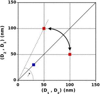
Following the example of 2D NMR with two-dimensional spectra pointing out the coupling between nuclei, [3] we propose to analyze a given set of NPs using a 2D plot
to extract information on the correlation between widths and lengths of anisotropic objects. Each particle is characterized by two sizes, noted D1 and D2, measured on
perpendicular axes. These two sizes generally correspond to the length and width of the nano-objects. For each particle and on a same graph, we plot D1 as a function of D2 and
also D2 as a function of D1 (see Figure II.1).
Figure II. 1. Schematic 2D size plot for two nanoparticles: an isotropic one (blue square)
with a size (D1 = D2) around 40 nm; one nanorod with a length of 100 nm (D1) and a
width of 50 nm (D2) associated to two points (red squares) on either side of the median
(see double arrow).
In such a plot, any isotropic nanoparticle (case D1 = D2) will correspond to one
diagonal point, lying on the median of the axes (see Figure II.1). On the contrary, anisotropic particles will be associated to two points (one for x = D1 and y = D2 and the
second one for x = D2 and y = D1), lying symmetrically on both sides of the median. In
fact, the aspect ratio, AR, of the particle (i.e. the ratio between its length and its width)
0 50 100 150 0 50 100 150 (D 2 , D 1 ) (nm ) (D 1 , D2) (nm) q
Chapter II Shape analysis of anisotropic nanoparticles
45
is equal to the tangent of angle q (see Figure II.1) defined by the x-axis and the line going through the origin of the graph and the upper point associated to the particle. q close to 90° corresponds to large aspect ratio while q close to 45° means aspect ratio near unity. Using this 2D size graph, it is therefore straightforward to distinguish groups of particles depending on their anisotropy.
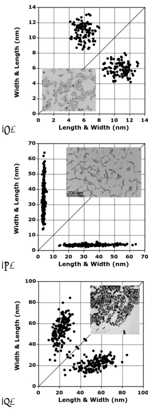
Figure II.2 reports such a 2D plot for a real mixture of nanoparticles: points in the red ellipse are associated to nanorods (average AR = 10.9 ± 2.5 corresponding to an average qangle of ca 85°) whereas the ones in the blue circle are related to smaller nanoparticles which could be considered isotropic (average AR = 1.25 ± 0.04 corresponding to an average qangle close to 51°). For this and the following image
analysis, Image J software was used manually or with a macro in order to extract width and length of nano-objects in various micrographs. The macro is based on the PSA macro, available at http://code.google.com/p/psa-macro. Overlapping nanoparticles are automatically rejected for the statistical analysis. The results were adjusted to account for the overestimation of particle size when fitting square or rectangular shapes with ellipses, according to Igathinathane et al. [4] The software leads to a table listing for
each particle of two perpendicular dimensions D1 and D2. No ordering of these values
is needed since this table is doubled by listing D2 as a function of D1. Plots of D1 as a
function of D2 and D2 as a function of D1 are combined in the 2D size plot analyzed in
Chapter II Shape analysis of anisotropic nanoparticles
46
Figure II.2. 2D size plot of the TEM picture (bar length: 100 nm) of Co nanoparticles
and nanorods.[5] The synthesis of this mixture was based on the procedure described in
reference 6.
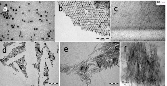
Besides the obvious advantage of the 2D plot to suggest various groups of NPs, characterizing the correlation between width and length of samples mainly constituted by nanorods could be highly enlightening. As an example, 2D size plots of various nanorod systems are given in Figure II.3. In Figure II.3A, corresponding to CdSe/CdS anisotropic NPs, length and width are both constant and present a polydispersity somewhat equivalent for both axes: the point cloud has a somewhat round shape. The average aspect ratio AR is close to 1.9 ± 0.3 corresponding to a qangle of 62°. On the contrary, Figure II.3B corresponds to a distribution of size of ZnO NPs where the width is more or less constant (ca 10-11 nm) and the length of the nanorods is totally independent on the width and is spread over the 15-65 nm range. The average aspect ratio AR is close to 9.4 ± 2.9 corresponding to a qangle close to 84°, demonstrating the large average anisotropy of the NPs. This case corresponds therefore to a total de-correlation between the two axes of the particles.
Chapter II Shape analysis of anisotropic nanoparticles
47 (A)
(B)
(C)
Figure II.3. 2D size plots of the TEM pictures: (A) bullet shaped CdSe/CdS
nanoparticles. The micrograph was extracted from Figure 5a in reference 7; (B). ZnO nanorods. The synthesis of these NPs is given in the main part of this chapter; (C). Au nanorods. The micrograph was extracted from Figure 8 in reference 8.
0 2 4 6 8 10 12 14 0 2 4 6 8 10 12 14 Wi dth & Le n g th ( n m )
Length & Width (nm)
0 10 20 30 40 50 60 70 0 10 20 30 40 50 60 70 Wi dth & Le n g th ( n m )
Length & Width (nm)
0 20 40 60 80 100 0 20 40 60 80 100 Wi dth & Le n g th ( n m )
Chapter II Shape analysis of anisotropic nanoparticles
48
Finally, the Figure II.3C shows an average variation of the width with the length, a partial correlation which can roughly be described by a linear dependency of length on width. Indeed, the data points from the clouds far from the diagonal domain suggest the relationship:
Width (nm) = 7.6 + 0.2574* length (nm).
Figure II.3 demonstrates therefore that the distribution in (width, length) and thus the correlation between both parameters could be strongly different from one sample to the other. In order to get a more quantitative analysis of these 2D plots, extraction of statistical parameters like average length and width and corresponding standard deviations should be performed. Nevertheless, this analysis is hampered by the presence of different sub-populations (such as the two in the red and blue ellipses in Figure II.2) that could necessitate tedious procedures to separate them, especially when these populations overlap each other.
In order to numerically identify the previous different sub-populations and to obtain their own statistical parameters, a multivariate analysis has been performed. This was done on the assumption that those clouds are issued from Gaussian probability densities and give us information about the average characteristics of those sub-populations (number, orientation, average length and width, corresponding standard deviation and correlation). Additionally, when a limited number of NPs can be counted, such an approach can also lead to improved accuracy for statistical analysis by helping to discard minor sub-population that are not representative of the samples. The multivariate analysis has been performed with the mixmod software (http://mixmod.org) using R package (http://www.r-project.org/). Apart from the Gaussian character of the probability densities, no assumption about the orientation, shape and volume of the different sub-populations was made during calculation. The number of sub-populations that composed the point clouds was fixed by the user or chosen numerically. Each sub-population was then characterized by the mean of the two studied variables (i.e. short and long axis lengths) as well as the corresponding standard deviations. Additionally, the correlation parameter between both variables was calculated. The correlation is
Chapter II Shape analysis of anisotropic nanoparticles
49
equal to zero when the two variables are totally independent and equal to 1 when they are affinely related to each other.
Applying such a procedure and as expected from the visual aspect of the plots, the NPs of Figure II.3A and II.3B show no statistical correlation between their length and width: the correlation parameter is found close to zero (respectively 0.05 and 0.09). However, in Figure II.3C, the expected partial correlation is confirmed by the value of the correlation parameter close to 0.33. Therefore, qualitatively and quantitatively, the correlation between length and width may strongly differ from one type of NPs to another. Importantly, this correlation may be a footprint of the nucleation-growth process occurring during the nanoparticle synthesis or of physical or chemical procedures used after synthesis. That is why 2D plots are of special interest to describe the modifications of structures occurring through time during growing or aging process.
II.2. Applications of the 2D plot analysis
The next examples will illustrate this point with the shape analysis of ZnO nanoparticles which were prepared through the following procedure: in the glove box, 92.68 mg of dodecylamine (DDA, 2 equivalents) was added in a small vial to 57.9 mg of zinc precursor [Zn(C6H11)2]. After complete mixing, the vial was taken out of the
glove box and exposed to the air. After various reaction times, part of the sample was analyzed by TEM. Samples for TEM studies were prepared by slow evaporation of droplets of colloidal solution deposited on carbon-coated copper grids.
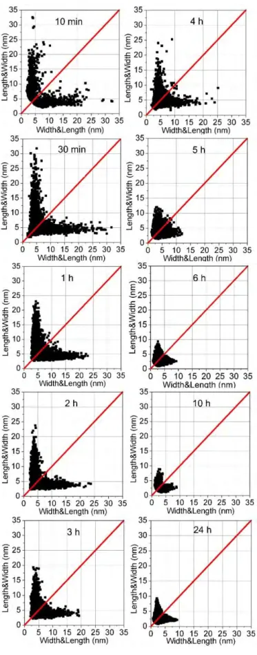
Figure II.4 shows the TEM pictures and corresponding 2D plots of ZnO NPs obtained by hydrolysis of an organometallic precursor as a function of the reaction time (from 2 to 80 hours). The point clouds obtained at 2 and 4 h are centered on the median (q = 45°). The 2D plots show clearly that the point clouds became rounder at 4 h than they were at 2 h. This corresponds to the presence of quasi isotropic nanoparticles, the average size of which evolves from ca 3.4 ± 1.7 to 5.3 ± 2.7 nm. The growth of ZnO NPs clearly starts by a nucleation step where pristine isotropic nanoparticles are formed.