HAL Id: hal-03004153
https://hal-cnrs.archives-ouvertes.fr/hal-03004153
Submitted on 20 Nov 2020
HAL is a multi-disciplinary open access
archive for the deposit and dissemination of
sci-entific research documents, whether they are
pub-lished or not. The documents may come from
teaching and research institutions in France or
abroad, or from public or private research centers.
L’archive ouverte pluridisciplinaire HAL, est
destinée au dépôt et à la diffusion de documents
scientifiques de niveau recherche, publiés ou non,
émanant des établissements d’enseignement et de
recherche français ou étrangers, des laboratoires
publics ou privés.
The Membrane-associated Form of the DNA Repair
Protein Ku is Involved in Cell Adhesion to Fibronectin
Sylvie Monferran, Catherine Muller, Lionel Mourey, Philippe Frit, Bernard
Salles
To cite this version:
Sylvie Monferran, Catherine Muller, Lionel Mourey, Philippe Frit, Bernard Salles. The
Membrane-associated Form of the DNA Repair Protein Ku is Involved in Cell Adhesion to Fibronectin. Journal
of Molecular Biology, Elsevier, 2004, 337, pp.503 - 511. �10.1016/j.jmb.2004.01.057�. �hal-03004153�
C
OMMUNICATIONThe Membrane-associated Form of the DNA Repair
Protein Ku is Involved in Cell Adhesion to Fibronectin
Sylvie Monferran, Catherine Muller*, Lionel Mourey, Philippe Frit and
Bernard Salles
Institut de Pharmacologie et de Biologie Structurale, CNRS UMR 5089, 205 route de Narbonne, 31077 Toulouse Cedex, FranceThe Ku heterodimer (Ku70/Ku80) plays a central role in DNA double-strand breaks recognition and repair. However, Ku is expressed also on the surface of different types of cells along with its intracellular pool within the nucleus and the cytoplasm. Participation of membrane-associ-ated Ku in cell – cell interaction has been reported recently. Here, we describe a novel function of cell-surface Ku as an adhesion receptor for fibronectin (Fn). The role of Ku in cell adhesion was investigated by com-paring the Ku80 deficient Chinese hamster ovary (CHO) cell line, xrs-6, with clones transfected stably with either the hamster or human Ku80 cDNA. Ku expression in transfectant cells resulted in a significant increased adhesion on Fn and type IV collagen as compared to control cells. The observed increase in cell adhesion relied on Ku cell-surface expression, since antibodies directed against Ku70 or Ku80 subunit inhibited adhesion on Fn of Ku80, but not control vector, transfected xrs-6 cells. In addition, both Ku70 and Ku80 present a structural relation-ship with integrin I (or A) domains and the A1 and A3 domains of von Willebrand factor, domains known to be involved in Fn binding. Both Ku70 and Ku80 exhibit a complete set of residues compatible in their position and chemical nature with the formation of a metal ion-dependent adhesion (MIDAS) site implicated in ligand binding and integrin activation. Taken together, these functional and structural approaches support a new role for Ku as an adhesion receptor for Fn.
q2004 Elsevier Ltd. All rights reserved.
Keywords: Ku heterodimer; DNA repair; cell adhesion; fibronectin; von Willebrand A domains
*Corresponding author
Ku is a complex composed of two tightly associ-ated subunits called Ku70 and Ku80.1 Ku is the
DNA-binding component of the DNA-dependent kinase (DNA-PK) complex. Ku binds to DNA double strand breaks (DSBs), recruits and activates the large catalytic subunit of DNA-PK, a member of the phosphatidylinositol 3-kinase superfamily.2
As a major component of the non-homologous end-joining (NHEJ) pathway, the entire DNA-PK
complex is required for the repair of DSBs and the rejoining of the DNA ends specifically generated during V(D)J recombination.3 Ku was originally
reported to be a nuclear protein, consistent with its function in DNA DSBs repair. On the other hand, several studies have revealed the cyto-plasmic and the cell-surface localization of Ku proteins in a variety of tumor cells, including leukemia, multiple myeloma and solid tumor cell lines.4 – 9 Furthermore, a recent work demonstrates
the localization of the whole DNA-PK complex in membrane lipid rafts of mammalian cells.10These
recent results suggest that the extra-nuclear localiz-ation of DNA-PK complex is serving additional role besides its main function in DNA DSBs repair. Indeed, the participation of Ku70 and Ku80 in cell –cell interaction has been described recently.6,8,9 Both Ku80 and Ku70 are up-regulated
0022-2836/$ - see front matter q 2004 Elsevier Ltd. All rights reserved.
E-mail address of the corresponding author: [email protected]
Abbreviations used: Fn, fibronectin; CHO, Chinese hamster ovary; MIDAS, metal ion-dependent adhesion; DNA-PK, DNA-dependent kinase complex; DSB, double strand break; NHEJ, non-homologous end-joining; MM, multiple myeloma; ECM, extracellular matrix; CD40L, CD40 ligand; vWA, von Willebrand factor A.
on the membrane of CD40-activated multiple myeloma (MM) cells9or in a variety of tumor cells
under hypoxic conditions.6 In these cells,
homo-typic and heterohomo-typic adhesion can be blocked specifically by Ku antibodies, suggesting the ability of Ku to function as a cell adhesion molecule.
Partial results also suggest a role of Ku in con-trolling cell adhesion to the extracellular matrix (ECM).9 In fact, antibodies directed against Ku70
or Ku80 specifically inhibit adhesion (70 – 80% decrease) of CD40 ligand (CD40L)-treated, but not untreated, MM cells to fibronectin (Fn). However, CD40L stimulation increases adhesion to Fn only slightly as compared to unstimulated cells despite a significant increase of Ku cell-surface expression.9
Exposure of MM cells to CD40L produces various effects on cellular function and it remains difficult to identify cell responses that are specifically attributable to the expression of Ku at the cell membrane. To explore the role of Ku as a cell-surface receptor to ECM, we decided to use Ku-deficient and proficient cell lines. Rodent cells deficient in Ku are viable, whereas human cells are not.11 Ku-deficient Chinese hamster ovary
(CHO) cells were chosen as a model for our study, since this type of cell has been used extensively to study cell adhesion receptors (in particular integrins) on diverse ECM components, including Fn.12Thus, the role of Ku in cell adhesion on ECM
proteins was evaluated using the Ku80-deficient CHO cell line, xrs-6, in comparison to clones stably transfected with either the hamster (xrs-6/Ha Ku80) or human (xrs-6/Hu Ku80) cDNA.
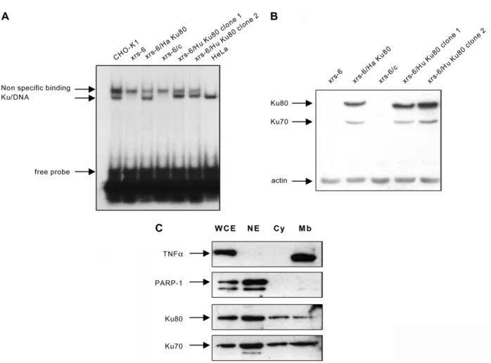
Biochemical characterization of the transfected cell lines
As described,13,14 xrs-6 cells showed a complete
lack of double-stranded DNA ends binding activity (Figure 1A). Full restoration to parental level (CHO-K1) was observed in xrs-6/Ha Ku80 as described,14 and in the two clones of xrs-6/Hu
Ku80 human examined. The mutant and com-plemented cell lines were examined by Western blotting using a polyclonal antibody Ku-154 that recognizes the human and hamster Ku80 protein. No material cross-reacting with antibody Ku-154 was detected in xrs-6 cells, and expression of Ku80 at similar levels was restored in the different transfected clones (Figure 1B). As described,13 – 16
xrs-6 cells show dramatically decreased levels of Ku70 protein, which was restored in the trans-fected cell lines. Since the Ku subunits stabilize each other, the absence of Ku80 from these cells has been shown to result also in loss of the Ku70 component.13 – 16 We then investigated the
sub-cellular distribution of both Ku70 and Ku80 subunits in Ku80 transfected xrs-6 cells using frac-tionated cell extracts. First, the different fractions were analyzed by immunoblotting for PARP-1 (a nuclear DNA-binding protein) and TNFa (a mem-brane-associated protein) in order to validate the quality of the subcellular fractionation. The results
obtained with xrs6/Ha Ku80 cells are shown in
Figure 1C. As expected, PARP-1 expression was
present in the nuclear fraction, while TNFa expression was highly predominant in the mem-brane fraction. The expression of Ku70 and Ku80 protein was observed in the three fractions with a higher level of expression in the nuclear fraction (Figure 1C). Similar results were obtained with the xrs-6/Hu Ku80 (data not shown). In these Ku80 xrs-6 transfected cells, Ku is present in the mem-brane fraction in addition to its intracellular pool within the nucleus and the cytoplasm, a sub-cellular distribution comparable to that observed in various human cell lines.17
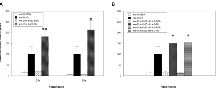
Cell surface-associated Ku supports adhesion of CHO cells on fibronectin
We then compared the adhesion of control vector and Ku80 transfected xrs-6 cells to immobilized ECM components, particularly Fn. We first deter-mined the concentration of substrate giving maxi-mal cell adhesion and the number of cells that gave a linear correlation between the cell number and the measured optical absorbance after the staining procedure (data not shown). Using these data (50 mg/ml of Fn, 5 £ 104 cells per 96 well
plate) the adhesion for both cell lines on the Fn substrate was assayed. As shown in Figure 2A, the xrs-6/Ha Ku80-competent cell line showed a significant increase in adhesion on Fn as compared to Ku80-deficient cells (182 ^ 27 relative to 100 ^ 35, p , 0:01), whereas no significant differ-ence between the adhesion of Ku-proficient and deficient cells on bovine serum albumin (BSA) was found (19 ^ 8 relative to 15 ^ 7). Increased cell adhesion on Fn was observed for the delayed adhesion response (six hours) as shown in
Figure 2A. A similar significant increase in cell adhesion to Fn, but to a lesser extent, was observed with the two clones transfected with the human Ku80 cDNA (150 ^ 30 (clone 1), 154 ^ 26 (clone 2) relative to 100 ^ 36) (Figure 2B). The observed differences between cells transfected with either hamster or human Ku80 cDNA might be due to alterations in the heterodimer conformation in the xrs-6/Hu Ku80 cells. Indeed, in these cells the two Ku proteins that form the heterodimer are from different species (human Ku80/hamster Ku70) and similar alterations in function have been described in the context of DNA repair.13 – 15Similar
experiments were performed using three other substrates, collagen I, collagen IV and laminin. As described, both control vector and Ku80 trans-fected xrs-6 cells CHO fibroblasts adhere on collagen IV.18As shown inTable 1, a statistical
sig-nificant difference was observed for hamster Ku80 cDNA transfected xrs-6 cells (p , 0:05 relative to control) whereas the increased adhesion of human Ku80 cDNA transfected cells to collagen IV was not significant. By contrast, no significant differences were observed between the adhesion of Ku-proficient and deficient cells on collagen I 504 Structural/Functional Homology of Ku with Integrins
and laminin, two poor adhesion substrates for CHO fibroblasts (seeTable 1).18
We then attempted to determine if the differ-ences in the adhesion between the Ku-deficient and proficient cells were due to Ku cell-surface expression. As a control, we first tested the cation chelator EGTA, known for its ability to inhibit integrins and therefore cell adhesion to the ECM component. As expected (Figure 3A and B), EGTA strongly diminished the adhesion of both deficient and proficient Ku80 cells to Fn. The effect of
anti-Ku antibodies on cell adhesion to Fn was examined. A polyclonal antibody Ku-172, generated in our laboratory, was used. This anti-body is able to recognize the Ku80 protein in a native conformation, as demonstrated by the results of the immunoprecipitation experiments shown in Figure 3C. As shown inFigure 3B, treat-ment with the antibody Ku-172 decreases adhesion of xrs6/Ha Ku80 cells to Fn significantly (,50%) as compared to pre-immune serum control. Similar inhibition was observed with a monoclonal
Figure 1. Biochemical characterization of xrs-6 and xrs-6 transfectants. A, DNA ends binding activity. Whole-cell extracts were prepared from: (i) CHO-K1 (wild-type cells, a kind gift from Dr P. Jeggo, Brighton, UK); (ii) xrs-6 (obtained from the ECACC, Salisbury, UK); (iii) xrs-6 transfected with either control vector (xrs-6/c) or vectors contain-ing either hamster Ku80 cDNA (xrs-6/Ha Ku80; obtained from the ECACC, Salisbury, UK) or human Ku80 cDNA (xrs-6/Hu Ku80 clone 1 and clone 2) obtained by transfection with the human full-length Ku80 cDNA using the Flp-In system (Invitrogen, Groningen, The Netherlands); or (iv) human HeLa cells. Cell extracts were incubated with an 18 bp radiolabeled double-stranded oligonucleotides probe (4 ng) in the presence of 1 mg of closed circular plasmid DNA, as a non-specific competitor. The electrophoretic mobility of the protein –DNA complexes was analyzed in 5% (w/v) polyacrylamide gel as described.33Note that the non-specific band observed is specific for hamster cell extracts.
B, Western blot: 100 mg of whole-cell extracts of CHO-K1 cells, xrs-6 cells and xrs-6 transfectants containing either hamster or human Ku80 cDNA (clone 1 and clone 2) were run on a 10% polyacrylamide gels under reducing con-ditions. The proteins were then blotted onto a nitrocellulose filter and the expression of Ku proteins was detected by Western blot as described.33Expression of Ku80 was detected with a rabbit polyclonal antibody, Ku-154 generated in
our laboratory against the recombinant human Ku heterodimer produced in SF9 cells. This antibody recognized both Ku70 and Ku80 in human cells (data not shown) and cross-reacts with the Ku80 protein in hamster cells. Ku70 expression was detected with a monoclonal antibody (clone N3H10, obtained from Neomarkers, Fremont, CA) that cross-reacts with rodent Ku70 protein. C, Expression of Ku in fractionated cell extracts. Whole-cell extracts (WCE), nuclear extracts (NE), water-soluble cytosolic extracts (Cy) or 0.5% (v/v) Triton-soluble crude membrane extracts (Mb) of xrs-6/Ha Ku80, prepared as described,34were analyzed for Ku70 (mAb N3H10, Neomarkers, Fremont, CA),
Ku80 (polyclonal antibody, Ku-154, our laboratory), PARP-1 (polyclonal antibody, Ab-2, Neomarkers, Fremont, CA) and TNFa (mAb, R and D Systems, Minneapolis, MN) expression by Western blotting.
antibody directed against the Ku70 subunit that cross-reacts with hamster Ku70 (see the Western blot inFigure 1B). By contrast, antibodies directed against Ku80 or Ku70 have no effect on the adhesion of control vector transfected xrs-6 cells to Fn (Figure 3A). Taken together, these results show that Ku, at the cell surface, plays a prominent role in regulating the adhesion of CHO fibroblasts on Fn. It is not known if the function of Ku in cell adhesion forms part of the cellular response to DSBs-inducing agents, along with its function in DNA repair. To address this question, experiments were performed with the enediyne antibiotic, cali-cheamicin g 1, a drug that induces DSBs with a
high level of efficiency in cellular DNA.19 As
expected, the xrs-6/c cell line exhibited a strong hypersensitivity (about tenfold) to this drug as compared to xrs-6/Ha Ku80 (data not shown). No modification in cell adhesion to fibronectin was observed when a Ku80 xrs6-complemented cell line was exposed for one hour to subtoxic concen-trations of calicheamicin g 1 (15 and 30 pM, which corresponded to IC10 and IC20, respectively) (data
not shown). These results suggest that the mem-brane-associated form of Ku performs functions in cell adhesion that are unrelated to DNA damage.
How might Ku contribute to increase cell adhesion on Fn? This alteration of cell adhesion
Figure 2. xrs-6 transfectant cells demonstrate enhanced binding to fibronectin as compared to xrs-6 mutant cells. A, xrs-6/control vector and xrs-6/Ku80 hamster cells were incubated for two hours or six hours at 37 8C in 96 wells coated with bovine serum albumin (BSA) or fibronectin (Fn). No difference in cell size (forward scatter (FS)) or com-plexity (side scatter (SS)) was observed between the Ku-deficient and proficient cell lines by means of flow cytometry analyses (BD Biosciences, Le Pont de Claix, France) (data not shown). Cell adhesion was measured using a standard colorimetric assay with slight modifications.35 Briefly, after extensive washing to remove non-adherent cells, cells
were fixed with 3.7% (v/v) paraformaldehyde then stained with 0.1% (w/v) crystal violet. Stained cells were solu-bilized by adding 100 ml of 0.1% (v/v) acetic acid and adhesion was quantified by measuring the absorbance of the samples at 595 nm. The adhesion of xrs-6/c and xrs-6 transfectant cells was always evaluated in parallel. The values obtained for Ku80-transfected cells were expressed relative to the values obtained for xrs-6/c arbitrarily set at 100. (*,**statistically significant by Student’s t-test; *p , 0:05 relative to control; **p , 0:01 relative to control). Data shown represent the mean and SD of at least five experiments performed in triplicate. B, Similar experiments were performed with xrs-6/c and xrs-6/Hu Ku80 clones incubated on Fn or BSA-coated wells for two hours (*statistically significant by Student’s t-test, p , 0:05 relative to control). Data shown represent the mean and SD of five experiments (clone 1) and four experiments (clone 2).
Table 1.xrs-6 transfectant cells demonstrate enhanced binding to collagen IV, but not to collagen I or to laminin, as compared to xrs-6 mutant cells
Type IV collagen ðn ¼ 3Þ Type I collagen ðn ¼ 4Þ Laminin ðn ¼ 3Þ BSA Substrate BSA Substrate BSA Substrate xrs-6/c 27 ^ 3 100 ^ 24 84 ^ 8 100 ^ 12 94 ^ 20 100 ^ 7 xrs-6/Ha Ku80 22 ^ 5 148 ^ 8* 92 ^ 13 100 ^ 11 96 ^ 19 91 ^ 4
xrs-6/Hu Ku80 clone 1 27 ^ 6 126 ^ 11 Nd Nd Nd Nd
xrs-6/Hu Ku80 clone 2 29 ^ 3 124 ^ 9 76 ^ 13 95 ^ 9 78 ^ 7 97 ^ 2 xrs-6/control vector and xrs-6 transfectant clones were incubated for three hours at 37 8C on 96 wells coated with 5 mg/ml of bovine serum albumin (BSA), or 50 mg/ml of type I or IV collagen or laminin. Cell adhesion was measured using a standard colorimetric assay.35The adhesion of xrs-6/control vector and xrs-6 transfectant cells was always evaluated in parallel. The values obtained for
Ku80 transfected cells were expressed relatively to the values obtained for xrs-6/c arbitrarily set at 100 (*statistically significant by Stu-dent’s t-test, p , 0:05 relative to control). Data shown represent the mean and SD of at least three experiments performed in triplicate.
properties could be due to either Ku modulation of integrin/Fn-binding activity or to a direct effect of Ku in Fn/cells interaction. In favor of the latter hypothesis, it is noteworthy that the N-terminal regions of Ku70 and Ku80 comprise a divergent member of the von Willebrand factor A (vWA) domain.20,21 The majority of well-characterized
vWA domains are found in cell adhesion and ECM proteins,22and the eponymous vWA domains
of von Willebrand factor play key roles in the link-age of platelets to damlink-aged blood vessels at sites of injury. The adhesion of platelets to the vessel wall is mediated by the first and third A domains of the protein, whose crystal structures have been determined.23 – 25 Integrins, heterodimers a and b
subunits, the major cell-surface receptors for ECM, contain a vWA-type domain in their b sub-units (bA)26 and some of them contain a second
such domain in their a subunits (aA). vWA domains are present in some intracellular
proteins.21 To date, according to the well-known
role of Ku in DNA repair, these vWA domains have been suggested to play a role as a protein – protein interacting module in order to sequester other proteins to sites of DNA damage.20
Accord-ing to our present data, which demonstrate the new role of Ku in cell adhesion on ECM, the Ku vWA domains are likely to participate in this latter process. As detailed below, structure comparison with well-known adhesion receptor proteins strongly supports our hypothesis.
The N-terminal part of Ku70 and Ku80 exhibit strong structural homology with vWA domain of integrins characterized as directly involved in Fn binding
The crystal structure of the Ku heterodimer has been determined both alone (PDB entry code 1JEQ) and bound to a 55 nt DNA element (PDB
Figure 3. Surface Ku mediates the adhesion of fibroblasts on Fn. EGTA (2 mM) or antibodies directed against either Ku80 (rabbit polyclonal antibody produced in our laboratory Ku-172; 1/50) or Ku70 (clone N3H10; 20 mg/ml) were added to cell suspensions (xrs-6/c, A; xrs-6/Ha Ku80, B) and incubated for one hour at 37 8C. A non-specific isotype-matched IgG (Sigma, Saint Louis, MO) and preimmune serum (our laboratory) were included as controls. Following the incubation with antibodies, cells were incubated on Fn for an additional two hours and was adhesion measured by a colorimetric assay. Data correspond to the mean and SD of three experiments performed in duplicate. (*,***statisti-cally significant by Student’s t-test, *p , 0:05 relative to control, ***p , 0:001 relative to control); C, pre-immune serum or polyclonal Ku-172 antibody (our laboratory) were coupled to magnetic anti-rabbit IgG beads (Dynabeads M-450, Dynal, Norway) according to the manufacturer’s recommendations. Immunoprecipitations were carried out in xrs-6/ c and xrs-6/HaKu80 extracts as described.33Immunoprecipitation products were analyzed for Ku70 (mAb N3H10,
Neomarkers, Fremont, CA) and Ku80 (polyclonal antibody, Ku-154, our laboratory) expression by Western blot. Input (xrs-6/c, 1; xrs6/Ha Ku80, 2), IP with control beads (xrs-6/c, 3; xrs6/Ha Ku80, 4), IP with beads coupled to Ku-172 antibody (xrs-6/c, 5; xrs6/Ha Ku80, 6).
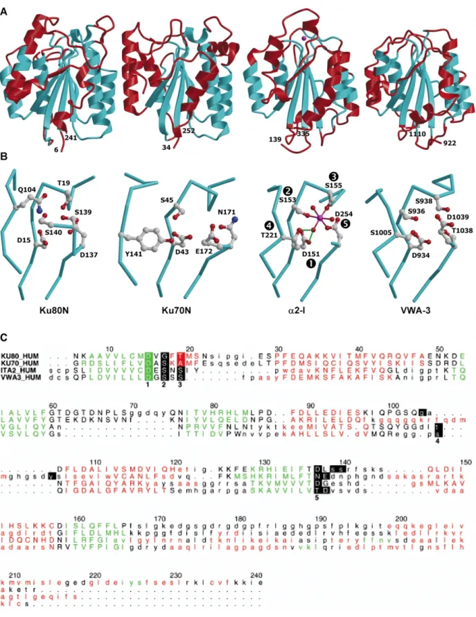
Figure 4. vWA-type domains and their MIDAS or pseudo-MIDAS sites. A, Ribbon diagrams comparing the overall structures of Ku80N, Ku70N, a2-I, and vWA-3 (from left to right) shown in the same orientation. The structural zones used for the superposition of Ku70N, a2-I, and vWA-3 on Ku80N are shown in cyan. The magnesium ion observed in the MIDAS site of a2-I is displayed as a magenta sphere. The rmsd values obtained after superposing are 1.3 A˚ for 109 matched Caatoms of Ku80N and Ku70N, 1.4 (1.6) A˚ for 98 (82) Ca atoms of Ku80N (Ku70N) and
a2-I, and 1.4 (1.6) A˚ for 97 (100) Ca atoms of Ku80N (Ku70N) and VWA-3. As expected from previous studies,25
VWA-3 and a2-I display a greater structural similarity with an rmsd value after superimposition of 1.2 A˚ for 151 Ca
atoms. B, Close-up view of the MIDAS motif of unliganded-a2-I domain and equivalent sites in Ku80N, Ku70N, and vWA-3. Coordinating side-chains of a2-I and corresponding residues in the other proteins are shown as ball-and-stick, with oxygen atoms in red, carbon in grey, and nitrogen in blue. The magnesium ion and the three coordinating
entry code 1JEY) at 2.7 A˚ and 2.5 A˚ resolution, respectively.1 The superimposition of the
DNA-free and DNA-bound structures indicates that the heterodimer maintains its conformation in the absence of DNA.1A search for structural homology
in other protein structures using the program DALI27 revealed a significant similarity of the
N-terminal domain of the Ku subunits with numerous a/b proteins, despite weak sequence homology. From 3241 protein chains in the data-base, integrin I (or A) domains and the A1 and A3 domains of von Willebrand factor display the highest Z values (,15). The overall structure of the N-terminal domains of Ku80 (Ku80N, residues 6– 241) and Ku70 (Ku70N, residues 34 –252) were compared to the structure of the I domain from the a-subunit of the integrin a2b128(a2-I, residues
139– 335, PDB entry code 1AOX) and to the struc-ture of vWA-325 (residues 922 –1110, PDB entry
code 1ATZ) (Figure 4A). All these structures adopt the so-called dinucleotide-binding fold consisting of an open-twisted almost parallel b-sheet flanked on both sides by a-helices (Figure 4A). The six-stranded central b-sheet is the most conserved structural feature, except for the shortest strand located at the C terminus of the proteins and which is absent from Ku70N. Differences occur in the loops and in the helices that surround the b-sheet, especially those at the C-terminal end. Such a variation in the position of the C-terminal region has been observed from the different crystal structures of integrin I domains.
In addition, the C-terminal region of integrins has been shown to undergo conformational changes upon ligand binding.29,30 These
con-formational changes propagate from the ligand-binding site cleft, located at the top of the sheet that contains a structural motif, termed MIDAS for metal ion-dependent adhesion site,31implicated
in ligand binding and integrin activation. The MIDAS motif is formed by an aspartate and two serine residues followed by a threonine and a second aspartate residue, found in three non-contiguous sequence elements. We refer to these residues as D1, S2, S3, T4 and D5 (Figure 4B). It is worth mentioning that a perfectly conserved MIDAS motif, i.e. as detected by “hand-edited alignments”, is not a prerequisite for metal bind-ing, which rather requires detailed structural analysis of a given vWA-type domain in different states.21,25 For instance, vWA-3, which differs from
the canonical MIDAS motif only at D5 (replaced by a threonine residue followed by an aspartate residue) has been shown to display conformational
incompatibility for metal binding.25In the structure
of unliganded a2-I, only S2, S3 and D5 are involved in direct coordination of the metal ion, whereas D1 and T4 are hydrogen bonded to one of the three water molecules that complete the coordination sphere (Figure 4B). However, upon formation of complexes, the ligand does coordinate the metal ion via a carboxylate group leading to a rearrangement of the MIDAS residues, where T4 now coordinates the metal ion, and to movements related to the biological function.29,30 The chemical
environment of Ku70N and Ku80N with respect to the MIDAS motif of integrins was examined
(Figure 4B and C). In Ku80N, both aspartate
residues D1 and D5 are conserved, and S3 is replaced by a threonine residue. S2 and T4 are not conserved in sequence but have structural counter-parts, Q104 and S140, respectively. Furthermore, the side-chain of S139 may provide an additional ligand by replacing one of the three water molecules found in a2-I. For Ku70N, D1 and S2 are conserved, whereas D5 is replaced by an aspar-agine residue. There is no residue equivalent in sequence or structurally to S3 but the side-chain of an acidic residue may instead participate in the coordination of the metal ion. T4 is not conserved in sequence but the hydroxyl group of the structu-rally equivalent Y141 occupies the same position as the hydroxyl group of the threonine residue. Thus, Ku70N and Ku80N both exhibit a complete set of residues compatible in their position and chemical nature with metal binding, provided the side-chains of these residues may change their conformation slightly to meet the metal ion coordi-nation requirements. The fact that these pseudo-MIDAS sites are located on both sides of the Ku heterodimer is particularly suited to a function in molecular recognition (data not shown).
In conclusion, using Ku-proficient and deficient cell lines, we have demonstrated for the first time the involvement of cell-surface Ku in the adhesion process on Fn. We demonstrated recently that cell-surface Ku is involved in adhesion and migration on Fn of monocytic cells, therefore establishing its role in cellular adhesion on ECM during physio-logical processes (our unpublished results). In agreement with this new role, the detailed analysis of three-dimensional structure of the N-terminal domains of both Ku sub-units strongly suggests that the Ku heterodimer may act as an adhesion receptor to fibronectin such as the heterodimeric integrins.32 Ku has been implicated in cell –cell
adhesion6,8,9 and cell adhesion on extracellular
matrixes (our present data). Thus, Ku could be
water molecules are shown as magenta and red spheres, respectively. C, Structure-based sequence alignment of Ku80N, Ku70N, a2-I, and VWA-3. Residues in a helices and b strands are colored red and green, respectively. Lower-case letters in the sequences denote residues that cannot be superimposed on a residue of Ku80N. Residues of a2-I defining the MIDAS motif are shown in bold with reverse video and numbered 1 to 5. Other residues mentioned in the text are shown as reverse video. Numbering is shown for the sequence of Ku80N. This Figure was made with the program ALSCRIPT.36
defined as a new adhesion molecule that contributes to establish a relationship between cells and host tissue microenvironment.
Acknowledgements
This work was supported by grants from the “Ligue Re´gionale Contre le Cancer” (Comite´ Haute-Garonne et Comite´ du Lot) and from the “Ligue Nationale Contre le Cancer” (Equipe labellise´e). S.M. is a recipient of a PhD fellowship from the “Ligue Nationale Contre le Cancer”. We thank Sophie Bouquet for helpful advice.
References
1. Walker, J. R., Corpina, R. A. & Goldberg, J. (2001). Structure of the Ku heterodimer bound to DNA and its implications for double-strand break repair. Nature, 412, 607–614.
2. Gottlieb, T. M. & Jackson, S. P. (1993). The DNA-dependent protein kinase: requirement for DNA ends and association with Ku antigen. Cell, 72, 131–142.
3. Lee, S. H. & Kim, C. H. (2002). DNA-dependent protein kinase complex: a multifunctional protein in DNA repair and damage checkpoint. Mol. Cell, 13, 159–166.
4. Dalziel, R. G., Mendelson, S. C. & Quinn, J. P. (1992). The nuclear autoimmune antigen Ku is also present on the cell surface. Autoimmunity, 13, 265–267. 5. Ginis, I. & Faller, D. V. (2000). Hypoxia affects tumor
cell invasiveness in vitro: the role of hypoxia-activated ligand HAL1/13 (Ku86 autoantigen). Cancer Letters, 154, 163–174.
6. Lynch, E. M., Moreland, R. B., Ginis, I., Perrine, S. P. & Faller, D. V. (2001). Hypoxia-activated ligand HAL-1/13 is lupus autoantigen Ku80 and mediates lymphoid cell adhesion in vitro. Am. J. Physiol. Cell Physiol. 280, C897–C911.
7. Prabhakar, B. S., Allaway, G. P., Srinivasappa, J. & Notkins, A. L. (1990). Cell surface expression of the 70-kD component of Ku, a DNA-binding nuclear autoantigen. J. Clin. Invest. 86, 1301–1305.
8. Tai, Y. T., Podar, K., Kraeft, S. K., Wang, F., Young, G., Lin, B. et al. (2002). Translocation of Ku86/Ku70 to the multiple myeloma cell membrane: functional implications. Expt. Hematol. 30, 212–220.
9. Teoh, G., Urashima, M., Greenfield, E. A., Nguyen, K. A., Lee, J. F., Chauhan, D. et al. (1998). The 86-kD subunit of Ku autoantigen mediates homotypic and heterotypic adhesion of multiple myeloma cells. J. Clin. Invest. 101, 1379–1388.
10. Lucero, H., Gae, D. & Taccioli, G. E. (2003). Novel localization of the DNA –PK complex in lipid rafts: a putative role in the signal transduction pathway of the ionizing radiation response. J. Biol. Chem. 278, 22136–22143.
11. Li, G., Nelsen, C. & Hendrickson, E. A. (2002). Ku86 is essential in human somatic cells. Proc. Natl Acad. Sci. USA, 99, 832–837.
12. Charo, I. F., Nannizzi, L., Smith, J. W. & Cheresh, D. A. (1990). The vitronectin receptor alpha v beta 3 binds fibronectin and acts in concert with alpha 5
beta 1 in promoting cellular attachment and spread-ing on fibronectin. J. Cell Biol. 111, 2795 –2800. 13. Taccioli, G. E., Gottlieb, T. M., Blunt, T., Priestley, A.,
Demengeot, J., Mizuta, R. et al. (1994). Ku80: product of the XRCC5 gene and its role in DNA repair and V(D)J recombination. Science, 265, 1442–1445. 14. Singleton, B. K., Priestley, A., Steingrimsdottir, H.,
Gell, D., Blunt, T., Jackson, S. P. et al. (1997). Molecu-lar and biochemical characterization of xrs mutants defective in Ku80. Mol. Cell. Biol. 17, 1264–1273. 15. Smider, V., Rathmell, W. K., Lieber, M. R. & Chu, G.
(1994). Restoration of X-ray resistance and V(D)J recombination in mutant cells by Ku cDNA. Science, 266, 288–291.
16. Boubnov, N. V., Hall, K. T., Wills, Z., Lee, S. E., He, D. M., Benjamin, D. M. et al. (1995). Complementa-tion of the ionizing radiaComplementa-tion sensitivity, DNA end binding, and V(D)J recombination defects of double-strand break repair mutants by the p86 Ku auto-antigen. Proc. Natl Acad. Sci. USA, 92, 890–894. 17. Koike, M. (2002). Dimerization, translocation and
localization of Ku70 and Ku80 proteins. J. Radiat. Res. 43, 223–236.
18. Cook, G. A., Wilkinson, D. A., Crossno, J. T., Jr, Raghow, R. & Jennings, L. K. (1999). The tetraspanin CD9 influences the adhesion, spreading, and peri-cellular fibronectin matrix assembly of Chinese hamster ovary cells on human plasma fibronectin. Expt. Cell Res. 251, 356–371.
19. Elmroth, K., Nygren, J., Martensson, S., Ismail, I. H. & Hammarsten, O. (2003). Cleavage of cellular DNA by calicheamicin gamma1. DNA Repair, 2, 363–374. 20. Doherty, A. J., Jackson, S. P. & Weller, G. R. (2001).
Identification of bacterial homologues of the Ku DNA repair proteins. FEBS Letters, 500, 186–188. 21. Whittaker, C. A. & Hynes, R. O. (2002). Distribution
and evolution of von Willebrand/integrin a domains: widely dispersed domains with roles in cell adhesion and elsewhere. Mol. Biol. Cell, 13, 3369–3387. 22. Tuckwell, D. (1999). Evolution of von Willebrand
factor A (VWA) domains. Biochem. Soc. Trans. 27, 835–840.
23. Emsley, J., Cruz, M., Handin, R. & Liddington, R. (1998). Crystal structure of the von Willebrand factor A1 domain and implications for the binding of plate-let glycoprotein Ib. J. Biol. Chem. 273, 10396–10401. 24. Bienkowska, J., Cruz, M., Atiemo, A., Handin, R. &
Liddington, R. (1997). The von Willebrand factor A3 domain does not contain a metal ion-dependent adhesion site motif. J. Biol. Chem. 272, 25162–25167. 25. Huizinga, E. G., Martijn van der Plas, R., Kroon, J.,
Sixma, J. J. & Gros, P. (1997). Crystal structure of the A3 domain of human von Willebrand factor: impli-cations for collagen binding. Structure, 5, 1147–1156. 26. Xiong, J. P., Stehle, T., Diefenbach, B., Zhang, R.,
Dunker, R., Scott, D. L. et al. (2001). Crystal structure of the extracellular segment of integrin alpha Vbeta3. Science, 294, 339–345.
27. Holm, L. & Sander, C. (1993). Protein structure comparison by alignment of distance matrices. J. Mol. Biol. 233, 123–138.
28. Emsley, J., King, S. L., Bergelson, J. M. & Liddington, R. C. (1997). Crystal structure of the I domain from integrin alpha2beta1. J. Biol. Chem. 272, 28512–28517. 29. Shimaoka, M., Xiao, T., Liu, J. H., Yang, Y., Dong, Y., Jun, C. D. et al. (2003). Structures of the alpha L I domain and its complex with ICAM-1 reveal a shape-shifting pathway for integrin regulation. Cell, 112, 99–111.
30. Emsley, J., Knight, C. G., Farndale, R. W., Barnes, M. J. & Liddington, R. C. (2000). Structural basis of collagen recognition by integrin alpha2beta1. Cell, 101, 47–56.
31. Lee, J. O., Bankston, L. A., Arnaout, M. A. & Liddington, R. C. (1995). Two conformations of the integrin A-domain (I-domain): a pathway for activation? Structure, 3, 1333–1340.
32. Hood, J. D. & Cheresh, D. A. (2002). Role of integrins in cell invasion and migration. Nature Rev. Cancer, 2, 91–100.
33. Muller, C., Monferran, S., Gamp, A. C., Calsou, P. & Salles, B. (2001). Inhibition of Ku heterodimer DNA end binding activity during granulocytic
differen-tiation of human promyelocytic cell lines. Oncogene, 20, 4373–4382.
34. Yu, Q. & Stamenkovic, I. (1999). Localization of matrix metalloproteinase 9 to the cell surface provides a mechanism for CD44-mediated tumor invasion. Genes Dev. 13, 35–48.
35. Hutchings, H., Ortega, N. & Plouet, J. (2003). Extra-cellular matrix-bound vascular endothelial growth factor promotes endothelial cell adhesion, migration, and survival through integrin ligation. FASEB J. 17, 1520 –1522.
36. Barton, G. J. (1993). ALSCRIPT: a tool to format multiple sequence alignments. Protein Eng. 6, 37–40.
Edited by M. Yaniv