UNIVERSITÉ DE MONTRÉAL
FABRICATION PAR ÉLECTROFILAGE D’UNE STRUCTURE À ÉLUTION DE FACTEURS DE CROISSANCE POUR CONTRÔLER LA DIFFÉRENCIATION DE
CELLULES SOUCHES NEURONALES EN NEURONES MOTEURS
LOÏC BINAN
INSTITUT DE GÉNIE BIOMÉDICAL ÉCOLE POLYTECHNIQUE DE MONTRÉAL
MÉMOIRE PRÉSENTÉ EN VUE DE L’OBTENTION DU DIPLÔME DE MAÎTRISE ÈS SCIENCES APPLIQUÉES
(GÉNIE BIOMÉDICAL) AOÛT 2013
UNIVERSITÉ DE MONTRÉAL
ÉCOLE POLYTECHNIQUE DE MONTRÉAL
Ce mémoire intitulé:
FABRICATION PAR ÉLECTROFILAGE D’UNE STRUCTURE À ÉLUTION DE FACTEURS DE CROISSANCE POUR CONTRÔLER LA DIFFÉRENCIATION DE
CELLULES SOUCHES NEURONALES EN NEURONES MOTEURS
présenté par : BINAN Loïc
en vue de l’obtention du diplôme de : Maîtrise ès sciences appliquées a été dûment accepté par le jury d’examen constitué de :
M. BUSCHMANN Michael, Ph. D, président
M. JOLICOEUR Mario, Ph. D, membre et directeur de recherche
M. DE CRESCENZO Gregory, Ph. D, membre et codirecteur de recherche M. AJJI Abdellah, Ph. D, membre et codirecteur de recherche
REMERCIEMENTS
Je souhaite remercier le Pr. Mario Jolicoeur pour m’avoir encadré et dirigé pendant ma maitrise ainsi que pour son aide financière.
Je souhaite aussi remercier mes co-directeurs Abdellah Ajji et Gregory De Crescenzo, notamment ce dernier pour toute l’aide qu’il m’a apportée dans ma rédaction.
Je voudrais aussi remercier les personnes qui travaillent dans les laboratoires où j’ai réalisé mes expériences et notamment Cédric Vallée pour l’aide qu’il m’a apportée au jour le jour.
RÉSUMÉ
Les blessures médullaires et les lésions étendues des nerfs handicapent des milliers de nouvelles personnes chaque année aux États-Unis seulement. Il n’y a actuellement aucun traitement permettant la guérison de ces blessures. Une approche envisagée est la greffe de structures chargées de cellules souches pour rétablir les fonctions originelles du tissu. Ce travail vise donc à développer une méthode de fabrication d’une structure qui puisse offrir un support mécanique aux cellules, tout en délivrant sur place le cocktail de biomolécules adéquat pour diriger la différenciation des cellules vers le phénotype de neurone moteur. Afin de préparer cette structure, de la gélatine et de l’acide polylactique-L ont été co-électrofilés. L’électrofilage permet la fabrication de fibres de diamètre dépendant de la concentration des solutions en polymère. Les fibres ainsi fabriquées ont été réticulées afin de ralentir et contrôler leur dégradation. Les biomolécules permettant de promouvoir la différenciation des cellules en neurones moteurs, l’acide rétinoïque et la purmorphamine, ont été incluses dans la partie extérieure des fibres, en gélatine. Ces molécules ont diffusé de manière continue à partir des fibres, dans le milieu liquide. Les cellules implantées sur cette structure ont proliféré et se sont différenciées en neurones moteurs. Leur phénotype a été caractérisé par immunofluorescence.
ABSTRACT
Spinal cord injury and extended nerve injury currently have no cure. These pathologies are responsible for the decrease in quality of life of thousands of new people every year in the US only, and are draining huge costs to the healthcare system. Current research in the area focuses on the grafting of an artificial structure loaded with stem cells to restore tissue functions. The objective of this work is to propose a structure that can offer mechanical support to the cells, favor their proliferation, and promote their differentiation into motor neuron, by delivering in
situ the appropriate cocktail of growth factors. Such structure was prepared by
co-electrospinning of poly L-lactic acid and gelatin. Fiber diameter can be adjusted by controlling the polymer concentration. These fibers were crosslinked to slow their degradation. Retinoic acid and purmorphamine were included in the outer layer of gelatin. These two growth factors are known to direct cell differentiation towards a motor neurons phenotype and were continuously released from the fibers in the medium. Cells proliferated on the structure and differentiated into motor neurons. Their phenotype was characterized by immunostaining using sample images.
TABLE DES MATIÈRES
REMERCIEMENTS ... III RÉSUMÉ...IV ABSTRACT………...V TABLE DES MATIÈRES ... VI LISTE DES TABLEAUX ... VIII LISTE DES FIGURES ... IX LISTE DES SIGLES ET ABRÉVIATIONS ... XI LISTE DES ANNEXES ... XII
INTRODUCTION ... 1
CHAPITRE 1 DÉMARCHE GÉNÉRALE ... 3
CHAPITRE 2 REVUE DE LITTÉRATURE ... 4
Article 1 Approaches for neural tissue regeneration………4
2.1 Introduction ... 5
2.2 Injection of free cells for in situ tissue regeneration ... 7
2.3 Use of growth factors to promote healthy tissue re-growth ... 9
2.4 Use of structures to mechanically support tissue growth ... 14
2.5 Use of conductive structures to electrically stimulate neuron differentiation ... 22
2.6 Conclusion ... 23
2.7 Bibliography ... 32
2.8 Acknowledgments ... 51
CHAPITRE 3 FABRICATION DES FIBRES ET OBTENTION DE NEURONES
MOTEURS………....52
3.1 Présentation ... ..52
3.2 Article 2 – Differentiation of neuronal stem cells into motor neurons using co-axial electrospun Poly-L-Lactic Acid/gelatin fibers as an instructive cue-delivering scaffold ... 53
3.2.1 Introduction ... 55
3.2.2 Materials and methods ... 58
3.2.3 Results ... 63 3.2.4 Discussion ... 68 3.2.5 Conclusions ... 72 3.2.6 Acknowledgment ... 72 3.2.7 References ... 73 3.2.8 Figures ... 77 3.2.9 Supplementary data ... 86
CHAPITRE 4 DISCUSSION GÉNÉRALE ... 88
CONCLUSION……...89
BIBLIOGRAPHIE ... 90
LISTE DES TABLEAUX
Table 1. Growth factors and their use in the context of neuronal disease treatment………….25 Table 2. Examples of tri-dimensional scaffolds generated by electrospinning………….……28 Table 3. Examples of techniques tested in vivo……….30
LISTE DES FIGURES
Fig 1. Hydrogel fabrication and application………..16 Fig 2. Co-electrospinning needles………..………...17 Fig 3. Electrospinning setup………..…18 Fig 4. Fiber morphology: SEM images of co-electrospun fibers corresponding to 10% PLLA
(core) and 7% gelatin (outer shell) (A, C) or 7% PLLA and 5% gelatin (B) without any instructive cues (A, B) or with retinoic acid and purmorphamine in the gelatin (C) and TEM image showing the core shell structure of the fibers (D)………...77
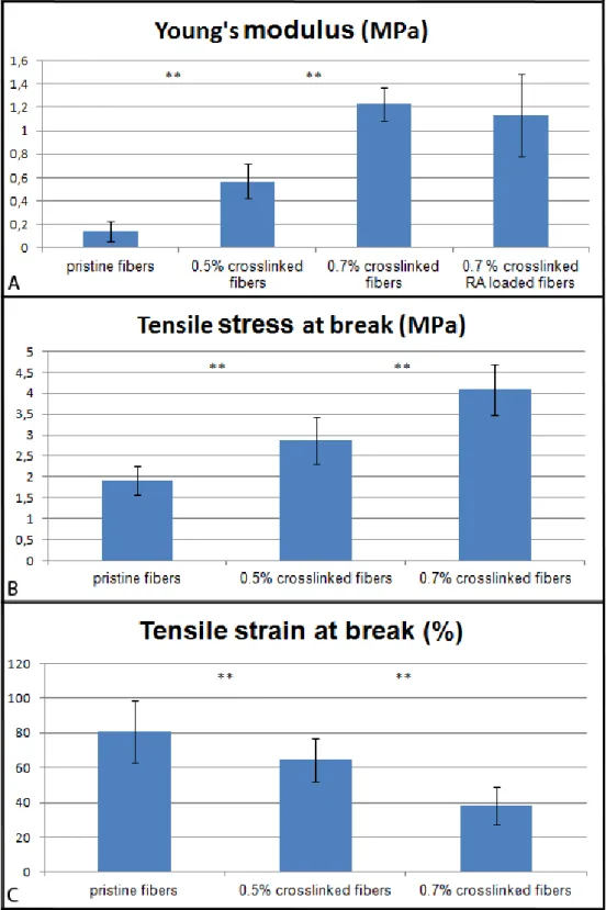
Fig 5. Young’s modulus (A), Tensile stress (B) and strain (C) at break of fibers crosslinked
with different concentrations of glyceraldehyde………..78
Fig 6. SEM images of untreated (A, C, E, G) and crosslinked (B, D, F, H) fibers at different
degradation times: day 0 (A, B), day 3 (C, D), day 5 (E, F) and day 7 (G, H)………...79
Fig 7. Evaluation of gelatin layer degradation by (A) fiber diameter measurements from SEM
imaging and (B) Orange II dye assay after various glyceraldehyde crosslinking treatments. (For panel A, * Indicates student test p<0.05, and ** indicates p<0.001)……….80
Fig 8. Concentration of retinoic acid released from the fibers in the culture medium. Medium
was changed at days 3, 6, 9, and 13.……… …..………...80
Fig 10. Nestin and SYTOX staining of cells cultured on control fibers at days 2, 7 and 14
(A, B, C) or RA- and purmorphamine-loaded fibers at day 2(D). Images are 40x (A, C, D) or 10X (B)………..82
Fig 11. Representative images of NSLCs at different time points in culture on RA and
purmorphamine loaded fibers (from A to I) and fibers without instructive cues (J, K, L, only at day 14). Cell morphology on 40x images obtained with Cells stained for Islet-1 at day 2 (A), 7 (D), 14 (G), for HB-9 at day 2 (B), 7 (E), 14 (H), and for Choactase at day 2 (C), 7 (F), and 14 (I)……….. ………...83
Fig 12. Morphological evaluation. Cell morphology on 40x images obtained with TUJ-1
staining and SEM images. Cells cultured on pure fibers (A, C, E, G) or RA- and
purmorphamine-loaded fibers (B, D, F, H) at day 2 (A, B, G), 7 (C, D), and 14 (E,F, H)…...…84
Fig 13. Neurite length on fibers with or without RA and purmorphamine.
(The value at day 14 for pure fibers is under detection limit capacity)……….85
Fig 14. Formation of neurospheres on tissue culture plate after seven days of culture………...86 Fig 15. Images of cells cultured on fibers and stained with SYTOX green for nuclei at
day 2 (A), day 7 (B) and day 14 (C)……….…86
Fig 16. Cell penetration: at least two different focal plans present on this 40x image obtained
LISTE DES SIGLES ET ABRÉVIATIONS
BDNF Brain derived neurotrophic factorCNTF Ciliary neurotrophic factor EGF Endothelial growth factor
ELISA Enzyme linked immunosorbent assay FGF Fibroblast growth factor
GDNF Glial cell line-derived neurotrophic factor
HA Hyalorunic acid
IGF Insulin-like growth factor
LOCS Linear ordered collagen scaffolds MSC Mesenchymal stem cell
NGF Nerve growth factor NSC Neural Stem Cell
NSLC Engineered Stem Like Cell NT-3 Neurotrophin 3
PEG Polyethylene glycol PES Poly ethersulfone PGA Poly glycolic acid
PLGA Poly lactic co-glycolic acid PLLA Poly L-lactic acid
RA Retinoic acid
Shh Sonic hedgehog
LISTE DES ANNEXES
ANNEXE 1 – Protocole Orange II.……….114 ANNEXE 2 – Protocole Immunocoloration………....115
INTRODUCTION
Une multitude d’accidents peuvent occasionner des lésions des nerfs, voire de la moelle épinière, entrainant une baisse notable de la qualité de vie des patients.
En effet, bien que des nerfs périphériques blessés sur de courtes distances puissent se régénérer, si la blessure est trop importante ou touche la moelle épinière, la régénération du tissu nerveux se fait difficilement. [1] Cette mauvaise régénération du tissu est due à la fois à une activité réduite des cellules souches neuronales, ce qui diminue la plasticité du tissu nerveux, et à la présence de tissu cicatriciel gênant la régénération du tissu originel. [2] Ces raisons font que la thérapie par cellules souches est toute indiquée pour pallier à ce genre de pathologie. En effet, ces thérapies impliquent l’injection de cellules souches pour pallier au manque du patient et restaurer la capacité du tissu à se régénérer.
Les cellules souches ont la capacité de se multiplier et de se différencier, théoriquement, selon les stimuli environnementaux, en n’importe quel type cellulaire. Par conséquent, il est nécessaire de pouvoir contrôler leur différenciation. Les principaux facteurs qui influencent le phénotype vers lequel une cellule souche évoluera sont les propriétés mécaniques de son environnement, la topologie de son support qui doit copier la matrice extracellulaire et les facteurs de croissance qu’elle reçoit. Il est désormais connu que des cellules souches neuronales se différencieront en neurones moteur si elles reçoivent les concentrations adéquates d’acide rétinoïque et de sonic hedgehog. [3] D’autre part, il a été récemment découvert que la purmorphamine a les mêmes effets sur les cellules que sonic hedgehog. [3]
L’utilisation de cellules souches pour de telles thérapies requiert la fabrication d’une structure support qui d’une part offre un milieu aux propriétés mécaniques satisfaisantes guidant l’étalement des cellules et d’autre part évite la migration anarchique des cellules souches injectées. Différentes techniques ont donc été développées pour permettre l’implantation d’une structure chargée de cellules souches dans la zone blessée. Un problème majeur des méthodes proposées jusqu’alors est que la différenciation des cellules souches implantées nécessite la présence de facteurs de croissance qui ne sont pas produits par le patient dans la zone blessée et dont la concentration doit être très finement contrôlée. La majorité des techniques mises au point à l’heure actuelle offrent bien un support mécanique aux cellules injectées, mais ne permettent
pas le contrôle de leur différenciation. Elles requièrent donc en général une injection externe de ces facteurs. [4] La mise en place de telles techniques sur l’homme n’est pas envisageable car elles demandent des opérations chirurgicales répétées beaucoup trop lourdes, et une administration très régulière de ces molécules signal, afin d’obtenir un stimulus contrôlé de leur concentration dans l’environnement entourant les cellules.
Dans ce contexte, les travaux les plus récents réalisés dans le domaine ont comme objectif la fabrication de structures qui puissent à la fois offrir un support mécanique aux cellules afin de permettre leur adhésion, d’organiser spatialement leur croissance, et, le cas échéant, de promouvoir leur multiplication avant implantation, mais qui puissent aussi diffuser directement in situ et à la bonne concentration les facteurs de croissance requis pour contrôler la différenciation desdites cellules. [5]
L’objectif de ce travail est d’utiliser l’électrofilage pour fabriquer une telle structure. Cette technique permet l’obtention de fibres de diamètre variable à partir de différents polymères. Nous proposons d’utiliser l’électrofilage concentrique de deux polymères, la gélatine (couche externe) et l’acide poly-lactique (cœur interne) afin de bénéficier à la fois des propriétés mécaniques des fibres en acide poly-lactique et des bonnes propriétés de contact et de biocompatibilité de la gélatine. En plus d’offrir un bon support mécanique, la structure développée doit pouvoir délivrer des facteurs de croissance.
La première partie de ce mémoire présente une revue de la littérature sur les techniques actuellement étudiées dans le but d’obtenir la régénération de tissus nerveux.
La seconde partie, reprenant les concepts les plus intéressants exposés dans notre revue de littérature, propose, à notre avis, la technologie la plus prometteuse pour fabriquer une structure permettant de promouvoir la prolifération et la différenciation des cellules souches en neurones moteurs. Dans cette partie, les propriétés mécaniques et la dégradation du matériel sont étudiées. Ensuite la vitesse d’élution des biomolécules est évaluée et, finalement, la capacité de ce nouveau matériel à promouvoir la différenciation des cellules souches neuronales en neurones moteurs est validée par l’analyse d’images obtenues par immunofluorescence.
CHAPITRE 1
DÉMARCHE GÉNÉRALE
Ce mémoire présente deux articles. Le premier, intitulé « Approaches for neural tissue regeneration » a été soumis dans Stem cells reviews and reports le 17 juillet 2013. Cet article permet d’exposer l’ensemble des techniques étudiées actuellement dans le but d’obtenir la génération de nouveaux tissus à partir de cellules souches. Les connaissances développées dans cette revue permettent de juger des avantages de chacune des techniques connues, et de faire un choix éclairé parmi celles-ci pour répondre à nos objectifs. Le second, intitulé “Differentiation of neuronal stem cells into motor neurons using co-axial electrospun Poly-L-Lacctic Acid/gelatin fibers as an instructive cue-delivering scaffold”, a été soumis dans Biomaterials le 20 Août 2013.
Ces journaux scientifiques ont été sélectionnés pour leur niveau d’impact dans le domaine étudié ; Biomaterials étant le journal de référence pour la fabrication de structures en génie tissulaire alors que Stem cells reviews and reports publie des protocoles et revues de littératures sur les techniques émergentes dans le domaine.
J’ai réalisé moi-même l’essentiel des travaux expérimentaux, une partie de la caractérisation de la dégradation des fibres a été réalisé sous ma supervision par Charlène Tendey au cours de son stage de fin d’étude. J’ai aussi bénéficié de l’expérience de mes directeur et co-directeurs au cours de ces travaux et de la rédaction des articles.
CHAPITRE 2
REVUE DE LITTÉRATURE
Il existe différentes approches étudiées dans le cadre du génie tissulaire appliqué aux tissus nerveux. Toutes ces approches ont un objectif similaire qui est de promouvoir la différenciation de cellules souches neuronales en un des types de neurones ou en cellules de Schwann. Elles visent généralement des applications soit dans le système nerveux périphérique ou la moelle épinière, afin le plus souvent de réparer des nerfs endommagés, soit dans le système nerveux central avec des applications plus tournées vers les maladies neuro-dégénératives. Bien que les champs d’application varient légèrement, les méthodes envisagées sont très similaires technologiquement, et une revue de l’ensemble de ces approches est proposée ici dans un manuscrit récemment soumis au journal Stem cells reviews and reports.
Article 1 – Approaches for neural tissue regeneration
Loïc BINAN, Abdellah AJJI, Gregory DE CRESCENZO, Mario JOLICOEUR
“Approaches for neural tissue regeneration”
Soumis dans «Stem cell reviews and reports » le17 juillet 2013. Manuscript ID. STCR-883
Abstract
There is currently no treatment for neurodegenerative diseases such as Parkinson’s or Alzheimer's diseases. While spinal cord injury has no treatment either, nerve injuries are being treated with autologous grafts, a procedure that in turn translates into a loss of function in the donor area. The development of therapies for these pathologies has become urgent as population keeps on ageing. A promising direction of investigation is the use of regenerative techniques to re-grow healthy and functional tissue in the injured area. In this review article, various approaches currently investigated to promote neural regeneration are covered. Those include approaches based on (and many times combining) stem cell therapy, scaffolds made of hydrogel, electrospun fibers and conductive materials as well as the use of soluble or non-diffusible growth factors.
Keywords
Nerve, spinal cord; stem cell, regeneration, biomaterial, scaffold, hydrogel, electrospinning, growth factor.
2.1 Introduction
16 million first-ever strokes occur every year, which makes it the second cause of death worldwide after heart diseases, with associated costs over 65 billion dollars in the United States only. [6] Also, 12,000 new cases of spinal cord injury (SCI) occur every year, with 270,000 people seeing their life quality highly affected. [7] Moreover, every year, 60,000 new cases of Parkinson’s disease are diagnosed, for a total of 7 to 10 million people worldwide. [8] However, there is currently no appropriate cure for all these affections and the research in this field is still in its infancy. Pathologies affecting neural tissues can be divided into two classes: those that come from an injury, where the neuronal tissue is physically damaged; and neurodegenerative diseases, where the tissue loses its functions with time, and ultimately dies. For the first class, the use of autologous grafts to repair damaged nerves is now possible in many cases. [9] This
surgical solution consists in taking a nerve, usually with a sensory function from a healthy area, and grafting it in the injured area to serve as a guide for the regrowth of the motoneurons. [9] However, this approach leads to a loss of function in the donor area, and, in most cases, function recovery at the site of implantation does not reach expected levels. [11] For the second class of diseases, there is no actual treatment targeting the cause of the symptoms. Current medical treatments are aimed at slowing down the disease progression and/or minimizing its impacts on patients’ quality of life. For instance, dopamine and dopamine agonists are administered to patients suffering from Parkinson’s disease in order to compensate for the lack of dopamine inside the brain and thus attenuate symptoms. [12] The difficulties faced when trying to provide an appropriate and complete treatment to those pathologies lie in the fact that it would require the restoration of tissue function, and re-growth of tissue replacing the dead one. By itself, current medication does not appear to be a solution for those two issues since, once those diseases are developed, the site of the pathology becomes hostile to tissue re-growth, which impedes the body regenerating healthy tissue. [13]
It has long been considered that once damaged, a neuronal tissue does not regenerate. Indeed, it was thought that, contrary to other tissues, neuronal tissue did not contain any stem cells, and therefore would not regenerate. Nevertheless, the recent discovery that nervous tissue is actually more dynamic than expected and that it contains stem cells has opened new therapeutic avenues based on a regenerative medicine approach. [14] This opportunity resides on the hypothesis that stem cells may have the same regenerative abilities in neural tissue as in other cases and are able to promote tissue regeneration. A remaining issue that may mitigate the use of stem cell-based therapies for neural tissues is that the number of active neural stem cells is thought to be very low. It is indeed admitted that stem cells do not divide continuously but that they specifically enter cycles of divisions, which prevents cell depletion in the tissue. It is thought that stem cells from the subventricular zone (SVZ) have a longer quiescence sequence between their cycles of proliferation than other stem cells. In fact, it has been suggested by Hwang and colleagues that glial cells might actually be the very stem cells of the nervous system. [15] Future stem cell-based regenerative strategies thus require either the use of stimuli in vivo or the addition of an in vitro step aiming at amplifying the patient's active neuronal stem cells prior to their
re-implantation as cells or tissue. Injuries that could be potentially treated with such a tissue regeneration or replacement technique are those related to nerve or spinal cord damages, or loss of function such as in Parkinson's, Huntington's or Alzheimer's diseases. For neurodegenerative diseases, the strategy may be directed toward the synthesis of a tissue presenting normal activity and thus re-establishing endogenous tissue function. For instance, in Parkinson’s disease, the new tissue would be expected to synthesize dopamine up to normal levels in the substancia nigra and improve the patient’s quality of life. [16] These approaches are particularly promising since damaged areas become hostile to the growth of new tissue, and would not allow for the re-growth of healthy tissue without any intervention except in highly rare cases. [13]
Although the discovery of neural stem cells has paved the way for innovative and efficient regenerative therapies, there are still several issues that remain to be solved before routinely applying such techniques. First, cell implantation and connection with host tissue need to be improved and cell growth and differentiation into desired phenotype(s) must be controlled, more likely through the use of growth factor(s) or related stimuli. Furthermore, structures supporting cell growth, possibly combined to growth factors delivery ability, still need to be developed. This review aims at presenting the various emerging techniques that are proposed for neural tissue regeneration.
2.2 Injection of free cells for in situ tissue regeneration
In 2006, experimental observations reported that stem cells are produced in specific regions of the brain and then migrate to their final destination, allowing for brain plasticity even at the adult stage. [14] This observation led to the conclusion that neuronal tissue is more likely to be able to regenerate, thus opening promising opportunities to cure neuronal diseases. The presence of
endogenous stem cells in neural tissue indeed suggests that it should be possible to obtain the same kind of tissue production in other parts of the brain or nerves by providing those progenitors cells. [14] Indeed, the injection of additional stem cells at the site of injury has first been proposed to enhance the regenerative effect of endogenous cells. In that endeavor, mesenchymal stem cells (MSCs) were selected since they are easy to amplify, protect themselves from the immune system, and can differentiate into neural lineage in vitro and in vivo. [17] MSC-derived
neural progenitors were injected directly in the diseased area of the brain in mice that had chronic experimental autoimmune encephalomyelitis. Those injections contributed to maintain normal phenotype of the tissue. [18] The beneficial effect of MSC upon new tissue formation was partly attributed to the extracellular matrix (ECM) produced by the MSCs, which created a reservoir of bioactive substances being responsible for this neuro-supportive environment at the injury site. [17, 19]
Of salient interest, the benefits of stem cell therapy may also be extended to spinal cord injuries as demonstrated by Cummings and coworkers: human neuronal stem cells (hNSCs) cultured as neurospheres were directly injected in spinal cord-injured mice. [20] After 17 weeks, locomotor recovery and remyelination were observed even in myelin-deficient mice. More interestingly, SEM microscopy showed evidence of synapse formation between injected and endogenous cells, and cells migrated in the gray and white matters without participating to the formation of scar tissue in the injured area.
Human embryonal carcinoma-derived cells have also been identified as a potential source of stem cells. Their transplantation has been shown to promote the recovery of normal motor capacity in a rat animal model. [21] More specifically, transplantation was performed in rats that demonstrated a deficiency in the passive avoidance test one month after the surgical injury of their neural tissue. Following cell transplantation, a partial recovery of the learning capacity was observed one month after treatment and lasted over 6 months. Heine and colleagues also promoted nerve regeneration in chronically denervated mice. [22] The team used an immortalized neural progenitor mouse cell line (C17.2 cells), to be transplanted into mice having a deficiency in the regeneration of their tibial nerve. Those mice were observed for four months and demonstrated improved physiological recovery when compared to control population. Their nerves presented increased amount of axons and reinervation of the muscular tissue of the foot. The team also reported that the transplanted cells were still present in the nerve region, but they were not presenting neuron phenotype. The latter observation strongly suggested that stem cells participated in the regeneration by providing growth factors to endogenous cells, and that those endogenous cells were the essential component of the newly generated nerve.
Munoz et al. injected human MSCs into the dentate gyrus of the hippocampus of mice and obtained a colonization of the whole dorsal area of the hippocampus. [23] Implanted cells were
shown to promote endogenous cell proliferation. New cells were also observed to migrate through the brain for 30 days. After this period, those migrated cells presented markers of oligodendrocytes and mature neurons. In addition, human MSCs were shown to stimulate the production of neuronal survival factors such as nerve growth factor (NGF), vascular endothelial growth factor (VEGF), basic fibroblast growth factor-2 (FGF-2) and ciliary neurotrophic factor (CNTF), as well as to promote the proliferation of endogenous cells. Altogether, these studies suggested that the benefic effect of stem cell injections might be attributed to their ability to produce the adequate growth factors on site, these instructive cues leading to the remodeling of surrounding endogenous tissues, rather than to the creation of de novo tissue from differentiating stem cells. These conclusions in turn suggest that the injection of the adequate set of instructive cues may be sufficient to obtain similar spectacular effects without stem cell injection.
2.3 Use of growth factors to promote healthy tissue re-growth
2.3.1 In situ growth factor injection
As illustrated in the previous section, stem cell injection has already given very positive results in terms of function recovery. These promising results may however be mitigated as cell injection may result in tumor formation. [24] In addition, most of the effects obtained with free stem cell injection have been attributed so far to the growth factors these cells produced rather than from their ability to differentiate and form new tissue. These conclusions have led to the hypothesis that the delivery of specific growth factors, rather than stem cells, may be favorable for regeneration. In support to this hypothesis is the fact that, in contrast to central nervous system (CNS) neurons, injured peripheral motoneurons are able to regenerate their axons thanks to neurotrophic factors provided by Schwann cells. Their ability to regenerate is however transient, probably because levels of neutrophic factors rapidly decline after injury. Schwann cell capacity to support axonal regeneration was also shown to be enhanced strongly when Schwann cells extracted from six-month chronically denervated sciatic nerve were cultured in vitro in the presence of transforming growth factor β (TGF-β) and forskolin prior to re-implantation. [25] Injection of growth factors and cytokines directly at the injury site has also been tested to
promote tissue regeneration. In that line of thinking, Kobayashi and colleagues demonstrated that specific neurotrophins, when continuously delivered to the damaged area by cannula, prevented the atrophy of axotomized neurons. [26] Growth factor infusion was correlated to the persistence of high mRNA levels for GAP-43, Tα1-tubulin and TrkB (the BDNF receptor). The effect of the treatment was still observed 14 days after the last injection, more likely due to the long-term stimulation of these regeneration-associated genes that acted in synergy with the delivered growth factors. Other synergistic effects mediated by the co-injection of growth factors have also been documented after sciatic nerve axotomy: Boyd and Gordon demonstrated that long-term co-administration of glial cell line-derived neurotrophic factor (GDNF) combined with BDNF efficiently promoted axonal regeneration of motoneurons. [27] When used in combination with BDNF, GDNF was found to increase the number of axons per Schwann cell in vivo. Growth factor effects were however only observable after seven days of continuous injection.
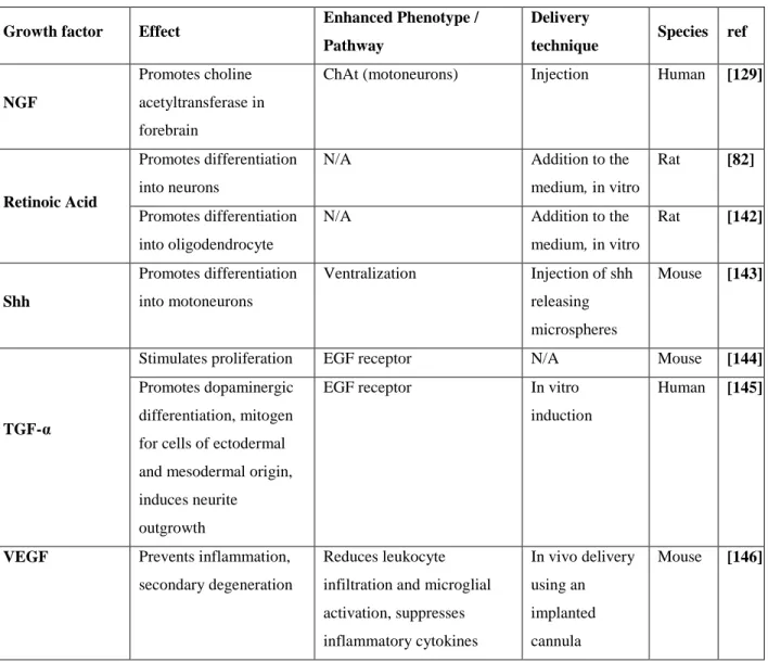
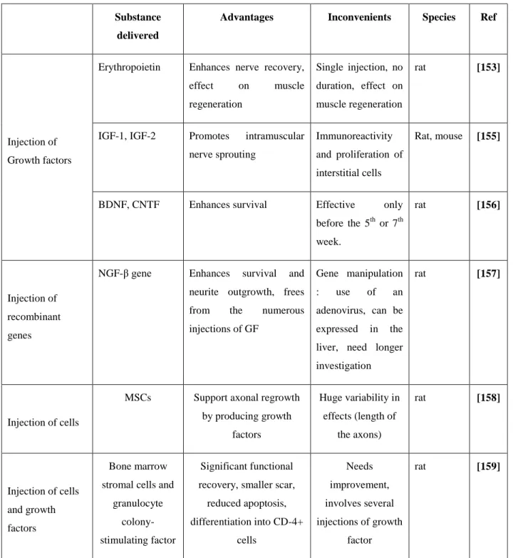
Strategies based on growth factors (Table 1) may thus be viable, although the need of continuous delivery by infusion, as originally tested in rat models, may represent a serious impediment for its translation to human. Therefore it appears necessary to develop novel experimental approaches that would allow for the growth factors to be delivered in situ in a spatio-temporally controlled fashion with minimal surgical intervention.
2.3.2 Use of encapsulated growth factors to promote axonal regeneration
and replace injured tissue
Classical engineered growth factors delivery systems are based on their diffusion from hydrogel or porous polymeric material, or on the degradation of a polymeric scaffold in which they are freely entrapped or, alternatively, at the surface of which they are grafted. Depending on the design of the system, growth factor delivery can be controlled via pure diffusion through porous polymeric material or through polymer degradation. [28-30] When designing such scaffolds, important characteristics to be taken into account and evaluated are the drug loading capacity of the polymer construct and its degradation time, if needed. Also, the biocompatibility and
biosafety of degradation products are strictly required. Among biodegradable polymers envisaged for brain-specific applications and currently used as delivery systems in vitro or in in vivo animal models, poly lactic co-glycolic acid (PLGA) has been widely studied. [31-33] This Food and Drug Administration (FDA)-approved biodegradable synthetic polymer is mainly used to mimic the extracellular matrix mechanical properties and shape, as well as to strengthen scaffolds originally engineered from natural materials such as gelatin or elastin. [34] VEGF and BDNF encapsulation in PLGA microspheres has been reported by Wang and colleagues using the
water-in-oil-in-water encapsulation method. [35] The microspheres were then embedded within a
crosslinked hyaluronic acid (HA) hydrogel to serve as scaffold for the culture of neural stem cells. The group demonstrated that such a design allowed for a slow and linear delivery of both growth factors for 6 days, with both encapsulated BDNF and VEGF maintaining their respectively known protective and pro-proliferative properties. The same strategy has been used to deliver CNTF from photopolymerizable nanoarray hydrogels. [36] However, a major issue related to the encapsulation of growth factors resides in the difficulty to obtain long-term delivery within a defined concentration range. Hydrogels indeed rapidly release most of the growth factors they contain in an initial burst that is higher than physiological levels, a phenomenon that reduces the duration of the delivery. [37] [38]
The most-often encountered problem is that a longer delivery time requires sufficient loading of the structure, which in turn usually results in unwanted burst of delivered growth factor just after hydrogel injection. In an attempt to address this shortcoming, Betram et al. obtained their best results for NGF delivery with microspheres of PLGA-PLLA-PEG. [37] This combination suppresses the burst in secretion observed with PLGA-only hydrogels by controlling growth factor delivery through continuous degradation rather than desorption process. With that enhanced system, NGF was delivered up to 65 days without any observable burst.
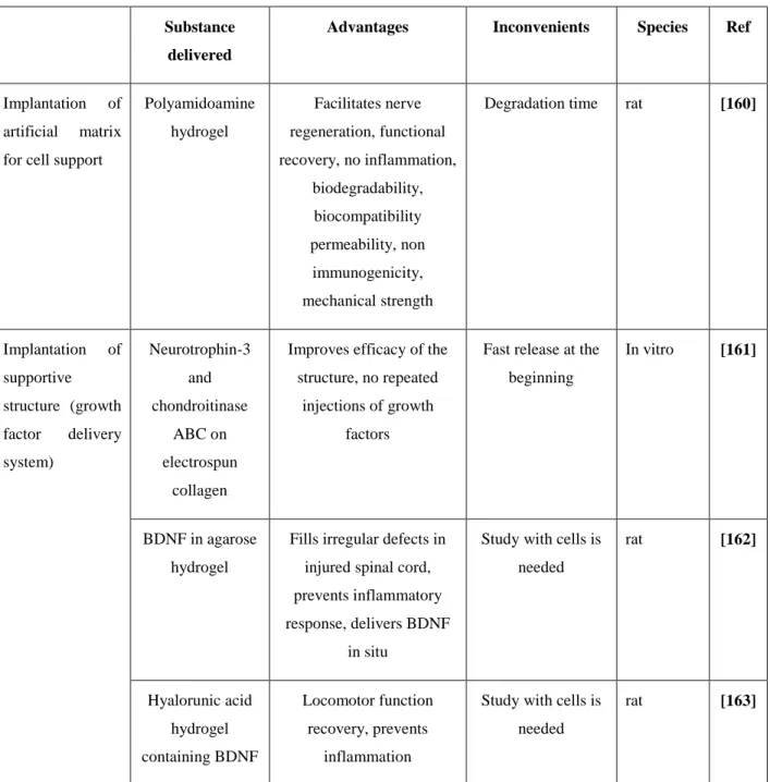
An alternative to growth factor encapsulation resides in growth factor direct blending with the polymer that constitutes the scaffold, during its fabrication. The scaffold will then release the included growth factors when degrading in the implantation area. In order to control cell differentiation, retinoic acid (RA)-containing poly(lactic-co-glycolic) acid electrospun fibers have been prepared by its incorporation with the polymer before electrospinning. RA was uniformly
present inside the construct and 80% of the loaded RA was released in 105 days at a constant rate. [39] Johnson et al. demonstrated the ability of scaffolds made of fibrin to deliver neurotrophin-3 (NT3) and platelet-derived growth factor (PDGF) in vivo. These scaffolds were prepared by the polymerization of an NT3/PDGF-polymer solution mixed with stem-cell derived neural progenitor cells. The presence of viable cells inside the tri-dimensional structure was confirmed, resulting in enhanced differentiation level of the cells into neurons due to the presence of NT3 and PDGF. In this study, the receptors for epidermal growth factor were neutralized by the addition of a blocking antibody to the structure, thus blocking the inhibitory pathways present in an injured area. Core shell fibers have also been used to deliver NGF for sciatic nerve regeneration in rats. NGF was loaded in the PEG core of the fiber, which was covered by a PLGA shell. After initial release of the NGF that migrated to the shell during the electrospinning process, NGF release was mainly due to diffusion through the shell layer, hence reducing the initial burst phenomenon. Grafting of this conduit for nerve guidance promoted functional recovery with the same efficiency as autografts 12 weeks after implantation. [40] A very similar technique has been used to deliver VEGF using coaxially electrospun fibers of hydroxyl-functionalized poly(ε-caprolactone) as a shell and VEGF-loaded BSA as a core. [41] Erythropoietin was delivered at stroke site using hyaluronan/methyl cellulose (HAMC) hydrogel. The latter was chosen based on its anti-inflammatory properties, mostly attributed to the presence of hyaluronan. Erythropoietin diffused from the implant to the ventricular zone of the stroke, reducing the cavity, and proved to have neuroprotective and neurogenerative properties. [42] Another advantage of this process may reside in its applicability to perform growth factor gradients. This aspect is particularly appealing since, in the adult nervous system, stem cells migrate to their final location by following growth factor gradients that are present in the tissue. [43-45] Axons are also known to grow along these gradients. More importantly, these gradients have an influence on the differentiation pathway followed by the cells. [46] Therefore, attempts have been made to mimic these natural gradients to spatially localize neurogenesis. [47] Growth factor gradients have been created either by assembling hydrogel pieces filled with growth factors at various concentrations [48] or by growth factor diffusion from wells being positioned within the hydrogel so as to result in the establishment of a gradient upon diffusion. [49] It was shown that gradients of the neurotrophic factors NGF and B27 used synergistically could multiply and
position synapses within the construct. [50] The gradients were created using channels connected with PDMS reservoirs that were filled at different time points to create a diffusion gradient. The synapse density increased proportionally with the NGF/B27 gradients.
2.3.3 Delivery of encapsulated growth factors to the native tissue
Scaffolds could also be applied to the delivery of growth factors in situ without any scaffold colonization by endogenous cells; the structure being used as a reservoir only. This use is believed to be of particular interest in the case of Alzheimer's disease where the patient is unable to maintain the adequate population of cholinergic neurons. That is, since NGF is a growth factor that stimulates the cholinergic function, its delivery to the brain of patients suffering from Alzheimer's disease has been investigated. In this specific case, the NGF be delivered passed the blood brain barrier, and the delivery must be very local to avoid any other regions of the brain to be impacted. Therefore, polymeric devices have been investigated to deliver NGF locally in rat brains. [51,52] While local delivery was successful, remaining limitations were the short half-life of those molecules and the short migration distance, hence the importance of the choice of the implantation site. [51,53] BDNF delivery, via hydrogels implanted into rat brains has also been investigated as BDNF could be used to treat major depression. In these studies, BDNF delivery was found to be successful as an antidepressant effect was observed. Once again, the efficacy of the treatment is closely related to the concentration of growth factors that reaches the targeted cells and the duration of the treatment. [54]
2.3.4 Use of cells to produce and deliver growth factors in situ
As emphasized in the preceding section, growth factor delivery is associated with many hurdles related to adequate spatio-temporal delivery. In order to overcome these issues, in situ synthesis of instructive cues by encapsulated cells has been investigated. In that respect, Shanbhag and colleagues encapsulated two lines of fibroblasts being genetically modified to produce either BDNF or NT-3. [55] The cell-containing scaffolds were demonstrated to enhance cell survival, migration distance of NPCs, and NPCs attachment to the scaffold as well as better survival when
compared to empty scaffolds. NT-3 secretion by modified fibroblasts was shown to promote neuron formation. In their study, Dey and colleagues injected MSCs genetically engineered to secrete BDNF or NGF in YAC 128 mice (a model for Huntington’s disease) and measured mice motor abilities. [56] MSCs secretion of BDNF improved neuronal survival and reduced clasping and behavioral deterioration after 9 months. Those results were not observed with NGF-producing MSCs or with pristine MSCs, although mice that had received unaltered MSCs also presented less clasping. This result was attributed to the secretion of anti-inflammatory cytokines by the injected MSCs, once again highlighting the advantages of MSC manipulation for this application. Such a strategy may however be inapplicable due to serious ethical concern.
Cells may also be used as an underlying layer to provide a favorable environment to regenerate neuronal structures ex vivo. The experimental approach would then rely on culturing a first layer of cells on top of which the cells of interest would grow. The first cell layer would provide trophic factor(s) to create a niche where the neuronal cells could optimally develop. In that endeavor, the use of a PA6 cell layer (a bone marrow-derived stromal cell line, precursor of adipocytes) was shown to provide an excellent environment in which human embryonic stem cells were able to differentiate in vitro into dopaminergic neurons in a highly specific way (87 %), prior to injection. [57] The exact role of PA6 cells was not fully understood, and was partly attributed to uncharacterized growth factors production. This technique has also been used to deliver growth factors to other cells directly inside the brain: NGF producing fibroblasts were implanted into the forebrains of six patients suffering from Alzheimer's and resulted in significant neuronal growth and slowed down the cognitive decline. [62]
2.4 Use of structures to mechanically support tissue growth
As discussed above, both stem cells and growth factors have shown promising results but their immediate use still presents limitations that prevent their routine implementation to the clinic. While injections of cells and/or growth factors promote tissue regeneration, a structure that offers an adequate mechanical support for the growth of endogenous or injected cells may be required when the injury site is large. [58] Filling the injury cavity with a mechanically relevant scaffold may prevent the formation of scar tissue; the latter being known as a hostile environment for
neurite growth due to its inherent structure and the presence of macrophages and other inhibitory factors. [59] Furthermore, in the case of neurodegenerative diseases, the pathological tissue is no longer able to adequately present the factors promoting proper differentiation of injected stem cells, as already observed when stem cells were injected to treat Alzheimer's disease. [13] [60] A scaffold that would support cell growth and differentiation, offering a protective and stimulating microenvironment may thus be required since the host tissue is no longer able to play that role.
2.4.1 Hydrogels to offer mechanical support to cells
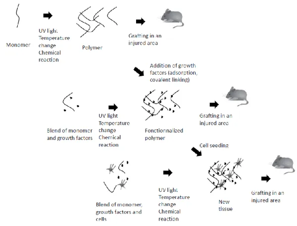
Hydrogels (Figure 1) have now emerged as a type of materials of choice for implant design. Hydrogels are usually defined as water-swollen polymer networks, often resulting from the polymerization of hydrophilic polymers. Such polymerization can be chemically driven, through reaction with a crosslinker, [61] or physically driven, through a change in reactivity or conformation that may be induced by pH or temperature changes [62] or even photo-activated. [63] Physically crosslinked hydrogels have the potential to be generated directly in situ; they do not require the use of photo-crosslinkers that are often toxic. However, these hydrogels are usually less stable than their chemically crosslinked counterparts. [62] Hydrogels present many advantages for neural tissue regeneration, as it is relatively easy to create a three-dimensional hydrogel structure harboring adequate mechanical properties. Furthermore, the inherent hydrophilicity of hydrogels - along with their ability to deliver growth factors - make them good candidates for neural cell supportive systems. [64] Amongst them, several polysaccharides including agarose, alginate, methylcellulose, dextran and chitosan as well as polysaccharide blends have been investigated. Soft, positively charged surfaces as those obtained with dextran/chitosan blends were reported to favor neuron attachment by Zudema and colleagues. [65] Hyaluronic acid (HA) was also reported to offer adequate mechanical support as well as good cell adhesion, proliferation and migration properties. [35]
The fact that physiological fluids penetrate hydrogels, combined to their softness and their three-dimensional structures, make them interesting candidates for tissue repair. As already demonstrated for vascular repair applications, hydrogels per se are presented by some authors as sufficient to allow neuronal cell growth and tissue formation. [66] On the same note,
differentiation of NSCs has been observed in three-dimensional porous chitosan scaffolds. On their own, these scaffolds were shown to promote differentiation to a higher extend than when soluble NGF was administered to cells cultured on two-dimension surfaces; the combination of three-dimensional scaffold with NGF however gave the best results. [67] The modulus of hydrogels was shown to be a critical parameter: for alginate-based constructs, a lower stiffness was reported to lead to better proliferation and differentiation of NSCs. [68]
Hydrogels have already been tested several times in animal models in vivo animal models for neuronal regeneration. [69-72] Their healing properties have been assayed when used as bridges after spinal cord or nerve sectioning. In such assays, Poly[N-(2-hydroxypropyl) methacrylamide] (PHPMA) hydrogels were successfully used in rats with sectioned spinal cord. [73] PHPMA constructs were reported to be well integrated, to prevent scar tissue formation and to promote axonal colonization. Such success was attributed to the good swelling and elastic properties of this type of hydrogels, combined to their high porosity that allowed neuronal and axonal growth through the structure. [73]
2.4.2 Electrospun fibers as a mechanical support for cell development
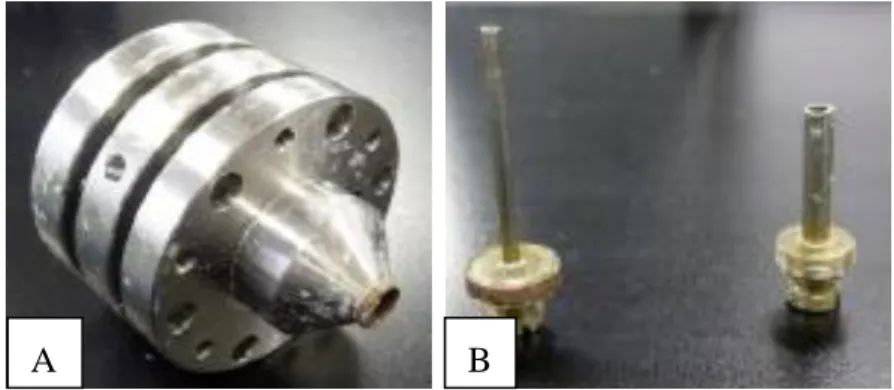
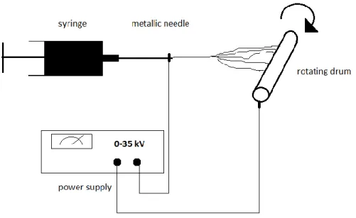
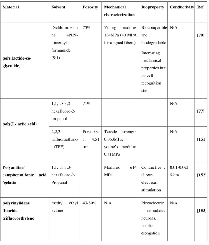
Another fabrication technique, namely electrospinning, has also been demonstrated to be successful for the generation of three-dimensional scaffolds made of micro- to nano- size fibers with controlled properties. This technique was originally developed to create textiles and filters and was then prop osed for wound dressing material. [74] In this process, a polymer solution is pushed through a metallic needle (Figure 2) while a strong electric field is imposed between the needle and the collector, charging the surface of the droplet at the extremity of the needle with static electricity (Figure 3). The polymer solution is then attracted by the electric field, and forms nanofibers (200-1500 nm of diameter) that dry during their flight between the needle and the collector. [75] This method is well adapted for neuronal scaffold production as fibrous scaffolds with high surface-to-volume ratios and structurally mimicking the extracellular matrix can be generated. [76] Furthermore, various polymers of interest, including biodegradable ones, can be used to create electrospun fibers. Those include poly lactic acid (PLLA), poly glycolic acid (PGA) and their copolymers, PLGA, poly(ether sulfone) (PES). [77-80] This high flexibility in terms of material selection, fiber diameter and orientation, porosity and mechanical properties offers a wide choice of possibilities so that biocompatible mats with adequate degradation rate and mechanical properties can be designed. [81] Examples are given in Table 2.Fig 2. Co-electrospinning needles: device allowing electrospinning of 3 different solutions with
a layered core shell structure (A). Needles used with this core shell structure. (B) Diameters are respectively 1.83 and 3mm.
B A
Fig 3. Electrospinning setup
Christopherson and coworkers investigated the influence of electrospun fiber diameter on rat NSCs, while the cells were cultured on PES fibers coated with laminin for cell attachment. [82] Of interest, different phenotypes were obtained with different types of fibers: fibers of small diameter (283 nm) were shown to promote NSCs differentiation into oligodendrocytes whereas larger fibers (749-nm diameter) promoted their differentiation into neurons. Wang et al. also showed that aligned tussah silk fibroin (TSF) fibers had a significant beneficial effect on neural differentiation of hESC. [83] They demonstrated cell migration along the fibers, and neurite growth with neurites following the fiber direction. In comparison, the cells cultured on random fibers migrated randomly with a shorter migration range. Neurites were longer on 400-nm aligned fibers, when compared to 800-nm ones. The authors nevertheless mentioned that, in some experiments, neurites happened to grow perpendicularly to fiber direction. They showed a 6% increase in the neuron amount on 400-nm aligned nanofibers when compared to 800-nm aligned fibers. Altogether, neurites were significantly longer and followed the material direction. In comparison, 800-nm random fibers showed a decrease both in number and total neurite length. Lower cell migration and proliferation were observed on these structures when compared to 2-D culture conditions. Cells were also shown to grow towards the fiber pattern: the thinner fibers directed the elongation of the cells, most likely explaining the increased level of differentiation into neurons. Of interest, cell culture on PLLA fibers was reported to abrogate the need of calf
serum in the culture medium. In the absence of calf serum, neurite alignment was increased. [84] Finally, the implantation of these structures in the area of sectioned nerves or degenerated tissues, as a support for the re-growth of host tissue, has been investigated. Nisbet et al. implanted electrospun fibers of poly(ε-caprolactone) in rat brains. [85] 60 days after implantation, scaffolds were not encapsulated in scar tissue, despite a recruitment of microglia and astrocytes. The structure promoted tissue growth as demonstrated by the presence of neurites inside the construct. Intriguingly, cell growth occurred perpendicular to fibers rather than in a parallel direction and randomly aligned fibers showed a better colonization by endogenous cells. The first observation was contradictory to experiments performed in vitro; further investigation is thus required to better understand and master this therapeutic approach.
Co-electrospinning may answer the dual needs of controlling the porosity and mechanical properties of materials, as well as allow drug delivery abilities of materials created with other techniques. In this approach, a core shell structure is created using a double coaxial needle. The core is made of the same polymeric materials as previously evoked. The outer part can be made of various materials such as laminin, which has been shown to favor cell attachment to the fibers. [86] Other proteins such as gelatin may also be used. [87] In such system, the core brings porosity and stiffness while the outer part of the fiber allows for a receptive contact of the cells on the scaffold surface; the outer layer may also be used for in situ growth factor delivery. As an alternative to co-electrospinning, electrospun fiber modification by hydrogel polymerization may be applied to create an outer shell to enhance cell contacts. Using this approach, Poly(ε-caprolactone) electrospun fibers were covered with poly(ethylene glycol)-poly(ε-Poly(ε-caprolactone) (PEGPCL) hydrogel, to support neural growth and deliver NGF in situ. [88] This construct was used to coat implanted electrodes. The mechanical support offered by the fibers reduced the degradation rate of the hydrogel, resulting in the elution of NGF, which increased the proximity of neurons and electrodes. An interesting technique has been developed by Huang et al. who electrospun fibers through a hole in a charged mask to create fiber patterns. Using this technique, fibers were patterned on a cell repellant hydrogel; regulating the voltage applied to the mask controlled the size and density of the fiber spots. This technique opens new avenues to control cell colonization through the creation of cell-receptive or -repulsive areas. [89]
Hydrogels and electrospun fibers have thus been demonstrated to favor stem cell growth and differentiation without any addition of growth factors; a very appealing property for the treatment of nerve injuries. However, in the case of degenerative diseases, the patient’s cells are usually unable to produce the required set of growth factors promoting regeneration; their inclusion in the implanted scaffold may thus be mandatory. Those factors may also be needed to counter the effects of molecules promoting the disease or, simply to promote the growth of functional tissue. [13] In addition to appropriate growth factor inclusion within a given scaffold, providing healthy stem cells to create tissue with restored functions may be desirable, as endogenous cells have lost this ability.
2.4.3 Use of structures to display bound growth factors in situ
As an alternative to growth factor diffusion from hydrogels or release from electrospun structures, growth factor immobilization through covalent grafting or non-covalent but stable tethering, has also been reported for both hydrogels and electrospun fibers. Covalent grafting of integrin binding peptides inside a collagen structure has been shown to enhance cell attachment to the substrate and improve cell viability [90]. Also, the stable tethering of BDNF onto collagen scaffold has also been assayed. The latter was achieved by designing a chimeric protein corresponding to BDNF fused to collagen binding domain. Scaffolds decorated with chimeric BDNF and seeded with dorsal root ganglia cells were implanted in rats and resulted in good recovery after spinal cord transection. [91] On the same note, the production of EGF fused to collagen binding domain has also been documented; its use for stem cells encapsulation within EGF-decorated collagen-based hydrogel resulted in better cell survival than in control collagen scaffold, while cells were observed to differentiate into various neural phenotypes. [92] Human recombinant BDNF and human recombinant NT-3 were also covalently immobilized on gelatin-based scaffolds using photo-polymerization: photocurable styrene derivatized gelatin was used to produce conduits on which extracellular matrix molecules along with rhBDNF and rhNT-3 were photo-co-immobilized. Dorsal root ganglia showed larger neurite extensions on structures displaying both neurotrophins than on structures displaying only one of the two proteins. [93]
NGF immobilization on chitosan scaffolds using genipin crosslinking was also applied to generate conduits later implanted in denervated rats. Such strategy was shown to lead to nerve reconstruction in vivo. [94]
Electrospun fibers were also used as displaying structures. [95] Amine terminated poly(ethylene glycol) and poly(ε-caprolactone) allowed Cho and colleagues to prepare amine functionalized electrospun fibers on which NGF was subsequently grafted. This strategy permitted to eliminate initial burst in released molecules, while increasing cell differentiation into neurons. Of salient interest, growth factor immobilization onto fibers led to more pronounced results than those related to cell culture on fibers in the presence of soluble cues. [96] In another work, covalent grafting of BDNF was performed on ethylene diamine-modified poly(ε-caprolactone) fibers. Cells proliferated more on these displaying structures even with half the amount of soluble BDNF. [97]
Spatially varying growth factor density during immobilization may also be of interest to explore the potential benefits of gradients of non-diffusible growth factor(s). This may be achieved thanks to recent technical developments now allowing for the creation of patterns of non-diffusible proteins: in that respect, laser-assisted protein adsorption by photobleaching is extremely promising. [98]
2.4.4 Use of anisotropic scaffolds to direct tissue growth
A desirable feature for smart engineered grafts would be to mimic patient’s tissue spatial organization in order to direct tissue regeneration, as nerve regeneration implies that new axons grow in the same direction. As previously mentioned, in order to meet this goal, growth factor gradients have been proposed, since they are responsible in vivo for directing the genesis of endogenous tissues. However, several attempts to create such gradients highlighted technical difficulties. [43-45] Therefore, as an alternative, the use of hydrogel- or electrospun fiber-based anisotropic scaffolds has been explored. Most hydrogels are obtained from the polymerization of monomers in solution and therefore show isotropy. Although this fabrication process is not adapted to generate structure determinism, hydrogels made of apo-collagen bear the potential to
be shaped to form aligned structures and membranes. [99] These structures, designated as linear ordered collagen scaffolds, were crosslinked with laminin to direct axon growth. [100] Implantation of this material allowed functional recovery in rats, with improved linear orientation of the newly generated tissue.
When topology needs to be controlled, electrospinning appears as the method of choice since the extent to which fibers are aligned can be varied with mandrel rotating speed. Electrospun fiber structures have been demonstrated to be extremely promising to direct axons growth and model newly grown tissue. In a previously evoked study, aligned PLLA fibers were used to grow motor and sensory neurons. Neurons were shown to grow following nano-fiber orientation and neurites grew along the fibers. [84] Aligned electrospun fiber structures may thus be a key to direct axonal growth and cell migration. [101]
2.5 Use of conductive structures to electrically stimulate neuron
differentiation
The neural tissue is electrically active, therefore attempts have been made to create conductive structures to allow electrical stimulation of the cells in order to improve the functionality of the graft. Cells that had been stimulated on a cover glass coated with conductive gold nanoparticles showed an increase in neurite length. [102] The design of a 3-D conductive scaffold made of freeze-dried collagen with polypyrrole, and presenting oriented micro channels has been reported. Electrical stimulation thanks to this structure increased neurite outgrowth in the direction of the electrical field in vitro and resulted in a better remyelination of the axons while Schwann cells were observed to migrate towards the anode. [103] In the case of electrospun fibers, conductivity may be obtained through the use of polypyrrole in the fiber core or as an outer layer being polymerized on electrospun fibers. [104]
Conductive electrospun nanofibers were also proved to enhance the rate of neurite growth and their total length. [105] When polypyrrole was polymerized onto PLA and PLGA electrospun fibers, the resulting structure was shown to be effective for dorsal root ganglia cell culture. Electrical stimulation resulted in 40% longer neurites. [105, 106] NGF has been incorporated in
such conductive constructs, in order to benefit from the growth factor effect as well as the stimulation. Longer neurites were obtained using this combination. [107]
Another way to add conductive properties to materials is to include carbon nanotubes or nanoparticles in the structure. [108] Carbon nanotube-mediated electrical stimulation was shown to promote neural maturity, and increase the speed of neurite outgrowth. [109] Carbon nanotubes were integrated in PLA electrospun fibers and conferred conductive properties. [110] Electrical stimulation resulted in increased levels of neuronal markers and stimulated neurons were more aligned along fibers. A difficulty for the use of nanotubes in electrospinning is the difficulty to spin them directly. To overcome this issue, Miao et al. co-electrospun multi-walled carbon nanotubes and poly(vinyl pyrrolidone) using co-axial electrospinning. The core of the fibers was composed of nanotubes while the sheath was made of poly(vinyl pyrrolidone) as a way to make the nanotube solution spinnable. [111]
Finally, carbon nanotubes can be used alone as conductive cell support. The main issue resides in their toxicity; their biofunctionalization is thus required. [112] NSCs were cultured on nano-ropes of carbon nanotubes: the structure strongly increased the maturation of NSCs into neuronal cells and promoted neurite elongation following the topography of the rope. [113]
2.6 Conclusion
Current techniques and strategies that may allow for neuronal tissue regeneration or replacement were reviewed. While stem cell injection was proven to promote functional recovery, it is now accepted that the effect is mainly due to growth factors and other instructive cues being secreted by these cells rather than injected cells differentiating into new tissue. These growth factors may thus be injected directly in situ to reach similar results, however, this requires heavy surgery. Their release from a scaffold implanted in the diseased area though diffusion or degradation, or alternatively, their attachment to an implantable scaffold, has been investigated with some noticeable success. Such a strategy is very attractive as it decreases the amount of growth factor needed and limits their effect to the injured area while taking advantage of the mechanical support provided by the scaffold itself.
Altogether, ideal nervous or neuronal implants should present several characteristics. The literature is now consensual upon the fact that, for an implant, mimicking the extracellular matrix features including porosity, pore size, and mechanical properties, while acting as a reservoir of instructive cues and harboring adequate topology for an appropriate spatial organization of the tissue, are all desirable traits. Therefore, despite all promising results presented therein, a significant amount of work remains to be done in order to fulfill all those requirements and enable the long-term success of regenerative medicine approaches. Above all, any technique should cope with surgeons’ constraints. In that endeavor, hydrogels present several interesting features as they are well characterized, commonly used as drug carriers, and since they can be prepared with a variety of chemicals. Also, as an alternative, electrospinning was proven to be very promising as it permits to control the mechanical properties of the scaffold while designing conductive materials. Alignment of fibers and incorporation of growth factors were also shown to be feasible for additional tailoring of electrospun scaffold, i.e., to direct tissue growth and control cell phenotype. Many other materials may also lead to the development of constructs with improved regenerative potential: polyurea silica aerogels, [114] phosphate-based glass fibers [115], to name a few, are indeed amenable to produce scaffolds with interesting properties regarding their biocompatibility, nano-porous structure, mechanical strength, directionality or surface functionalization. Matrigel, which is made of extracellular proteins secreted by Engelbreth-Holm-Swarm (EHS) mouse sarcoma that easily self assemble to form a matrix, has been assayed to regenerate nerve tissue and gave good results regarding the differentiation of dopaminergic neurons. [116] Finally, self-assembled fibers made of spider silk have been reported to spatially support neuronal and astrocytic differentiation. [117] [118]Scaffold design could also benefit from a other fabrication techniques such as oriented growth factors immobilization strategies [119] , layer-by-layer lithography [120], microcontact [121] [122] or inkjet printing [123] [124] to give a few examples of very promising approaches being currently explored. Finally, recent research is now pointing towards alternatives to electrical stimulation to promote cell differentiation: sub sonic vibrations [125], stretching [126], cyclic tensile loading [127] and low level LASER (LLL) stimulation [128] are promising avenues that may impact significantly future research in neural regeneration.
Growth factor Effect Enhanced Phenotype / Pathway
Delivery
technique Species Ref
BDNF
Increases number of neurons in striatum, septum, thalamus and hypothalamus N/A Intraventricular administration Rat [129] Promotes myelinisation, prevents migration
TrkB and p75 pathways for cell death, Rho kinase for migration Injection Rat [130] GDNF Regulates proliferation, differentiation and survival of neurons
Pathways associated to Ret and GDNF family receptor α1 (GFR α1), on the surface of migrating enteric neural crest–derived cells
N/A Chick [131]
Erythropoietin
Prevents apoptosis, anti-oxydant, promotes differentiation into dopaminergic neurons Mimics lower O2 environment Addition to the medium, in vitro Rat [132] EGF Enhances NSC proliferation
Neural stem cell Released from implanted hydrogel
Mouse [133]
FGF-2
Maintains cell viability, increases cell
proliferation
N/A Addition to the medium, in vitro Rat [82] FGF-4 Promotes serotoninergic differentiation
N/A Addition to the medium, in vitro Rat [134] FGF-8 Promotes dopaminergic and serotoninergic differentiation
Effect on antero posterior position Addition to the medium, in vitro Rat [134] Mitogen, promotes differentiation into mesencephalic precursors
Effect on antero posterior position
Addition to the medium, in vitro
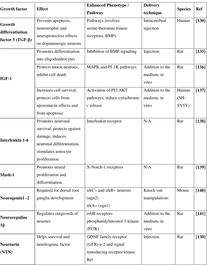
Table 1. Growth factors and their use in the context of neuronal disease treatment (continues)
Growth factor Effect Enhanced Phenotype /
Pathway
Delivery
technique Species Ref
Growth differentiation factor 5 (TGF-β) Prevents apoptosis, neurotrophic and neuroprotective effects on dopaminergic neurons Pathways involves serine/theronine kinase receptors, BMPs Intracerebral injection Human [130] IGF-1 Promotes differentiation into oligodendrocytes
Inhibition of BMP signaling Injection Rat [135]
Protects motor neurons, inhibit cell death
MAPK and PI-3K pathways Addition to the medium, in vitro
Rat [136]
Increases cell survival, protects cells from epoxomicin effects and from apoptosis
Activation of PI3/AKT pathways, reduce cytochrome c release Addition to the medium, in vitro Human (SH-SY5Y) [137] Interleukin 1-6 Promotes neuronal survival, protects against damage, induces neuronal differentiation, stimulates astrocyte proliferation
Interleukin receptor N/A Rat [138]
Mash-1
Promotes neural proliferation and differentiation
X-Notch-1 receptors N/A Rat [139]
Neurogenin1 -2
Required for dorsal root ganglia development trkC+ and trkB+ neurons (ngn2) trkA+ (ngn1) Knock-out manipulations Mouse [140] Neuroreguline 1β Regulates outgrowth of neurites erbB receptors phosphatidylinositol 3-kinase (PI3K) Addition to the medium, in vitro Rat [141] Neurturin (NTN)
Helps survival and neuritogenic factor
GDNF family receptor (GFR) α-2 and signal transducing receptor kinase Ret