HAL Id: dumas-01623795
https://dumas.ccsd.cnrs.fr/dumas-01623795
Submitted on 13 Nov 2017HAL is a multi-disciplinary open access archive for the deposit and dissemination of sci-entific research documents, whether they are pub-lished or not. The documents may come from teaching and research institutions in France or
L’archive ouverte pluridisciplinaire HAL, est destinée au dépôt et à la diffusion de documents scientifiques de niveau recherche, publiés ou non, émanant des établissements d’enseignement et de recherche français ou étrangers, des laboratoires
Impaired cerebrovascular reactivity assessed by BOLD
hypercapnic fMRI is associated with increased risk of
stroke in patients with symptomatic intracranial
atherosclerotic stenosis
Jérémie Papassin
To cite this version:
Jérémie Papassin. Impaired cerebrovascular reactivity assessed by BOLD hypercapnic fMRI is asso-ciated with increased risk of stroke in patients with symptomatic intracranial atherosclerotic stenosis. Human health and pathology. 2017. �dumas-01623795�
AVERTISSEMENT
Ce document est le fruit d'un long travail approuvé par le
jury de soutenance et mis à disposition de l'ensemble de la
communauté universitaire élargie.
Il n’a pas été réévalué depuis la date de soutenance.
Il est soumis à la propriété intellectuelle de l'auteur. Ceci
implique une obligation de citation et de référencement
lors de l’utilisation de ce document.
D’autre part, toute contrefaçon, plagiat, reproduction illicite
encourt une poursuite pénale.
Contact au SID de Grenoble :
bump-theses@univ-grenoble-alpes.fr
LIENS
LIENS
Code de la Propriété Intellectuelle. articles L 122. 4
Code de la Propriété Intellectuelle. articles L 335.2- L 335.10
http://www.cfcopies.com/juridique/droit-auteur
UNIVERSITE GRENOBLE ALPES FACULTE DE MEDECINE DE GRENOBLE
Année : 2017 N°
Altération de la vasoréactivité cérébrale en IRMf hypercapnique chez
les patients présentant une sténose artérielle intracrânienne
symptomatique d’origine athéromateuse à haut risque de récidive.
THESEPRESENTEE POUR L’OBTENTION DU DOCTORAT EN MEDECINE DIPLÔME D’ETAT
PAPASSIN Jérémie
THESE SOUTENUE PUBLIQUEMENT A LA FACULTE DE MEDECINE DE GRENOBLE*
Le : 12 Octobre 2017
DEVANT LE JURY COMPOSE DE
Président du jury : M. le Professeur Mikael MAZIGHI Membres
M. le professeur Tae-Hee CHO M. le Docteur Olivier DETANTE
M. le Professeur Alexandre KRAINIK, directeur de thèse [Données à caractère personnel]
REMERCIEMENTS Aux membres du jury,
Au Professeur Mikael Mazighi, chercheur hors pair et neurologue interventionnel. Vous m’avez fait l’honneur de quitter la capitale pour venir présider ce jury.
Au Professeur Tae-Hee Cho, neurologue et excellent pédagogue. Je tenais absolument à ce que vous fassiez parti de ce jury pour apporter votre expertise à ce travail. Vos présentations en congrès ou en cours de DES sont toujours claires et pertinentes.
Au Docteur Olivier Detante, neurologue, show-man et chercheur passionné. Merci pour tes enseignements neurologiques et extra-neurologiques, tu as su me transmettre ton intérêt pour la science ainsi que ton dynamisme.
Au Professeur Alexandre Krainik, neuroradiologue et chercheur polyglotte. Toujours le mot pour rire. Merci pour la confiance que tu m’as accordé pour ce projet. Tu as su à la fois me laisser indépendant et être présent lorsqu’il le fallait.
Aux collègues et amis,
A Olivier Casez, ancienne star des terrains de foot, maintenant amateur de vin et d’arabica. Tu aurais fait aussi un excellent directeur de thèse. A tes côtés, j’ai appris à être médecin et (presque) neurologue, depuis le début de l’externat.
A Mathieu Vaillant. Les personnes qui t’ont élu neurologue le plus gentil du monde n’ont sûrement jamais couru ou monter à ski de rando avec toi, la cadence est insoutenable. Merci pour tes conseils et ta bienveillance.
A Gérard Besson. Homme érudit et musicien acharné vous avez façonné une véritable légende. J’ai hâte d’être CCA chez vous et d’apprendre à écrire mon prénom en hiéroglyphe. A Katia Garambois. Je scrute maintenant avec attention le moindre ostium vertébral.
A Isabelle Favre « la dominatrice », assistante méticuleuse et avisée, tu aurais dû lire nos courriers d’hospitalisation jusqu’au bout…
A Anne So, archétype de la femme du XXIème, capable de tout faire en même temps et très bien, à mes yeux (et à ceux de bon nombre d’hommes), c’est une chose incroyable.
A l’équipe médicale de neurologie A, merci de votre soutien pendant ces semestres au pays des stimulés et des implantés.
A l’équipe médicale de psychiatrie. Stage très enrichissant alors que je venais à reculons. J’ai appris les rudiments de la psychopathologie en abandonnant ma blouse de « somaticien » et mes objets contraphobiques (marteau réflexe et sthéto).
A l’équipe 5 du GIN dirigée par Emmanuel Barbier. Merci pour l’accueil que vous m’avez réservé dans le monde parallèle de la recherche. Pensées particulières à Claire, Nora et Ligia. A Monsieur Tarel, neurologue passionné et homme juste, vous êtes un exemple pour moi. Merci pour les leçons du soir, vos expressions Shadoks et vos répliques à la Audiard.
A Marie Barre, une neurologue toujours classe en chaussures à paillettes. Jamais de fashion faux pas, Cristina Cordula serait fière de toi.
A Julien Gere. Merci à toi je sais dorénavant ce qu’est un waketeu.
A Nicolas Miret. Ton sens clinique aiguisé et ton bronzage de ski de rando m’ont toujours énervé (ceux sont plus des aveux que des remerciements, désolé…).
A toute les équipes paramédicales et secrétaires, merci pour votre attention et soutien tout au long de ces années. On s’est bien marré !!
Aux co-internes et amis,
Dans l’ordre d’apparition, Anne cath’ ou Bernard pour les intimes, tu as donné le ton et le rythme (au pas de course) à cet internat ! Marine alias mariiiinnnnneeee a ensuite apporté sa douceur et son sourire. Alors que le bon Moumoun faisait semblant d’être un clinicien, mais tu ne trompes personne, on s’est que tu bluffes Martonni. Sérieusement, merci mec pour les bons et mauvais moments qu’on a passé ensemble. Formation par la suite du duo de choc avec Pauline, une co-interne brillante, esthète et championne toutes catégories confondues du radio-potin, j’ai hâte de reformer la team. Descente d’un étage et changement de pays, on se retrouve avec Emma (pulcino de la Basilicata) sous l’œil attentif de Raul, à parler rigidité, voyages et fromages. Puis transfert chez l’ennemi voisin. Pendant que Bibi le bo goss, venu à Grenoble pour faire ses preuves en mouvement (pour finalement faire plomberie à Dijon), l’ami Seb assurait l’intendance… Qui aurait parié qu’avec ce zigoto chevelu nous allions trouver un remède miracle pour tous les zinzins, notre classique valium-tercian. La Ponce quant à elle affinait sa technique pour repousser les avances de nombreux prétendants et la Maze élaborait un sujet d’étude sur les trans-genres (bref des psychiatres, des vraies). Pour continuer sur le rythme effréné de la psychiatrie, rien de mieux qu’une année recherche. C’est avec Ligia (Lig’ Lig’) que j’apprenais à dompter les rongeurs. A l’écart du tumulte grenoblois, la bande de joyeux lurons composée d’Aurèle le mélenchoniste, Juju la présidente, la grosse Margo, Hélène IPA, Elo-psy et Louise le fou du volant collectionnaient les histoires de dingues !! Tout ça pour qu’au final, on décide de me faire redoubler… Heureusement, Catalina et Hugo bite de fer tiennent la boutique pendant que je finalise ce manuscrit. Pensées à tous les autres petits copains : Clem la pile électrique, Col-Col, le bon Gab’, Marie la rêveuse, Nasty l’autobus, Anne la cycliste, Gio le plus grand neurologue, Rig’, Thomas (je te prends quand tu veux au squash gros), la bonne Hélène, Guillaume et son baobab, Salma, Loïc le babyfooteux et Lucie (et son absence de second degré). Merci d’avoir été là, l’histoire aurait été moins drôle sans vous !
Aux copains,
Aux pedz’, Badar, Vinc’, Bilou, Malik et Valer, au plaisir de se matter une énième fois le pari et les 3 frères, ou de se retrouver pour un petit resto chinois à volonté et aux copines des bancs de l’école, Sophie, Mytien, Sandar’ et Aurore. Amies de toujours et pour toujours. Aux colocs de l’île verte. La bière, les nouilles chinoises, les cloisons en polystyrène et Fifa n’auront pas eu raison de nous. Alex&Aude vous êtes toujours prêts à m’accueillir sur Paris pour un F.I.E.A.L.D. ou un concert mais pour ce qui est de la grimpe… on a parlera à Peter. Ciol&Poupette, même en déménageant on arrive à être voisins, c’est con mais c’est très sympa. Merci Amel’ pour ce que t’as fait pour la Grande et la Petite Jojo, et merci Gros pour la relecture de la thèse, je pense que tu seras la seule personne, peut être avec les membres du jury, à avoir lu ce papier.
Aux colocs de SMH. Nico l’Œdipe et Flo le poulpe, merci votre reprise de Toujours pas d’amour de Priscilla et vos fouines empaillées. Moumoun t’as bien essayé de incruster dans cette magnifique maison, on s’est que la moquette aux murs ça donne du cachet !
A la fine équipe, Ludo, Astrid, Yohan, Marjo’, Flo & Flo, Martin, Jess, Nico, Krousti-Bat’ et à tous les potes de promo, Marie Peg’, la brune, Rom’ et ses crocodiles, Z, Sof’, Jean Luc, Manon, Jano, Simon. Je ne sais toujours pas pourquoi j’ai choisi neuro, à vous voir le cerveau n’est clairement pas un organe essentiel.
Pensée spéciale à Ludobite et la grosse Dièg’, vous formez une excellente base arrière et de très bons con-pagnons de révision. « On s’est pas comment ça va finir, mais au moins on aura bien rigolé »
A la bande de mecs (et meufs) sympas, Reda et Isle 187, La truite et MauMaud la Milf, Le nain et Bébéééee, Max et Alizée les petites personnes, Mat’ Taïchi et Popo, Fred le romantique & Eva la teutonne. Vous êtes de grands malades ! Unis dès la P1 puis dispersés aux 4 coins de l’Europe et de la Savoie, nos retrouvailles donnent toujours mal au crâne.
A ma famille,
A Poup’ et Mum, je ne serais pas là sans vous mais surtout je n’aurai jamais été celui que je suis sans votre soutien et votre amour. Je vous dois tout.
A ma tante Vivi, tu es la personne la plus drôle et aimante que je connaisse. Je te considère comme ma deuxième maman.
Aux frérots, Raph’ et Small T, mes complices. A courir derrière un ballon ou à de se mettre sur la gueule, je n’ai que de bons souvenirs avec vous.
A mes tantes, oncles, cousins et cousines et à tous les moments partagés ensemble. D’avoir une si belle famille est, à mes yeux, la plus grande des richesses.
A mes grands-parents, qui doivent sûrement me regarder d’où ils sont. Je pense souvent à vous, j’espère vous rendre fier.
A belle famille, qui me traîne à ski en haut de la Saulire ou dans les arcos. C’est toujours un plaisir de partager votre table! Vous faites beaucoup pour nous. Merci.
A Philippine.
Ravissante, pétillante, candide, tu es plus que ma moitié, je n’irai pas bien loin sans toi. Même s’il nous a fallu plus de 7 ans pour se rencontrer, j’espère ne plus perdre un seul instant et passer le restant de mes jours à tes côtés. Je t’aime ma belle.
TITLE:
Impaired cerebrovascular reactivity assessed by BOLD hypercapnic fMRI is associated with increased risk of stroke in patients with symptomatic intracranial atherosclerotic stenosis
AUTHORS:
Jérémie Papassina, Olivier Heckbc, Naïla Boudiafcd, Eric Condaminee, Isabelle Favrea, Sébastien Marcelf, Kamel Boubagrab, Katia Garamboisa, Florence Tahonb, Johan Pietrase, Olivier Detanteac, Alexandre Krainikbce
Stroke Unit, University Hospital Grenoble Alpes, 38000 Grenoble, Francea
Department of Neuroradiology, University Hospital Grenoble Alpes, 38000 Grenoble, Franceb Univ. Grenoble Alpes, Inserm, Grenoble Institute of Neurosciences, 38000 Grenoble, Francec Univ. Grenoble Alpes, CNRS, LPNC, 38000 Grenoble, Franced
Univ. Grenoble Alpes, Inserm, CNRS, University Hospital Grenoble Alpes, IRMaGe, 38000 Grenoble, Francee
Stroke Unit, Hospital Savoie Metropole, 73000 Chambery, Francef
*corresponding author: alexandre.krainik@univ-grenoble-alpes.fr
Key words: brain; stroke; intracranial arterial stenosis; DSC perfusion; BOLD fMRI;
LIST OF ABBREVIATIONS
ACA: anterior cerebral artery AIF: arterial input function ASL: arterial spin labelling
BOLD: blood oxygenation level dependent CBF: cerebral blood flow
CBV: cerebral blood volume CVR: cerebrovascular reactivity EPI: echo-planar imaging
fMRI: functional magnetic resonance imaging FOV: field of view
GM: gray matter
GRE: gradient recalled echo
IAS: intracranial atherosclerotic stenosis ICA: internal carotid artery
LDLc: low-density lipoprotein cholesterol LI: laterality index
MCA: middle cerebral artery MNI: Montreal Neurologic Institute mRS: modified Rankin scale
MTT: Mean Transit Time
NIHSS: National Institutes of Health Stroke Scale PCA: posterior cerebral artery
PET: positron emission tomography PetCO2: end-tidal CO2 pressure
RECURRENT: recurrent ipsilateral ischemic event ROI: regions of interest
SINGLE: single ischemic qualifying event
SPECT: single-photon emission computed tomography TDS: transcranial Doppler sonography
TIA: transient ischemic attack WI: weighted images
ABSTRACT:
Introduction: Despite intensive medical management, intracranial atherosclerotic stenosis
(IAS) remains at risk of recurrent ischemic events. Impaired cerebrovascular reserve is suggested to explain hemodynamic stroke. Cerebrovascular reactivity assessed by hypercapnic challenge using BOLD functional MRI (CVR BOLD fMRI) has been proposed to estimate cerebrovascular reserve and to identify patients at higher risk. To discuss patients’ selection for additional therapy such as intracranial angioplasty and stenting, we studied the relationships between baseline characteristics of patients with unilateral symptomatic IAS, recurrence of ischemic events, and CVR BOLD fMRI.
Subjects and methods: Nineteen patients with symptomatic unilateral IAS of middle cerebral
artery (MCA) or internal carotid artery were selected. We calculated statistical relationships between individual clinical and biological baseline characteristics, recurrent ischemic events, basal perfusion estimated by dynamic susceptibility contrast MRI, and CVR BOLD fMRI measured in MCA territories (CVRMCA), and reported using laterality indices (LI).
Results: Eight patients (42%) had an abnormal LI CVRMCA (|LI| CVRMCA ≥ 0.08). During a mean
follow-up of 3.6 years, recurrent ischemic events occurred within the first year. They were more frequent in impaired CVRMCA group (n=6/8) than in normal CVRMCA group (n=1/11),
with different survival curves (log rank, p=0.003). Baseline characteristics were similar in both groups.
Conclusion: Impaired CVR assessed by BOLD hypercapnic fMRI is associated with increased
risk of stroke in patients with symptomatic IAS. CVR mapping should be proposed to select high-risk patients in order to discuss additional treatment.
INTRODUCTION:
Intracranial atherosclerotic stenosis management
Intracranial atherosclerotic stenoses (IAS) account for approximately 10% of cerebrovascular ischemic strokes, and up to 50% in Asian populations1–3. IAS are associated with high rates of recurrent ischemic event and cerebrovascular mortality when compared with other stroke subtypes4,5. The recurrence rate of ischemic events in the territory of the stenotic artery remains high6,7, with a major risk of subsequent disabling stroke8, despite optimal medical treatment.
Large randomized trials demonstrated better outcomes with aggressive medical management compared to IAS recanalization using intracranial stenting9,10 or surgical extracranial-intracranial bypass11,12 to prevent recurrent stroke because of the high rate of procedural complications. Actually, a combination of antiplatelet therapies (aspirin 325mg per day plus clopidogrel 75mg per day for 90 days) is recommended with an intensive medical management of risk factors: high blood pressure, elevated low-density lipoprotein cholesterol (LDLc) levels, diabetes, smoking, overweight, and insufficient exercise13. When life style adaptation is insufficient, antihypertensive drug and statin should be used to obtain a systolic blood pressure below 140 mmHg and LDLc level below 0.7g/l, respectively.
Vitamin K antagonists were associated with higher rates of adverse events and without benefit over aspirin4. No clinical benefit was demonstrated for direct oral anticoagulant (direct IIa and Xa factor inhibitors) or other anti-platelet agents such as cilostazole or dipyridamole.
Despite optimal medical management, IAS remains associated with a recurrence rate of stroke events at 2 years of 15-25%4,9. The identification of risk factors of stroke recurrence among IAS patients under aggressive medical management would be helpful to consider alternative therapy14,15. Indeed, old infarct in the territory of the stenosis, new stroke presentation, and absence of statin use at enrollment were independently associated with high recurrence rates of stroke events in the medical group in the SAMMPRIS trial16. Additionally, impaired cerebrovascular reserve was suggested as a risk factor of stroke recurrence17.
Cerebrovascular functions
The cerebral vasomotricity leads to active changes of vessels resistance, through dilation and contraction, in order to adjust cerebral blood flow (CBF) in response to variations of neural activity (neurovascular coupling), to cerebral perfusion pressure (autoregulation), and to circulating gases and metabolites (vasoreactivity).
The neurovascular coupling adapts the cerebral blood flow to local neuronal activity providing oxygen, glucose and adenosine triphosphate to neurons and glia, and removing heat and catabolites18.
The cerebral autoregulation refers to the ability of vasculature to maintain CBF constant, despite changes of perfusion pressure, to prevent ischemia-related brain damage. Combination of myogenic, humoral and neurogenic feedback mechanisms are implicated in the regulation of adequate CBF maintaining constant distal perfusion pressure in brain capillaries during large arterial pressure variations19,20.
The cerebrovascular reactivity (CVR) modulates CBF in response to changes of circulating stimulus. Circulating gases such CO2 and O2 elicit vascular dilation and contraction,
respectively, to maintain brain oxygenation.
Magnitude of the cerebrovascular properties is modulated by physiological changes (e.g. age, sex, hypoxia, arterial pressure, basal perfusion…), brain diseases (e.g. Alzheimer’s disease; stroke,…), and structural vascular damage such as atherosclerosis (chronic arterial hypertension, diabetes,…)18,21.
CVR assessment
When perfusion pressure decreases downstream the IAS, autoregulation may maintain CBF by vasodilation and cerebral blood volume increase. This additional dilation, known as cerebrovascular reserve, might be insufficient to compensate further perfusion pressure decrease. When cerebrovascular reserve is exhausted, patients are at risk of low grade ischemia and recurrent hemodynamic events22,23. Thus, the estimation of the cerebrovascular reserve using perfusion measures before and after vasodilatory stimulus has been proposed to identify patients at risk of ischemia 17,24.
While Positron Emission Tomography (PET) remains the “gold standard” technique25, non-invasive alternative methods have been advocated, such as transcranial Doppler sonography (TDS)26, xenon-enhanced computed tomography27, single-photon emission computed tomography (SPECT)28, and more recently by functional magnetic resonance imaging (fMRI) using blood oxygenation level dependent (BOLD) contrast or arterial spin labelling (ASL)24. Most common vasodilatory stimuli are acetazolamide and CO217,18.
Among these techniques, BOLD CVR fMRI is an accessible, non-invasive, and reproducible whole brain imaging method29,30. Among vasoactive stimuli, CO2 challenge is safer when
compared to hypotensive provocation. Intravenous acetazolamide administration is exposed to side effects, contraindications, and large inter-subject variability31. Thus, well tolerated CO2 inhalation with immediate and transient vasodilation is now advocated to be the most
suitable stimulus29–31.
Objectives
Our objective was to investigate the relationships between clinical and biological characteristics of patients with unilateral symptomatic IAS, recurrence of ischemic events, basal perfusion parameters, and individual CVR mapping using BOLD hypercapnic fMRI. The study allowed to estimate: 1°) CVR and basal perfusion characteristics among patients with recurrent ischemic event, and 2°) occurrence of recurrent ischemic event in patients with CVR impairment.
SUBJECTS AND METHODS
Study participants
We conducted an observational study on adult patients referred at the University Hospital Grenoble Alps between February 2011 and August 2016 for transient ischemic attack (TIA) or non-disabling stroke that was attributed to 50–99% unilateral IAS of internal carotid (ICA) or first segment of the middle cerebral artery (MCA) assessed by catheter angiography or brain CT angiography32. Optimal medical treatment, consisting of dual antiplatelet therapy aspirin (75-300 mg per day) plus clopidogrel (75 mg per day), and management of risk factors (high blood pressure, hypercholesterolemia, diabetes, smoking, overweight) was implemented for each patient.
We excluded patients with tandem extracranial stenosis (70–99%) or occlusion, intraluminal thrombus proximal to or at the target lesion, any hemorrhagic infarct within 14 days before enrolment, non-atherosclerotic causes of intracranial stenosis, presence of a cardiac source of embolus, history of stent placement, cervical endarterectomy or extra-intracranial bypass surgery, contraindication to MRI and Gadolinium-Dota intravenous administration. We also excluded patients with incomplete MRI protocol due to excessive head motion (translation or rotation >2mm or >2° in any direction, respectively) or inappropriate hypercapnic stimulus. The study protocol was approved by our institute’s committee on human research (B120620-10).
Clinical data collection and follow-up
Qualifying ischemic event-related data was obtained from medical records and included stroke risk factors, medical history, National Institutes of Health Stroke Scale (NIHSS; 0 to 42,
higher score indicating greater severity) at hospital admission, modified Rankin Scale (mRS; 0: no symptom to 6: death) at discharge, serum creatinine, glycosylated haemoglobin and lipid testing. Clinical follow-up period ranged from qualifying event until death, loss of follow-up or study end. All patients were seen yearly or more frequently if needed. Recurrent stroke or TIA, adverse events and assessment of the patient’s functional abilities and mobility using the mRS were conducted by a neurologist (JP, IF, KG, SM, OD) during visits or by telephone interview. Any stroke or TIA in the territory of the qualifying artery was considered to be a recurrent ischemic event. Ischemic events out of the territory of the qualifying artery or death were also analyzed.
MRI Protocol
All participants were scanned using a 3T ACHIEVA TX MR scanner (Philips Healthcare®, the Netherlands) at University Hospital Grenoble Alps, using a whole-body RF transmit and 8 channels head receive coils. All participants had anatomical, basal perfusion, and BOLD CVR studies.
Anatomical study
Anatomical study consisted in a 3D gradient recalled echo (GRE) T1-weighted image (WI) (160 contiguous slices, slice thickness=1mm, TR/TE/α: 9.9/4.6ms/8°, 256x256 matrix, field of view (FOV) of 256x160 mm for 4.5 min acquisition time).
Basal perfusion study
Perfusion-WI was performed using dynamic T2*-WI GRE echo-planar imaging (EPI) (25 contiguous slices, thickness=4mm, TR/TE/α: 1660/40ms/75°, 112x112 matrix, FOV=224x184mm), tracking a bolus of 0.1mmol/kg of Gadolinium (Guerbet®, France),
was flushed by a bolus of physiological saline (60mL). To obtain an accurate estimate of the baseline MR signal intensity S(0) prior to the arrival of contrast agent an injection delay of 10 seconds was applied. Eighty single-shot, gradient-echo, echo-planar images, were obtained in each of the slices during the 2’11 minute acquisition time.
CVR study
CVR imaging consisted in BOLD sensitive T2* WI GRE EPI acquisition (32 axial slices, slice thickness=4mm, TR/TE/α: 3000/35ms/90°, 64x64 matrix, FOV=256x256 mm, 240 dynamics) with a whole brain coverage. Hypercapnic challenge was conducted according to the following block-designed paradigm [air(1′)–hypercapnia(2′)–air(1′)]×3, for a total duration of 12 minutes. Each participant was exposed alternatively to medical air and to a hypercapnic normoxic gas mixture (CO2 (8%), O2 (21%), and N2 (71%) through a non-rebreathing face
mask. Physiological parameters, including end-tidal CO2 pressure (PetCO2), arterial oxygen
saturation, breath frequency and heart rate were monitored using a MR compatible device (Maglife, Schiller medical). This protocol induces an increase PetCO2 about 10 mm Hg,
recorded via nasal cannula.
Images processing and analyses
Data analysis was performed with Amigo, an in-house post-processing suite written in Python 3.6 using Matlab (MathWorks, Inc., Natick, MA, USA) and SPM12 (SPM, Wellcome Department of Imaging Neuroscience).
Anatomical data
White and gray matters (GM) were segmented through the 3D GRE T1 sequence using the standard space tissue probability maps from SPM12. GM maps were spatially normalized to the Montreal Neurologic Institute (MNI) T1-weighted template and resampled at 2x2x2mm
resolution. GM maps were further smoothed with a 6-mm Gaussian Kernel for Regions of Interest (ROI) analyses.
Basal perfusion data
Basal perfusion images preprocessing consisted in coregistration, normalization, resampling onto anatomical images, and spatial smoothing with a 6-mm Gaussian Kernel. The arterial input function (AIF) was estimated by averaging 5 voxels from all slices of the perfusion scan during the dynamics. These 5 voxels were automatically detected by optimizing a function with a signal evolution characterized by the higher peak height, a faster dropout, concomitant to a smaller full-width-half-maximum and bolus arrival time (brain-feeding artery detection). Mean Transit Time (MTT) maps were then calculated pixel-wise with a deconvolution approach based on a singular value decomposition using a tracer arrival timing insensitive method and an automatic regularization of oscillations33. The relative Cerebral Blood Volume (CBV) was estimated from the area under the curve of the voxel concentration. To obtain quantitative maps, the mean brain CBV was normalized to 5%. The MTT maps were estimated from the area under the curve and from the time to maximum of the deconvolved function respectively. CBF was calculated as the ratio CBV/MTT from the central volume theorem. The excluded voxels from the computation were averaged with the nearby voxels.
CVR data
BOLD CVR images preprocessing consisted in coregistration, normalization, resampling onto anatomical images, and spatial smoothing with a 6-mm Gaussian Kernel. Statistical analysis was conducted using a general linear model and the individual PetCO2 in mmHg as a
Figure 1: FLAIR-T2 weighted image (A), Time-Of-Flight weighted image (B), basal perfusion
parameters and CVR maps in 68-year woman referred for symptomatic right MCA stenosis in red arrow, without ischemic recurrence (radiological convention).
ROI analyses
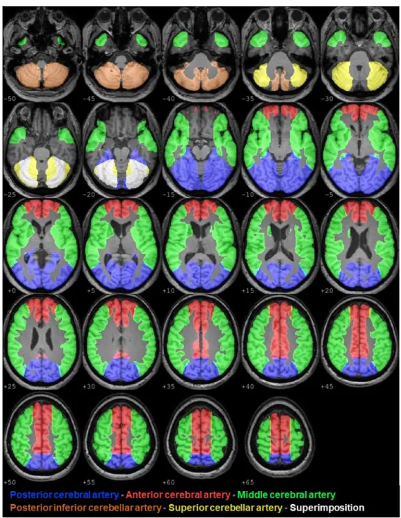
Vascular territories ROIs were obtained by using a standard set of vascular ROIs34 masked by smoothed GM maps, giving individual ROIs in the territories of the middle (MCA), anterior (ACA), and posterior (PCA) cerebral arteries (Figure 2).
We conducted ROI analyses on each basal perfusion parameter and CVR maps. For each
parameter, we calculated mean values and a laterality index (LI), e.g. for CVR in MCA territory LI CVRMCA = (Left_CVRMCA – Right_CVRMCA) / (Left_CVRMCA + Right_CVRMCA). A
positive value of LI representing left-hemisphere dominance and negative value a right-hemisphere dominance. The intermediate values reflect varying degrees of laterality. In our analyses we used the absolute values of LI (|LI|). We also defined 2 groups based on |LI| CVRMCA: NORMAL CVRMCA for |LI| CVRMCA < 0.08; IMPAIRED CVRMCA for |LI| CVRMCA ≥ 0.0835.
Calculation of a LI was also used for all basal perfusion parameters (CBF, CBV, or MTT) and in other ROI.
Statistical analyses
Analyses were performed using the SPSS 20.0. Non-parametric statistics were conducted because of small sample sizes. Intergroup comparisons were conducted using Mann-Whitney U tests. Relationship between CVRMCA status and recurrent ipsilateral ischemic
event was determined using Kaplan–Meier analysis with log rank comparison. Correlations between basal perfusion parameters and CVRMCA were assessed by Spearman’s ρ correlation
coefficient. All tests were two-tailed and a p value < 0.05 was considered statistically significant.
RESULTS:
Twenty-one patients satisfied inclusion criteria. No adverse reaction to the Gadolinium-dota administration and to hypercapnic stimulus was reported. Two patients were excluded because of incomplete protocol realization secondary to discomfort due to the face mask within the head coil (n=1) and inappropriate hypercapnic stimulus (n=1), leaving 19 patients for further analyses. Finally, 13 men and 6 women were included, with a mean age of 61.0 years (SD ±12.5). Clinical and biological baseline characteristics are presented in Table 1. IAS were located on right MCA (n=9), left MCA (n=5), right ICA (n=3), left ICA (n=2). Qualifying events were stroke (n=15) and TIA (n=4).
IAS patients (n=19)
Age in years 61.0±12.5
Men 13
History of hypertension 10
History of lipid disorder 11
Smoking 8
Diabetes 5
History of coronary artery disease 2 Low-density lipoprotein cholesterol (g/L) 1.1±0.4 High-density lipoprotein cholesterol (g/L) 0.5±0.3
Total cholesterol (g/L) 1.9±0.4
Triglycerid (g/L) 1.4±0.6
Glycosylated haemoglobin (%) 6.9±2.5 Overweigh or obesity (body-mass index > 25) 5 Qualifying event
- Stroke 15
- TIA 4
NIHSS at entrance (median [IQR]) 2 [0.5-3] mRS at discharge (median [IQR]) 2 [0-2] On antithrombotic therapy at qualifying event 4 On antihypertensive therapy at qualifing event 9 On lipid-lowering therapy at qualifiying event 5 On diabetic therapy at qualifiying event 4 Symptomatic qualifying artery
- ICA 5
- MCA 14
Follow-up duration in years 3.6±1.4 mRS follow-up (median [IQR]) 1 [0-1.5]
Table 1: Clinical and biological baseline characteristics. Data are mean ±standard deviation. IAS=intracranial atherosclerotic stenosis, TIA=transient ischemic attack, MCA=first segment of middle cerebral artery, ICA=internal carotid artery, NIHSS=National Institutes of Health Stroke Scale, mRS=modified Rankin Scale, [IQR]=interquartile range.
During follow-up (mean±SD=3.57±1.43 years), we identified 2 groups: 12 patients had a single ischemic qualifying event (SINGLE), and 7 patients experienced a recurrent ipsilateral ischemic event (RECURRENT), of which 3 strokes and 4 TIA (Table 2). Overall annual risk of recurrent ipsilateral ischemic event was 18.4% per year. An ischemic event out of the qualifying IAS territory occurred in 3 patients (2 in RECURRENT group and 1 in SINGLE group). No intracranial hemorrhage was detected on follow-up. One death caused by acute pulmonary edema was reported at 3.5 years follow-up for a patient from SINGLE group. Annual risk of recurrent combination of all ischemic events and death was 24.2% per year.
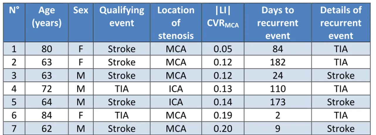
N° Age (years) Sex Qualifying event Location of stenosis |LI| CVRMCA Days to recurrent event Details of recurrent event
1 80 F Stroke MCA 0.05 84 TIA
2 63 F Stroke MCA 0.12 182 TIA 3 63 M Stroke MCA 0.12 24 Stroke
4 72 M TIA ICA 0.13 110 TIA
5 64 M Stroke ICA 0.14 173 Stroke
6 84 F TIA MCA 0.19 2 TIA
7 62 M Stroke MCA 0.20 9 Stroke
Table 2: Summary details of RECURRENT patients. |LI|CVRMCA=absolute value of laterality
index of cerebrovascular reactivity in middle cerebral artery territories, other abbreviations are defined in Table 1.
Comparisons between SINGLE and RECURRENT groups
When compared with SINGLE, RECURRENT patients were older (69.5±9.1. vs 56.1±11.8 p=0.04). There were no differences among other clinical and biological data (Table 3).
SINGLE group (n=12) RECURRENT group (n=7) p value Age in years 56.1±11.8 69.5±9.1 0.04 Men 9 4 0.54 History of hypertension 5 5 0.3
History of lipid disorder 8 3 0.43
Smoking 6 2 0.48
Diabetes 3 2 0.9
History of coronary artery disease 1 1 0.84 Low-density lipoprotein cholesterol (g/L) 1.2±0.4 1±0.3 0.17 High-density lipoprotein cholesterol (g/L) 0.4±0.1 0.6±0.4 0.2 Total cholesterol (g/L) 1.9±0.5 1.9±0.3 0.86 Triglycerid (g/L) 1.4±0.6 1.5±0.7 0.84 Glycosylated haemoglobin (%) 7.0±2.9 6.9±1.7 0.48 Overweigh or obesity (body-mass index > 25) 4 1 0.54 Qualifying event
- Stroke 10 5
0.71
- TIA 2 2
NIHSS at entrance (median [IQR]) 3[1-1.5] 1[0-2.5] 0.23 mRS at discharge(median [IQR]) 2[0.8-2] 1[0-2] 0.3 On antithrombotic therapy at qualifying event 3 1 0.71 On antihypertensive therapy at qualifing event 5 4 0.59 On lipid-lowering therapy at qualifiying event 4 1 0.54 On diabetic therapy at qualifiying event 2 2 0.71 Symptomatic qualifying artery
- ICA 3 2
0.9
- MCA 9 5
Follow-up duration in years 3.8±1.3 3.2±1.7 0.38 mRS follow-up (median [IQR]) 0.5[0.5-1.3] 1[0.5-1.5] 0.59
Table 3: Clinical and biological baseline characteristics of SINGLE and RECURRENT groups.
Patients who experiment recurrent ipsilateral ischemic event are significantly older than SINGLE patients. Data are mean ± standard deviation. Abbreviations are defined in Table 1.
RECURRENT patients had higher |LI| CVRMCA (0.14±0.05 vs 0.05±0.03, p=0.001). There were
no differences among |LI| CVR in other arterial territories. Subtle intergroup differences in basal perfusion parameters did not reach significance (Table 4).
SINGLE group (n=12) RECURRENT group (n=7) p value |LI| CVR MCA 0.05±0.03 0.14±0.05 0.001 ACA 0.04±0.04 0.05±0.06 0.23 PCA 0.04±0.03 0.04±0.02 0.54 |LI| CBF MCA 0.07±0.05 0.11±0.06 0.23 ACA 0.07±0.05 0.04±0.02 0.38 PCA 0.03±0.02 0.02±0.02 0.97 |LI| CBV MCA 0.04±0.03 0.03±0.03 0.54 ACA 0.04±0.03 0.03±0.03 0.26 PCA 0.03±0.03 0.03±0.02 0.97 |LI| MTT MCA 0.07±0.03 0.10±0.07 0.43 ACA 0.05±0.03 0.05±0.04 0.65 PCA 0.03±0.03 0.02±0.02 0.38
Table 4: Laterality indices between SINGLE and RECURRENT groups for each vascular ROI and
each hemodynamic parameter (cerebrovascular reactivity, basal perfusion parameters). The |LI| CVRMCA is significantly increased in patients who experiment recurrent ipsilateral
ischemic event, other hemodynamic parameters differ between these two groups. Data are mean ± standard deviation. |LI|=absolute value of laterality index, CVR=cerebrovascular reactivity, CBF=cerebral blood flow, CBV=cerebral blood volume, MTT=mean time transit, ACA=anterior cerebral artery territories, PCA=posterior cerebral artery territories, other abbreviations are defined in Table 1.
Illustrative cases are shown in Figure 3.
Figure 3: Basal perfusion parameters and CVR maps in patients referred for symptomatic
right MCA severe stenosis (radiological convention). Upper row: 65-years man without ischemic recurrence (SINGLE group). Both basal perfusion (CBF, CBV, MTT) and CVRMCA were
symmetrical: |LI| CBFMCA=0.00, |LI| CBVMCA=0.00, |LI| MTTMCA=0.01, |LI| CVRMCA=0.02
(NORMAL CVRMCA). Lower row: 83-years woman with 2 recurrent transient ischemic attacks
(RECURRENT group). Autoregulation with increased CBV failed to maintain CBF symmetrical. Additionally, CVR decreased downstream MCA stenosis: |LI| CBFMCA=0.18, |LI| CBVMCA=0.06,
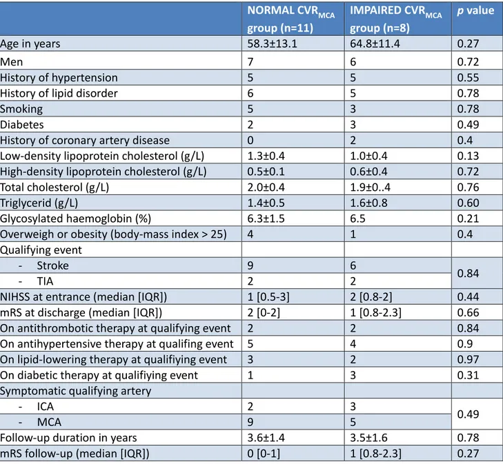
Comparisons between IMPAIRED and NORMAL CVRMCA groups
Among all patients, 11 patients had NORMAL CVRMCA, and 8 had IMPAIRED CVRMCA. There
were no intergroup differences among clinical and biological baseline data (Table 5).
NORMAL CVRMCA group (n=11) IMPAIRED CVRMCA group (n=8) p value Age in years 58.3±13.1 64.8±11.4 0.27 Men 7 6 0.72 History of hypertension 5 5 0.55
History of lipid disorder 6 5 0.78
Smoking 5 3 0.78
Diabetes 2 3 0.49
History of coronary artery disease 0 2 0.4 Low-density lipoprotein cholesterol (g/L) 1.3±0.4 1.0±0.4 0.13 High-density lipoprotein cholesterol (g/L) 0.5±0.1 0.6±0.4 0.72 Total cholesterol (g/L) 2.0±0.4 1.9±0..4 0.76
Triglycerid (g/L) 1.4±0.5 1.6±0.8 0.60
Glycosylated haemoglobin (%) 6.3±1.5 6.5 0.21 Overweigh or obesity (body-mass index > 25) 4 1 0.4 Qualifying event
- Stroke 9 6
0.84
- TIA 2 2
NIHSS at entrance (median [IQR]) 1 [0.5-3] 2 [0.8-2] 0.44 mRS at discharge (median [IQR]) 2 [0-2] 1 [0.8-2.3] 0.66 On antithrombotic therapy at qualifying event 2 2 0.84 On antihypertensive therapy at qualifing event 5 4 0.9 On lipid-lowering therapy at qualifiying event 3 2 0.97 On diabetic therapy at qualifiying event 1 3 0.31 Symptomatic qualifying artery
- ICA 2 3
0.49
- MCA 9 5
Follow-up duration in years 3.6±1.4 3.5±1.6 0.78 mRS follow-up (median [IQR]) 0 [0-1] 1 [0.8-2.3] 0.27
Table 5: Baseline characteristics of NORMAL and IMPAIRED CVRMCA groups. Clinical and
biological data are similar between patients with and without CVR impairment downstream the intracranial stenosis. Data are mean ± standard deviation. CVRMCA=cerebrovascular
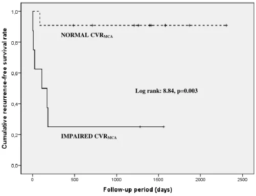
The occurrence of recurrent ipsilateral ischemic event in IMPAIRED CVRMCA (n=6/8) was
more frequent than in NORMAL CVRMCA group (n=1/11) with different survival curves
(p=0.003) (Figure 4).
Figure 4: Kaplan-Meier survival curves illustrating the relationship between CVRMCA and the
occurrence of ipsilateral ischemic event. The vertical cross-lines indicates patient whose data were censored due to study end, death, or recurrent ischemic event.
NORMAL CVRMCA
Log rank: 8.84, p=0.003
The annual risk of recurrent ipsilateral ischemic event was higher in IMPAIRED CVRMCA group
compared to NORMAL CVRMCA group (39.1%/year vs 3.4%/year, p=0.012). There were
neither differences among |LI| CVR in other arterial territories nor in other basal perfusion parameters (Table 6). NORMAL CVRMCA group (n=11) IMPAIRED CVRMCA group (n=8) p value |LI| CVR MCA 0.04±0.02 0.14±0.04 <0.001 ACA 0.04±0.04 0.06±0.05 0.44 PCA 0.04±0.03 0.04±0.02 0.17 |LI| CBF MCA 0.08±0.05 0.10±0.06 0.49 ACA 0.07±0.05 0.04±0.02 0.55 PCA 0.03±0.02 0.02±0.02 0.44 |LI| CBV MCA 0.03±0.02 0.05±0.03 0.27 ACA 0.04±0.03 0.03±0.03 0.78 PCA 0.03±0.03 0.03±0.02 0.90 |LI| MTT MCA 0.07±0.03 0.11±0.06 0.11 ACA 0.06±0.03 0.04±0.04 0.24 PCA 0.03±0.03 0.02±0.02 0.44
Table 6: Laterality indices between NORMAL and IMPAIRED CVRMCA groups for each vascular
ROI and each hemodynamic parameter (cerebrovascular reactivity, basal perfusion parameters). Except |LI| CVRMCA, no other hemodynamic parameters differ significantly
between these two groups. Data are mean ± standard deviation. Abbreviations are defined in Table 4.
CVRMCA and basal perfusion parameters correlations
We found a positive correlations between LI CVRMCA and LI CBFMCA (ρ=0.89, p=0.007 vs
ρ=0.59, p=0.043) for RECURRENT and SINGLE patients, respectively (Figure 5). A significant negative correlation was found between LI CVRMCA and LI MTTMCA in RECURRENT group
(ρ=-0.86, p=0.014) but not significant in SINGLE group (ρ=-0.47, p=0.12). For both groups, LI CVRMCA was not significantly correlated with LI CBVMCA.
Figure 5: Positive correlations between LI CBF and LI CVR from middle cerebral artery
territories (ρ=0.89, p=0.007 vs ρ=0.59, p=0.043) for RECURRENT and SINGLE patients with intracranial stenosis, respectively.
DISCUSSION:
In patients with symptomatic intracranial atherosclerotic stenosis, recurrent ischemic events occur despite optimal medical therapy. Additional treatments such as endovascular angioplasty and stenting are still not validated. Indeed, large trials failed to demonstrate significant clinical benefits when compared to higher risks of mordibity and mortality. To further address additional therapy, the estimation of cerebrovascular reserve may help to better identify patients at higher risk of stroke recurrence. In our observational study, we showed that 1°) patients with recurrent ischemic events have an impaired CVR downstream the IAS, using BOLD fMRI with hypercapnic challenge; 2°) patients with abnormal CVR defined by a laterality index ≥0.08 are at higher risk of recurrent ischemic events. These results emphasized the potential role of CVR fMRI to select patients for additional treatment.
In our study, the global rate of recurrence of ischemic event was 37%, similar to 38% observed in GESICA study6. The annual rate of stroke downstream the qualifying IAS was also close to 18% reported in the severe IAS subgroup of WASID trial, but higher than 12% in the SAMMPRIS trial4,9. We used identical restrictive inclusion and exclusion criteria. This led to analyze a much smaller population in our monocentric study. However, clinical baseline characteristics were similar. In SAMMPRIS, a more intensive control of cardiovascular risk factors, based on a tight clinical and biological follow-up and a lifestyle modification program may explain this difference9.
CVRMCA patients. The 13x higher rate in patients with CVR impairment advocates for a tight
relationship. Reduced CVR has been identified as a strong and independent predictor on stroke recurrence in steno-occlusive diseases, using multivariate analysis among other variables (cardiovascular risk factors, degree of stenosis)28,36,37. Most of these CVR studies used TDS with several advantages, such as high temporal resolution, arterial velocity quantification, non-invasiveness, cost, and availability, and limitations such as insonation window access, interoperator variability, absence of parenchymal imaging. Functional brain imaging techniques using SPECT, and MRI have been proposed to explore whole brain CVR17,18. However, fMRI studies on steno-occlusive diseases were heterogeneous with extracranial and/or stenosis or occlusion, and with different etiologies such as atheromatous or moya-moya15,24,29,30,38,39. Our study is the first one to evaluate CVR using fMRI in a homogeneous group of patients with symptomatic atherosclerotic stenosis of a major intracranial artery, like in large randomized trials4,9.
BOLD fMRI with hypercapnic challenge has been shown as a safe and reliable brain imaging technique to estimate cerebrovascular reserve29,30. However BOLD signal, which reveals changes in blood oxygenation, remains an unclear combination of variation in oxygenation consumption, CBF, CBV, and vasomotricity. Perfusion MRI techniques such as ASL were also proposed to monitor quantitative CBF changes to vasomotor stimuli24,40. In our experience, CVR fMRI using ASL remains difficult to implement in clinical work-flow, mostly limited by a poor signal to noise ratio, a lower temporal resolution (8 sec between 2 pairs of tag-control images), a critical sensitivity to labeling temporal parameters with aging and arterial transit time asymmetry a fortiori downstream IAS, sophisticated postprocessing that requires parametric assumptions and signal corrections for “absolute” CBF quantification41,42.
Because of the variability of basal perfusion and CVR, we chose to estimate these parameters independently, to calculate LI to minimize interindividual amplitude effects, and to test their relationships23. Using classical LI, as defined for hemispheric lateralization for language, we previously shown that 95% of 100 healthy subjects, had CVR |LI|<0.0835. In the present study, all RECURRENT patients but one had a CVR |LI|≥0.08. Based on both of these results, IMPAIRED CVRMCA had a much higher risk of ischemic event, suggesting a reliable
threshold to select patients at high risk in a future prospective study. However, the asymmetry characterization is less relevant and limits results interpretation in case of bilateral stenosis. Thus, absolute CVR quantification would be helpful to further address these limitations, although physiological and pathological confounds account for a large inter-individual variability 18,24,35.
In a previous study, basal CBF decrease and CVR impairment explained decreased parenchymal oxygenation, suggesting chronic low grade ischemia in patients with symptomatic IAS23. Here, CBF and CVR asymmetry were correlated in both RECURRENT and SINGLE patients. The correlation coefficient was higher in the RECURRENT group. However the small groups’ size did not allow to conduct appropriate statistical comparison. Despite these relationships, CVR asymmetry was the only significant parameter which differed across RECURRENT and SINGLE groups.
In fact, most recurrent ischemic events downstream IAS occurred during the first year after qualifying event, in line with large clinical trials4,9. This observation suggests time-dependent changes of the plaque under aggressive medical management, collateral supply
degree of stenosis may progress or regress over time43. The risk of new stroke is attenuated after 1 year by the stabilization of vulnerable plaque, and development of collateral circulation44. Intensive medical management should therefore accelerate these different protective processes45,46. Beside structural changes of vascularization, functional changes of arterial supply may also occur. Several cases of spontaneous improvement of cerebrovascular reserve were reported using SPECT47. However, temporal changes of cerebrovascular reserve remain to be documented in larger studies. A systematic CVR mapping and basal perfusion imaging follow-up would be interesting in order to test spatial agreement between CVR impairment and recurrent stroke, and to monitor natural evolution of CVR and basal perfusion.
Additionally to hemodynamic ischemic event related to CVR impairment, artery-to-artery embolism, in situ thrombo-occlusion, and occlusion of perforating arteries are other key pathophysiological mechanisms underlying cerebral ischemia in patients with IAS48. Dual antiplatelet therapy and restrictive medical management of cardiovascular factors decrease the risk of disability and death following recurrent stroke in and out the stenosis territory over extended follow-up9. Indeed, stroke out the qualifying artery territory and cardiovascular death also occurred, as in large trials4,6–9, in 4 patients out of 19, independently of CVRMCA status.
Given to our results, prevention of stroke recurrence within the first year remains the most critical therapeutic challenge after the acute stroke management. CVR fMRI would be useful to select high-risk patients and to may help to justify peri-procedural adverse events related to additional invasive treatment. We suggest that future therapeutic trials on endovascular
techniques using stents49 or angioplasty alone50, should be first conducted on IAS patients with CVR impairment, only.
CONCLUSION:
Symptomatic intracranial atherosclerotic stenosis is associated with high rates of subsequent stroke and cerebrovascular mortality. Prevention of recurrent ischemic events remains challenging. Using noninvasive CVR BOLD fMRI, LI CVRMCA derived from asymmetric BOLD
signal change, elicited by hypercapnic stimulus, shows that CVR impairment is observed in more than 40% of patients with unilateral symptomatic IAS. An increased |LI| CVRMCA is
associated with a major risk of ipsilateral recurrent ischemic event. CVRMCA impairment is
more frequent in patients with recurrent versus single event. CVR impairment significantly shortens event-free survival during the first year, despite optimal medical management. CVR status should be considered to identify among IAS patients those at a higher risk of recurrent ischemic event and to select those who may benefit from additional treatment, such as endovascular procedures.
BIBLIOGRAPHY
1. Sacco, R. L., Kargman, D. E., Gu, Q. & Zamanillo, M. C. Race-ethnicity and determinants of intracranial atherosclerotic cerebral infarction. The Northern Manhattan Stroke Study. Stroke 26, 14–20 (1995).
2. White, H. et al. Ischemic stroke subtype incidence among whites, blacks, and Hispanics: the Northern Manhattan Study. Circulation 111, 1327–1331 (2005).
3. Wong, L. K. S. Global burden of intracranial atherosclerosis. Int. J. Stroke Off. J. Int.
Stroke Soc. 1, 158–159 (2006).
4. Chimowitz, M. I. et al. Comparison of warfarin and aspirin for symptomatic intracranial arterial stenosis. N. Engl. J. Med. 352, 1305–1316 (2005).
5. Holmstedt, C. A., Turan, T. N. & Chimowitz, M. I. Atherosclerotic intracranial arterial stenosis: risk factors, diagnosis, and treatment. Lancet Neurol. 12, 1106–1114 (2013). 6. Mazighi, M. et al. Prospective study of symptomatic atherothrombotic intracranial
stenoses: the GESICA study. Neurology 66, 1187–1191 (2006).
7. Weber, R., Kraywinkel, K., Diener, H.-C. & Weimar, C. Symptomatic Intracranial Atherosclerotic Stenoses: Prevalence and Prognosis in Patients with Acute Cerebral Ischemia. Cerebrovasc. Dis. 30, 188–193 (2010).
8. Famakin, B. M. et al. Causes and severity of ischemic stroke in patients with symptomatic intracranial arterial stenosis. Stroke J. Cereb. Circ. 40, 1999–2003 (2009). 9. Chimowitz, M. I. et al. Stenting versus Aggressive Medical Therapy for Intracranial
Arterial Stenosis. N. Engl. J. Med. 365, 993–1003 (2011).
10. Zaidat, O. O. et al. Effect of a balloon-expandable intracranial stent vs medical therapy on risk of stroke in patients with symptomatic intracranial stenosis: the VISSIT randomized clinical trial. JAMA 313, 1240–1248 (2015).
11. The EC/IC Bypass Study Group. Failure of extracranial-intracranial arterial bypass to reduce the risk of ischemic stroke. Results of an international randomized trial. The EC/IC Bypass Study Group. N. Engl. J. Med. 313, 1191–1200 (1985).
12. Powers, W. J. et al. Extracranial-intracranial bypass surgery for stroke prevention in hemodynamic cerebral ischemia: the Carotid Occlusion Surgery Study randomized trial.
JAMA 306, 1983–1992 (2011).
13. Hoak, D. A. & Lutsep, H. L. Management of Symptomatic Intracranial Stenosis. Curr.
Cardiol. Rep. 18, 83 (2016).
14. Attyé, A. et al. Normalization of cerebral vasoreactivity using BOLD MRI after intravascular stenting. Hum. Brain Mapp. 35, 1320–1324 (2014).
15. Mandell, D. M. et al. Quantitative Measurement of Cerebrovascular Reactivity by Blood Oxygen Level-Dependent MR Imaging in Patients with Intracranial Stenosis: Preoperative Cerebrovascular Reactivity Predicts the Effect of Extracranial-Intracranial Bypass Surgery. Am. J. Neuroradiol. 32, 721–727 (2011).
16. Waters, M. F. et al. Factors Associated With Recurrent Ischemic Stroke in the Medical Group of the SAMMPRIS Trial. JAMA Neurol. 1–8 (2016).
doi:10.1001/jamaneurol.2015.4315
17. Gupta, A. et al. Cerebrovascular reserve and stroke risk in patients with carotid stenosis or occlusion: a systematic review and meta-analysis. Stroke J. Cereb. Circ. 43, 2884–2891 (2012).
18. Krainik, A. et al. Functional imaging of cerebral perfusion. Diagn. Interv. Imaging 94, 1259–1278 (2013).
19. Mchedlishvili, G. Physiological mechanisms controlling cerebral blood flow. Stroke
20. Powers, W. J. Cerebral hemodynamics in ischemic cerebrovascular disease. Ann.
Neurol. 29, 231–240 (1991).
21. Krainik, A., Hund-Georgiadis, M., Zysset, S. & von Cramon, D. Y. Regional
impairment of cerebrovascular reactivity and BOLD signal in adults after stroke. Stroke J.
Cereb. Circ. 36, 1146–1152 (2005).
22. Conklin, J. et al. Mapping white matter diffusion and cerebrovascular reactivity in carotid occlusive disease. Neurology 77, 431–438 (2011).
23. Bouvier, J. et al. Reduced CMRO₂ and cerebrovascular reserve in patients with severe intracranial arterial stenosis: a combined multiparametric qBOLD oxygenation and BOLD fMRI study. Hum. Brain Mapp. 36, 695–706 (2015).
24. Smeeing, D. P. J., Hendrikse, J., Petersen, E. T., Donahue, M. J. & de Vis, J. B. Arterial Spin Labeling and Blood Oxygen Level-Dependent MRI Cerebrovascular Reactivity in Cerebrovascular Disease: A Systematic Review and Meta-Analysis.
Cerebrovasc. Dis. Basel Switz. 42, 288–307 (2016).
25. Herold, S. et al. Assessment of cerebral haemodynamic reserve: correlation between PET parameters and CO2 reactivity measured by the intravenous 133 xenon injection technique. J. Neurol. Neurosurg. Psychiatry 51, 1045–1050 (1988).
26. Chen, J. et al. Impaired dynamic cerebral autoregulation and cerebrovascular reactivity in middle cerebral artery stenosis. PloS One 9, e88232 (2014).
27. Pindzola, R. R., Balzer, J. R., Nemoto, E. M., Goldstein, S. & Yonas, H.
Cerebrovascular Reserve in Patients With Carotid Occlusive Disease Assessed by Stable Xenon-Enhanced CT Cerebral Blood Flow and Transcranial Doppler. Stroke 32, 1811– 1817 (2001).
28. Ogasawara, K., Ogawa, A. & Yoshimoto, T. Cerebrovascular Reactivity to
Acetazolamide and Outcome in Patients With Symptomatic Internal Carotid or Middle Cerebral Artery Occlusion. Stroke 33, 1857–1862 (2002).
29. Donahue, M. J. et al. Routine Clinical Evaluation of Cerebrovascular Reserve Capacity Using Carbogen in Patients With Intracranial Stenosis. Stroke 45, 2335–2341 (2014).
30. Spano, V. R. et al. CO2 blood oxygen level-dependent MR mapping of cerebrovascular reserve in a clinical population: safety, tolerability, and technical feasibility. Radiology 266, 592–598 (2013).
31. Fierstra, J. et al. Measuring cerebrovascular reactivity: what stimulus to use? J.
Physiol. 591, 5809–5821 (2013).
32. Samuels, O. B., Joseph, G. J., Lynn, M. J., Smith, H. A. & Chimowitz, M. I. A
standardized method for measuring intracranial arterial stenosis. AJNR Am. J. Neuroradiol.
21, 643–646 (2000).
33. Wu, O. et al. Tracer arrival timing-insensitive technique for estimating flow in MR perfusion-weighted imaging using singular value decomposition with a block-circulant deconvolution matrix. Magn. Reson. Med. 50, 164–174 (2003).
34. Tatu, L., Moulin, T., Vuillier, F. & Bogousslavsky, J. Arterial territories of the human brain. Front. Neurol. Neurosci. 30, 99–110 (2012).
35. Boudiaf, N. et al. BOLD fMRI of cerebrovascular reactivity in the middle cerebral artery territory: A 100 volunteers’ study. J. Neuroradiol. J. Neuroradiol. 42, 338–344 (2015).
36. Markus, H. & Cullinane, M. Severely impaired cerebrovascular reactivity predicts stroke and TIA risk in patients with carotid artery stenosis and occlusion. Brain J. Neurol.
37. Reinhard, M. et al. Cerebrovascular reactivity predicts stroke in high-grade carotid artery disease. Neurology 83, 1424–1431 (2014).
38. Goode, S. D., Altaf, N., Munshi, S., MacSweeney, S. T. R. & Auer, D. P. Impaired Cerebrovascular Reactivity Predicts Recurrent Symptoms in Patients with Carotid Artery Occlusion: A Hypercapnia BOLD fMRI Study. Am. J. Neuroradiol. 37, 904–909 (2016). 39. Haller, S. et al. Reduced cerebrovascular reserve at CO2 BOLD MR imaging is
associated with increased risk of periinterventional ischemic lesions during carotid endarterectomy or stent placement: preliminary results. Radiology 249, 251–258 (2008). 40. Villien, M. et al. Changes in cerebral blood flow and vasoreactivity to CO2 measured
by arterial spin labeling after 6days at 4350m. NeuroImage 72, 272–279 (2013).
41. Vincent, T. et al. Bayesian joint detection-estimation of cerebral vasoreactivity from ASL fMRI data. Med. Image Comput. Comput.-Assist. Interv. MICCAI Int. Conf. Med.
Image Comput. Comput.-Assist. Interv. 16, 616–624 (2013).
42. Villien, M. et al. Per-subject characterization of bolus width in pulsed arterial spin labeling using bolus turbo sampling. Magn. Reson. Med. 69, 1677–1682 (2013).
43. Akins, P. T., Pilgram, T. K., Cross, D. T. & Moran, C. J. Natural history of stenosis from intracranial atherosclerosis by serial angiography. Stroke 29, 433–438 (1998). 44. Liebeskind, D. S. et al. Collaterals dramatically alter stroke risk in intracranial
atherosclerosis. Ann. Neurol. 69, 963–974 (2011).
45. Sander, K., Hof, U., Poppert, H., Conrad, B. & Sander, D. Improved Cerebral Vasoreactivity After Statin Administration in Healthy Adults. J. Neuroimaging 15, 266– 270 (2005).
46. Sterzer, P. et al. Pravastatin Improves Cerebral Vasomotor Reactivity in Patients With Subcortical Small-Vessel Disease. Stroke 32, 2817–2820 (2001).
47. Hasegawa, Y., Yamaguchi, T., Tsuchiya, T., Minematsu, K. & Nishimura, T.
Sequential change of hemodynamic reserve in patients with major cerebral artery occlusion or severe stenosis. Neuroradiology 34, 15–21 (1992).
48. van den Wijngaard, I. R. et al. Treatment and imaging of intracranial atherosclerotic stenosis: current perspectives and future directions. Brain Behav. 6, n/a-n/a (2016).
49. Ding, D., Starke, R. M., Crowley, R. W. & Liu, K. C. Role of stenting for intracranial atherosclerosis in the post-SAMMPRIS era. BioMed Res. Int. 2013, 304320 (2013). 50. Dumont, T. M. et al. Submaximal angioplasty for symptomatic intracranial
atherosclerosis: a prospective Phase I study. J. Neurosurg. 1–8 (2016). doi:10.3171/2015.8.JNS15791
THESE SOUTENUE PAR :
PAPASSIN Jérémie
TITRE:
Altération de la vasoréactivité cérébrale en IRMf hypercapnique chez les patients présentant une sténose artérielle intracrânienne symptomatique d’origine athéromateuse à haut risque de récidive.
RESUME :
Introduction: Malgré un traitement médical optimal, le risque de récidive dans les sténoses
artérielles intracrâniennes symptomatiques (SAIS) d’origine athéromateuse est élevé. Une altération de la vasoréactivité cérébrale (CVR) pourrait être impliquée. L’étude en IRM fonctionnelle BOLD hypercapnique de la CVR permettrait d’identifier les patients à haut risque de récidive, afin de proposer des traitements complémentaires plus invasifs comme l’angioplastie transluminale percutanée avec stent intracrânien.
Méthodes: Nous avons étudiés les relations statistiques entre les données cliniques et
biologiques individuelles, la récidive d’évènement ischémique et la CVR dans les territoires de l’artère cérébrale moyenne (ACM) rapportée par un index de latéralité (IL), pour 19 patients avec une SAIS de l‘ACM ou de l’artère carotide interne.
Résultats: Huit patients (42%) avaient un IL CVRACM anormal (|IL| CVRACM ≥ 0.08). Avec un
suivi moyen de 3.6 ans, la récurrence d’évènement ischémique était plus fréquente et plus précoce dans le groupe CVRACM altérée (n = 6/8) que dans le groupe CVRACM normale (n =
Conclusion: Une altération de la CVR en IRMf hypercapnique est associée à un risque élevé
de récidive pour les patients avec une SAIS d’origine athéromateuse. La cartographie de la CVR pourrait être utilisée pour sélectionner les patients à haut risque de récidive, afin de discuter un traitement complémentaire.
VU ET PERMIS D'IMPRIMER Grenoble, le 21/09/2017
LE DOYEN LE PRESIDENT DE LA THESE
Impaired cerebrovascular reactivity assessed by BOLD hypercapnic fMRI is associated with increased risk of stroke in patients with symptomatic intracranial atherosclerotic stenosis
Introduction: Despite intensive medical management, intracranial atherosclerotic stenosis
(IAS) remains at risk of recurrent ischemic events. Impaired cerebrovascular reserve is suggested to explain hemodynamic stroke. Cerebrovascular reactivity assessed by hypercapnic challenge using BOLD functional MRI (CVR BOLD fMRI) has been proposed to estimate cerebrovascular reserve and to identify patients at higher risk. To discuss patients’ selection for additional therapy such as intracranial angioplasty and stenting, we studied the relationships between baseline characteristics of patients with unilateral symptomatic IAS, recurrence of ischemic events, and CVR BOLD fMRI.
Subjects and methods: Nineteen patients with symptomatic unilateral IAS of middle cerebral
artery (MCA) or internal carotid artery were selected. We calculated statistical relationships between individual clinical and biological baseline characteristics, recurrent ischemic events, basal perfusion estimated by dynamic susceptibility contrast MRI, and CVR BOLD fMRI measured in MCA territories (CVRMCA), and reported using laterality indices (LI).
Results: Eight patients (42%) had an abnormal LI CVRMCA (|LI| CVRMCA ≥ 0.08). During a mean
follow-up of 3.6 years, recurrent ischemic events occurred within the first year. They were more frequent in impaired CVRMCA group (n=6/8) than in normal CVRMCA group (n=1/11),
with different survival curves (log rank, p=0.003). Baseline characteristics were similar in both groups.
Conclusion: Impaired CVR assessed by BOLD hypercapnic fMRI is associated with increased
risk of stroke in patients with symptomatic IAS. CVR mapping should be proposed to select high-risk patients in order to discuss additional treatment.