Contribution of tachykinin and kinin receptors in central autonomic control of blood pressure and behavioural activity in hypertensive rats
par
Helaine De Brito Pereira
Département de physiologie Faculté de Médecine
Thèse présentée à la Faculté des études supérieures et postdoctorales en vue de l’obtention du grade de
Philosophia Doctor (Ph.D.) en Physiologie
Mai, 2010
Université de Montréal
Faculté des études supérieures et postdoctorales
Cette thèse intitulée:
Contribution of tachykinin and kinin receptors in central autonomic control of blood pressure and behavioural activity in hypertensive rats
Présentée par : Helaine De Brito Pereira
a été évaluée par un jury composé des personnes suivantes : Dr. Jean-Louis Schwartz, président-rapporteur
Dr. Réjean Couture, directeur de recherche Dr. Madhu B. Anand-Srivastava, membre du jury
Dr. Pedro D'Orléans-Juste, examinateur externe Dr. Hélène Girouard, représentant de la FESP
I want officially thank God, for giving me the patience, health, strength, and intelligence to do my doctoral studies.
I am thankful for the kindest, most understanding and loving husband in the world. I cannot even put into words the kind of man he is. I am blessed that God brought him into my life and thankful for the almost six years that we have shared so far.
I would like to thank my siblings Renato and Karina, and my brother-in-law Fabio for treasured memories and loving support throughout my education and graduation.
Aos meus pais,
Que me deram a vida e me ensinaram a vivê-la com dignidade,
não bastaria um obrigado.
Que iluminaram os caminhos obscuros com afeto e dedicação para que os trilhássemos sem medo e cheios de esperanças,
não bastaria um muito obrigado.
Que se doaram inteiros e renunciaram aos seus sonhos, para que, muitas vezes, pudésse realizar os meus. Pela longa espera e compreensão durante minha longa
ausência, não bastaria um muitíssimo obrigado.
A vocês, pai e mãe por natureza, por opção e amor, não bastaria dizer, que não tenho palavras para agradecer tudo isso. Mas é o que me acontece agora, quando
procuro arduamente uma forma verbal de exprimir uma emoção incomparavel. Uma emoção que jamais seria traduzida por palavras.
A
Acknowledgements
I want first to thank my reseach director, Dr. Rejean Couture, for welcoming me in Canada and in his laboratory. During more than a decade of knowing him, he has helped me to see life and science in their full depth, and taught me how to appreciate the good scientific work that helps other researchers to build on it.
I warmly thank my laboratory friends and colleagues, Sébastien Talbot, Dr. Brice Ongali, Dr. Frank Cloutier, Jenny Pena-Dias, Mahmud Ismael, Emna Chahmi, Nejla Tidjane, Dr. Karim Lahjouji, and Jacques Sénécal.
Thank you to all members of the Physiology department, and especially to Johanne Payette and Marjolaine Homier, for their help and support.
I would like to say thank you to Paula Jansen Santana and Claude Gauthier for the graphic and artistic work on the figures.
In addition, I thank my Brazilian friends who encouraged, supported me and have always been present in hard times, Janaina Suckow, Simone Carrer, Angela Piovesan, Billa Furlanetto, THANK YOU and I love you girls!
Finally, I thank the Quebec Society of Hypertension (SQHA) and the Faculty of Graduate Studies (FESP), which have offered me travel grants and awards of excellence, respectively. The financial support of this work was provided by research grants from the Canadian Institutes of Health Research (CIHR) to Dr. Réjean Couture and National Council of Technological and Scientific Development (CNPq) (proc. 201428/2003-2) Brazil to H. De Brito Pereira.
Abstract
This work aims at studing the role of tachykinin NK-3 receptor (R) and kinin B1R in central autonomic regulation of blood pressure (BP) and to determine whether the B1R is overexpressed and functional in rat models of hypertension by measuring the effect of a B1R agonist on behavioural activity. Assumptions: (1) NK-3R located in the ventral tegmental area (VTA) modulates the mesolimbic dopaminergic system and has a tonic activity in hypertension; (2) B1R is overexpressed in the brain of hypertensive rats and has a tonic activity, which contributes to hypertension via a dopamine mechanism; (3) the inhibition of NK-3R and B1R with selective antagonists, reduces central dopaminergic hyperactivity and reverses hypertension. A model of genetic hypertension and a model of experimental hypertension were used: spontaneously hypertensive rats (SHR, 16 weeks) and Wistar-Kyoto (WKY) rats infused for 14 days with angiotensin II (Ang II) (200 ng / kg / min, subcutaneous (s.c.) with Alzet mini pump). The age-matched untreated WKY rats served as common controls. In the first study (article # 1), the cardiovascular response in SHR was evaluated following intracebroventricular (i.c.v.) and/or intra-VTA injection of an agonist (senktide) and antagonists (SB222200 and R-820) of NK-3R. These responses have also been characterized using selective dopamine antagonists DA-D1R (SCH23390), DA-D2R (raclopride) or non-selective dopamine DA-D2R (haloperidol). Also the VTA has been destroyed by ibotenic acid. The pressor response induced by senktide and the anti-hypertensive response induced by SB222200 or R-820 were more pronounced by intra-VTA. These responses were prevented by pre-treatment with raclopride and haloperidol. The lesion of the VTA has prevented the pressor response relayed by senktide (i.c.v.) and the anti-hypertensive effect of R-820 (i.c.v.). In addition, SB222200 (intra-VTA) prevented the pressor response of senktide (i.c.v.) and conversely, senktide (i.c.v.) prevented the antihypertensive effect of SB222200 (intra-VTA). The second study (article # 2) showed that the B1R antagonist (SSR240612)
administered by gavage or i.c.v. reverses hypertension in both models. This anti-hypertensive effect was prevented by raclopride and haloperidol. In contrast, the two B1R antagonists (R-715 and R-954) injected s.c., which do not cross the blood-brain barrier reduced weakly blood pressure in hypertensive rats. In the third study (article # 3), the i.c.v. injection of a selective kinin B1R agonist Sar[DPhe8][des-Arg9]BK caused behavioural responses in SHR and Ang II-treated rats and had no effect in control WKY rats . The responses elicited by B1R agonist were blocked by an antagonist of NK-1 (RP67580), an antagonist of NMDA glutamate receptor (DL-AP5), an inhibitor of nitric oxide synthase (NOS) (L -NNA) as well as raclopride and SCH23390.The responses were modestly affected by the inhibitor of inducible NOS (iNOS). The B1R mRNA (measured by RT-PCR) was significantly increased in the hypothalamus, the VTA and the nucleus accumbens of hypertensive animals (SHR and treated with Ang II) compared with control rats.
These neuropharmacological studies suggest that: (1) the NK-3R from the VTA is involved in the maintenance of hypertension in SHR by increasing DA transmission in the midbrain; (2) the B1R in SHR and Ang II-treated rats contributes to hypertension via a central mechanism involving DA-D2R; (3) the central B1R increases locomotor activity and nocifensive behaviours via the release of substance P (NK-1), DA and nitric oxide in both rat models of hypertension.
Thus, the brain tachykinin NK-3R and kinin B1R represent potential therapeutic targets for the treatment of hypertension. The modulation of the mesolimbic/mesocortical dopaminergic pathway by these receptors suggests their involvement in other physiological functions (pleasure, motor activity, coordination of the response to stress) and pathophysiology (anxiety, depression).
Keywords: Tachykinin NK-3R, kinin B1R, dopamine, ventral tegmental area, hypertension, behavior.
Résumé
Ce travail vise à étudier le rôle du récepteur NK-3 des tachykinines (NK-3R) et du récepteur B1 des kinines (B1R) dans la régulation autonomique centrale de la pression artérielle et de déterminer si le B1R est surexprimé et fonctionnel chez le rat hypertendu en mesurant l’effet d’antagoniste B1R sur l’activité comportementale. Hypothèses: (1) le NK-3R localisé dans l’aire tegmentale ventrale (VTA) module l’activité dopaminergique du système mésolimbique et possède une activité tonique dans l’hypertension; (2) le B1R est surexprimé dans le cerveau du rat hypertendu et possède une activité tonique qui contribue à l’hypertension via un mécanisme dopaminergique; (3) l’inhibition des NK-3R et B1R avec des antagonistes sélectifs réduit l’hyperactivité dopaminergique centrale et renverse l’hypertension. Un modèle d’hypertension génétique et un modèle d’hypertension expérimentale ont été utilisés: le rat spontanément hypertendu (SHR, 16 sem) et le rat Wistar Kyoto (WKY) infusé pendant 14 jours avec l’angiotensine II (Ang II) (200 ng/kg/min, s.c. avec mini pompe Alzet). Le rat WKY non traité du même âge a servi de témoin commun. Dans la première étude (article # 1), la réponse cardiovasculaire des SHR a été évaluée à la suite de l’injection i.c.v. et/ou intra-VTA d’un agoniste (senktide) et d’antagonistes (SB222200 et R-820) du NK-3R. Ces réponses ont aussi été caractérisées en utilisant des antagonistes sélectifs des récepteurs D1R (SCH23390), D2R (raclopride) ou non-sélectif DA-D2R (halopéridol). Aussi le VTA a été détruit par l’acide iboténique. La réponse pressive induite par senktide et la réponse anti-hypertensive induite par SB222200 ou R-820 étaient plus marquées par la voie intra-VTA. Ces réponses ont été prévenues par un pré-traitement avec le raclopride et l’halopéridol. La lésion du VTA a prévenu la réponse pressive relayée par le senktide (i.c.v.) ainsi que l’effet anti-hypertenseur du R-820 (i.c.v.). De plus, le SB222200 (intra-VTA) a prévenu la réponse pressive du senktide (i.c.v.) et inversement, le senktide (i.c.v.) a prévenu l’effet anti-hypertenseur du SB222200 (intra-VTA). La deuxième étude
(article # 2) a montré que l’antagoniste du B1R (SSR240612) administré par gavage ou i.c.v. renverse l’hypertension artérielle dans les deux modèles. Cet effet dépresseur a été prévenu par le raclopride ainsi que l’halopéridol. Par contre, le traitement avec deux antagonistes du B1R (R-715 et R-954) qui ne traversent pas la barrière hémo-encéphalique a réduit faiblement la pression artérielle chez les rats hypertendus. Dans la troisième étude (article # 3), l’injection i.c.v. d’un agoniste sélectif du B1R, le Sar[DPhe8][des-Arg9]BK a causé des réponses comportementales typiques chez le SHR et le rat traité à l’Ang II mais il n’a pas eu d’effet chez le rat témoin WKY. Les réponses induites par l’agoniste B1R ont été bloquées par un antagoniste du récepteur NK-1(RP67580), un antagoniste du récepteur NMDA du glutamate (DL-AP5), un inhibiteur des synthétases du monoxyde d’azote (NOS) (L-NNA) ainsi qu’avec le raclopride et le SCH23390. Les réponses ont été modestement influencées par l’inhibiteur de la NOS inductible (iNOS). L’ARNm du B1R (mesuré par RT-PCR) était significativement augmenté dans l’hypothalamus, le VTA et le noyau accumbens des animaux hypertendus (SHR et traités à l’Ang II) comparativement aux rats témoins.
Ces études neuropharmacologiques suggèrent : (1) que le NK-3R du VTA est impliqué dans le maintien de l’hypertension chez le SHR en augmentant la transmission DA au niveau du mésenséphale. (2) Le B1R chez le SHR et les rats traités à l’Ang II contribue à l’hypertension artérielle via un mécanisme central impliquant le DA-D2R. (3) le B1R central augmente l’activité locomotrice et les comportements défensifs, via la relâche de substance P (NK-1), de DA et de NO dans un modèle d’hypertension génétique et expérimental chez le rat.
Ainsi, les récepteurs cérébraux NK-3 des tachykinines et B1 des kinines représentent des cibles thérapeutiques potentielles pour le traitement de l’hypertension artérielle. La modulation de la voie dopaminergique mésolimbique/mésocorticale par ces récepteurs suggère une participation
dans d’autres fonctions physiologiques (plaisir, activité motrice, coordination de la réponse au stress) et en pathophysiologie (anxiété, dépression).
Mots clés : Tachykinine NK-3R, kinine B1R, dopamine, aire tegmentale ventrale, hypertension, comportement.
Index Dedication ... iii Acknowledgements ... v Abstract ... vi Résumé ... viii Index ... xi List of Figures ... xv
List of Tables ... xxiii
List of Abbreviations ... xxiv
Chapter I ... 1
1. General introduction ... 2
1.0.1 The autonomic nervous system: A strategic role in blood pressure regulation ... 2
1.0.2 Hypertension ... 3
1.0.3 The tachykinins and their receptors: regulation of central cardiovascular function in hypertension ... 3
1.0.4 The kinins and their receptors: regulation of central cardiovascular function in hypertension ... 6
1.1 Tachykinins ... 7
1.1.2 Tachykinins: biosynthesis and distribution ... 11
1.2 The NKA and NKB ... 16
1.3 Tachykinin receptors ... 18
1.4 Tachykinins: agonists and antagonists ... 19
1.4.1 NK-1R ... 19
1.4.2 NK-2R ... 26
1.4.3 NK-3R ... 29
1.5 Molecular biology of tachykinin receptors ... 34
1.6 Intracellular signaling pathways ... 35
1.7 Tachykinins in cardiovascular regulation ... 37
1.8 Tachykinins in spinal nociception ... 40
2. The ventral tegmental area ... 42
2.1 Location in the CNS ... 42
2.2 Roles of the VTA ... 42
2.2.1 Neuronal efferent and afferent of the VTA ... 44
2.2.1.1 Major efferents ... 44
2.2.1.2 Major afferents ... 47
2.3 Main receptors and neurotransmitters found in the VTA ... 47
2.4 Location and function of tachykinins in the VTA ... 49
3. Centrally acting anti-hypertensive agents ... 51
3.1 Clonidine ... 53 3.2 Guanabenz ... 54 3.3 Moxonidine ... 54 3.4 Methyldopa ... 54 4. Dopamine ... 55 4.1 Dopamine receptors ... 56
4.2 Dopamine: molecular signaling pathways ... 60
4.3 Dopamine in cardiovascular regulation ... 63
5. The kallikrein-kinin system ... 65
5.1 Brief history ... 65
5.2 Kininogens ... 68
5.3 Formation of kinins by kallikreins ... 69
5.4 Kallikreins distribution ... 71
5.5 Kininases ... 72
6. Kinin receptors ... 75
6.1 B1 receptors ... 75
6.1.2 Mechanism of the B1R induction ... 78
6.1.3 B1R agonists ... 80 6.1.4 B1R antagonists ... 81 6.2 B2 receptors ... 85 6.2.1 B2R agonists ... 85 6.2.2 B2R antagonists ... 86 6.2.3 B2R expression ... 91
6.3 Cloning of kinin receptors ... 92
6.3.1 B1R cloning ... 92
6.3.2 B2R cloning ... 93
6.3.3 Intracellular signaling pathways ... 93
7. Cardiovascular effects of kinins ... 97
8. Kinins receptors in the CNS ... 100
9. Kinins in the CNS ... 103
10. Action of kinins on body temperature ... 103
Chapter II ... 105 11. Objectives ... 106 11.1. First objective ... 106 11.2. Second objective ... 106 11.3 Third objective ... 107 Chapter III ... 108 12. Articles ... 109
12.1 Blockade of tachykinin NK-3 receptor reverses hypertension through a dopaminergic mechanism in the ventral tegmental area of spontaneously hypertensive rats ... 110
12.2 Contribution of the central dopaminergic system in the anti-hypertensive effect of kinin B1 receptor antagonists in two rat models of hypertension ... 159
12.3 Mechanism underlying behavioural activity induced by activation of brain kinin B1 receptor in hypertensive rats ... 187
Chapter IV ... 217
13. General discussion ... 218
13.1 Central tachykinin NK-3R in hypertension ... 218
13.2 Central kinin B1 receptors in hypertension and behavioural activity ... 223
13.3 Other drugs with central effects ... 230
14. Perspectives ... 232
15. General conclusion ... 237
List of Figures
Chapter I
Figure 1. Schematic representation of the TAC1, TAC3 and TAC4 genes
expression and their mRNA messengers ... 12
Figure 2. Distribution of immunoreactive-SP in the rat hypothalamus ... 14
Figure 3. Schematic representation of the rat and human tachykinin NK-1R ... 20
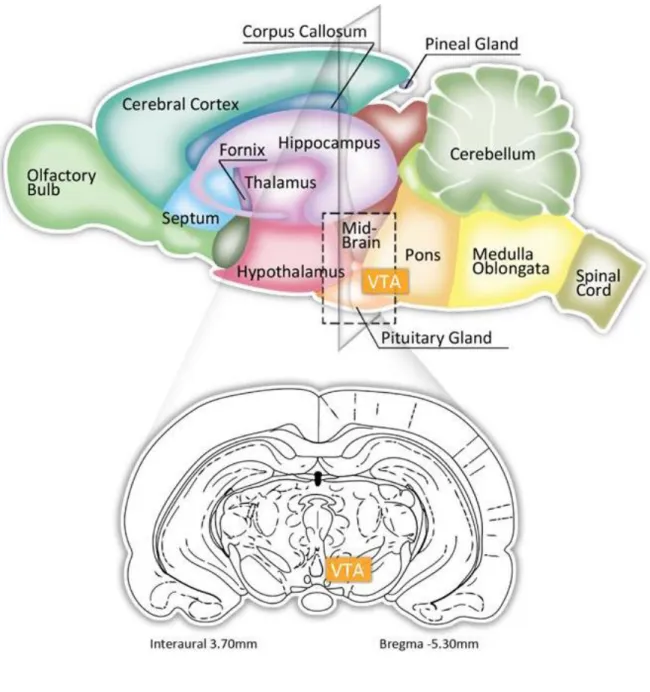
Figure 4. Location of the VTA in the rat brain ... 43
Figure 5. Schematic midsagittal section of a rat brain, showing the locations of the most important groups of dopaminergic neurons and the distribution of their axons and terminal buttons ... 46
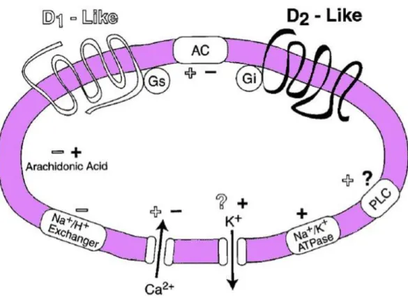
Figure 6. Schematic representation of the DA receptors in the human brain . ... 59
Figure 7. Signal transduction of D1R-like and D2R-like receptors... 62
Figure 8. The kallikrein-kinin system ... 70
Figure 9. Sites of proteolytic enzymes of kinins ... 74
Figure 10. Signaling mechanism of kinin B1R and B2R ... 95
Figure 11. Diagram of the cardiovascular effects of BK on B2R in the spinal cord ... 99
Artcle #1
Figure 1. Identification of the VTA as microinjection site following post-mortem histological examination of microinjected Evans’blue. A rat was considered to be correctly injected when a black spot was seen in the VTA without any evidence of haemorrhage or necrosis. Diagram was modified from the atlas of Paxinos and Watson (1998). Abbreviations: VTA, ventral tegmental area; SNR, substantia nigra reticular; ml, medial lemniscus; RMC, red nucleus magnocellular; MS, microinjection site. Scale: 0.5 mm ... ... 147
Figure 2. Time-course effects on changes in mean arterial blood pressure (ΔMAP) and heart rate (ΔHR) induced by i.c.v. (a) or VTA (b) injection of 500 pmol of SB222200 or its inactive enantiomer SB222201. Areas under the curves (AUC) were measured for a period of 0-10 h for i.c.v. and 0-8 h for VTA (small insets). Values represent the mean ± s.e.m. of n rats. Statistical comparison to aCSF is indicated by *P<0.05; **P<0.01; ***P<0.001. ... 148
Figure 3. Time-course effects on changes in mean arterial blood pressure (ΔMAP) and heart rate (ΔHR) induced by i.c.v. injection of (a) SB222200 (500 pmol) and (b) R-820 (500 pmol) before and 30 min after treatment with the DA-D2R antagonist raclopride (0.16 mgkg-1, i.v.) or the DA -D1R antagonist SCH23390 (0.2 mgkg-1, i.v.). Areas under the curves (AUC) were measured for a period of 0-8 h (SB222200) or 0-72 h (R-820) as shown in small insets. Values represent the mean ± s.e.m. of n rats. Statistical comparison to aCSF values (*) or NK3R antagonist alone (†) is indicated by * P<0.05, ** ††P<0.01; *** †††P<0.001. ... 149
Figure 4. Time-course effects on changes in mean arterial blood pressure (Δ MAP) (a) and heart rate (ΔH) (b) induced by R-820 (500 pmol) injected into the VTA before and 30 min after treatment with the DA-D2R antagonist raclopride (0.16 mgkg-1, i.v.). Areas under the curves (AUC) were measured for a period of 0-5 h (small insets). Values represent the mean ±
s.e.m. of n rats. Statistical comparison to aCSF values (*) or R-820 (†) is indicated by * †P<0.05; ** ††P<0.01; *** †††P<0.001. ... 150
Figure 5. Time-course effects on changes in mean arterial blood pressure (ΔMAP) and heart rate (ΔHR) induced by i.c.v. injection of 500 pmol R-820, once daily for a period of 5 days in SHR. Values represent the mean ± s.e.m. of 4 rats. Statistical comparison to pre-injection values is indicated by
*P<0.05; **P<0.01. ... 151
Figure 6. Time-course effects on changes in mean arterial blood pressure (ΔMAP) and heart rate (ΔHR) following i.c.v. (a) or VTA (b) injection of four increasing doses (10, 25, 65 and 100 pmol) of senktide in SHR. Areas under the curves (AUC) were measured for a period of 0-1 h (open bars for VTA and closed bars for i.c.v.). Values represent the mean ± s.e.m. of 6 rats per injection site. Statistical comparison to aCSF values (*) or i.c.v. (†) is indicated by ** ††P<0.01; *** †††P<0.001. ... 152
Figure 7. Time-course effects on changes in mean arterial blood pressure (ΔMAP) and heart rate (ΔHR) produced by senktide (25 pmol) injected into the VTA prior to (first day) and 1 h after VTA injection of R-820 (second day). Each point represents the means ± s.e.m. of 4 rats. Statistical comparison to aCSF (*) or senktide (†) values is indicated by * †P<0.05; **
††P<0.01; *** †††P<0.001 ... 153
Figure 8. Time-course effects on changes in mean arterial blood pressure (ΔMAP) produced by i.c.v. senktide 65 pmol (baseline: 161±5 mmHg) prior to and after treatment with (a) the DA-D1R antagonist SCH23390 (0.2 mgkg-1 i.v.) and the DA-D2R antagonist raclopride (0.16 mgkg-1 i.v.) (baseline: 167±4 mmHg); and (b) the non-selective DA-D2R antagonist haloperidol (10 mgkg-1 s.c.) (baseline: 164±2.4 mmHg) in SHR. In (c) are shown the Areas under the curves (AUC) measured for a period of 0-1 h. Values represent the mean ± s.e.m. of n rats. Statistical comparison to aCSF
(*) or senktide (†) values is indicated by * †P<0.05; ** ††P<0.01; *** †††P<0.001. .... ... 154
Figure 9. Time-course effects on changes in mean arterial blood pressure (ΔMAP) produced by treatments with the DA-D1R antagonist SCH23390 (0.2 mgkg-1 i.v.), DA-D2R antagonist raclopride (0.16 mgkg-1 i.v.) and the non-selective DA-D2R antagonist haloperidol (10 mgkg-1 s.c.) in SHR. Different time scales were used: in (a) (min), and (b) (hours).Values represent the mean ± s.e.m. of 4 rats. Statistical comparison to saline is indicated by *P<0.05; **P<0.01; ***P<0.001. ... 155
Figure 10. Time-course effects on changes in mean arterial blood pressure (Δ MAP) produced by senktide injected i.c.v. on the (a) ipsilateral and (b) contralateral side before and after SB222200 (500 pmol) injected into the VTA. The anti-hypertensive effect elicited by the VTA injection of SB222200 (500 pmol) was measured before and after i.c.v. injection of senktide (65 pmol) on the ipsilateral (c) and contralateral (d) side in SHR. Areas under the curves (AUC) for MAP are shown in (e and f). Values represent the mean ± s.e.m. of n rats. Statistical comparison to aCSF (*) and senktide alone or SB222200 alone (†) is indicated by * † P<0.05; ** ††P<0.01; *** ††† P<0.001. ... 156
Figure 11. Time-course effects on changes in mean arterial blood pressure (Δ MAP) produced by i.c.v. injected 65 pmol senktide (a, b) or 500 pmol R-820 (c, d) in SHR which underwent an ipsilateral (a, c) and contralateral (b, d) lesion of the VTA with ibotenic acid (IBO), 5 days earlier or a sham-operation. Areas under the curves (AUC) for MAP are shown in (e, f). Each point represents the mean ± s.e.m of n rats. Statistical comparison to aCSF (*) or sham-operated rats (†) is indicated by * †P<0.05; ** ††P<0.01; *** †††P<0.001. ... 157
Figure 1. Time-course effects on changes in mean arterial blood pressure (ΔMAP) induced by three doses of SSR240612 (1, 5 and 10 mg/kg, gavage) in AngII-treated rats (a). Raclopride (0.16 mg/kg, i.v.) was injected alone or 30 min prior to 5 mg/kg SSR240612 to assess the participation of DA-D2R in the anti-hypertensive effect of SSR240612 (b). Control WKY also received 5 mg/kg SSR240612 (a) or 0.16 mg/kg raclopride (b). Area under the curves (AUC) was measured for a period of 0-6 h (c) or 0-8h (d). Data are means ± s.e.m. of values obtained from (n) rats. Statistical comparison with vehicle values (*) or SSR240612 (†) is indicated by **P<0.01; ***, †††P<0.001 ... ... 181
Figure 2.Time-course effects on changes in mean arterial blood pressure (ΔMAP) induced by three doses of SSR240612 (1, 5 and 10 mg/kg, gavage) in SHR (a). Raclopride (0.16 mg/kg, i.v.) was injected alone or 30 min prior to 5 mg/kg SSR240612 to assess the participation of DA-D2R in the anti-hypertensive effect of SSR240612 (b). Control WKY also received 5 mg/kg SSR240612 (see Fig. 1a) or 0.16 mg/kg raclopride (b). Area under the curves (AUC) was measured for a period of 0-6 h (c,d). Data are means ± s.e.m. of values obtained from (n) rats. Statistical comparison with vehicle values (*) or SSR240612 (†) is indicated by *P<0.05; **P<0.01; ***, †††P<0.001. ... 182
Figure 3. Time-course effects on changes in mean arterial blood pressure (ΔMAP) induced by SSR240612 (5mg/kg, gavage) in AngII-treated rats (a) and SHR (b) before and 30 min after administration of the dopamine antagonist haloperidol (10 mg/kg, s.c). Hypertensive rats and control WKY also received 10 mg/kg haloperidol alone. Area under the curves (AUC) was measured for a period of 0-8 h (c) or 0-6 h (d). Data are means ± s.e.m. of values obtained from (n) rats. Statistical comparison with vehicle values (*) or SSR240612 (†) is indicated by *P<0.05; **, ††P<0.01; ***, †††P<0.001 ... 183
Figure 4. Time-course effects on changes in mean arterial blood pressure (ΔMAP) induced by SSR240612 (1μg, i.c.v.) in AngII-treated rats (a) and SHR (b) before and 30 min after administration of the DA-D2R antagonist raclopride (0.16 mg/kg, i.v.). Control WKY also received i.c.v. SSR240612. Raclopride alone was assessed in previous figures. Area under the curves (AUC) was measured for a period of 0-6 h (c, d). Data are means ± s.e.m. of values obtained from (n) rats. Statistical comparison with aCSF values (*) or SSR240612 (†) is indicated by *, †P<0.05; **, ††P<0.01; ***, †††P<0.001 ... 184
Figure 5. Time-course effects on change in mean arterial blood pressure (ΔMAP) induced by R-715 and R-954 (2mg/kg, s.c.) in AngII-treated rats (a) and SHR (b). Effects of both B1R antagonists were also tested in control WKY (a). Area under the curves (AUC) was measured for a period of 0-10 h (c) or 0-12 h (d). Data are means ± s.e.m. of values obtained from (n) rats. Statistical comparison with vehicle values (*) is indicated by **P<0.01; ***P<0.001. ... 185
Article #3
Table 1. PCR primer pairs used in this study ... 201
Figure 1. B1R gene expression in hypothalamus, ventral tegmental area (VTA) and nucleus accumbens of SHR and Ang II-treated rats. B1R mRNA levels were measured relative to 18S level. Data represent the mean ± s.e. mean of values obtained in groups of 4 rats. Statistical comparison to control WKY is indicated by *P<0.05; **P<0.01 or ***P<0.01 ... 212
Figure 2. Behavioural responses to 1 μg Sar[DPhe8][des-Arg9]BK injected i.c.v. in SHR and WKY. Each bar represents the mean ± s.e.mean of values obtained in 4 rats per group. Comparison to WKY is indicated by ***P < 0.001. ... 213
Figure 3. Inhibition of the behavioural responses to 1 μg Sar[DPhe8 ][des-Arg9]BK injected i.c.v. in SHR (a, c) and Ang II-treated rats (b, d) before and 3h after treatment with the kinin B1R antagonist SSR240612 administered either i.c.v. (10 μg) or by gavage (10 mg∙kg−1). Each bar represents the mean ± s.e. mean of values obtained in 4 groups of 6 rats. Comparison to aCSF (*) and to the B1R agonist (†) is indicated by *†P<0.05; **††P<0.01; ***P< 0.001 ... 214
Figure 4. Inhibition of the behavioural responses to 1 μg Sar[DPhe8 ][des-Arg9]BK injected i.c.v. in SHR before and 30 min after treatment with the
tachykinin NK-1R antagonist RP67580 (a), the NMDA receptor antagonist DL-AP5 (b), the NOS inhibitor L-NNA (c), the iNOS inhibitor 1400W (d), the dopamine D1R antagonist SCH23390 (e), the dopamine D2R antagonist Raclopride (f). Each bar represents the mean ± s.e. mean of values obtained in 6 groups of 6 rats. Comparison to the B1R agonist before treatment is indicated by *P<0.05, **P<0.01. ... 215
Figure 5. Inhibition of the behavioural responses to 1 μg Sar[DPhe8 ][des-Arg9]BK injected i.c.v. in Ang II-treated rats before and 30 min after treatment with the tachykinin NK-1R antagonist RP67580 (a), the NMDA receptor antagonist DL-AP5 (b), the NOS inhibitor L-NNA (c), the iNOS inhibitor 1400W (d), the dopamine D1R antagonist SCH23390 (e), the dopamine D2R antagonist Raclopride (f). Each bar represents the mean ± s.e.mean of values obtained in 6 groups of 6 rats. Comparison to the B1R agonist before treatment is indicated by *P<0.05, **P<0.01 ... 216
Figures – General discussion
Figure 12. Schematic representation showing tachykinin NK-3R agonist/antagonists injected i.c.v. (A) and VTA (B) in rats ... 219
Figure 13. Schematic representation of the NK-3R agonist and antagonists in the CNS. In A, upper panel shows that NKB is released and caused high occupancy of NK-3R. DA neurons are excited by NKB, resulting in an increase of DA release in the nucleus accumbens and a high level of DA-D2R occupancy post-synaptically. In B, lower panel illustrates the inhibition of NKB effect by NK-3R antagonist. As consequence, the DA neurons are less excited and the release of DA in the nucleus accumbens is strikingly decreased ... 222
Figure 14. Schematic representation showing the CSF circulation of kinin B1R agonist and antagonists injected i.c.v. in rats ... 225
Figure 15. Kinin B1R antagonists (SSR240612, R-924, R-715) injected by gavage, i.c.v. and s.c. decreased body temperature in SHR and Ang II-treated rats ... 236
List of Tables
Table I. Chemical formulae of endogenous tachykinins in mammals ... 10
Table II. Tachykinin NK-1R-selective agonists ... 24
Table III. Chemical formulae of tachykinin NK-1R antagonists ... 25
Table IV. Chemical formulae of tachykinin NK-2R antagonists ... 28
Table V. Tachykinin NK-3R in rat brain ... 32
Table VI. Chemical formulae of tachykinin NK-3R antagonists ... 33
Table VII. Chemical formulae of B1R antagonists ... 84
List of Abbreviations
ACE: angiotensin converting enzyme
ACEI: angiotensin converting enzyme inhibitor ACE-1: angiotensin converting enzyme-1 Ach: acetylcholine
ADHD: attention deficit hyperactivity disorder Ang: angiotensin
ANS: autonomic nervous system ANP: atrial natriuretic peptide BBB: blood-brain barrier BK: bradykinin
cAMP: cyclic adenosine monophosphate cGPM: cyclic guanosine monophosphate CPM: carboxypeptidase M
CPN: carboxypeptidase N BP: blood pressure
CGRP: calcitonin gene related peptide CNS: central nervous system
CREB: cAMP response element binding protein DA: dopamine or dopaminergic
DA-D1R: dopamine receptor type D1 DA-D2R: dopamine receptor type D2 DAG: diacylglycerol
EGF: epidermal growth factor GABA: γ-aminobutyric acid
GDNF: glial cell-derived neurotrophic factor GPCR: G protein-coupled receptors
HR: heart rate
HMWK: high molecular weight kininogen
HPLC: high-performance liquid chromatography HBECs: human brain endothelial cells
i.c.v.: intracerebroventricular IP3: inosito1-1,4,5-triphosphate
IRAK: interleukin-1 receptor-associated kinase 1 i.t.: intrathecal
ITIM: immunoreceptor tyrosine-based inhibitory pattern i.v.: intravenous
KD: kallidin KO: knockout
LMWK: low molecular weight kininogen LPS: lipopolysaccharides
MAP: mean arterial blood pressure
MAPK: mitogen-activated protein kinases mPFC: medial prefrontal cortex
mRNA: messenger ribonucleic acid NE: noradrenaline
NK-1R: tachykinin receptor type-1 NK-2R: tachykinin receptor type-2 NK-3R: tachykinin receptor type-3 NKA: neurokinin A
NKB: neurokinin B NO: nitric oxide
NOs: nitric oxide synthases NPγ: neuropeptide gamma NPK: neuropeptide K
NTS: nucleus of the solitary tract PB: parabrachial nucleus
Pa5: paratrigeminal nucleus
PDGF: platelet-derived growth factor PDTC: pyrrolidine dithiocarbamate PKA: protein kinase A
PKC: protein kinase C PLC: phospholipase C PLD: phospholipase D PPT: preprotachykinin gene
RT-PCR: polymerase chain reaction R: receptor
RVLM: rostral ventrolateral medulla s.c.: subcutaneous
SNS: sympathetic nervous system SN: substantia nigra
SO: supraoptic nucleus of the hypothalamus SP: substance P
SP5: trigeminal nucleus STZ: streptozotocin
TRKA: high affinity nerve growth factor WKY: Wistar-Kyoto rat
VTA: ventral tegmental area 6-OHDA: 6-hydroxydopamine
Chapter I
1. General introduction
1.0.1 The autonomic nervous system: A strategic role in blood pressure regulation
The autonomic nervous system (ANS) is controlled by central autonomic nuclei of the diencephalon (hypothalamus nuclei), forebrain (amygdala), midbrain (periaqueductal gray region) and brainstem (nucleus of the solitary tract, ambiguous nucleus, and dorsal motor of the vagus, rostral and dorsal ventrolateral medulla). These regions of the central nervous system (CNS) control pre-ganglionic sympathetic and parasympathetic efferent viscerimotor fibers (Benarroch, 1993). This system is very complex and provides precise control of cardiovascular function in normal physiological situations (standing, intense exercise).
In addition, the ANS provides a fast and sensitive regulation of arterial pressure due to the baroreflex. Through arterial baroreceptors and chemoreceptors, the cardiovascular centers situated in the brainstem and hypothalamus received constant information on the status of the peripheral circulation (de Champlain, 2001). Several studies supported the major role of ANS in controlling cardiac function and peripheral resistance (de Champlain et al., 1998). Clinical and experimental studies demonstrated that sympathetic activity was increased in hypertensive patients and in several models of experimental hypertension (Julius et al., 1988; de Champlain et al., 1998; Laflamme et al., 1998a, b; Grassi, 1998). Moreover, decreased parasympathetic tone was observed in several cases of clinical
1.0.2 Hypertension
According to 2008 statistics, provided by the Heart and Stroke Foundation of Canada, hypertension affects one in five Canadians. Hypertension is a risk factor that can triple the chance of developing cardiovascular diseases. It is the number one risk factor for stroke and a major risk factor for heart disease. However, since there are no symptoms, 42% of Canadians with high blood pressure (BP) do not even know that they have it. This is why hypertension is known as the silent killer.
The five major classes of anti-hypertensive agents are: diuretics, beta-blockers, calcium channel beta-blockers, angiotensin converting enzyme (ACE) inhibitors, and angiotensin AT1 receptor antagonists (Plante, 1999).
The causes of hypertension are highly diverse and provide an immense challenge for fundamental research, because it involves the kidneys, vascular, cardiac, nervous and endocrine systems.
1.0.3 The tachykinins and their receptors: regulation of central cardiovascular function in hypertension
Tachykinins are one of the largest families of neuropeptides. They can be found in species from amphibians to mammals. The tachykinin family is characterized by a common C-terminal sequence, Phe-X-Gly-Leu-Met-NH2. Therefore, the tachykinin N-terminal determines the receptor selectivity, while the conserved C-terminal sequence is responsible for the receptor
activation (Lucas et al., 1992). This family is named tachykinins, which means “fast movement" on the contraction of smooth muscles.
In mammals, the family members are substance P (SP), neurokinin A (NKA), neurokinin B (NKB), neuropeptide K (NPK) and neuropeptide (NP ). They are distributed in both the central and peripheral nervous systems. These have a wide range of biological effects that follow the activation of three types of trans-membrane receptors (R) called 1, NK-2 and NK-3, which are G protein-coupled receptors (GPCRs). Due to their vast distribution in the CNS, they play a major role in many essential functions such as learning, memory and emotional processes. This also includes stereotyped behaviours, anxiety, stress and pain. Thus, the tachykinins are known to exert central control over many functions of the ANS (Severini et al., 2002).
The potential role of tachykinins in central cardiovascular regulation has been suggested by several studies. Increases in blood pressure and heart rate (HR) accompanied by stereotyped behaviours were shown by tachykinin agonists injected into the i.c.v. and ventral tegmental area (VTA) (Picard et al., 1994; Takano et al., 1990; Cellier et al., 1997; Deschamps and Couture 2005). These cardiovascular effects are dependent on the activation of the sympathetic nervous system as well as the increased release of vasopressin from the neurohypophysis (Unger et al., 1981, Polidori et al., 1989, Takano et al., 1990). However, the specific neuronal circuits involved in cardiovascular and behavioural effects of tachykinins are still largely unknown.
A strategic central dopaminergic (DA) site is the VTA. It is located at the midbrain and composed mainly of dopaminergic neurons forming the area A10. Considering its projections to the limbic system and cerebral cortex, the VTA is more known for its role in the regulation of behavioural activity in response to stress, psychologic diseases, drug addiction and drug withdrawal. However, compelling experimental results suggest its involvement in cardiovascular control. Some studies suggest that the DA system also participates in these events. Indeed, when the three tachykinin NK-1R, NK-2R and NK-3R agonists were injected into the VTA of normotensive rats, they affected the autonomic control of BP and HR by increasing midbrain DA transmission (Deschamps and Couture 2005). Substance P (SP) injected into the i.c.v. improved the baroreflex sensitivity, while the injection of antibodies against SP (into the i.c.v. or directly into the nucleus of the solitary tract (NTS)) reduced the baroreflex (Chan et al., 1990; Appenrodt et al., 1993). Moreover, when SP or a selective NK-1R agonist was injected into the NTS, it induced hypotension and bradycardia. Electrical or chemical stimulation of the VTA with the injection of SP analogue DiMe-C7 produced an increase in BP and locomotor activity. The increases in locomotor activity induced by intra-VTA DiMe-C7 were blocked by the DA-D1R and DA-D2R antagonists, SCH23390 and haloperidol, respectively (Kubos et al., 1987; Cornish et al., 1994; Placenza et al., 2004). When the NK-3R agonist senktide was injected into the VTA, it caused several behaviours such as yawning and chewing known to be evoked by DA (Stoessl et al., 1991). Furthermore, the
activation of NK-3R by endogenous tachykinins in the substantia nigra (SN), an important DA center, stimulated the cardiac function. The NK-3R antagonist (SB222200) significantly reduced mean arterial pressure (MAP) for more than three hours when injected into the SN in spontaneously hypertensive rats (SHR) (Lessard et al., 2001, 2003, 2004). However, the function of NK-3R in the CNS during pathophysiological situations remains poorly understood.
1.0.4 The kinins and their receptors: regulation of central cardiovascular function in hypertension
Kinins are a small family of peptides (9 to 11 amino acids), including bradykinin (BK), kallidin (KD; Lys-BK) and T-kinin (Ile-Ser-BK) (Gabra et al., 2003). The most studied members of this family are BK and KD. Their active metabolites are produced by the enzymatic action of kininase I (des-Arg9-BK, des-Arg10-KD). These peptides produced their biological effects via the activation of two types of GPCRs, named B1 and B2. The B2R is activated by BK, KD and T-kinin (rats only) while the B1R is activated by des-Arg9-BK and des-Arg10-KD (Leeb-Lundberg, 2001). As opposed to B2R, which is expressed in most tissues, the B1R is usually absent in healthy tissues. The B1R is inducible and its expression is increased in the presence of cytokines, bacterial lipopolysaccharides (LPS) and after tissue injury (Marceau et al., 1998b).
Kinins have been identified in the CNS and sufficient evidence suggests that kinins play a role as neuromodulators in central cardiovascular regulation, pain and inflammation (Couture and Lindsey, 2000; Couture et al., 2001). In freely behaving normotensive rats, BK caused a pressor response when it was injected into the i.c.v. or directly into the NTS and paratrigeminal nucleus (Pa5) (Corrêa and Graeff, 1974; Fior et al., 1993; Lindsey et al., 1997; Couture and Lindsey 2000).
The thoracic spinal cord is another site of central cardiovascular regulation. Previous studies from our laboratory have shown that BK injected intrathecally (i.t.) increased BP via the activation of the sympatho-adrenal system and B2R (Lopes et al., 1992; Lopes et al., 1993). This was substantiated by autoradiographic studies, which revealed the presence of B2R binding sites in the spinal cord (Lopes et al., 1995; Couture and Lindsey, 2000).
Emanueli et al. (1999) reported that B1R agonists injected into the i.c.v. in SHR and Wistar Kyoto rats (WKY) caused increases in BP, while the B1R antagonist R-715 caused a small decrease of BP in SHR. The latter findings were not reproduced with the identical B1R agonists and antagonists in SHR (Cloutier et al., 2004). Thus, the role of central kinin B1R in hypertensive rats is still controversial.
1.1 Tachykinins
The name tachykinin resulted from the pharmacological similarity of these peptides to bradykinin. The tachykinins, while having generally the same
pharmacological profile, induced a fast contraction of the tissue (Khawaja and Rogres, 1996).
Tachykinins are present in several peripheral tissues where they perform many functions on the cardiovascular, respiratory and gastro-intestinal systems. Their effects include vasodilatation, increased vascular permeability, bronchoconstriction and contraction of smooth muscle cells. Neurogenic inflammation involved in diseases such as asthma is induced following the release of tachykinins from peripheral sensory C-fibers (Harrison and Geppetti, 2001).
The discovery of SP, by von Euler and Gaddum in 1931, introduced a new era of peptides in neuroscience. This previously unidentified substance was found in alcoholic extracts of the equine brain and horse intestine. It was isolated and characterized as an eleven amino acid peptide, which had a potent stimulant action on the jejunum and also caused hypotension (Severini et al., 2002).
Later, neuromedin K and neuromedin L were isolated from the porcine spinal cord (Kangawa et al., 1983; Minamino et al., 1984) and neurokinin and neurokinin from guinea-pig ileum and rat duodenum (Kimura et al., 1984). Substance K, neurokinin , and neuromedin L were re-named neurokinin A (NKA), while neurokinin and neuromedin K were called neurokinin B (NKB) (Maggi, 2000).
Two other mammalian tachykinins, neuropeptide K and neuropeptide , were isolated from porcine brain (Tatemoto et al., 1985) and the rabbit
intestine (Kage et al., 1988), respectively. These last two tachykinins were the products of the same gene (preprotachykinin A), which encodes SP and NKA. They were identified as the N-terminal elongated forms of NKA (Table 1).
Table I. Chemical formu lae of endogeno us tachykinins in mam m als References Chang and Leeman, 19 70 Nawa et a l. , 1984a Kangawa et a l. , 1983 Tatemoto et a l. , 1985
Kage et al., 1988 Zhang
et a l. , 2000 Pag e et a l., 2003 Sequences Ar g -Pr o -Lys-Pr o -Gln-Gln-P he-Phe -Gly -Leu -M et -NH 2 His-Lys-T hr -Asp-S er -P he-Val-Gly -Le u-M et -N H2 Asp-Met -His-Asp -Phe-P he-Val-Gly -Leu-Met -N H2 Asp-Ala-Asp-Ser -Ser -I le-Glu-Lys -Gln -Val-Al a-Leu-Leu -Lys- Ala-Leu-Tyr -Gly -Hi s-Gly -Gln-Ile -S er -His -Lys-Ar g -His-Lys- Thr -Asp-Ser -Phe-Val -G ly-Leu-Met -NH 2 Asp-Ala-Gly -Tyr -Gly -Gln-Ile -S er -His-Lys -Ar g -His-Lys-T hr -Asp-Ser - Phe-Val-G ly-Leu-Met -NH 2 Ar g -Se r-Arg-Thr-Ar g-Gl n-Phe-Tyr -Gly-Leu-M et -NH 2 Gly -Lys-Ala-Ser -Gln- Phe-Phe-Gly-Leu-Met -N H2 Subs tanc e P
NKA NKB Neuropeptide K Neuropeptide γ Hemokinin-1 Endok
1.1.2 Tachykinins: biosynthesis and distribution
Tachykinins were encoded by two genes that, according to the Human Genome Organization Gene Nomenclature Committee, are termed TAC1 (SP, NKA) and TAC3 (NKB), which replace the previously used terms PPT-A and PPT-B (Patacchini et al., 2004).
The precursor RNA from TAC1 was alternatively processed to yield four different mRNAs (α, β, γ and δ) (Figure 1). SP was encoded by all four mRNAs. NKA was encoded by β- and γ-PPT, neuropeptide K by β-PPT, and neuropeptide-γ by γ-PPT. The TAC3 gene encoded the precursor for NKB, which derived from α- and β-mRNAs (Severini et al., 2002). Recently, TAC4, a third tachykinin gene preprotachykinin-c (PPT-C) has been discovered in lymphoid B hematopoietic cells of mouse bone marrow (Zhang et al., 2000). TAC4 encoded hemokinin-1 and endokinin (Page, 2004). The expression of TAC1 and TAC3 genes which vary between tissues and species determined the distribution of the classical tachykinins (Page, 2005). Although the β-TAC1 mRNA was the predominant type expressed in the human basal ganglia, α-TAC1 mRNA was abundant in the mammalian brain (Bannon et al., 1992). The β- and γ- TAC1 mRNAs were found mainly in peripheral tissues (Nakanishi, 1987).
The most studied member among the tachykinins is unquestionably SP. The distribution of SP is greatly expressed in CNS areas that are involved in the regulation of pain transmission, affective behaviour and stress (De Felipe et al., 1998; Kramer et al., 1998).
• SP • SP • NKA • NPK • SP • NKA • NPγ • NKB • NKB
TAC4
TAC3
TAC1
• SP Nerve transmission Nerve transmissionNK
-1, N
K-2,
NK
-3 receptors
Figure 1. Schematic representation of the TAC1, TAC3, and TAC4 genes expression and their mRNA messengers. Adapted from Guard and Watson, 1991.
SP is present in the limbic system, including the hypothalamus and the amygdala, areas associated with emotional behaviour (Stahl, 1999). In the mesolimbic brain areas, SP is believed to be involved in reward, opioids withdrawal (Murtra et al., 2000) and memory-promoting effects (Huston and Hasenohrl, 1995). Immunoreactive-SP nerve fibers were found in the rat cortex and hippocampus, and were lightly distributed all over the layers of the neocortex. Studies revealed that the localization of these cells was a subset of the glutamic acid decarboxylase containing neurons supposedly γ-aminobutyric (GABA)-ergic neurons (Penny et al., 1986). In some neurons of the raphe nuclei of the rat medulla, SP co-existed with serotonin and GABA (Chan-Palay, 1978; Magoul et al., 1986).
SP has been found in dorsal root ganglia and sensory neurons as well as in the most autonomic ganglia and intrinsic enteric neurons in the peripheral nervous system (Hokfelt et al., 1977). The highest concentrations of immunoreactive-SP fibers were found in the SN and dorsal horn of the spinal cord and in nerve terminals of the hypothalamus in rats (Figure 2) (Kanazawa and Jessell, 1976; Douglas et al., 1982; Ljungdahl et al., 1978). Substantial, but lower levels were present in other regions including amygdala, caudate putamen, and globus pallidus (Kertes et al., 2009). The distribution of immunoreactive-SP neurons and fibers in the human brain showed considerable similarity to the rat brain, including dense distribution of immunoreactive-SP in the SN, the caudate putamen, and the
Figure 2: Distribution of immunoreactive-SP in the rat hypothalamus (bregma: -1.8 mm (Paxinos and Watson, 1998). Scale of density: green, high; blue, moderate and plum, low. CA3, CA3 region of the hippocampus; CP, caudate nucleus and putamen of the striatum, GP, globus pallidus; HL, lateral hypothalamus; PV, paraventricular nucleus of the hypothalamus; PO, piriform cortex; SO, supraoptic nucleus of the hypothalamus. Adapted from Shults et al., 1984.
brain stem (Chahl, 2006). However, the human cortex and hippocampus contained more abundant SP cells and fibers than rat brain (Hokfelt et al., 1976; Mai et al., 1986; Del Fiacco et al., 1987). The other classical tachykinins were generally found in lower concentrations than SP in the CNS. Most studies on the distribution of tachykinins have used antisera directed towards the C-terminal region of the peptides which differentiate weakly between the family members. The recent discovery of hemokinin-1 and endokinin and their cross-reactivity with anti-SP antibodies were questioning several of the older studies on the peripheral and central distribution of the classical tachykinins (Page, 2004). Recent studies using molecular biological techniques have confirmed that TAC1 and TAC3 mRNAs were strongly expressed in the brain (Pinto et al., 2004) and peripheral tissues, as described in previous studies. Since the classical tachykinins were synthesized in the cell soma and transported to the nerve terminals (Krause et al., 1987), precursor mRNAs were present in the cell soma, whereas the peptides were predominantly present in the nerve terminals. Thus, immunocytochemical methods will detect tachykinins in tissues that do not necessarily contain the mRNAs.
SP has been shown to preferentially activate mesocortical DA neurons in a similar way (Tamiya et al., 1990). SP injected directly into the VTA increased the levels of DA, its metabolites or both, in the prefrontal cortex and the nucleus accumbens, suggesting that SP increased the release of DA from mesocortical and mesolimbic DA neurons (Deutch et al., 1985; Cador et al., 1989; Barnes et al., 1990; Elliott et al., 1992).
SP has been shown to preferentially activate mesocortical DA neurons in a similar manner to acute stressors such as mild foot shock or restraint (Elliott et al., 1986b). Furthermore, evidence suggested that the increased turnover of DA in the prefrontal cortex and the nucleus accumbens in response to stress may have been mediated by SP. Increases in DA metabolism in the prefrontal cortex in response to foot shock stress was blocked by an SP antibody in the VTA (Bannon et al., 1983). These findings suggested that SP not only activated mesocorticolimbic DA neurons, but the activation of these DA neurons in response to stress may have been mediated by the endogenous SP system. The activation of VTA DA neurons has been implicated in the mediation of relapse, mainly in drug-induced (McFarland and Kalivas, 2001), and perhaps foot shock-drug-induced relapse as well (Capriles et al., 2003).
1.2 The NKA and NKB
Immunohistochemical studies and in situ hybridization showed a high distribution of NKA, NKB and their mRNA in each major subdivision of the brain (Minamino et al., 1984; Nagashima et al., 1989; Nawa et al., 1984b), including the hypothalamus (Tateishi et al., 1989; Larsen, 1992; Merchenthaler et al., 1992), midbrain (Arai and Emson, 1986) cerebral cortex and medulla (Minamino et al., 1984). NKA and SP were both localized to the same region. This was consistent with the fact that these two neuropeptides were synthesized from the same precursor (Ribeiro-da-Silva et al., 2000).
In humans and monkeys, the mRNA gene expression of the TAC1 responsible for the synthesis of SP and NKA was detected in the striatum, hypothalamus, periaqueductal gray region and the amygdala (Arai and Emson, 1986; Larsen et al., 1992; Sergeyev et al., 1999).
Immunocytochemical studies revealed high density of NKB, in contrast to a low density of SP, in the olfactory bulb, cerebral cortex and hippocampus in rats (Lucas et al., 1992). Although the raphe nucleus possesses a high density of SP, NKB was not detected (Warden and Young, 1988). In addition to these regions, NKB was found in the striatum and several nuclei of the hypothalamus, including the lateral and paraventricular nucleus (Merchenthaler et al., 1992).
The gene expression of c-fos was upregulated in rat brain areas activated by tachykinins (Spitznagel et al., 2001). Increased numbers of c-fos positive neurons were detected in the septum, amygdala, paraventricular nucleus, supraoptic nucleus, lateral hypothalamus, thalamus, VTA, and SN following i.c.v. injection in rats of NKB or senktide, a NK-3R agonist (Smith and Flynn, 2000). Using the same approach in rat, i.c.v. injected SP showed small expression of c-fos in the paraventricular and mediodorsal nuclei of the hypothalamus, the parabrachial nucleus and the medial thalamus. C-fos expression was reduced significantly in the presence of tachykinin NK-1R antagonist, RP67580 (Spitznagel et al., 2001). It is tempting to suggest that SP and NKB may activate distinct neural pathways, and consequently, that they are involved in different physiological functions. Further studies are necessary to confirm this hypothesis.
1.3 Tachykinin receptors
The existence of multiple receptors for tachykinins emerged with the work of Erspamer et al. (1981) showing considerable differences between the pharmacological profiles of tachykinins. The distribution of tachykinin receptors has been examined after identification and molecular cloning of the three tachykinin receptors (Yokota et al., 1989; Hershey and Krause, 1990; Nakanishi, 1991).
The three tachykinin receptors (NK-1, NK-2 and NK-3) have a very high homology between them (from 53.7 to 66.3%) in the seven transmembrane domains cytoplasmic or intracellular regions (Shigemoto et al., 1990). Lee et al. (1982) were the first to suggest the existence of several types of receptors for tachykinins.
The criteria originally used to distinguish the three receptors were; (1) the order of affinity of mammalian and non-mammalian tachykinins; (2) the activity induced by different fragments of tachykinins and, (3) the development of selective agonists (Maggi, 1995). Thanks to the first generation of SP antagonists and subsequently, to the discovery of NKA and NKB, the existence of three distinct receptors was suggested and later confirmed by molecular cloning. These receptors belonged to the “class A” family of seven-transmembranes G-protein-coupled receptors. It was not until 1986, at an international symposium held in Montreal on tachykinins, until the official nomenclature became neurokinin-1 (NK-1), neurokinin-2 (NK-2) and neurokinin-3 (NK-3) (Henry et al., 1987).
All endogenous tachykinins can act as full agonists at the three receptors, yet SP and hemokinin-1 are preferential agonists at NK-1 receptors (R), whereas NKA (NKA) and NKB (NKB) are preferential agonists at NK-2R and NK-3R, respectively (Regoli et al., 1994; Patacchini et al., 2004).
1.4 Tachykinins: agonists and antagonists 1.4.1 NK-1R
The first tachykinin receptor to be cloned was the NK-1R from the brain and the submandibular gland of the rat (Yokota et al., 1989; Hershey and Krause, 1990). It was identified in neurons and glial cells in the rat CNS (Buck et al., 1986), and in human amygdala and forebrain (Weidenhofer et al., 2006; Draganic et al., 2007) (Figure 3).
NK-1R was highly expressed in the hypothalamus, pituitary gland and amygdala, which are the brain regions that are critical for the regulation of affective behaviour and neurochemical responses to stress (Kramer et al., 1998).
Furthermore, the neural pathways that respond to stress, noxious or aversive stimulation were associated with the amygdala (Kramer et al., 1998).
Figure 3:
Schematic
representation of the rat
and human tachykin
in
NK
-1R
.
Ada
pted from Regoli et al., 1994.
F
ig
ure
3:
Schematic
representation of the rat
and human tach
y kin in NK -1 R . A da pte d from Re go li et a l., 199 4 .
The important role of NK-1R in neurogenic inflammation has been confirmed by studies using NK-1R knockout (KO) mice (Cao and Rodgers, 1998). The observation that tachykinin expression in both sensory neurons and hematopoietic cells is needed for the development of inflammation following antigen-antibody complex formation, at least in the airways, has been highlighted by the availability of the SP/NKA KO-mice (Chavolla-Calderon et al., 2003).
Preclinical research has implicated NK-1R in several pathological disorders, including emesis, asthma, psychiatric and gastrointestinal disorders, pain, migraine, inflammation and urinary bladder disorders (Lindstrom et al., 2007).
The oxidations of methionine at the C-terminal as well as the N-methylation of residue Gly9 of SP have greatly improved the selectivity of the natural peptide to NK-1R (Mussapi et al., 1993). The Sar9, Met (O2)11 -SP was a relatively stable agonist with a high affinity and selectivity for the NK-1R (Regoli et al., 1988). The existence of a subtype of NK-1R has been suggested in several studies; at least some agonists such as septide can bind to dissimilar sites of the SP receptor. It was proposed that these binding sites were located on different conformers at the NK-1R (Beaujouan et al., 2000). Agonists that have a pharmacological profile similar to SP were therefore, called "classical" while the agonists, which showed a similar profile to septide were called "septide-like agonists” (Maggi, 1995) (Table II). The first selective peptide antagonist for NK-1R was L-668169 developed in the late 1980s. However, like many others that
followed, this antagonist had low affinity and selectivity and displayed side effects (Maggi, 1995).
Other peptide antagonists (FK888 and Cam-2445) have subsequently been developed with higher affinity and selectivity (Fujii et al., 1992; Chan et al., 1996) (Table III). To increase the resistance of the antagonists to tissue peptidases, CP-96345, the first non-peptide antagonist, was developed (Snider et al., 1991). Conversely, it had a very low affinity for the NK-1R in mouse and rat and induced several side effects, among others, on the cardiovascular system and neural transmission by interacting with ionic channels (Wang and Hakanson, 1992).
RP67580, widely used in rats and mice to evaluate the physiological roles of NK-1R, has also some side effects including blocking calcium channels (Rupniak et al., 1993). In order to reduce nonspecific effects while maintaining a high affinity and selectivity, other non-peptide antagonists were developed such as LY303870, a potent antagonist (Iyengar et al., 1997) (Table III).
The possibility that NK-1R antagonists could be valuable as antipsychotic drugs was addressed based on evidence that SP modulates the activity of the mesolimbic DA system, which is the site of action of antipsychotic drugs. Consistent with this interpretation, the locomotor hyperactivity and changes in accumbens cell firing induced by intra-VTA infusion of SP were blocked by the DA-receptor antagonist haloperidol, an antipsychotic drug (Elliott et al., 1991). However, the lack of effect for the NK-1R antagonists in
these studies suggested that the effects of SP in rat VTA was mediated by the stimulation of NK-3R, rather than NK-1R, as suggested by anatomical (Saffroy et al., 1988), electrophysiological (Seabrook et al., 1995) and behavioural studies (Stoessl et al., 1991). Additionally, an exploratory trial with MK-869, which is a non-peptide antagonist with a high affinity and selectivity for the human NK-1R, showed a lack of efficacy in schizophrenic patients (Kramer et al., 1998; Rupniak et al., 1999).
To date, little is known about the way antagonists interact with NK-1R and even less is known about the mechanisms that govern the duration of their effects in vivo. The in vivo efficacy of an antagonist and its duration of action can sometimes be difficult to predict based only on potency values obtained by in vitro assays (Copeland et al., 2006)
Table II. Tachykinin NK-1R-selective agonists
Classical agonists "Septide-like agonists"
[Pro⁹]SP [Glp⁶, Pro⁹]SP (septide) [Pro⁹]SP sulfone SPOMe
[Sar⁹]SP sulfone [Apa⁹¯¹⁰]SP Physalaemin [Pro⁹¯¹⁰]SP
[Gly⁹Ψ(CH₂CH₂)-Leu¹⁰]SP [Glu(OBz)¹¹]SP
γ-aminovalery [pro⁹, NMeLeu¹⁰]SP(7-11)
(GR73,632)
[Gly⁹Ψ(CH₂CH₂)-Gly¹⁰]SP
γ-aminovalery [Pro⁹, NMeLeu¹⁰]SP(7-11)
Table III.
Chemical formu
lae of tac
hykinin NK
-1R antagonists
References Maggi, 1995 Maggi, 1995 Maggi, 1995 Snider
et a l., 1991 G arret et a l. , 1991 Iy engar et a l., 1997 Sequences Cyclo(Gln,DTrp,(NMe )P he(R) Gly ANC-2 Le u,Met (2 -(N-Me)in dolil)-CO-Hyp-Nal-NMeBzl [4 -Me]Z-(R)uMeTrpN H(S)CHMePh (2S, 3S )-cis-2-(diphenyl methyl)-N-(2 -methoxyphenyl)- met1-azabicyclo 2.2.2 octan-3-amine (3aR,7aR)-7,7-diphenyl -2-[1-i mi no-2-(2 -methoxyphenyl)- ethyl perhydroisoindo l-4-one (R)-1-N -(2-methoxybe nzyl)ac etylam ino -3-(1 H -indol-3-y l) - 2- N-(2 -(4 -( piperidin-1 -yl)piperidi n-1-yl)a cetyl)amino propane Antagonists L-668169 FK 888 Cam-2445 CP-96345 RP67580 LY 303870
1.4.2 NK-2R
The NK-2R was cloned from the bovine stomach and rat brain (Masu et al., 1987; Sasai and Nakanishi, 1989). It had 48% homology with the NK-1R (Yokota et al., 1989). The distribution of NK-2R has been studied mainly in the periphery, where high densities of binding sites were found (Mantyh et al., 1989; Tsuchida et al., 1990). These receptors have been localized on astroglial cells in rat spinal cord using electron microscopy (Zerari et al., 1998). However, the NK-2R were also located in myenteric of neurons descending colon of rats (Mantyh et al., 1989). The presence of NK-2R in the CNS has long been disputed; even autoradiography could not unequivocally prove their existence (Bergstrom et al., 1987; Saffroy et al., 1988; Mantyh et al., 1989; Nakanishi, 1991). Conversely, other studies have reported the existence of NK-2R at low concentrations in the striatum, hippocampus, hypothalamus, cerebral cortex and SN of rats and humans, by measuring mRNA by in situ hybridization or RT-PCR (Whitty et al., 1995; Bensaid et al., 2001). Moreover, autoradiography studies using as radioligand [3H]-SR48968 (a selective non-peptide antagonist NK-2R) showed the presence of NK-2R in discrete brain areas, including frontal cortex, hippocampus, amygdala, striatum, hypothalamus, thalamus and SN in rats (Saffroy et al., 2001, 2003). Several NK-2R agonists were developed based on observations that the fragment NKA (4-10) was more active and selective than NKA for the NK-2R (Drapeau et al., 1987). The selectivity of NKA (4-10) was improved by replacing the residue Met11 by Nle, ( Nle10 -NKA (4-10)) or by replacing the residue Gly8 by the β-Ala
residue which yielded the selective agonist β-Ala8 -NKA (4-10) (Rovero et
al., 1989). The agonists Lys5, MeLeu9, Nle10 -NKA (4-10) and Lys3, Gly8
-R-γ-lactam-Leu9 -NKA (3-10) (GR-64349) also have a high affinity and selectivity for NK-2R (Chassaing et al., 1991, Hagan et al., 1991).
The cyclic hexapeptide L-659877 was one of the first antagonists with high affinity and selectivity for the NK-2R. MEN10627 was synthesized in order to optimize the properties of L-659877. Polycyclic hexapeptides were more stable and retained the active conformation. MEN10627 was a competitive and reversible antagonist with potency comparable to SR48968 (Maggi et al., 1994). SR48968 was the first non-peptide antagonist developed to have a very high affinity and selectivity for the NK-2R. SR48968 was also a competitive antagonist also known for its long duration in vivo (Advenier et al., 1992; Emonds-Alt et al., 1992). GR-159897 was another good non-peptide NK-2R antagonist (Beresford et al., 1995) (Table IV).
Table IV. Chemical fo rmu lae of tac hykinin NK-2R antagonists References Maggi et a l. , 1992 Maggi et a l., 1994 Advenier et a l., 1992 Beresf ord et a l., 1995 Sequences Cyclo(Gln-Trp-Phe-Gl y-Leu-Met) Cyclo(Met -Asp-Trp-Ph e-Dap -Leu )c yclo(2 bet a -5 beta) (S)-N-methyl-N-4-acet ylamino -4-pheny lpipe ridino -2-(3,4-dic hlorophenyl)bu tyl benzamide [( R )- 1-[2 -(5-fluo ro -1 H -indol-3-yl)ethyl]-4-met hoxy -4[(phenylsu lfinyl) - me thyl]piperidine] A ntagonists L-659877 MEN10627 SR 48968 GR-159897
1.4.3 NK-3R
The NK-3R was cloned shortly after the NK-1R and NK-2R (Shigemoto et al., 1990). NKB is the endogenous ligand that has the greatest affinity for the NK-3R. The rank order of affinity of tachykinins for the NK-3R were NKB>>NKA>SP (Smith and Dawson, 2008).
Studies of function and localization have demonstrated the presence of NK-3R in mammalian central, peripheral and enteric nervous systems where its activation influences the release of a variety of neurotransmitters (for review, see Maggi, 1995). NK-3R were expressed in almost the entire CNS and spinal cord in rats (Almeida et al., 2004).
NK-3R was located in the medial prefrontal cortex (mPFC), thalamus, and amygdala. It was present in the midbrain DA nuclei (VTA) and SN, locus coeruleus, and septal and basal nuclei, which suggested its role in modulating central monoaminergic systems (Langlois et al., 2001). NK-3R was also present in dopamine (DA), and non-DA neurons of the VTA (Chen et al., 1998; Lessard et al., 2007).
NK-3R were located in mesolimbic and mesocortical pathways of VTA, which were known to be related to behaviours and cognitive functions, as well as to the pathophysiology of schizophrenia (Spooren et al., 2005; Lessard et al., 2009). This is suggested, in part, by the clinical efficacy of two non-peptide NK-3R antagonists (osanetant and talnetant) on the positive symptoms of schizophrenia (Meltzer, 2004; Spooren et al., 2005). This idea was also consistent with the hyperactivity of mesolimbic DA neurons in schizophrenia and the NK-3R was also distributed in other brain areas
(Table V). Activation of NK-3R located on the cell body of presynaptic neurons may have enhanced DA, acetylcholine (ACh), norepinephrine (NE) and GABA efflux at the nerve terminals (Marco et al., 1998; Jung and Bennett, 1996; Preston et al., 2000). These data suggested that NK-3R may be a target for treatment of psychiatric disorders (for review see Spooren et al., 2005).
Several peptide and non-peptide NK-3R agonists and antagonists have been developed. Selectivity and affinity for NK-3R were noticeably increased once the agonist MePhe77 -NKB (4-10) replaced Val7 with MePhe in the fragment NKB (4-10) (Drapeau et al., 1987). Another agonist that also had a good affinity for the NK-3R were the Pro77 -NKB. However, senktide [(succinyl-[Asp9, MePhe8])-SP(6–11)] was considered the best NK-3R agonist due to its high affinity and selectivity. Indeed, to activate NK-1R or NK-2R, the concentration of senktide had to be at least 60,000 times stronger than that required to activate the NK-3R (Wormser et al., 1986). R-486 and R-487 belonged to the first generation of antagonists selective for NK-3R, yet they maintained agonist activity to NK-1R and NK-2R (Drapeau et al., 1990) (Table VI). Being more selective, R-820 was a semi-peptide antagonist that had a good affinity in rats (Regoli et al., 1994). The first human non-peptide selective NK-3R antagonist was SR142801 (Emonds-Alt et al., 1995; Oury-Donat et al., 1995). However, in rat SR142801 and its (R)-enantiomer (SR 142806) act as agonists at spinal and supra-spinal levels (Cellier et al., 1997; Couture et al., 2000). SB222200 had a very
high affinity and selectivity for NK-3R in rat and mouse, and it crosses the blood-brain barrier (BBB) (Sarau et al., 2000).


![Table IV. Chemical formulae of tachykinin NK-2R antagonists References Maggi et al., 1992 Maggi et al., 1994 Advenier et al., 1992 Beresford et al., 1995 SequencesCyclo(Gln-Trp-Phe-Gly-Leu-Met)Cyclo(Met-Asp-Trp-Phe-Dap-Leu)cyclo(2 beta-5 beta) (S)-N-methyl-N-4-acetylamino-4-phenylpiperidino-2-(3,4-dichlorophenyl)butylbenzamide [(R)- 1-[2-(5-fluoro-1H-indol-3-yl)ethyl]-4-methoxy -4[(phenylsulfinyl)- methyl]piperidine] Antagonists L-659877 MEN10627 SR 48968 GR-159897](https://thumb-eu.123doks.com/thumbv2/123doknet/2165493.9856/55.892.299.578.117.1085/antagonists-references-sequencescyclo-phenylpiperidino-dichlorophenyl-butylbenzamide-phenylsulfinyl-antagonists.webp)
![Table VI. Cheminal formulae of tachykinin NK-3R antagonists References Drapeau et al., 1990 Drapeau et al., 1990 Regoli et al., 1994 Beaujouan et al., 1997 Sarau et al., 2000 Hay et al., 2002 Smith et al., 2009 Dawson et al., 2010 Dawson et al., 2010 Sequences H-Asp-Ser-Phe-Trp--Ala-Leu-Met-NH2H-Asp-Ser-Phe-Phe-Ala-Leu-Met-NH23-Indolyl-carbonyl-Hyp-Phg-N(Me)-Bzl(S)-(N)-(1-(3-(1-benzoyl-3-(3,4-dichlorophenyl)piperidin-3-yl) propyl) -4-phenylpiperidin-4-yl)-N-methylacetamide (S)-(_)-N-(a-ethylbenzyl)-3-methyl-2-phenylquinoline- 4-carboxamide])(−)-(S)-N-(α-ethylbenzyl)-3-(carboxymethoxy)-2-phenylquinoline-4-carboxamide (S)-(−)-N-(α-ethylbenzyl)-3-hydroxy-2-phenylquinoline-4-carboxamide3-amino-N-[(S)-cyclopropyl (phenyl)methyl]-2-(3-fluorophenyl)-4-quinolinecarboxamide N-[(S)-cyclopropyl (3-fluorophenyl)methyl]-3-{[methyl(methylsulfonyl)amino]methyl}-2-ph enyl-4-quinolinecarboxamide AntagonistsR-486 R-487 R-820 SR142801SB222200 SB235375 SB223412 GSK172981GSK256471](https://thumb-eu.123doks.com/thumbv2/123doknet/2165493.9856/60.892.184.810.113.953/dichlorophenyl-phenylpiperidin-methylacetamide-phenylquinoline-phenylquinoline-phenylquinoline-quinolinecarboxamide-quinolinecarboxamide.webp)

![Table VII. Chemical formulae of B1R antagonists References Regoli and Barabe, 1980 Wirth et al., 1991 Gobeil et al., 1999a Gobeil et al., 1996b Gabra and Siriois, 2005b Porreca et al., 2006 Gougat et al., 2004 SequencesLeu8]des-Arg9-BK Des-Arg9-D-Arg-[Hyp3,Thi5, D-Tic7,Oic8]-BK AcLys-Lys-[α(Me)Phe5, D-βNal7,Ile8]des-Arg9-BKAcLys-[D-βNal7,Ile8]des-Arg9-BK Ac-Orn-[Oic2,α (Me)Phe5, D-βNal7,Ile8]des-Arg9-BK N-[[4-(4,5-dihydro-1H-imidazol-2-yl)phenyl]methyl]-2-[2-[[(4-methoxy-2,6-dimethylphenyl)sulfonyl]methylamino]ethoxy]-N-methylacetamidefumarate [(2R)-2-[((3R)-3-(1,3-benzodioxol-5-yl)-3-[[(6-methoxy -2-naphthyl)sulfonyl]amino]propanoyl)amino]-3-(4-[ [2R,6S)-2,6-dimethylpiperidinyl]methyl]phenyl)-N-isopropyl-N-methylpropanamide hydrochloride]Antagonists Leu8-des-Arg9-BK [des-Arg10]-Hoe 140R-914 R-715 R-954 LF-22-0542 SSR240612](https://thumb-eu.123doks.com/thumbv2/123doknet/2165493.9856/111.892.227.720.58.931/sequencesleu-dimethylphenyl-methylacetamidefumarate-benzodioxol-dimethylpiperidinyl-methylpropanamide-hydrochloride-antagonists.webp)
