o
Epigenetic reprograrnrning of irnprinted genes in embryonic stem celis, fertilized and cloned ernbryos
Par Senan Baqir
Centre de recherche en reproduction animale (CRRA) Faculté de médecine vétérinaire
Thèse présentée à la Faculté des études supérieures en vue de l’obtention du grade de Philosophiae Doctor (Ph.D.)
En Sciences Vétérinaires (Reproduction)
Janvier 2003 © Senan Baqir, 2002
o
Ubi
x3
4h
de Montréal
Direction des bibliothèques
AVIS
L’auteur a autorisé l’Université de Montréal à reproduire et diffuser, en totalité ou en partie, par quelque moyen que ce soit et sur quelque support que ce soit, et exclusivement à des fins non lucratives d’enseignement et de recherche, des copies de ce mémoire ou de cette thèse.
L’auteur et les coauteurs le cas échéant conservent la propriété du droit d’auteur et des droits moraux qui protègent ce document. Ni la thèse ou le mémoire, ni des extraits substantiels de ce document, ne doivent être imprimés ou autrement reproduits sans l’autorisation de l’auteur.
Afin de se conformer à la Loi canadienne sur la protection des
renseignements personnels, quelques formulaires secondaires, coordonnées ou signatures intégrées au texte ont pu être enlevés de ce document. Bien que cela ait pu affecter la pagination, il n’y a aucun contenu manquant. NOTICE
The author of this thesis or dissertation has granted a nonexclusive license allowing Université de Montréal to reproduce and publish the document, in part or in whole, and in any format, solely for noncommercial educational and research purposes.
The author and co-authors if applicable retain copyright ownership and moral
rights in this document. Neither the whole thesis or dissertation, nor
substantial extracts from t, may be printed or otherwise reproduced without the author’s permission.
In compliance with the Canadian Privacy Act some supporting forms, contact information or signatures may have been removed from the document. While this may affect the document page count, it does not represent any Ioss of content from the document.
II
(D
Université de Montréalfaculté des études supérieures
Cette thèse intitulée:
Epigenetic reprogramming of irnprinted genes in embryonic stem ceils, fertilized and cloned embryos
Présentée par Senan Baqir
a été évaluée par un jury compose des personnes suivantes:
Président dujury: Dr. Alan K Goff
Directeur de recherche: Dr. Lawrence C. $mith Membre du jury: Dr. Bruce Murphy
Examinateur externe: Dr. Andrew Watson Représentant du doyen: Dr.
Thèse acceptée le:
Mots clés: Empreinte génomique, Clonage, Reprogrammation épigénétique, Cellules souches embryonnaires, Méthylation
L’empreinte génomique est un processus épigénétique au cours duquel l’allèle maternel ou paternel est exprimé. La perturbation de la machinerie épigénétique qui reprogramme l’expression du gène soumis à l’empreinte conduit dans plusieurs cas à des anomalies de développement du foetus, à un fort taux de mortalité et à une augmentation marquée de la taille des organes internes. Les animaux qui survivent présenteront des problèmes de croissance. Bien qu’on ne connaisse pas exactement l’assise moléculaire de la coordination du programme épigénétique des gènes soumis à l’empreinte, il a été démontré que la méthylation de l’ADN et l’acetylation de l’histone jouait un rôle dans la régulation de la répression allélique des gènes soumis à l’empreinte.
En dépit de certains succès récents dans le domaine du clonage animal chez différentes espèces, le mécanisme moléculaire dont dépend la réussite du clonage demeure largement obscur. Bien que plusieurs facteurs soient susceptibles de contribuer à la réussite de cette technique, dont la synchronisation des cycles cellulaires, l’âge et le type de cellules donneuses et les conditions de culture, bien peu a été fait au chapitre de l’optimisation du profil de l’expression génique particulièrement des gènes soumis à l’empreinte. Le traitement préalable des cellules donneuses en vue de reprogrammer le noyau a soulevé plusieurs problèmes pratiques tels que la capacité épigénétique d’une cellule donneuse à soutenir le développement à terme sans engendrer d’anomalies de croissance. Peu d’études ont porté à ce jour sur la question de savoir si le patron de l’expression d’un gène soumis à l’empreinte est affectéparla privation de sérum, les inhibiteurs de la
Iv
(
méthylation de l’ADN et la présence d’histone désacétylase dans les fibroblastessomatiques, fréquemment utilisés comme cellules donneuses dans le clonage animal. Étant donné que les cellules souches (ES) qui proviennent du bouton embryonnaire du blastocyste sont différentes des fibroblastes embryonnaires au chapitre de la méthylation de l’ADN et de l’activité de l’enzyme méthyletransférase, l’objectif général de ma thèse était de caractériser le patron d’expression d’une série de gènes soumis à l’empreinte dans les cellules ES de la souris de même que dans les embryons fécondésin vitropuis clonés, cellules et embryons ayant été préalablement traités avec des agents de
reprogrammation epigénétique de la chrornatine.
La première série d’expériences (article 1) portait plus particulièrement sur la
détermination du profil d’expression de deux gènes de régulation de la croissance soumis à l’empreinte soit l’1gf2 et l’H19 dans des cultures de cellules ES dont la croissance était inhibée par privation de sérum et par confluence en plus du vieillissement in vitro
(plusieurs passages). Nos données démontrent que la culture des cellules ES dans un milieu de culture exempt de sérum et où la densité cellulaire est élevée provoque une augmentation de l’expression des deux gènes soumis à l’empreinte et se traduit par un profil de méthylation aberrant d’une région de régulation cruciale de l’1gf2, la DMR2. En outre, après plusieurs passages, la culture des cellules ES a provoqué une réduction marquée de l’expression des deux gènes soumis à l’empreinte. Cette étude démontre donc que les contraintes de croissance imposées à des cellules ES mises en culture provoquent une altération de l’expression des gènes soumis à l’empreinte et du statut de méthylation d’un locus de régulation crucial.
d’expression d’un groupe de gènes soumis à l’empreinte de même que le patron de méthylation de la région DMR2 du gène Igf2 et de les relier au statut de différenciation des cellules ES de souris exposées à la ISA, un inhibiteur de l’histone désacétylase et à la 5AzaC, un inhibiteur de la méthylation de l’ADN. Il s’est avéré qu’à l’exception du H19, l’expression des autres gènes soumis à l’empreinte (Igf2, p57K1P2
Pegi et Igf2r) subissait une régulation positive à la suite du traitement à la TSA des cellules ES.
L’addition simultanée de la 5AzaC et de la TSA amenait une augmentation synergique de l’expression des gènes soumis à l’empreinte. Cependant, l’altération du patron
d’expression génique imputable au traitement à la ISA n’était pas associé à la
modification du statut de méthylation de la région DMR2 du gène Igf2. En outre, bien que les deux traitements entraînent une différenciation partielle des cellules ES et
l’apparition de certaines colonies à la morphologie irrégulière, le traitement à la ISA et à la 5AzaC n’a pas provoqué une élimination complète des cellules indifférenciées dans les colonies ES tel qu’indiqué par la coloration à l’AP et l’expression de l’Oct4, qui n’ont jamais été complètement abolis par le traitement. Dans l’ensemble, ces résultats
confirment que tout comme la méthylation de la séquence CpG, l’acétylation de l’histone joue un rôle crucial dans la machinerie épigénétique qui gouverne l’expression des gènes
soumis à l’empreinte dans les cellules ES de souris.
L’objectif de la troisième ronde d’expériences (article 3) était de décrire les patrons d’expression relatifs de plusieurs gènes soumis ou non à l’empreinte pendant le développement embryonnaire en période de préimplantation de souris fécondées ou activées par parthénogenèse puis cultivées in vitro en présence de TSA et de 5AzaC.
VI
(
Grâce au PCR quantitatif en temps réel, nous avons démontré que le patrond’expression des gènes soumis à l’empreinte, soit le Igf2, Pegi, p57KIP2 et Igf2r, était soumis à une régulation positive à la suite d’un traitement à la TSA et à la 5AzaC. De manière similaire, le traitement à la ISA a conduit, chez des embryons
parthénogénétiques, à l’induction de gènes soumis à l’empreinte qui étaient présumément sous l’influence d’un silenceur. Bien que le traitement avec ces deux inhibiteurs ait causé la dégénérescence de plusieurs embryons, le traitement à la TSA a permis d’accélérer la division cellulaire et a hâté la différenciation comme le prouve l’expression en Oct4 chez certains embryons. Ces données indiquent que l’acétylation de l’histone joue
véritablement un rôle dans la régulation des voies d’expression des gènes soumis à l’empreinte au stade de la préimplantation chez les embryons de souris.
Enfin, la dernière série d’expériences (article 4) avait pour but de déterminer si le clonage permet l’expression fidèle des gènes soumis à l’empreinte et également la
reprogrammation de l’expression des gènes soumis à l’empreinte des noyaux donneurs (cellules somatiques et ES) exposés à des traitements qui modifient le niveau
d’acétylation de l’histone et de méthylation de l’ADN. Il s’est avéré que certains gènes soumis à l’empreinte étaient partiellement programmés alors que d’autres n’étaient pas complètement corrigés en cours de développement avant le stade de blastocystes des embryons clonés. En effet, le passage du noyau ES dans le cytoplame de l’oocyte était incapable d’amener l’expression des gènes soumis à l’empreinte dans les cellules ES au nouveau biologique. En outre, les blastocystes clonés issus des cellules ES traitées avec la ISA et la 5AzaC présentaient des altérations supplémentaires de l’expression des gènes soumis à l’empreinte : le mauvais réarnorçage du programme épigénétique après
méthylation et d’acétylation.
Globalement, ces études démontrent que l’expression des gènes soumis à l’empreinte est sensible à la privation de sérum, à la confluence et à l’altération de la méthylation de l’ADN et de l’acétylation de l’histone dans les cellules ES chez les embryons au stade de préimplantation. En outre l’oocyte n’arrive pas à reprogrammer correctement
l’expression de tous les gènes soumis à l’empreinte chez les donneurs nucléaires soumis au traitement à la TSA et à la 5AzaC du moins pas avant le stade blastocyste. Des études supplémentaires devront être menées afin d’étudier la reprogrammabilité épigénégtique des gènes soumis à l’empreinte dans les cellules ES si on espère utiliser ces dernières comme source de noyaux pour le clonage animal ou de matériel thérapeutique pour la transplantation médicale.
VIII
C
Summary
Key Words: Genomic imprinting, Cioning, Epigenetic reprogramming, Embryonic stem ceils, Methylation.
Genomic imprinting is an epigenetic process by which either the paternai or the maternai aliele is expressed. Perturbation ofthe epigenetic machinery that reprograms imprinted gene expression in many cases leads to fetai developmental abnormalities, high mortality rate, disproportionate enlargement of internai organs and those animais that survive display growth disorders. Although the exact moiecular basis which coordinates the epigenetic program of imprinted genes is unknown, it has been shown that DNA methylation and histone acetylation are involved in regulating the allelic repression pathway of imprinted genes. Despite recent success in cioning animais of various species, the molecular mechanism needed for successfuÏ cioning remains largely obscure. Whiie many factors are likely to contribute to the success including ceil cycle synchronization, donor ceil (type and age) and cuiture conditions, littie has been done to coordinate an optimum profile of gene expression, especiaiiy those that are imprinted. Prior treatment of donor celis for nuclear reprogramming purposes bas raised many practical concems such as the epigenetic suitability of a donor ceii to support deveiopment to term without growth abnormaiities. Few studies bave investigated whether the pattem of imprinted gene expression is affected by serum starvation, inhibitors of DNA methyiation and histone deacetyiases in somatic fibrobiast ceiis, which in many cases are used as a donor ceil for animai cloning. Given the fact that embryonic stem (ES) ceils, which are derived
general objective of my thesis was to characterize the expression pattern of a range of imprinted genes in mouse ES celis, fertilized and cloned embryos, ail ofwhich have been pretreated with nuciear reprogramming agents.
The first set of experiments (article 1) focused on determining the expression profile oftwo growth regulatory imprinted genes, namely 1g12 and H19 in ES cells grown under growth inhibitory conditions, specifically serum starvation and confluency in addition to in vitro aging (at several passages). Our data showed that culture of ES cells with serum-depleted media and at high ceil density increased the expression of both imprinted genes and led to an aberrant methylation profile of a key 1gf2 regulatory region (DMR2). Moreover, culture of ES ceils for several passages resuited in markedly reduced expression ofboth imprinted genes. Thus, this study demonstrates that growth
constrained cultures of ES cells are associated with alterations in imprinted gene expression in addition to the methylation status of a crucial regulatory locus.
The goal ofthe second set ofexperiments (article 2) was to characterize the expression pattern of a group of imprinted genes as well as the methylation profile of DMR2 of Igf2 and relate it to the differentiation status of mouse ES ceils exposed to ISA, an inhibitor of histone deacetylases and 5AzaC, a global inhibitor of DNA
methylation. In this experiment we report that with the exception of H 19, other imprinted genes (Igf2 p57KIP2 Pegi and Igf2r) expression were upregulated following TSA
treatment in ES cells. Simultaneous addition of 5AzaC and TSA synergistically increased the expression of imprinted genes. However, the alteration of gene expression pattem due to ISA treatment was flot associated with modification in the methylation status of the
X DMR2 of Igf2. Moreover, although both drugs caused partial differentiation of ES celis and the appearance of some morphologically irregular colonies, ISA and 5AzaC
treatment did flot fully elirninate undifferentiated celis within ES colonies as indicated by AP staining and Oct4 expression, which were neyer completely abolished afier treatment. Taken together, these findings confirm that, like CpG methylation, histone acetylation plays a pivotai role in the epigenetic machinery that governs imprinted gene expression in mouse ES celis.
The objective ofthe third experiments (article 3) was to establish the relative expression pattems of several imprinted and non imprinted genes during preimplantation development of fertilized and parthenogenetic mouse embryos cultured in vitro in the presence of TSA and 5AzaC. By the use of quantitative real time PCR analysis we showed that the expression pattem ofthe imprinted genes Igf2, Pegi, p57ll)2 and Igf2r was upregulated foiiowing ISA and 5AzaC treatment. Similarly, TSA led to the
induction ofpresumably silenced imprinted genes in parthenogenetic embryos. Although treatment with both inhibitors caused degeneration of severai embryos, ISA treatment ied to accelerated ceii division and early differentiation evident by Oct4 expression of some embryos. These data indicate that histone acetylation is indeed involved in regulating the expression pathway of imprinted genes in preimplantaion stage mouse embryos.
Finally, the iast set of experiments (article 4) was designed to determine whether the cioning procedure is capable of supporting faithful expression of imprinted genes and also whether cloning is able to reprogram imprinted gene expression of donor nuciei (ES and somatic ceils) exposed to drugs that modify the levels of histone acetylation and
partially reprogrammed, others remain flot ftflly corrected during development to the blastocyst stage of cÏoned embryos. This was evident by the failure of the oocyte to bring about the high levels of irnprinted gene expression in donor ES ceils to biological levels. Moreover, cloned blastocysts derived from ES celis treated with TSA and 5AzaC show additionaÏ alterations in imprinted gene expression, suggesting that improper resetting of the epigenetic program afier nuclear transfer is directly related to changes in methylation and acetylation patterns.
Collectively, these studies show that imprinted gene expression is susceptible to serum starvation, confluency and alterations in DNA methylation and histone acetylation levels in ES ceils and preimplantation stage embryos. Moreover, the oocyte fails to correctly reprogram ail imprinted gene expression of TSA and 5AzaC treated nuclear donors at least tili the blastocyst stage. Further studies are required to investigate the epigenetic reprogrammability of imprinted genes in ES celis should they be used as a source of nuclear donor in animal cloning or therapeutic material in transplantation medicine.
Ç
Acknowledgments
Completing a PhD degree is truly a marathon event, and I would flot have been able to complete this
j
ourney without the aid and support of countless people over the past few years. firstly, I would like to express my gratitude to my supervisor, Dr. Lawrence Smith, whose expertise, encouragement, support and patience added considerably to my graduate experience.I wish to thank members of my PhD committee Dr. Bruce Murphy and Dr. Amir $lim, for their sincere advice and valuable comments on manuscripts.
Also warmest thanks must go to current aiid previous members of our team namely, Carmen Léveilleé, Jacinthe Therrien, Flâvio Meirelles, Vilceu Bordignon and france filon for their technical assistance.
T am also grateful to ail my other friends and colleagues at the CRRA who have made this work possible, namely, the researchers, secretaries and fellow graduate students. Thank you for creating a very positive atmosphere that makes it easier to withstand the
difficulties that sometimes arise.
I would also like to express my special gratitude to my family specially my parents for the support they provided me through rny entire life and in particular through my PhD studies.
List ofjury II
Sommaire III
$ummary VIII
Acknowledgments XII
Table of contents XIII
List of figures XV
List of Tables XVIII
List of abbreviations XIX
1. Introduction 1
2. Literature review 4
2.1. Preimplantation embryo development and embryonic stem cells 4
2.2. Genomic imprinting 6
2.2.1. Epigenetic characteristic and the role of imprinted genes $
2.3. Genomic repression of imprinted genes 11
2.3.1. Epigenetic repression via DNA methylation 12 2.3.1.1. Dynamics ofDNA methylation in the embryo 13
2.3.1.2. DNA methyltransferases 17
2.3.1.3. Affect of DNA methyltransferases inhibitors (5AzaC) 20 2.3.2. Epigenetic repression via histone deacetylation 21 2.3.2.1. Affect of histone deacetylase inhibitors (HDACs) 23 2.3.3. Epigenetic repression of imprinted genes via DNA 24
.
XIV 2.4. Effect of serum starvation (Medium) on imprinted gene expression 272.5. Imprinted gene expression in ES ceils 29
2.6. Epigenetic reprogramming in cloned embryos 32 2.7. Aberrant regulation of imprinted genes and growth disorders 35
3. Rational, Hypothesis and objectives 39
4. Articles
4.1. Article one: 41
Growth Restricted In Vitro Culture Conditions Alter the Imprinted Gene Expression Patterns of Mouse Embryonic Stem Cells
4.2. Article two 75
Acetylation and Methylation Alter Imprinted Gene Regulation in Embryonic Stem Cells
4.3. Article three 107
Alteration of imprinted gene expression in preimplantation embryos exposed to inhibitors of histone deacetylases and DNA methyltransferases
4.4. Article four 133
Partial reprogramming of imprinted gene expression in cloned embryos reconstructed with embryonic stem and fetal fibroblast celis conditioned with 5AzaC and TSA
5. General Discussion 158
6. General Conclusion 164
L
1. Literature Review
Figure 1. List and chromosomal location of ail imprinted genes
identified as ofAugust, 2002. 7
Figure 2. Examples of imprinted gene expression in association with
DNA methylation. 9
Figure 3. Dynamic changes in DNA methytransferase activity in the
pre and postimplantation mouse embryo. 14
Figure 4. Dynamics of global DNA demethylation patterns in early
embryos. 15
Figure 5. Dynamics of de novo and maintenance DNA methylation
pattems in dividing ceils. 18
Figure 6. Models for active (euchromatin) and repressed (heterochromatin) structures, including histone tau modifications such as methylation
and acetylation. 22
Figure 7. Diagram ofthe repression process caused by interactions
between a highly methylated DNA sequence and HDACs. 26 2. Article One
Figure 1. The effect of serum starvation (0.5% v/v) on mouse ES
ceil cultures. 69
Figure 2. Effects of ES cell serum starvation on gene expression Igf2,
H19, Gas6 and Oct4. 70
C
XVI Figure 4. Effects of ES ceil confluence on the gene expression. 72 Figure 5. Effect of ES ccli density and serum starvation on Igf2 andH19 expression. 73
Figure 6. Effect of ES passage number (in vitro aging) on gene expression. 74 3. Article Two
Figure 1. Morphological and differentiation assessment by AP staining. 102 Figure 2. Effect of 5AzaC on the expression profile of a group of
imprinted and non-imprinted genes in ES ceils. 103 Figure 3. Effect of TSA on the expression profile of a group of imprinted
and non-imprinted genes in ES celis. 104
figure 4. Effect of combined and reciprocal addition of TSA and 5AzaC or control ES medium on the expression pattem of a group of imprinted
and non-imprinted genes in ES cells. 105
Figure 5. Melting peak analysis ofthe DMR2 oflgf2. 106
4. Article Three
Figure 1. Expression profile of a group of non-imprinted genes, Gapdh, Gas6 and Oct4, In in vivo produced embryos and in vitro cultured ernbryos
treated with ISA and 5AzaC. 12$
Figure 2. Changes in the expression ofmatemally expressed imprinted genes
57K1P2
1119 and Igf2r expression in preimplantation stage embryos
treated with TSA and 5AzaC. 129
o
and Pegi in preimplantation stage embryos treated with ISA
and5AzaC. 130
Figure 4. Expression pattern oflgf2 and Pegi in parthenogenetic derived
embryos 131
5. Article F our
Figure 1. Nuclear transfer embryos arrested at the 2-3 ccli stage, stained
withhoechst. 155
figure 2. Expression levels of several imprinted and non-imprinted genes in
ES celis and somatic fibroblast ceils. 156
Figure 3. Expression levels of several imprinted and non-imprinted genes in nuclear transfer embryos derived from ES celis and fF celis pretreated
with ISA and 5AzaC. 157
o
XVIII
List of Tables
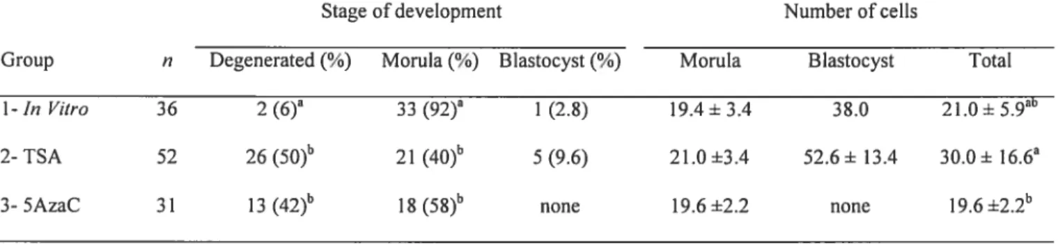
1. Article three
Table 1. Developmental stages and mean ccli numbers of early morula stage embryos exposed for 12 h to TSA and
5AzaC. 132
2. Article four
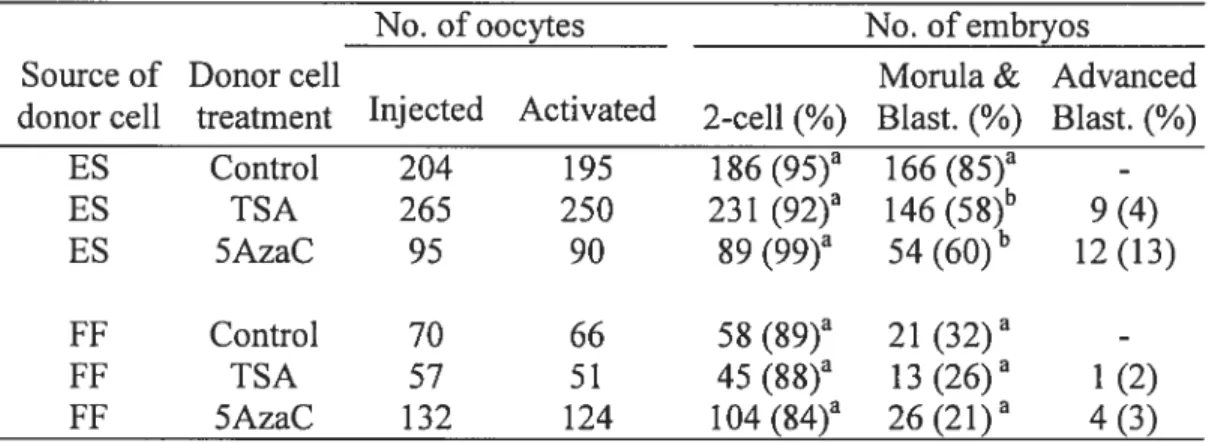
Table 1. Developmental potential of cloned embryos generated from ES and FF cells pretreated with TSA and 5AzaC. 157 Table 2. Nuclear abnormalities ofanested nuclear transfer ernbryos. 157
Abbreviations
5AzaC 5 Aza-Cytidine
cDNA Complementary deoxyribonucleic acid DMRs DifferentialÏy methylated regions
DNA deoxyribonucleic acid
FCS Fetal caif serum
Gapdh Glyceraldehyde-3 -phosphate dehydrogenase Gas6 Growth arrest specffic 6
h Hour
hCG Human chorionic gonadotropin
HDACs Histone deacetylases
1CM limer celi mass
1CR Imprinting control region
Igf2 Insulin like growth factor 2
kb Kilobase
LIF Leukemia inhibitory factor
LOI Loss ofimprinting
M Mole
MeCP2 Methyl cytosine binding proteins
ng Nano gram
Pegi Patemally expressed genes 1
xx
PGC Primordial germ celis
PMSG Pregnant mare’s serum gonadotropin
RT-PCR Reverse transcriptase polymerase chain reaction TRD Transcriptional repression domain
T$A Trichostatin A
1. Introduction
Despite recent success in cloning animais ofvarious species the moiecular rnechanisms needed for successful cloning remains largely obscure. While many factors are likeiy to contribute to the success, including celi cycle synchronization (Campbell et al., 1996), donor age, ceil type (Hill et al., 2000; Kato et al., 2000; Shiota et al., 2002) and medium conditions (Betts et al., 2001), little has been done to coordinate an optimum profile of gene expression, especially those that are imprinted.
It is increasingly evident that the in vitro production of morphologically normal pre-implantation embryos does not guarantee that their post-implantation development and post-natal life will be normal. During early embryonic development, extensive modifications in global methylation take place. Therefore any perturbation ofthe methylation process can cause epigenetic deregulation of developmentally important genes. $uch epigenetic alterations could affect in particular the expression of genes that are subject to genornic imprinting resulting in growth anomalies (Dean et al., 199$).
Culture conditions such as serum starvation of donor cells is a common practice in embryo cloning (Cibeili et al., 1992; Jones et al., 2001; $higa et al., 1999; Wilmut et al., 1997), whereby such a process could interfere with components ofthe ceil cycle resulting in improper maintenance of epigenetic tags and chromatin imprints (Hayashida et al., 1997; Rideout et al., 2001). Sirnilarly, addition or removal of serum in the culture ofpreimplantation mouse embryos was associated with an aberrant expression pattern of imprinted genes, improper methylation ofkey imprinting control regions (DeChiara et al.,
1990; Doherty et al., 2000; He et al., 1992) and reduced developmental potential afier embryo transfer (Khosla et al., 200 la). Moreover, it has been shown that leveis ofDNA
C
2
methyltransfereses 1 and 3h were downregulated when celis were arrested (serum deprivation) at G0/G1 stage ofthe celi cycle (Robertson et al., 2000b). Furthermore, studies monitoring the differences in the epigenetic profile between various celi origins has revealed that DNA mathylation patterns in ES celis are different than those observed in somatic tissues (Shiota et al., 2002), flot to mention dissimilarities in the levels ofDNA methyltransferase activity between ES and somatic celis (Jabner et al., 1982; Lei et al., 1996; Li et al., 1992; Palmiter et al., 1982; Robertson et al., 1999; Stewart et al., 1982). These dissimilarities between sornatic versus ES celis were more evident in celis
deficient of Dnmtl which showed that unlike somatic cells, ES cells are viable in culture although they contain minimal levels of genomic methylation (Lei et al., 1996; Nan et al., 1996). Similarly, ES celis lacking MeCP2, a methyl cytosine binding protein whose activity is associated with histone deacetylases, have no effect on survivability (Tate et al., 1996).
Prior treatment of cells as source of donor nuclei has raised many practical questions such as, increased abortion rates and aberrant phenotype. These abnormalities are characterized by placental defects, cartridge and skeletal deformation, respiratory difficulties in addition to fetal overgrowth and disproportionate enlargement of internal organs (organomegaly). Together, these symptoms were designated the “large offspring syndrome” (LOS), which has been observed in cattle, sheep (Farin and Farin, 1995; McEvoy et al., 1998; McEvoy et al., 2000; Schnieke et al., 1997; Sinclair et al., 2000; Thompson et al., 1995; Young and Fairbum, 2000; Young et al., 1998) and mice (Tanaka et al., 2001; Wakayama et al., 1998; Wakayama et al., 1999). Several studies have
epigenetic reprogramming of donor DNA (Humpherys et al., 2001; Lanza et al., 2000; Tanaka et al., 2001). In fact, epigenetic errors ofthe donor celi were carried to a later stage of development and was exerted in the placenta and the skin of cloned animais which again reveaied inappropriate reestablishment of methylation profiles in these tissues (Humpherys et al., 2001; Ohgane et al., 2001).
Other donor ceil pre-conditioning agents might be required to epigenetically reprogram the nuclei. For example, 5AzaC, a demethylation compound, which leads to reduced methylation levels of donor ceils so as to “simulate” genomes found in gametes or blastomeres. Similarly, trichostatin A (ISA) an inhibitor ofhistone deacetylases, can give rise to positive epigenetic alterations by rendering the chromatin easily
reprogrammable by the oocyte environment.
Taken together, it seems that a proper genomic “ingredient” ofthe donor celi is a prerequisite to maximize survivability and minimize fetal abnormalities associated with cloned embryos. Therefore, new methods are required to epigenetically synchronize ES celis should they be used in animal cloning or in transplantation therapy applications.
4
2. Literature Review
(
2.1. Preimplantation embryo development and embryonic stem celis
Embryonic development starts with fertilization of the egg by the sperm. Afier fertilization, the embryo divides slowÏy with the absent of size increase. Up until the 2-celi stage, the embryo relies exclusively on the oocyte recourses for development. By the end ofthe 2-ceil stage (species specific) the embryo starts activating local genes required for early growth and development. As cleavage proceeds, compaction of the embryo is evident by the late 8-celi stage embryo and the formation ofthe morula. The following stage (blastocyst) is characterized by the graduai formation of two distinct celi iineages, namely, the trophectoderm (TE) and the muer celi mass (1CM). Eventually, the embryo starts to expand at 4.5 days post fertilization in the mouse, whereby hatching occurs, enabling the embryo to be implanted in the uterus.
ES ceils are derived from the undifferentiated inner ceil mass (1CM) ofthe 3.5 day blastocyst, and are capable of maintaining an undifferentiated state indefinitely in culture in the presence of leukemia inhibitory factor (LIF). ES cells express markers of pluripotent undifferentiated celis such as Oct4, SSEA1 and aikaline phosphatase (Berstine et al., 1973; Solter and Knowles, 1978; Wobus et ai., 1984). Upon LIF withdrawal, they underexpress these markers, lose their pluripotent capacity and differentiate into various celi types. ES celi pluripotency has been manifested by the production of chimeric, tetraploid and cloned mice (Bradley et al., 1984; Wakayama et al., 1999).
Recent work showed the possibility of directing the fate of these undifferentiated ES celis in vitro into a variety of highly specialized differentiated ce!! types including neurons, islet celis, hepatocytes and cardiac muscle cel!s (fuchs and Segre, 2000; Wobus, 2001). Moreover, gene targeting in ES cells through recombinant mutation has been widely used to create genetically modified ES ce!! unes (Capecchi, 1989; Ramirez-Solis et al., 1993) and to repair spontaneous mutations (Doetschman et al., 1987). Together, these possibilities made ES ceils a useful source oftherapeutic material in transplantation medicine/therapeutic cloning to treat diabetes, Alzheimer and Parkinson disease patients (Colman and Kind, 2000).
The controversial use and derivation ofhuman ES ceils (hES) fromin vivolin
vitro produced embryos lcd scientists to develop a new concept whereby ES celis are derived from cloned embryos of somatic donor ce!!, designated as ntES celis. In fact mouse ntES cel!s have been derived from cloned blastocysts generated by somatic donor celis (Kawase et al., 2000; Munsie et al., 2000). Important!y, mouse ntE$ celis have been shown to differentiate into severa! ceil lineages (Wakayama et al., 2001) and late!y, genetic defects in mice have been corrected by the combination of ntES cells and gene therapy (Rideout et al., 2002). As oftoday, few hE$ ceil unes are estab!ished
(http ://escr.nih. gov/index.htrnl), however, it remains to be seen whether therapeutic cloning can be applied with the use ofhE$ cel!s.
6
2.2. Genomic Imprinting
Imprinting in mammais is a unique epigenetic process, whereby either the paternal or the maternai allele of the gene is expressed. This phenomenon was observed as eariy as 2 decades ago through elegant pronuclear transplantation experiments by McGrath and Solter, which dernonstrated that the parental genomic contribution to the embryo are non-equivalent (McGrath and $olter, 1984; Surani et al., 1984). This can be manifested by the failure ofparthenogenetic and androgenetic embryos to develop to term. So far a total of 66 imprinted genes (31 maternally and 35 paternally expressed; Fig. 1) have been identified in the mouse
(http ://www.mgu.har.mrc.ac.uk/imprinting/imptables). Imprinted genes are estimated to constitute 1% (around 150-200) of total genes in the mammalian genome, they generate considerable interest due to their unique profile of expression and the criticai role they play in prenatal (DeChiara et al., 1991) and postnatal (Itier et al., 1998; Lefebvre et al.,
199$) growth and development. An additional role for imprinted genes has ernerged in live animais lately, whereby inactivation of some imprinted genes in female mice caused abnormal maternai behavior (Lefebvre et al., 1998; Li et al., 1999). Not to mention that deregulation of imprinted genes ieads to several growth disorders and abnormalities. Although the imprinting mechanism has flot been defined compietely, the
accomplishment of monoallelic expression requires each gene to be “tagged” and subjected to germ line-specific epigenetic modifications such as DNA methylation, which are propagated subsequently from sperm and egg to the fertilized embryo then to the fetus and tbroughout adulthood.
Fig. 1 (Beechey, 2002)
ci
Chromosome:C
2 6 . ca’Iy €riibrjJrii Asb4 Icthiity ‘:Masgce GtLb( ar L U ig7 1Mai final II13o1aaI Nospas -Nosp & utlialiiy tr” iFvi.it & Pat) Copg2 r€Ia rialIorcopg2as Ma: Mit 1&b9 PegliMest .‘ Z,rn I necnalalPegslPwi [eiïaity Usp29 iMufl Zim3 • zrp2LN (I viabirty — FPii prst Ube3a iiutui• Ube3aas iMa * MOI 1-52 *MB11-8S Snrpn uat Snu% iaat Çcrt gra.vth Magei2 Mit} Z(pt27fMkrn3 Pasg I Z(p 127n’Mkrn3, FraI3 Obph T hep US Tssc&lpI S1c22a11 ‘ Pistil! P5T /Cdknlc Kvlqt t/Kcnq 1KvlqIl.n Tssc4 Topal?CdBI MashflAsc(2 Ins2 iqf2as 1012 RIS
iris! emtryonic clhality HtOa l qr)wtFl Dlk (M:i. Meg3J 0112 & Pi)
*
MB! 1426 MB11-19 Mai * 41811-343 * MOu-78 * M81148 * MB! 149 lmprinrcd genes w’th.n clusters are net necessartiy in correct ardu imçxintodgnr.es in ru] are matornaflyexpressed irnçrirtcd genes ir bitte are atentaIIy etpressed * rnaternally exprosscd small nueleolar RNAs * palcma’iy expressod sniall nucleolar RNAs ragions wah nhrnrmal imprintmg tenotypes with mnlernnl (Mat) rir painma (Pat) diiIrraiinn Fig. 1. List and chromosomal location of ail imprinted genes identifies as of August, 2002. Adapted from{
Beechey 2002) 7 rwIaI viati; it’ & crawL rM.at:. ‘iacertal sZe << (rrtat), :‘> IpLI:I 10 11 12a
. ei4eciS U2afl-nl &Pat) 19 14 15 17 18 aa. .w
61c22fl halai 9eoaata 31c22a2 graMti etwW — Ig(2raWAi retLrdalj Ofl Pat: 1912f (MU & Pt3 impact Dan t t LI ti1i ‘‘ty & RdII
I
2.2.1 Epigenctic characteristics and the role of imprinted genes
While CpG islands in non-imprinted housekeeping genes are hypomethylated,
hyperacetylated and transcriptionally active (Bird, 1986; Stem et ai., 1983; Tazi and Bird, 1990), imprinted genes contain highly methylated CpG islands (Surani et ai., 1990b) ofien located on the repressed allele thereby designating them as differentially methylated regions (DMR5), and in many cases, configured heterochromatically (Jones, 1999). Almost ail imprinted genes contain DMRs, while some ofthese DMRs are established either in the oocyte or the sperm and are called primary DMRs, secondary DMRs are usually produced afier fertilization and may resuit ftom methylation spreading from a prirnary DMR (Bartolomei et al., 1993; Bird, 1999; Brandeis et al., 1993; Eversole-Cire et al., 1993; feu et al., 1994; ferguson-Smith and Surani, 2001; Neumann and Barlow, 1996; Olek and Walter, 1997; Plass et al., 1996; Razin and Cedar, 1994; Stoger et al., 1993; Surani, 199$; Tremblay et al., 1997). Although DNA methylation is attributed to the allelic repression observed in imprinted genes, some imprinted genes however have their DMRs on the active allele (Fig. 2) (Bestor, 2000; Constancia et al., 199$; Reik et al., 2001). Furthermore, imprinted genes are physically linked in clusters that contain both maternally and patemally expressed genes (Wutz et al., 1997; Zemel et al., 1992). Interestingly, many of these clusters contain at least one imprinted gene that encodes an untranslated RNA such as H19 and Snrpn (Brannan et al., 1990; Ripoche et al., 1997; Wutz et al., 1997). Moreover, imprinted genes are oflen enriched with CpG islands (Paulsen et al., 2000) and similarities in specific short repetitive sequences in DMRs between numerous imprinted genes has been well established (Bartolomei et al., 1993;
Ç
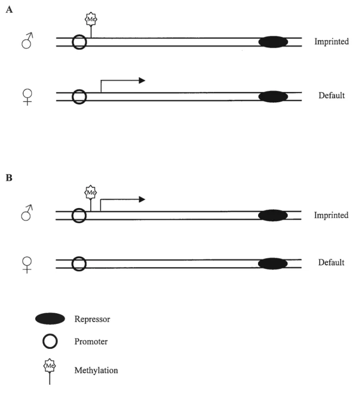
A B
g
g
— Repressoro
Promoter Methylation Imprinted Default Imprinted Defaulto
Fig. 2. Examples of imprinted gene expression in association with DNA
methylation. Imprints (methylation) can be located on the repressed allele (A) or on the expressed allele (B). Adapted with modifications from Sieutel et al., 2000.
p.
z
o
z
Me
o
E
Hatada et al., 1995; Neumann et ai., 1995; Stoger et ai., 1993). Furthermore, it seems iikeiy that differences in chrornatin structure as an epigenetic modification, influence the expression of irnprinted genes (Feu and Keisey, 1997). These modifications inciude asynchronous repiication aiso known as replication timing between the maternai and patemal aliele (Izumikawa et ai., 1991; Kitsberg et ai., 1993; Knoii et ai., 1994; Ripoche et aI., 1997) and frequencies ofmeiotic recombination rates during male and female meiosis in gerrn ceils (Paidi et ai., 1995; Robinson and Lalande, 1995; Simon et ai.,
1999). However, it is worth noting that asynchronous replication aiso occurs in non imprinted genomic regions (Chess et ai. 1994). Other chromatin structure modifications, such as differentiai histone acetyiation levels between parental aileles bas been reported as weli (Hu et al., 199$; Khosla et al., 200 lb; Saitoh and Wada, 2000), this is manifested by the fact that the heaviiy methyiated inactive X chromosome is depleted of the acetyl moiety on histone H3 and H4 and simiiar to heterochromatic regions (Jeppesen and Tumer, 1993).
A substantial number of imprinted genes have been functionally implicated in the reguiation ofernbryonic, fetai and postnatal growth and development (Barton et al., 1991; Itier et al., 199$). Usualiy, paternally expressed genes are growth enhancers while
maternaiiy expressed genes are growth repressors. Moreover, whereas embryos carrying oniy maternai contribution such as parthenogenetic embryos poorly develop
extraembryonic tissues, androgenetic embryos which contain soieiy patemal genomes, negatively contribute to the formation of the embryo proper. The Igf system is the most documented feature of ail imprinted genes that bas been correlated with embryonic
- deveiopment. This system inciude insulin-iike growth factor II (Igf2), and embryos
10 deficient of this paternally expressed gene show fetal lethality and are significantly smaller than control littermates (DeChiara et al., 1990). Similarly, conversely however, mutant mice lacking Igf2r/mannose-6-phosphate receptor, a maternally expressed gene, are approximately 30% larger at birth than controïs (Barlow et al., 1991; Lau et al., 1994; Wang et al., 1994) and this overgrowth can be equilibrated by combining Igf2 and Igf2r mutations (Wang et al., 1994). It is worth noting that the main function of Igf2r is to degrade extra-cellular Igf2 via receptor-rnediated endocytosis (Czech, 1989; Oka et al.,
1985). Like Igf2, H19 maps to chromosome 7 in the mouse but is matemally expressed (Bartolomei et al., 1991), both genes are physically linked (Zemel et al., 1992) with a reciprocal allelic expression. Newbom mice that carry the 1119 nuli mutant are 28% larger than normal littermates; however, the size increase can be eliminated by double mutation of 1119 and Igf2 simultaneously (Leighton et al., 1995). Two other imprinted genes are involved in fetal growth control, nameÏy the Mas proto-oncogene (Villar and Pedersen, 1994) and insulin 2 (Ins2) (Giddings et al., 1994).
Interestingly, most imprinted genes are expressed and play an important role in the formation of the placenta (www.mgu.har.mrc. ac.uk!imprinting), therefore indirectly contributing to fetal growth by regulating nutrient transfer to the fetus. Two major imprinted genes have so far been implicated in placenta! development namely, Mash2, a gene that encodes basic-helix-loop-helix transcription factors which, is responsible for the formation ofthe spongiotrophoblast layer in the placenta (Guillemot et al., 1995; Guillemot et al., 1994), whereas Igf2 transcript is expressed specifically in the
labyrinthine trophoblast lineage (Constancia et aÏ., 2000). Further evidence demonstrating the importance ofthese genes in placental formation comes ftom androgenetic embryos
which poorly develop spongiotrophoblast tissue due to the loss of Mash2 (Guillemot et al., 1994). It is noteworthy to mention that Mash2 imprinting is independently regulated from DNA methylation (Caspary et al., 199$; Tanaka et al., 1999).
Imprinted genes have been shown to be involved in celi cycle regulation. This was demonstrated by the identification of p57 (currently Cdknlc) (Hatada and Mukai, 1995; Matsuoka et al., 1996; Taniguchi et aÏ., 1997), a maternally expressed gene that encodes a tissue specific cycÏin-cdk inhibitor (Chung et al., 1996; Lee et al., 1995; Matsuoka et aÏ., 1995). Biallelic expression ofthis gene leads to decreased celluÏar proliferation, such as those observed in ceils derived from parthenogenetic tissues. Conversely, androgenetic ceils which presumably are nuli for p57, have an increased rate ofcellular division (Matsuoka et al., 1995; O’Keefe et al., 1997). Furthermore, it has been demonstrated that imprinted genes are responsible for postnatal behavior (Isles and Wilkinson, 2000). Females that lack a functional Pegi and Peg3, which are paternally expressed genes, neglect feeding their offspring (Kuroiwa et al., 1996; Lefebvre et al., 199$; Li et aI., 1999; Nishita et al., 1996). Both genes are expressed abundantly in the mouse fetus, particularly in mesodermal tissues and the brain. Similarly, behavior abnormalities were observed in mice lacking Grfl and Ube3a genes, which were
characterized by defects in contextual leaming and lack of long term memory (Brambilla et al., 1997; Jiang et al., 199$).
2.3. Genomic repression of imprinted genes
12
o
It has been known for some time that DNA methylation is inversely correlatedwith gene activity, meaning that hypermethylation of DNA sequences are associated with gene repression while hypomethylation correlates with gene activation. A large body of evidence suggests that DNA methylation is involved in a variety of biological processes that include ceil differentiation, establishment and maintenance of genomic imprinting, X chromosome inactivation, stability of chromatin structure, DNA replication and
carcinogenesis (Hall, 1991; Junien, 1992; Leonhardt et al., 1992; Li et al., 1993b; Rastan, 1994; Razin and Kafti, 1994; Surani et al., 1990b; Surani et al., 1988). Genomic DNA is methylated and maintained in vitro with the use of a group of enzymes called DNA methyltransferases which were identified in mice (Bestor et al., 198$; Okano et al.,
199$a; Okano et al., 199$b; Yoder and Bestor, 199$; Yoder et al., 1997) and humans (Finnegan and Deimis, 1993; Yen et al., 1992). DNA methyltransferases target the cytosine residues of CpG islands in key regulatory sites, such as promoters or the
imprinting control region (1CR) in the case of imprinted genes, and add a methyl group to the 5-carbon position to suppress transcription (Bestor et al., 1928; Bestor and Ingram,
1983; Pfeifer et al., 1983). Moreover, the role ofmethylation in genomic imprinting is supported by the demonstration that methyltransferase nuil mice lack proper expression ofimprinted genes (Li et al., 1993a; $urani et al., 1990a). In addition to the fact that removal and disruption of primary DMR sequences in mice resulted in deregulation of imprinted gene expression (Thorvaldsen et al., 199$).
2.3.1.1 Dynamics of UNA methylatïon in the embryo
C
The genomic DNA undergoes dynamic alterations in the methylation pattern during early embryonic development (Fig. 3). These changes are described in three steps, firstly the SO called erasure, which is characterized by an active wave of DNA
demethylation that occurs afier fertilization tili the blastocyst stage, in which the majority of CpG islands in the embryo are void of methylation (Howlett and Reik, 1991; Kafti et al., 1992; Monk, 1987; Oswald et al., 2000). Recently however, it has been shown that demethylation occurs at a much faster rate whereby genomic demethylation is complete by the morula stage (Santos et al., 2002). It should be noted that imprinted alleles are protected from the genome wide demethylation activity during preimplantation development (Brandeis et al., 1993; Olek and Walter, 1997; Shemer et al., 1996; Tremblay et al., 1997). The physiological importance of the wave of global demethylation in early embryogenesis is unknown. It is thought however, that this process is required to prevent transmission of aberrant epigenetic modifications to subsequent generations and also as a prerequisite for the formation of pluripotent
embryonic stem (ES) celis at the blastocyst stage. Two types of DNA demethylation (Fig. 4) have been documented to occur in the early embryo namely, active and passive
demethylation. While the first does not require DNA replication, it is a must for the latter (Reik and Walter, 2001; Rougier et al., 1998). Secondly the establishment process, which takes place at the time of implantation throughout gastrulation, is characterized by a wave of global de novo methylation resulting in a rapid increase in DNA methylation levels
Sperm 1 CeIl 8 CeJ1 Morula Blast. Egg cylinder PGC +d. . .. . . . u u. u u u u .. u u u u u u u u u u u. u u u. u u u u u u u. u u.
‘t
D3.5
Implantation D5.5Erasure
Establishment
Fig.
3.
Dynamic
changes
in
DNA
methytransferase
activity
in
the
pre
and
postimplantation
mouse
embryo.
DNA
dernethylation
occurs
shortly
afler
fertilization
tilT
the
blastocyst
stage.
After
implantation
a
wave
of
de
novo
methylation
take
place
throughout
gastmlation.
Adapted
from
Li
et
al
1997,
with
slight
modifications.
Oocyte _ — _ JEscape
.— ri)ii
44
44
s
I
I
D7.5El
• Methyl group (Ch3)
Highly methylated double strand DNA
o
fig. 4. Dynamics of global DNA demethylation pattems in early embryos. Demethylation occurs in the absence ofDnmt 1 with continued rounds of DNA replication (passive demethylation) or actively without DNA replication (active demethylation). Adapted with modifications from Reik and Walter 2001.
Active
demethylation
Passive demethylation
16
during post-implantation development (Chaillet et al., 1991; Jalmer et al., 1982; Kafri et al., 1992; Monk, 198$; Razin and Shemer, 1995; $anford et al., 1987; Stoger et al., 1993; Tremblay et al., 1995). Recent work indicates that the de novo methylation wave take place as early as the blastocyst stage and is restricted to theinnerceli mass (1CM) (Santos et al., 2002). Thirdly, the so called gametogenesis escape, whereby the primordial germ cells (PGC5) of both sexes of the developing embryo escape the wave of de novo
methylation and remain highly undermethylated (Monk et al., 1987; Sanford et al., 1987) until they begin to differentiate as gametes (sperm and oocyte) (Kafri et al., 1992). It is widely believed that perturbation in methylation dynamics in preimplantation embryos leads to developmental abnormalities and growth disorders.
2.3.1.2 UNA methyltransferases
The importance of DNA methyltransferases in genomic imprinting was
demonstrated by the fact that mice lacking maintenance methyltranferase (Dnmt 1) gene fail to show allele-specific methylation, thereby disrupting the expression profile of irnprinted genes (Li et ai., 1 993b). Furthermore, ernbryos deficient in DNA
methyltransferase have hypomethylated genomes and die early in embryogenesis due to abnormal expression of imprinted genes (Caspary et al., 199$; Li et al., 1993b; Li et al.,
1992). DNA methyltransferases (Fig. 5) play a pivotai role in the establishment and maintenance of imprinting during early pre- and postimplantation stages (Li et al., 1 993b; Surani et al., 1990b).
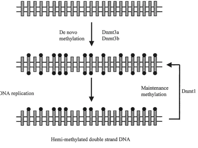
So far few methyltransferases (for reviews see Bestor, 2000) have been identified namely; Dnmtl, the maintenance methyltransferase which can recognize hemi
methylated CpGs, as DNA replicates it adds a methyl group to the daughter DNA strand (Bestor and Ingram, 1983; Gruenbaum et al., 1982; Leonhardt et al., 1992; Pfeifer et al., 1983). Although Dnmtl is known for maintenance DNA methylase activity, it was demonstrated that it has a significant de novo methylase activity (Yoder et al., 1997). The second DNA methyltransferase was called Dnmt2 (Okano et al., 199$b; Yoder and Bestor, 199$) which was found in prokaryotic and eukaryotic organisms. Dnmt2 knock out ES celis failed to interrupt either de novo methylation or maintenance of
hemimethylated DNA sequences, suggesting a limited role in genomic de novo and global maintenance methylation in ES celis (Okano et al., 1 998a). A third
18
Non-methylated double strand DNA
-I
‘I
o
CpG residuesHemi-methylated double strand DNA
• Methyl group (Ch3)
Fig. 5. Dynamics of de novo and maintenance DNA methylation pattems in dividing celis. Methylation is introduced into unmethylated DNA by de novo methylation enzymes Dnmt3a, Dnmt3b and Dnmtl. Following DNA replication, Dnmtl recognize the hemi methylated DNA sequence and introduce a methyl group on the opposite strand. Adapted with modifications from Bird 1999.
o
De novo methylation Dnmt3a Dnmt3b 1-MaintenanceDNA replication
I
methylation Dnmtlthat are present at low levels in somatic and differentiated ES celis (Okano et al., 1 99$b). Dnmt3a and Dnmt3b lack the capacity to maintain the methylation ofhemimethyiated DNA sequences but interestingly, have high affinity for de novo methylating “blank” genomic DNA rendering them as a prime candidate for de novo methylase (Okano et al., 1 998a). This was evident by experiments demonstrating that Dnmt3a was responsible for de novo methylation of DNA sequences in Drosophila, which usually lack any
methylated regions (Lyko et al., 1999), and also in experiments of bi-knock-out ES celis of both forms which showed absence of newly methylated regions and early death at gastrulation (Okano et al., 1999). While Dnmt3a knockout mice develop to term but die around 3-4 weeks afier birth, embryos deficient in Dnmt3b fail to develop beyond 9.5 days afier fertilization. Moreover, it bas been shown that Dnmt3a and Dnmt3b have an overlapping function in preimplantation embryos since their expression was found localized mainly in the embryonic ectoderm (Okano et al., 1999). furthermore, it bas been shown recently that Dnmt3L, a protein sharing homology with DNA
methyltransferases, interacts with Dnmt3a and Dnmt3b to carry out de novo methylation ofimprinted genes in germ celis (Bourc’his et al., 2001; Hata et al., 2002). It is interesting to note that together with Dnmt3L, other proteins have been identified lately that interact directly with DNA methyltransferases including PCNA, DMAP 1, HDAC 1, HDAC2, pRB and RP5b, however, neither their cooperative function nor the mechanism by which they target DNA methyltransferases bas been determined yet (Bachrnan et al., 2001; Fuks et al., 2001; Robertson et al., 2000a). With ail the methyltransferases and the interacting proteins known SO far, it is possible that the maintenance and establishment ofmethyl
20
imprints in the sperm, oocyte and fertilized embryo may stiil depend on additional novel DNA methyltransferases yet to be identffied (Howeli et al., 2001; Oswald et al., 2000).
2.3.1.3 Affect of UNA methyltransferase inhibitors (5AzaC)
5-Azacytidine (5AzaC) is a potent chemical agent that is known to induce global DNA demethylation (Creusot et al., 1982; Juttermann et al., 1994; Michalowsky and Jones, 1987). The drug 5AzaC is a nucleoside analog which incorporates into cellular DNA and irreversibly inhibits DNA methyltransferases by forming a covalent binding complex on the DNA methyltransferase enzyme (Bender et al., 199$; Juttermaim et al., 1994). It has a short life span and is typically administered to patients with carcinogenic illnesses by continuous subcutaneous injections over several days (Chitambar et al., 1991; Glover and Leyland-Jones, 1987; Goldberg et al., 1993). Treatment ofsomatic female celi unes with 5AzaC induces partial reactivation ofthe inactive X chromosome, evident by the expression ofX-linked genes, decompaction ofthe heterochromatin site and advanced replication timing (Jablonka et al., 1985; $cbmidt et al., 1985).
Moreover, it is worth noting that drugs that inhibit DNA methylation are highly toxic, due to their interference with the methylation machinery (Davidson et al., 1992; Juttermann et al., 1994; Yoshida et al., 1990).
Since imprinted gene expression is dependent on hypermethylation of DMRs, treatment of celis with 5AzaC results in many cases in upregulation of imprinted genes due to loss of imprinting (LOI). While several imprinted genes were overexpressed in response to 5AzaC exposure in preimplantation stage embryos (Baqir and $mith, 200 la),
embryonic (Nishita et al., 1999) and gastric (Kang et al., 2000) carcinoma ceils, mouse fibroblast celis (E! Kharroubi et al., 2001; Grandjean et al., 2001; Pedone et al., 1999) and human fibroblast celis (Hu et al., 1998), some imprinted genes, such as p57, were flot induced in embryonic rhabdomyosarcoma celis (Chung et al., 1996). Moreover, it is interesting to note that 5AzaC can bring about demethylation of highly methylated sequences in silenced endogenous non-imprinted genes (Jones and Taylor, 1980; Jones et al., 1982), retroviruses (Groudine et al., 1981) and genes involved in tumor gastric celis (Kang et al., 2000).
2.3.2. Epigenetic repression via histone deacetylation
A large body of evidence indicates that epigenetic control of gene expression in
mammals is achieved by a combination of DNA methylation and chromatin modification (for reviews see Cheung et al., 2000; Kouzarides, 1999; Spencer and Davie, 1999). It has been known for some time that chromatin structure is associated with the acetylation pattern of the histones at individual lysine residues by regulating the interaction between nucleosomes (Grunstein, 1997). Histone acetylation is ofien associated with activated transcription and deacetylation conelates with transcriptional repression (Struhi, 1998; Tazi and Bird, 1990). Evident by the fact that euchromatins are usually open and oflen hyperacetylated thereby enabling transcription factors to access the DNA and form an environment that is active (Grunstein, 1997; Kuo and Allis, 199$). Conversely, closed configured chromatin (heterochromatin), which is hypoacetylated would block
transcription factors and render the chromatin in a repressive state (Fig. 6) (Berger, 1999;
C
Euchromatin
o
Heterochromatin
f ig. 6. Models for active chromatin (euchromatin), which is open and ofien hyperacetylated and repressed chromatin (heterochromatin) which is structurally closed and hypoacetylated thereby blocking transcription factors. Both models shows histone tau modifications such as methylation and acetylation. Adapted with
Kuo and Allis, 1992). This model was supported by the fact that, while transcriptionaly suppressed regions including telorneres, centromeres, heterochrornatin and methylated sites are found to be acetylation free (Brown et al., 1997; Cortes et al., 1999; Ekwall et al., 1997; Grunstein, 1997; Nan et al., 1992), active genomic sequences are euchromatic and harbor highly acetylated histones with no CpG methylation (Grunstein, 1997). Another piece of evidence cornes from the fact that X-linked genes that are expressed have their histone H4 hyperacetylated, while X-linked genes that are silenced are usually hypoacetylated (Gilbert and Sharp, 1999; Jeppesen, 1997; Jeppesen and Turner, 1993). It should be noted however, that in few rare cases, it has been shown that acetylation of specific lysine residues is associated with gene repression (De Rubertis et al., 1996; Pazin and Kadonaga, 1997; Turner, 1991). Repressed chrornatin structures usually recruits histone deacetylases (HDACs) to prornoter regions or key regulatory sites to repress genornic transcription (De Rubertis et al., 1996).
2.3.2.1. Affect of histone deacetylase inhibitors (HDACs)
Histone deacetylases are the enzymes responsible for the removal of acetyl rnoieties from specific lysine residues of core histones, thereby creating a positively charged environment. The interaction between the positively charged lysine residues reduces nucleosome rnobility on the DNA strand rendering it inaccessible to the
transcriptional machinery (Cheung et al., 2000; Wolffe and Hayes, 1999). The inhibitory affect of HDACs on gene expression can be reversed by use of drugs that inhibit HDACs, such as sodium butyrate, trapoxin and trichostatin A (TSA). Moreover, it is worth noting
24
Q
that drugs that inhibit HDACs are highly toxic due to their interference with thearchitectural structure ofthe chromatin (Davidson et al., 1992; Yoshida et al., 1990), and might cause cellular anest in G1/G2 ofthe celi cycle (Sugita et al., 1992; Yoshida and Beppu, 1988), apoptosis (Medina et al., 1997) and antiproliferative effects (Sambucetti et al., 1999; Sugita et al., 1992). Although imprinted gene expression is controlled by DNA methylation, recent reports have shown that histone deacetylation is involved in the silencing pathway of genomic imprinting. This was demonstrated by experiments showing that several imprinted genes were overexpressed in response to HDAC inhibitors exposure in various cell types including human fibroblast (Ru et al., 1998), mouse embryonic stem cells (Baqir and $mith, 2000), preimplantation stage embryos (Baqir and $mith, 200 la), embryonic fibroblast tEl Kharroubi et al., 2001; Yoshioka et al., 2001) and cells ftom newbom mice (Pedone et al., 1999). However, it should be noted that some imprinted genes, including Snrpn, U2afl-rsl and H19, appear to remain unaltered by TSA exposure tEl Kharroubi et al., 2001; Gregory et al., 2002; Pedone et al.,
1999; $aitoh and Wada, 2000; Yoshioka et al., 2001).
2.3.3. Epigenetic repression of imprintcd genes via DNA methylation and histone deacetylation
While it is evident that beside DNA methylation in imprinted genes (Brandeis et al., 1993; Ferguson-Smith and Surani, 2001; Neumaim and Barlow, 1996), other
mechanisms are involved in genomic repression pathways, such as those caused by HACDs in plants (Pazin and Kadonaga, 1997; Tian and Chen, 2001). The two silencing
processes were brought together by experiments showing that the inactive X chromosome
t
is hypoacetyÏated (Jeppesen and Turner, 1993; Keohane et al., 1996; Wakefield et al., 1997), in addition to the fact that differential histone H3 and H4 acetylation has been found at the CpG island of DMR1 of SNRPN gene (Saitoh and Wada, 2000). Further evidence cornes from experirnents that show HDAC inhibitors reactivated the
transcription of sequences repressed normally by DNA methylation. For instance, ISA was responsible for the increased expression of a transfected methylated gene in mouse fibroblast celis (Eden et al., 1998) and sodium butyrate in the case of methylated non imprinted episomal reporter gene (Bender et al., 199$). Similarly, both sodium
butyrate/ISA, like 5AzaC were able to restore the expression of a methylated transgene (Pikaart et al., 199$). In addition, it has been reported that TSA can substitute for 5AzadC in restoring the transcriptional silencing from previously methylated plant ribosomal
RNAgenes (Chen et al., 1997). Moreover, it has also been shown more specifically that the alteration in gene expression was due to genome wide or site-specific DNA
demethylation caused by HDACs in various organisms namely, fungus (Selker, 1998), virus (Szyf et al., 1985), transfected plasmid (Cervoni and Szyf 2001) and in mouse skin fibroblast celis (Hu et al., 2000). Conversely, another form of interplay between DNA methylation and histone deacetylation was demonstrated by recent studies showing that the methyltransferase inhibitor 5Aza-dC induced hyperacetylation of a hypermethylated region (Saitoh and Wada, 2000; Takebayashi et al., 2001). Much ofthe methylation acetylation coimection was pointed at a recently identified group of methyl-cytosine specific binding proteins (MeCP 1 and MeCP2) that are mainly localized in
- heterochromatin sites and capable ofrecruiting histone deacetylases (Fig. 7)
26
o
*
o
Acetyl group CpG residues Ao
• Methyl group (Ch3)Fig. 7. Diagram ofthe repression process. The repression complex is formed initially by the interaction of MeCP2 with highly methylated CpG residues which then recruits a co-repressor (mSin3A) to which HDACs are associated. The deacetylases removes acetyl moieties ftom lysine residues ofhistone H3 and H4. Adapted with modifications from Razin 199$.
via a transcriptional repression domain (TRD) (Cross et al., 1997; Hendrich and Bird, 199$; Jones et al., 199$; Nan et al., 1997; Nan et al., 199$). Indeed, it has been demonstrated that the repression of H 19 caused by the methylation of the imprinting control region (1CR) is associated with MeCP2 and presumably forming a repressive complex with deacetylase activity (Dreweli et al., 2002).
Furthermore, it is intriguing to note that the link between DNA methylation and histone acetylation was further strengthened by studies that showed that flot only DNA
methyltransferases, the enzyme required to methylate genomic sequences, but also Dnmt3L, a protein that interacts with de novo methyltransferases, to be actively associated with HDACs (Aapola et al., 2002; Fuks et al., 2000; Fuks et al., 2001; Robertson et al., 2000a; Rountree et al., 2000; Wade et al., 1999).
2.4. Effect of serum starvation (medium) on imprinted gene expression
During early embryogenesis, extensive genomic modifications such as DNA methylation occur to accommodate the requirement of later developmental stages. Perturbation of this process would normally lead to deregulation of imprinted genes, growth disorders, pregnancy complications and unexpected miscaniages. There is increasing evidence that environmental factors such as in vitro culture and manipulation of preimplantation stage embryos can give rise to phenotypic, genetic and epigenetic abnormalities at postimplantaion and postnatal stages of development (Walker, 1996; Young et al., 199$). Much ofthe blame was assigned to the use ofserum and amino acids in culture medium. For instance mouse embryos cultured in Whitens medium showed
C
2$
H19 LOI, whereas K$OM AA medium had no affect on H19 expression (Erbach et al., 1994). Similarly, augmentation of amino acids in KSOM AA compared to Whitens medium increased the expression of 1gf2 and Igf2r in preimplantation mouse embryos (Ho et al., 1995). More recently, it has been shown that culture ofmouse embryos in serum free medium led to Hi 9 overexpression and was associated with undermethylation ofthe imprinting control region (Doherty et al., 2000). Conversely, the addition of serum to mouse embryo culture resulted in alteration in the expression of two important growth regulatory imprinted genes namely, 1gf2 and GrblO (DeChiara et al., 1990; He et al.,
1998), and was associated with reduced developrnental potential afier embryo transfer (Khosla et al., 200 la). Moreover, culture ofpreimplantation bovine embryos in serum restrictive medium, resulted in major alterations in Igf2 expression in the liver and skeletal muscle in day 70 fetuses (Blondin et al., 2000). Much ofthe deregulation of imprinted genes caused by serum deprivation was attributed to aberrant methylation and acetylation profile ofthese ceils (Ferguson-Srnith et al., 1993; Jones et al., 2001; Knosp et al., 1991). This is flot surprising though, since it has been demonstrated that serum starvation was capable of altering the enzymatic activity ofDnmtl and Dnmt3a in vitro (Robertson et al., 2000b), and depletion of polyamine in culture was associated with an alteration in methyltransferase activity in teratocarcinoma stem celis (Frostesjo et al.,
1997).
Celi cycle synchronization is required to facilitate the nucleo-cytoplasmic interaction between donor nuclei and the recipient oocyte in animal cloning (Szollosi et al., 198$; Wilmut and Campbell, 199$). Serum-depleted culture conditions are commonly used as a prerequisite to synchronize donor ceils before nuclear transfer (Wakayama et
ai., 199$; Wilmut et al., 1997). The fact that several cloned animais were derived successfully from serum starved donor celis confirm this notion (Wilmut et al., 1997). However, the resulting pregnancies are oflen associated with increased miscarriages and post natal mortality, in addition to pronounced developmental malformations such as increased body and organ size (Nagy et al., 1993; Wakayama et al., 1999; Wang et al.,
1997). One school ofthought have suggested that the observed growth abnormalities in cloned animais are triggered by incomplete epigenetic reprogramming of imprinted genes in the donor celi due to culture condition thereby carrying these alteration to the fetus. This was demonstrated by chimeric experiments, whereby alterations in the epigenetic profile ofimprinted genes in ES celis were carried to the fetus (Dean et al., 199$). Yet alone, serum starvation induced alterations in the expression pattern of several imprinted genes in mouse (Baqir and Smith, 2000; Eversole-Cire et al., 1993; Eversole-Cire et al.,
1995; Hayashida et al., 1997), rat (Ungaro et al., 1997) and rabbit (Han et al., 1996) somatic cells. Not to mention that serum-constrained culture conditions caused an
aberrant methylation profile (Eversole-Cire et al., 1995; Ferguson-Smith et al., 1993) and acetylation pattern of core histones in fibroblast ceils (Knosp et al., 1991).
2.5. Imprinted gene expression in ES ceils
ES cells are derived from the blastocysts 1CM, they are pluripotent and give rise to the embryo proper, amnion, yolk sac and the chorioallantoic portion of the placenta (for review see (Rossant, 2001). Unlike embryonic germ celis which are totipotent, ES cells retain their appropriate parental imprints, as assessed by the developmental potential
C
j
ofchimeras (Allen et al., 1994; Mann et al., 1995). ES celi culture require the presence of exogenous factors such as leukernia inhibitory factor tUF) ($mith et al., 198$; Williams et al., 1988), they lose their totipotency in the event that LIF is withdrawn from the medium (Stewart et al., 1992). Moreover, ES ccli derivation and unlimited capability for division make them more vuinerable to epigenetic changes, and therefore unstable in culture (Dean et al., 199$; Humpherys et al., 2001). Evidently by the fact that allele specific methylation and expression pattems of imprinted genes in ES celis are unstable with progressive passage in culture (Baqir and Smith, 2000; Nagy et al., 1993; Szabo and Maiin, 1994). This was confirmed by the failure of late passage ES celis to produce live offspring by tetraploid embryos (Nagy et al., 1990; Nagy et al., 1993; Wang et aI., 1997) and by nuclear transfer (Rideout et al., 2000).
Recently it has been demonstrated that DNA mathylation profiles in ES ceils are different than those observed in somatic tissues (Shiota et al., 2002) not to mention that differences in the levels ofDNA methyltransferase activity between ES and somatic ceils exists. For instance, unlike somatic cells in which Dnmtl is shown to be the most
abundant methyltransferase, ES celis and early stage embryos are known to contain high levels of de novo methylation activity (Jahner et al., 1982; Lei et al., 1996; Li et al., 1992; Palmiter et al., 1982; Robertson et al., 1999; Stewart et al., 1982). Moreover, in contrast to somatic cells, ES cells deficient of Dnmtl are viable in culture, although they contain minimal levels of genomic methylation (Lei et al., 1996; Nan et al., 1996). $imilarly, ES cells lacking MeCP2, a methyl cytosine binding protein whose activity is associated with histone deacetylases, have no effect on the survivability of ES cells (Tate et al., 1996). Moreover, it seems that Dnmt2 has a limited role in genomic methylation in ES cells due