Université de Montréal
Systematic analysis of protein complexes involved in the human RNA polymerase II machinery
par: Racha Al-Khoury
Département de biochimie Faculté de médecine
Mémoire présenté à la Faculté des études supérieures en vue de l’obtention du grade de Maitre ès Sciences
en biochimie Fèvrier, 2009 ©, Racha Al-Khoury, 2009
Université de Montréal Faculté des études supérieures
Ce mémoire intitulé:
Systematic analysis of protein complexes involved in the human RNA polymerase II machinery
présenté par: Racha Al-Khoury
a été évalué par un jury composé des personnes suivantes: Pascale Legault président-rapporteur Benoit Coulombe directeur de recherche Jacques Archambault membre du jury
Résumé
La transcription, la maturation d’ARN, et le remodelage de la chromatine sont tous des processus centraux dans l'interprétation de l'information contenue dans l’ADN. Bien que beaucoup de complexes de protéines formant la machinerie cellulaire de transcription aient été étudiés, plusieurs restent encore à identifier et caractériser.
En utilisant une approche protéomique, notre laboratoire a purifié plusieurs composantes de la machinerie de transcription de l’ARNPII humaine par double chromatographie d’affinité "TAP". Cette procédure permet l'isolement de complexes protéiques comme ils existent vraisemblablement in vivo dans les cellules mammifères, et l'identification de partenaires d'interactions par spectrométrie de masse. Les interactions protéiques qui sont validées bioinformatiquement, sont choisies et utilisées pour cartographier un réseau connectant plusieurs composantes de la machinerie transcriptionnelle. En appliquant cette procédure, notre laboratoire a identifié, pour la première fois, un groupe de protéines, qui interagit physiquement et fonctionnellement avec l’ARNPII humaine. Les propriétés de ces protéines suggèrent un rôle dans l'assemblage de complexes à plusieurs sous-unités, comme les protéines d'échafaudage et chaperonnes.
L'objectif de mon projet était de continuer la caractérisation du réseau de complexes protéiques impliquant les facteurs de transcription. Huit nouveaux partenaires de l’ARNPII (PIH1D1, GPN3, WDR92, PFDN2, KIAA0406, PDRG1, CCT4 et CCT5) ont été purifiés par la méthode TAP, et la spectrométrie de masse a permis d’identifier de nouvelles interactions.
Au cours des années, l’analyse par notre laboratoire des mécanismes de la transcription a contribué à apporter de nouvelles connaissances et à mieux comprendre son fonctionnement. Cette connaissance est essentielle au développement de médicaments qui cibleront les mécanismes de la transcription.
Mots clés : Machinerie transcriptionnelle, ARN polymérase II humaine, purification TAP, partenaires d'interactions, réseau d’interactions.
Abstract
Genomes encode most of the functions necessary for cell growth and differentiation. Gene transcription, RNA processing, and chromatin remodeling are central processes in the interpretation of the information contained in genomic DNA. Although many protein complexes forming the cellular machinery that interprets mammalian genomes have been studied, a number of additional complexes remain to be identified and characterized.
Using proteomic approaches, Dr. Benoit Coulombe’s laboratory purified many components of the RNAPII transcription machinery using tandem affinity purification (TAP), a procedure that allows the isolation of protein complexes as they likely exist in live mammalian cells, and the identification of interaction partners using mass spectrometry. High confidence interactions were selected computationally and used to draw the map of a network connecting many components of the mRNA transcriptional machinery. By applying this procedure, our lab has identified, for the first time, a group of proteins, that interacts both physically and functionally with human RNAPII, and whose properties suggest a role in the assembly of multi-subunit complexes, acting as RNAPII-specific scaffolding proteins and chaperones.
The aim of my project was to continue the characterization of the network of protein complexes involving transcription factors, and thus, further pursuing our survey of protein complexes in whole cell extracts. Eight novel RNAPII interaction partners (PIH1D1, GPN3, WDR92, PFDN2, KIAA0406, PDRG1, CCT4 and CCT5) were purified using the tandem affinity purification (TAP) method, and their interaction partners were identified by mass spectrometry.
Over the years, our lab’s analysis of transcriptional regulation and mechanisms has contributed novel and important knowledge that provided better understanding of mRNA synthesis. This knowledge is paramount to the development of therapeutics that will target transcriptional mechanisms.
Key words: Transcription machinery, human RNA polymerase II, TAP purification, interaction partners, interaction networks.
Table of contents
RÉSUMÉ III
ABSTRACT IV
TABLE OF CONTENTS V-IX LISTE OF TABLES X LISTE OF FIGURES XI-XII
ABBREVIATIONS XIV-XVIII
AKNOWLEDGEMENTS XIX
SECTION #1: INTRODUCTION 1 1-1) From genes to proteins 1-2 1-1.1) Transcription process 2
1-1.1.1) RNA polymerase II (RNAPII) 2-3 1-1.1.2) Recruitment of the RNAPII to promoter 3-5
1-1.1.2.1) The de-condensation of chromatin 5-6
1-1.1.2.1.1) Histones 7
1-1.1.2.1.2) Chromatin remodeling 7 1-1.1.2.1.2.1) Chromatin remodeling complexes 7-8 1-1.1.2.1.2.2) Histone modifications 8-9 1-1.1.2.2) Assembly of the RNAPII Pre-Initiation Complex (PIC) 9 1-1.1.2.2.1) Core promoter 9-11
1-1.1.2.2.2) Recruitment of the RNAPII to the core promoter 11-13
1-1.1.3) Initiation 13
1-1.1.3.1) mRNA processing prior to productive elongation 13
1-1.1.4) Elongation 14
1-1.1.5) Termination and 3’ end mRNA processing 14-15
1-1.2) mRNA export 15 1-1.3) Protein synthesis 15-16 1-1.4) Protein folding 16-17 1-1.4.1) Molecular chaperones 17 1-1.4.1.1) Hsp40 family 17 1-1.4.1.2) Hsp70 family 17-18 1-1.4.1.3) Hsp90 family 18 1-1.4.1.4) Hsp60 family/Chaperonin family 18- 19 1-1.4.1.4.1) GroEL family 19 1-1.4.1.4.2) TCP-1 ring complex family 21 1-1.4.1.4.2.1) The prefoldin complex 21-22 1-1.4.1.4.3) The AAA+ superfamily of ATPases 22
1-2) Protein complexes 22-24
1-2.1) Methods for studying protein-protein interactions 24 and protein complexes in eukaryotes
1-2.1.2) Phage display 25-26 1-2.1.3) Co-immunoprecipitation (Co-IP) method 26 1-2.1.4) BRET and FRET methods 26-27
1-2.1.5) LUMIER 27-28
1-2.1.6) The PCA approach 28-29
1-2.1.7) The yeast two-hybrid method 29-30
1-2.1.8) The tandem affinity-purification method coupled to 30-31 mass spectrometry
SECTION # 2: THE PROJECT AND ITS OBJECTIVE 32 2-1) Previous work in Dr. Coulombe’s laboratory 32-34 2-2) Objective of my project 35-36 SECTION #3: MATERIALS AND METHODS 37 3-1) The TAP purification method 37-39 3-1.1) Constructing a vector encoding the protein of interest 40-41 fused at its C-terminus to the tag
3-1.1.1) PCR 41-42
3-1.1.2) Enzymatic digestion 42
3-1.1.3) Ligation 42
3-1.1.4) Transformation into XL-1 cells 42-43 3-1.2) Creating Human cell lines carrying the tagged protein of 43 Interest
3-1.3) Screening Tests 44 3-1.4) Purifying the complex in which our protein of interest is 44-45 present by double affinity purification
3-1.4.1) First affinity column 45
3-1.4.2) Second Affinity column 45
3-1.5) Identifying the interaction partners of the protein of 46 interest by mass spectrometry
3-1.6) Applying computational tools to select high confidence 47 interactions SECTION #4: RESULTS 48 4-1) Purification of GPN3/MGC14560 49-51 4-2) Purification of WDR92/LOC116143 52-54 4-3) Purification of PDRG1 55-57 4-4) Purification of NOP17/PIH1D1 57-60 4-5) Purification of PFDN2 60-62 4-6) Purification of the URI/Prefoldin complex 63 4-7) Purification of CCT4 and CCT5 63-68 4-8) Purification of KIAA0406 68-71 4-9) Graphical representation of the presented TAP 72 results in this project
SECTION #5: DISCUSSION 74 5-1) The AP-MS method 74-75 5-2) Purification of the 8 newly-identified RNAPII 75-77 interaction partners
5-3) PFDN2, PDRG1, PIH1D1, WDR92 and 77-78 the URI/Prefoldin complex
5-3.1) First Hypothesis: URI/Prefoldin complex might
be involved in the proper folding of Rpb5 or in 78 the assembly of RNA polymerases
5-3.2) Second Hypothesis: The URI/Prefoldin complex
might be involved in the regulation of transcription 79 by the RNA polymerase II
5-3.3) Third Hypothesis: The URI/ Prefoldin complex 80-81 might be involved in RNA processing
5-3.4) Fourth Hypothesis: The URI/Prefoldin complex 81-82 might play an important role in apoptosis
SECTION #6: CONCLUSION AND PERSPECTIVES 83-84
REFERENCES 85-106
List of tables
Table I: Summary of the general transcription factors 4 Table II: Histone post-translational modifications affecting 9 transcription
List of figures
Figure 1: Schematic representation of a proposed model 6 for the activation of class II gene transcription
Figure 2: The core promoter 10
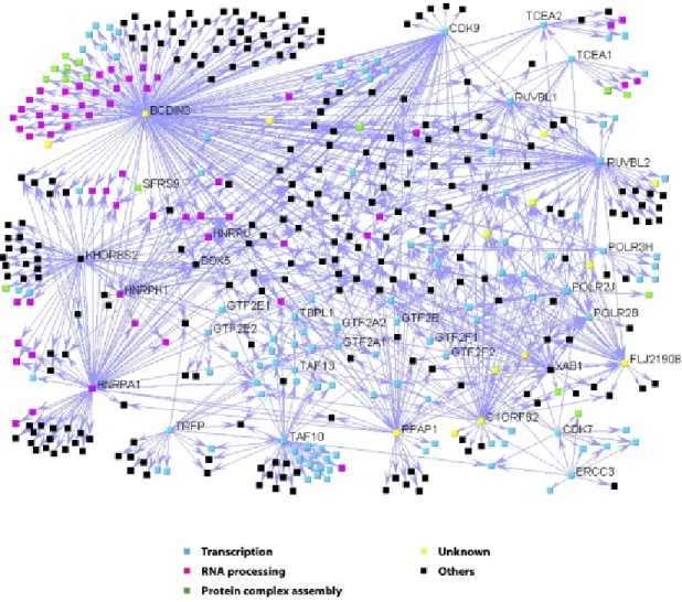
Figure 3: The GroEL and the TCP-1 ring complex families 20 Figure 4: The network of protein complexes involving the
RNAPII basal transcription machinery according 33 to Jeronimo et al., 2007
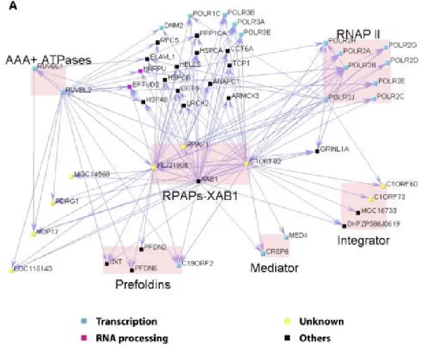
Figure 5: Network highlighting the interactions of RPAPs-XAB1
with RNAPII, the regulatory complexes integrator and 34 mediator and a group of proteins with chaperone/
scaffolding activity (Jeronimo et al., 2007)
Figure 6: Schematic representation of the TAP procedure 38 Figure 7: Schematic representation of the ecdysone-inducible 39 system in the mammalian EcR293 cells
Figure 8: The pMZI vector 40 Figure 9: Schematic representation of the tandem-affinity 41 purification tag fused to a protein at its C-terminus
Figure 10-A: Linear representation of GPN3 cDNA showing 49 the conserved domains
Figure 10-C: TAP gel (of GPN3) 50 Figure 10-D: Mascot results (of GPN3) 51 Figure 11-A: Linear representation of WDR92 cDNA showing 52 the conserved domains
Figure 11-B: Screening test (of WDR92) 53 Figure 11-C: TAP gel (of WDR92) 53 Figure 11-D: Mascot results (of WDR92) 54 Figure 12-A: Linear representation of PDRG1 cDNA 55 Figure 12-B: Screening test (of PDRG1) 56 Figure 12-C: TAP gel (of PDRG1) 56 Figure 12-D: Mascot results (of PDRG1) 57 Figure 13-A: Linear representation of PIH1D1 cDNA showing 58 the conserved domain
Figure 13-B: Screening test (of PIH1D1) 58 Figure 13-C: TAP gel (of PIH1D1) 59 Figure 13-D: Mascot results (of PIH1D1) 59 Figure 14-A: Linear representation of PFDN2 cDNA showing 60 the conserved domain
Figure 14-B: Screening test (of PFDN2) 61 Figure 14-C: TAP gel (of PFDN2) 61 Figure 14-D: Mascot results (of PFDN2) 62 Figure 15-A.1: Linear representation of CCT4 cDNA showing 64 the conserved domain
Figure 15-A.2: Linear representation of CCT5 cDNA showing 64 the conserved domain
Figure 15-B.1: Screening tests for CCT4 65 Figure 15-B.2: Screening tests for CCT5 65 Figure 15-C.1: TAP gel for CCT4 66 Figure 15-C.2: TAP gel for CCT5 66 Figure 15-D.1: Mascot results for CCT4 67
Figure 15-D.2: Mascot results for CCT5 67 Figure 16-A: Linear representation of KIAA0406 cDNA 69 Figure 16-B: Screening test (of KIAA0406) 69 Figure 16-C: TAP gel (of KIAA0406) 70 Figure 16-D: Mascot results (of KIAA0406) 71 Figure 17: Interaction map showing the eight purified proteins, 73 in this project, and their high confidence interactions
Abbreviations
Å Ångström
ADP Adenosine diphosphate ATP Adenosine triphosphate
ATPase Adenosine triphosphate hydrolase AT rich Adenine-Thymine rich
BiP Immunoglobulin heavy chain-binding protein bps Base pairs
BRE TFIIB recognition element
BREu TFIIB recognition element upstream BREd TFIIB recognition element downstream BRET Bioluminescence resonance energy transfer CBP Calmodulin binding protein
CCT Chaperonin containing TCP1 CDK7 Cyclin dependant kinase 7 cDNA coding deoxyribonucleic acid CHX cycloheximide
CFP Cyan fluorescent protein Co-IP Co-immunoprecipitation CTD C-terminus domain C- terminus carboxyl-terminus DCE Downstream core element
DNA Deoxyribonucleic acid DPE Downstream promoter element DSIF DRB sensitive inducing factor EcR Ecdysone response
FACT Facilitates chromatin transcription FBS Fetal bovine Serum
Fmol femtomole
FRET Fluorescence resonance energy transfer GAL Galactose
G- protein Guanine nucleotide-binding protein GTFs General transcription factors HAT Histone acetyltransferases HEK Human embryonic kidney HDAC Histone deacetylases
H1 Histone 1
H2A Histone 2 A H2B Histone 2 B
H3 Histone 3
H4 Histone 4
HSP Heat shock protein i.e Example
IgG Immunoglobulin G
Inr Initiator
ISWI Imitation switch
Kb Kilobase
LUMIER Luminescence-based mammalianinteractome mapping MAT1 ménageà trois 1
m7G cap 7-methyl guanosine cap mRNA messenger ribonucleic acid mRNPs mRNA ribonucleoprotein particles MS Mass spectrometry
MTE Motif ten element
NELF Negative elongation factor
nm Nanometer
N- terminus Amino-terminus
NTPs Nucleoside triphosphates
LC- MS/MS Liquid chromatography tandem mass spectrometer OD Optical density
PCA Protein-fragment complementation assay PDRG1 p53 and DNA damage regulated 1 PIC Pre-initiation complex
PIH1D1 PIH1 domain containing 1 PFDN Prefoldin
pmol Picomole
Poly(A) Poly(Adenosine)
Pre- mRNAs Precursor-messenger ribonucleic acids P-TEFb Positive transcription elongation factor b
R2TP Rvb1-Rvb2-Tah1-Pih1
RAP RNA polymerase II associated factor RNA Ribonucleic acid
RNAP RNA polymerase
RNAPI RNA polymerase I RNAPII RNA polymerase II
RNAPIIA RNA polymerase II in the A form RNAPIIO RNA polymerase II in the O form RNAPIII RNA polymerase III
RPAP RNA polymerase II associated protein rRNA Ribosomal ribonucleic acid
SDS Sodium dodecyl sulfate
SDS- PAGE Sodium dodecyl sulfate polyacrylamide gel electrophoresis SNF2 Sucrose non fermenting 2
snoRNAs Small nucleolar ribonucleic acid SnRNAs Small nuclear ribonucleic acids SWI/SNF Switch/ Sucrose non fermenting TAF TBP associated factor
Tah1 Tpr-containing protein associated with Hsp90 TAP Tandem affinity purification
TBP TATA box binding protein TCP-1 T-complex polypeptide 1 TEV Tobacco etch virus TNF-α Tumor necrosis factor-α
TRAP TNF receptor-associated protein TRNAs Transfer ribonucleic acids UV Ultraviolet
VP16 Herpes simplex virion protein 16 WDR92 WD repeat domain 92
Acknowledgements
First and foremost, I would like to thank GOD for his grace and guidance throughout my life, and for giving me the strength and courage to do this work.
I would also like to thank my supervisor Dr. Benoit Coulombe for providing me with the opportunity to work in his lab, as well as guiding and advising me throughout my work towards this thesis.
Thanks to all of the members of our laboratory with whom I have enjoyed working these past two years, especially Annie Bouchard and Philippe Cloutier for their help and advice, you both have taught me a lot.
A huge “thank you” goes to my sister Diala, my brothers, Firas and Nawras, and friends for all of their support and encouragement. I love you guys and I am so grateful for having you in my life.
I dedicate my Master’s thesis to my parents, Wafaa Butros and Imad Al-Khoury, who have always provided me with their support, constant encouragement and their unconditional love, and who have always given me the strength to follow my dreams. Thank you and I love you dearly.
Section 1: Introduction
1-1) From genes to proteins:
Our genome, made of DNA (deoxyribonucleic acid), is the storehouse that contains all of the required information necessary to build an organism. It is arranged into 24 distinct chromosomes each of which contains many genes which encode proteins. Less than 2% of our genome encodes for genes, the equivalent of about 30000 genes, while the rest of our genome consists of non-coding regions which are thought to play a role in maintaining the integrity of chromosomes and regulating the expression of proteins.
DNA consists of 4 different nucleobases, adenine, guanine, cytosine and thymine, and one DNA molecule can contain hundred million nucleotides. Nucleotides polymerize together, via phosphodiester bonds, to form nucleic acids, the linear representation of which constitutes the primary structure of nucleic acids. In fact, DNA is made up of two associated polynucleotide strands forming a double helix, where the two strands are complementary and oriented antiparallel to each other (Harvey Lodish et al., 4th edition).
This genetic material, DNA, carries the necessary information to specify the amino acid sequences of proteins each of which will have a specific function in the cell (Harvey Lodish et al., 4th edition). To interpret the information encoded by DNA, a transcription process occurs in which an enzyme, RNA polymerase, is recruited to the DNA and transcribes the information into RNA (ribonucleic acid). This RNA can then serve as template for the synthesis of proteins since each set of three ribonucleotides encodes an amino acid, the building block of proteins. This process occurs with the help of ribosomes which consist of ribosomal RNA (rRNA) associated to a set of proteins (Harvey Lodish et al., 4th edition). All of these processes will be
explained in greater details next, starting with the transcription of DNA to RNA.
1-1.1) Transcription process:
Transcription is the process by which DNA is translated into RNA with the help of RNA polymerase enzymes. In eukaryotes, there exist three kinds of RNA polymerases (RNAP), RNAPI, RNAPII and RNAPIII; this is why genes are classified into three classes depending on the RNA polymerase enzyme that transcribes them. RNAPI, localized within the nucleoli, is the enzyme responsible for the transcription of ribosomal RNAs (rRNAs). On the other hand, RNAPII, which is localized within the nucleoplasm, transcribes all protein-encoding genes and certain small nuclear RNAs (snRNAs) genes. Finally, RNAPIII, which is also localized within the nucleoplasm, is the enzyme responsible for the transcription of transfer RNAs (tRNAs) and 5S rRNA (reviewed by Archambault et al., 1993).
RNAPII transcription is the first step in gene expression and a focal point of cell regulation. It is atarget of many signal transduction pathways, and a molecular switchfor cell differentiation in development(Cramer et al., 2001). 1-1.1.1) RNA polymerase II (RNAPII):
RNAPII is the enzyme responsible for the transcription of mRNA in eukaryotes. It is composed of 12 subunits, often referred to as Rpb1 to Rpb12 by decreasing order of their molecular mass. Five of these subunits are common to all three RNA polymerases and they are Rpb5, 6, 8, 10 and 12, and only Rpb4, 7, 9 and the CTD of Rpb1 (see below) are unique to RNAPII. Rpb1 and Rpb2 consist the two largest subunits and are responsible for most of the catalytic activity of polymerase (reviewed by et al.,, 2006).
Rpb1 also contains the CTD which consists of a tandem repeat of a heptapeptide: Tyr-Ser-Pro-Thr-Ser-Pro-Ser which appears 52 times in humans.
The CTD is an essential feature of RNAPII and is involved in transcription and mRNA processing. The CTD is disordered in RNAPII structures and tends to be degraded by proteases (reviewed by Thomas and Chiang, 2006). Depending on its phosphorylation state, RNAPII can exist in two forms; either the RNAPIIA, with an unphosphorylated CTD, involved in pre- initiation complex (PIC) assembly and transcription initiation, or RNAPIIO form, with a phosphorylated CTD, and is implicated in transcript elongation and termination (reviewed by Thomas et al., 2006).
Rpb4 and Rpb7 are two subunits of the RNAPII known to form a heterodimer required for the formation of the pre- initiation complex (PIC), the initiation of transcription, and RNA chain elongation (reviewed by Thomas et al., 2006).
Finally Rpb9, unique to RNAPII, plays an important role in transcription elongation, transcription-coupled DNA repair, and helps in the selection of the correct transcription start site and in maintenance of transcription fidelity (Chen et al., 2007).
1-1.1.2) Recruitment of RNAPII to the promoter:
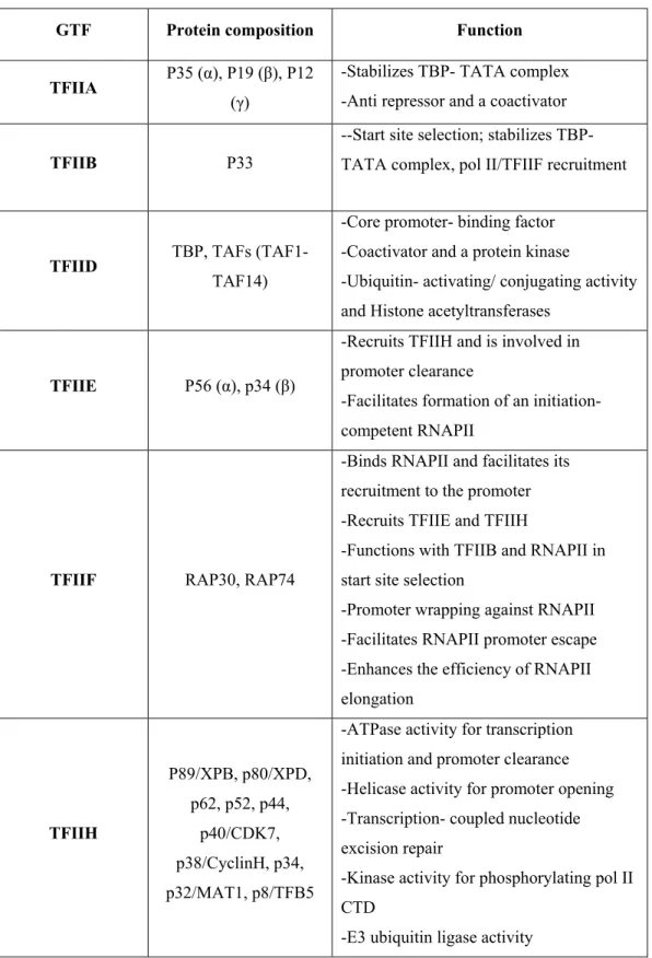
On its own, RNAPII is unable to initiate transcription but rather requires other proteins, such as the general transcription factors (GTFs), involved in the recognition of promoter sequences, the response to regulatory factors, and conformational changes essential to the activity of the polymerase during the transcription cycle (reviewed by Hahn, 2004). The general transcription factors TFIIA, TFIIB, TFIID, TFIIE, TFIIF and TFIIH assemble the RNAPII onto promoter DNA to form the Pre-Initiation complex (PIC). Refer to table 1 for a summary about the general transcription factors, their composition and function (adapted from a review by Thomas et al., 2006).
GTF Protein composition Function TFIIA P35 (α), P19 (β), P12
(γ)
-Stabilizes TBP- TATA complex -Anti repressor and a coactivator
TFIIB P33
--Start site selection; stabilizes TBP-TATA complex, pol II/TFIIF recruitment
TFIID TBP, TAFs
(TAF1-TAF14)
-Core promoter- binding factor -Coactivator and a protein kinase
-Ubiquitin- activating/ conjugating activity and Histone acetyltransferases
TFIIE P56 (α), p34 (β)
-Recruits TFIIH and is involved in promoter clearance
-Facilitates formation of an initiation- competent RNAPII
TFIIF RAP30, RAP74
-Binds RNAPII and facilitates its recruitment to the promoter -Recruits TFIIE and TFIIH
-Functions with TFIIB and RNAPII in start site selection
-Promoter wrapping against RNAPII -Facilitates RNAPII promoter escape -Enhances the efficiency of RNAPII elongation TFIIH P89/XPB, p80/XPD, p62, p52, p44, p40/CDK7, p38/CyclinH, p34, p32/MAT1, p8/TFB5
-ATPase activity for transcription initiation and promoter clearance -Helicase activity for promoter opening -Transcription- coupled nucleotide excision repair
-Kinase activity for phosphorylating pol II CTD
-E3 ubiquitin ligase activity
Other proteins also regulate transcription, such as the mediator which is a large multiprotein complex that communicates directly with many gene-specific regulators (Blackwell et. al., 2006), and sequence-gene-specific DNA-binding transcription regulators (i.e., activators and repressors) (Martinez, 2002).
Transcription initiation proceeds through a number of specific steps. First, the packed chromatin must be de-condensed so that the transcriptional machinery could have access to the promoter region of a gene. Second, the binding of transcriptional activators to enhancer elements located close to or many thousands of base pairs away from the transcription start site. Finally, co-activators are recruited to bring the RNAPII PIC to the site where the transcription activators are bound to enhancers (reviewed by Gross et al, 2006).
1-1.1.2.1) The de-condensation of chromatin:
Inside the non-dividing eukaryotic nuclei, huge DNA molecule is packaged, into filament with the help of proteins to form the 30 nm chromatin. This chromatin structure plays an important role in gene regulation since it presents an obstacle that must be overcome by transcription machineries to access the underlying DNA (reviewed by Ruthenberg et al., 2007).
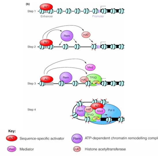
The first step in the transcription process is the binding of activators to enhancers which will recruit histone- modifying enzymes helping in the displacement of nucleosomes, as well as target the basal transcription machinery to the core promoter (discussed later on). The mediator, a co-activator, is also involved and will serve as a bridge between the enhancer-bound activators and the pre- initiation complex at the core promoter (reviewed by Szutorisz et al., 2005). The mediator is a multi- subunit complex, discovered by Roger D. Kornberg and R. Young at the same time, which is essential for transcription of class II genes. Figure 1 summarizes these processes (adapted from a review by Szutorisz et al., 2005).
Figure 1: Schematic representation of a proposed model for the activation of class II gene transcription. In the first step, the activator binds to the enhancer element and then recruits chromatin remodeling complexes to decondense the chromatin structure in step two. This is followed by the recruitment of the transcription machinery to the core promoter in step three. Finally in step 4, the enhancer- bound activator is brought into closer proximity, with the help of the mediator complex, to the transcription machinery at the core promoter.
1-1.1.2.1.1) Histones:
Histones are the main protein components of chromatin and play a major role in the structure of chromatin. There are five major types of histones, called H1, H2A, H2B, H3 and H4, and these histones are present in almost all cell types. Four of these five types of histones, H2A, H2B, H3 and H4, specifically bind to DNA and they are the called “core” histones (reviewed by Lusser et al., 2003). Two of each assemble to form an octameric structure around which 146 base pairs of DNA are wrapped to form a structure called the nucleosome core, which is separated from other nucleosome cores by a thread of DNA referred to as the linker DNA. Together, one nucleosome core with a linker DNA, they form a complete chromatin subunit. H1, on the other hand, interacts with the nucleosomal core and the adjoining linker DNA (reviewed by Lusser et al., 2003). Histones protect the DNA, wrapped around them, from cleavage by endonucleases, whereas the linker DNA region is susceptible to endonucleolytic cleavages.
Chromatin condensation creates an important obstacle for transcription, therefore, chromatin remodeling complexes and histone-modifying enzymes have to be recruited, by gene-specific regulatory factors, to de-condense the region where transcription is to be potentiated.
1-1.1.2.1.2) Chromatin remodeling
1-1.1.2.1.2.1) Chromatin remodeling complexes:
One class of chromatin remodeling complexes is the ATP-dependent molecular machine, which uses the hydrolysis of ATP to modulate the interactions between DNA and histones in the chromatin. Although they are very different in composition and in function, these complexes share an ATPase subunit which belongs to the Snf2-like family (reviewed by Lusser et al., 2003).
ATP-dependent chromatin remodeling complexes are divided into three groups depending on their biochemical properties and the sequence similarity of their ATPase subunits.
The first group is the SWI/SNF group, which plays a role in the activation of transcription. Then there are the ISWI and the Mi-2/CHD groups, both of which are involved in the repression of transcription (reviewed by Peterson, 2002).
The most studied of these groups is the SWI/SNF group, which can modulate chromatin structure by helping DNA-bending proteins facilitate nucleosomal sliding, as well as disrupting high-order chromatin folding and nucleosomes (reviewed by Peterson, 2002, and by Vignali et al., 2000).
1-1.1.2.1.2.2) Histone modifications:
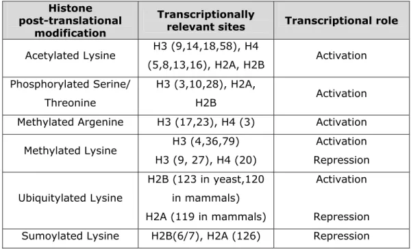
Histones can also undergo posttranslational modifications that can alter their interaction with DNA. Different histone modifications can have a role in different biological processes such as, gene regulation, DNA repair or DNA chromatin condensation. Some of these modifications include the acetylation of lysines, the methylation of lysines and arginines, the phosphorylation of serines and threonines, the ubiquitination of lysines, the sumoylation of lysines, and the ADP-ribosylation of glutamic acids (reviewed by Khorasanizadeh, 2004). The enzymes responsible for these modifications are the histone acetyltransferases (HAT), histone deacetylases (HDAC), histone methyltransferases, kinases for phosphorylation, ubiquitinases and poly-(ADP-ribose) polymerases (reviewed by Davie, 1996). Most modifications localize to the amino- and carboxy-terminal histone tails, and a few localize to the histone globular domains (reviewed by Berger, 2007). As to the modifications that are involved in the regulation of transcription, they are divided into two groups, either activating or repressing transcription. These modifications are summarized in table 2 (adapted from a review by Berger, 2007).
Histone post-translational
modification
Transcriptionally
relevant sites Transcriptional role
Acetylated Lysine H3 (9,14,18,58), H4 (5,8,13,16), H2A, H2B Activation Phosphorylated Serine/ Threonine H3 (3,10,28), H2A, H2B Activation
Methylated Argenine H3 (17,23), H4 (3) Activation
Methylated Lysine H3 (4,36,79) H3 (9, 27), H4 (20) Activation Repression Ubiquitylated Lysine H2B (123 in yeast,120 in mammals) H2A (119 in mammals) Activation Repression
Sumoylated Lysine H2B(6/7), H2A (126) Repression
Table II: Histone post- translational modifications affecting transcription
1-1.1.2.2) Assembly of the RNAP II Pre- Initiation Complex (PIC): Once the chromatin has de-condensed to a more accessible structure, the gene- specific regulatory factors, already bound to the site of transcription initiation, will now recruit the RNA polymerase II to the core promoter where the pre-initiation complex can assemble.
1-1.1.2.2.1) Core promoter:
A core promoter (Figure 2) is defined as the minimal portion of DNA sequence, located upstream of a gene, required to properly initiate transcription. It most commonly consists of elements such as the TATA box, which is present 25- 30 bps upstream of the transcription start site (+1), and is an AT rich sequence that is recognized by the TBP protein (TATA-binding protein),
Figure 2: The core promoter
Schematic representation of the core promoter needed for basal transcription by the RNAPII enzyme. What is shown are the elements that most commonly consist the core promoter (TATA box, BREu and BREd, Inr, DPE, MTE and finally DCE).
a subunit of the TFIID general transcription factor. The initiator element (Inr), which has a conserved sequence, is located at the +1 site. The Inr is functionally similar to the TATA box (-25 to -30 bps away from the Inr position at +1), and can function independently of the TATA box when they are more than 30 bps apart, otherwise, these two elements function synergistically (reviewed by Smale et al., 2003). The Inr element is also recognized by TFIID, through its TAF (TBP associated protein) subunits, TAF1 and TAF2 (reviewed by Smale, 1997, and by Thomas and Chiang, 2006). Another element is the DPE (downstream promoter element), present at +28 - +32 relative to the +1 position. This element is conserved from Drosophila to humans and is required for the binding of purified TFIID (through its TAF6 and TAF9 subunits) to a subset of TATA-less promoters. The DPE element also acts in conjunction with the Inr element (reviewed by Smale et al., 2003, and by Thomas and Chiang, 2006).
In addition to DPE, two other core promoter elements are present downstream of the transcription start site, and they are the MTE and DCE
elements. The MTE (Motif Ten Element) is positioned between +18 to +29, and normally functions in conjunction with Inr to enhance transcription by RNAPII. It can also work synergistically, in an Inr-dependent manner, with the TATA box and DPE, or without them (when they are not present), to strengthen the promoter activity. DCE (Downstream Core Element), on the other hand, is found between +6 to +34 and is recognized by the TAF1 subunit of TFIID (reviewed by Thomas et al., 2007, and by Thomas and Chiang, 2006). The DCE contributes to the transcriptional activity and binding of TFIID (reviewed by Smale et al., 2003).
The core promoter also consists of a BRE (TFIIB recognition element) which could be present at two positions, upstream of the TATA box (BREu) and downstream (BREd) of the TATA box. BREd, bound to TFIIB, was shown to modulate promoter strength and act as a positive element for transcription when the promoter contains only the BREd element. However, in the presence of BREu, BREd has a negative effect on transcription. BREu, on the other hand, might have a positive or negative effect on transcription of TATA-less promoters likely to contain BREu, rather than the TATA-containing promoters (Deng et al., 2006).
Although most core promoter studies were conducted on TATA box containing promoters, it turns out that the majority of mammalian genes have TATA-less promoters within which are present multiple start sites, generating diversity and complexity. This indicates that TATA-driven PIC assembly is the exception, rather than the rule, in eukaryotic transcription (reviewed by Sandelin et al., 2007).
1-1.1.2.2.2) Recruitment of RNAPII to the core promoter:
This process starts with the formation of the PIC, which consists of the RNAPII and several general transcription factors including TFIIA, TFIIB,
TFIID, TFIIE, TFIIF and TFIIH. The assembly of the PIC is thought to proceed in two different pathways.
The first pathway is called the “sequential assembly pathway” where the TBP protein of TFIID first binds to the promoter region at the TATA box, resulting in the bending of the DNA at an 80° angle (Kim J. L. et al., 1993, Kim Y. et al., 1993). This event is followed by the entry of TFIIA and TFIIB, both of which will help in stabilizing the TFIID bound to promoter. TFIIB will help in facilitating the recruitment of the RNAPII- TFIIF complex (reviewed by Thomas et al., 2007). The entry of TFIIF induces a second bending of the DNA, close to the +1 site, causing the wrapping of the promoter region around the RNAPII (Robert et al., 1998, reviewed by Coulombe and Burton, 1999). This event is finally followed by the entry of TFIIE and TFIIH.
The second pathway is known as the “RNAPII holoenzyme pathway”, where the RNAPII is present in a pre-assembled holoenzyme complex containing the RNAPII and suppressors of RNAPII mutations, with or without a subset of general transcription factors. In addition, other proteins associated with chromatin remodeling, DNA repair, and mRNA processing, are involved. This holoenzyme complex would be recruited to a transcription site by TFIID bound to a promoter (reviewed by Thomas et al., 2007). Evidences supporting both models have been reported. Thus, it is likely that both assembly pathways exist in vivo.
In both pathways, it is clear that each general transcription factor involved plays an essential role in transcription. For example, when the TBP subunit of TFIID binds to the TATA box of the promoter, it bends the DNA around the RNAPII bringing it closer to the transcription start site. Furthermore, when TFIIH is recruited, it introduces negative superhelical tension in the DNA, through its ATPase/Helicase subunit, producing a transient bubble.
Finally initiation and RNA synthesis can be potentiated with the addition of two nucleoside triphosphates (NTPs) (reviewed by Kornberg, 2007).
1-1.1.3) Initiation:
The initiation process does not start smoothly; up until positions +8/+10 multiple rounds of abortive initiation occur resulting in the production of short (2- 9 nucleotides) transcripts. These rounds proceed until the complex reaches a point where it is stabilized, usually at position +9, and this transition is referred to as “promoter escape”. Soon after promoter escape, RNAPII pauses again and this phenomenon is called the “promoter-proximal pausing”, in which a large number of genes have been shown to be regulated (reviewed by Margaritis et al., 2008).
1-1.1.3.1) mRNA processing prior to productive elongation:
The transcription of mRNA is tightly coupled to the recruitment of the mRNA processing machinery. The initial transcripts synthesized by the RNAPII, called pre-mRNAs, have to be processed to form the mRNA before getting exported to the cytoplasm. The first processing step in all eukaryotic cells occurs as soon as the transcript is ~25 bases long. An m7G cap is added to the 5'-end of the transcript by a capping enzyme bound to the phosphorylated CTD. This cap modification has been shown to occur during the arrest of the transcription process, prior to its entry in the productive elongation phase. This suggests that the capping enzyme might play an important role as a checkpoint, ensuring that uncapped transcripts are not elongated. Once the 5'-end is capped, introns are excised with the help of the spliceosome, one of the largest macromolecular machines in the cell, also brought to the transcription site through interaction with the CTD of RNAPII (reviewed by Almeida et al., 2008).
1-1.1.4) Elongation:
Once the RNAPII has escaped the promoter it enters the elongation phase. At this stage all of the general transcription factors dissociate to form the “transcription elongation complex”, except for TFIIF. This phenomenon is referred to as the “Promoter clearance step”.
Promoter clearance has been shown to coincide with phosphorylation of the CTD of RNAPII, which plays an important role in recruiting protein factors involved in elongation, as well as in mRNA maturation, surveillance, and export. Other elongation factors are recruited at this stage to the RNAPII to modulate its catalytic activity and form the transcription elongation complex. This includes factors such as Elongins, P-TEFb, DSIF and NELF, which can all influence the pausing of the RNAPII, either positively or negatively. FACT, a histone chaperone, is another elongation factor recruited to help facilitate the elongation process by remodeling the chromatin (reviewed by Sims et al., 2004).
1-1.1.5) Termination and 3'-end mRNA processing:
Termination is the last step of transcription by the RNAPII and it is a process that depends on pre-mRNA 3'-end processing signals, such as the poly(A) signal. Therefore, once the poly(A) signal has been read by the RNAPII, it is recognized by a multi-component cleavage/polyadenylation complex recruited to the RNAPII through the CTD of Rpb1. This complex will direct the endonucleolytic cleavage and the polyadenylation of the free 3'-end (reviewed by Lykke-Andersen et al., 2007, and Kim et al., 2003). Poly(A)-binding proteins are then recruited to the 3’end of the processed transcript to help protect it from exonucleolytic degradation. Other signals also exist on different transcripts, such as the snRNAs and the snoRNAs, and are recognized
by other protein complexes, also recruited to RNAPII through the CTD of Rpb1, which contain the RNA binding proteins Nrd1p, Nab3p, and additional co-factors (reviewed by Lykke-Andersen et al., 2007, and Kim et al., 2003).
1-1.2) mRNA export:
The transcription process is also coupled to the nuclear export of mRNAs, which is an essential process for the expression of genes in all eukaryotic cells. Therefore, as soon as the mRNA is synthesized, proteins are recruited forming the mRNA ribonucleoprotein particles (mRNPs). Export machineries will recognize signals within the proteins of mRNPs rather than within the mRNA itself, and will export the mRNA to the cytoplasm where the proteins synthesis occurs (reviewed by Iglesias et al., 2008).
1-1.3) Protein synthesis:
Translation is the process by which mRNAs are used to direct the synthesis of polypeptide chains in three different steps, including initiation, elongation, and termination. This process is catalyzed by a complex called the ribosome which is, in fact, a ribozyme with RNA at the heart of its enzymatic activity (reviewed by Steitz, 2008 and by Culver, 2001). Mammalian ribosomes are only found in the cytosol and in the mitochondria (reviewed by Hebert et al., 2007).
The eukaryotic ribosome is a complex that consists of two asymmetric ribonucleoprotein subunits, the 60s and the 40s subunits, which will bind the mRNA and use it as a template to catalyze the correct assembly of amino acids into polypeptide chains. tRNAs are the adaptor molecules used by ribosomes to decode the information in the mRNA. They are present at the interface between the mRNA and the growing amino acid chain.
There exits three tRNA-binding sites in a ribosome, the A, P and E sites. The A site is the site of entry of a cognate aminoacyl tRNA. The P site is the site of the peptidyl tRNA, where the elongation of the polypeptide chain occurs. The amino acid bound to the tRNA in the A site is transferred to the growing polypeptide chain bound to the tRNA in the P site. The E site is the exit site, occupied by the deacylated tRNA. An exception occurs during the initiation step where the cognate aminoacyl tRNA carrying a methionine enters the P site rather than the A site and base pairs with the start codon of the mRNA (reviewed by Culver, 2001). The translation ends once a stop codon is read by the tRNA; at this point the polypeptide chain separates form the tRNA, which itself separates from the ribosome. Finally the ribosomal subunits separate from the mRNA.
1-1.4) Protein folding:
Following the synthesis of a new polypeptide chain, its correct folding will convert it into the mature, active protein which can then be localized to its appropriate location (reviewed by Hebert et al., 2007). This process will normally take place either in the cytoplasm, for cytoplasmic proteins, or in the endoplasmic reticulum, for transmembrane and secretory proteins (reviewed by Paulsson et al., 2003). The information needed for the correct folding of a protein is encoded by its amino acid sequence. The protein folding process requires most of the time the assistance of a network of molecular chaperones to support the folding in vivo. This is important to prevent aggregation in the crowded intracellular environment, and prevent inappropriate inter- and intra-molecular interactions (reviewed by Deuerling et al., 2004). As soon as the nascent polypeptide emerges from the ribosome, the ribosome-associated chaperones guide its folding. Finally, once a protein has been properly folded by a chaperone, it must be localized to its appropriate subcellular compartment.
Chaperones will help these proteins find their way to the place where they can display their appropriate function (Lund et al., 2003).
1-1.4.1) Molecular Chaperones:
Molecular chaperones exist in all organisms and in all cellular compartments. They are defined as a large group of proteins that share the property of assisting the folding, unfolding, assembly and disassembly of other macromolecular structures (reviewed by Ellis, 2006). Furthermore, chaperones have also been shown to play a role in the translocation of newly synthesized proteins (reviewed by Nicoll et al., 2005). The main chaperone classes are the Hsp40 (the DnaJ family), Hsp60 (includes GroEL and T-complex polypeptide 1 (TCP-1) ring complexes), Hsp70, Hsp90 (reviewed by Fink, 1999) and the AAA+ superfamily of ATPases.
1-1.4.1.1) Hsp40 family:
The best defined role for Hsp40 is as a cochaperone for the Hsp70. This family of proteins consists of over 100 members, defined by the presence of three distinct domains. The first is a highly conserved J- domain of ~70 amino acids near its N-terminus, responsible for the interaction with Hsp70. The presence of a J- domain in a protein defines it as a DnaJ-like protein, and is involved in regulating the ATPase activity of the Hsp70 (Hennessy et al., 2000). The second domain is a glycine and phenylalanine rich region that acts as a flexible linker. Finally, the third domain is a cysteine-rich, zinc finger-containing C-terminus domain (Cheng et al., 2008, and reviewed by Fink 1999).
1-1.4.1.2) Hsp70 family:
Members of this family help to protect cells from stress, aside from playing an important part in protein folding. They are characterized by a very
weak ATPase activity which is stimulated by the binding of the Hsp40 cochaperone. They are composed of two major functional domains; an N-terminal ATPase domain and a C-N-terminal domain that is responsible for the binding of polypeptides. Some of the better known mammalian members of this family include the constitutive cytosolic member HSC70, the stress- induced cytosolic form HSP70, the ER form BiP and the mitochondrial form mHSP70 (Morano KA, 2007, and reviewed by Fink 1999).
1-1.4.1.3) Hsp90 family:
Hsp90 is one of the most abundant chaperones in the cell found to be upregulated in response to stress. It also pays an important role, under normal conditions, in protein folding, intracellular transport, and stability of proteins many of which are critical for signal transduction. Mammalian Hsp90 exist as dimers and is often found in complexes with other chaperones. Members of this family of proteins are highly conserved in all organisms from bacteria to humans and are essential. Examples of the Hsp90 family include the cytosolic form, HSP90A, the endoplasmic reticulum form, HSP90B and the mitochondrial form, TRAP (reviewed by Fink, 1999, and by Pearl et al., 2008).
Hsp90 has an ATPase activity and contains three functional domains. The first is the ATP binding domain near the N- terminus. The other two domains are the protein binding and the dimerization domains located near the C-terminus of the Hsp90 protein. All of these domains play an important role in the function of the protein (Southworth et al., 2008, Barginear et al., 2008 reviewed by Fink, 1999).
1-1.4.1.4) Hsp60 family/ Chaperonin family:
This family of proteins is the best studied of the chaperones, and is divided into two families/ groups; the GroEL (also called group I) and the TCP-1 ring complex families (also called group II), both of which are represented
schematically in Figure 3 (Figure 3 is adapted from Martin- Benito et al., 2007, Martin- Benito et al., 2002, and Roseman et al,. 1996). Members of this family are involved in the assembly of large multiprotein complexes (reviewed by Fink, 1999). Hsp60 genes are highly conserved, indicating a central role in cell viability (Lund et al., 2003).
1-1.4.1.4.1) GroEL family:
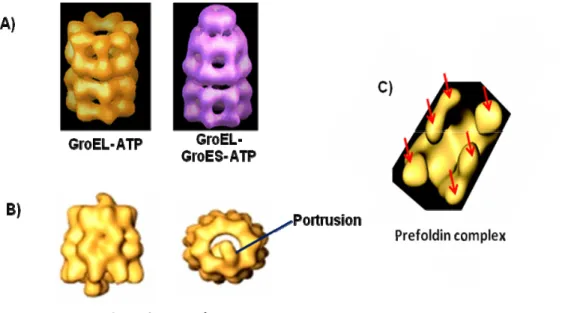
This family of proteins is found in prokaryotes, chloroplasts, and mitochondria. The vast majority of organisms contain at least one gene for this protein. GroEL requires the assistance of a cochaperone called GroES “lid” and the presence of ATP to facilitate the folding of newly synthesized polypeptides. Substrates of this group of chaperones include several metabolic enzymes, RNA polymerase II subunits, and other proteins involved in transcription and translation (reviewed by Fink, 1999). GroEL chaperones have also been shown to play a role in conformational maintenance of pre-existing proteins, secretion, and proteolysis (Houry et al., 1999, Kusukawa et al., 1989, Kandror et al., 1994, and reviewed by Deuerling et al., 2004). GroEL is composed of two heptameric rings, of the large subunit GroEL, stacking back to back and forming a 14-subunit hollow cylinder (Lund, 2001). Binding of ATP and GroES induces large conformational changes in GroEL providing a polar environment favorizing the folding of the encapsulated protein. Then ATP hydrolysis primes the GroEL to release GroES “lid”, allowing the folded substrate to exit the chaperone (Lund et al., 2003, reviewed by Fink, 1999 and reviewed by Deuerling et al., 2004).
Figure 3: The GroEL and the TCP-1 ring complex families
A) Left: A side view of the GroEL chaperone in the ATP bound state. This view shows the two heptameric ring. Right: A side view of the GroEL chaperone in the ATP bound state in the presence of the GroES cochaperone.
B) Left: A side view of the TCP-1 ring complex chaperone showing the two octameric rings. Right: A top view of the TCP-1 ring complex chaperone showing the protrusion “lid”.
C) The prefoldin complex with its six subunits (red arrows point to the six subunits).
1-1.4.1.4.2) TCP-1 ring complex family:
The TCP-1 complex is an ATP-dependent complex found in the eukaryotic cytosol, and is composed of two identical stacked rings, each composed of eight different proteins referred to as CCT1 (also referred to as TCP-1) to CCT8 encoded by different genes. This family of proteins does not require the assistance of a cochaperone such as the GroES, but is rather characterized by the presence of a portrusion “lid” on the TCP-1 ring complex, which is actually part of the CCT protein itself. TCP-1 ring complexes are chaperones that assist in the folding of a small number of proteins, mainly actin and tubulin (Yam et al., 2008, Lund et al., 2003, reviewed by Fink, 1999, and Llorca et al., 1999).
1-1.4.1.4.2.1) The prefoldin complex:
The TCP-1 ring complex does not function alone in the folding of actin and tubulin, but rather requires the assistance of another molecular chaperone named prefoldin. The prefoldin complex itself is a heterohexameric complex found in archaeabacterial and eukaryotic organisms (Martin- Benito et al., 2002). In eukaryotes, the prefoldin complex is an oligomer composed of six different proteins (PFDN1- PFDN6). The archaeal prefoldin chaperone, on the other hand, is an oligomer composed of two different proteins, two α-subunits and four β-subunits (Martin- Benito et al., 2002).
Actins and tubulins are very abundant and highly conserved proteins involved in processes which are essential and unique to eukaryotes. These processes include muscle contraction, segregation of chromosomes, stabilization and alteration of cell shape, endocytosis and exocytosis to mention a few (Leroux et al., 2000, Gu et al., 2008). Once the N-terminus (~145 amino acids) of the actin or tubulin nascent chains have been synthesized, the prefoldin complex binds to it and keeps the polypeptide chain in an unfolded
state until its synthesis is complete. After which, the polypeptide is delivered to the TCP-1 complex for proper folding (Hansen et al., 1999, Gu et al., 2008).
1-1.4.1.4.3) The AAA+ superfamily of ATPases:
AAA stands for ATPases Associated with diverse cellular Activities (Kunau et al., 1993). The AAA+ superfamily is a large and functionally diverse superfamily of NTPases that are characterized by a conserved nucleotide-binding and catalytic module, the AAA+ module (Snider et al., 2008). Members of the AAA+ superfamily of ATPases are found in all three kingdoms of life and function in diverse cellular processes, often via chaperone-like activities (Zhang et al., 2002; Ogura and Wilkinson, 2001). AAA+ proteins typically assemble into hexameric ring complexes that are involved in the energy-dependent remodeling of macromolecules (Iyer et al., 2004, Snider et. al. 2008). The defining feature of AAA+ proteins is a structurally conserved ATP-binding module of 200-250 amino acids, that oligomerizes into active arrays (reviewed by Erzberger et al., 2006, Ogura and Wilkinson 2001). ATP hydrolysis by the AAA+ protein is coupled to physical contact with the target substrate molecule and requires that specific interaction for remodeling of the substrate to take place (Zhang et al., 2002).
1-2) Protein complexes:
Many cellular functions are performed by multiprotein complexes. For example, the RNA pol II enzyme which itself is composed of 12 subunits, that need to come together and interact with each other to form the enzyme. Once formed, this enzyme requires the assistance of the general transcription factors which will be recruited, and interact with it, to facilitate the transcription process. Another example of multiprotein complexes is the chaperones themselves. Chaperones are often composed of different proteins that interact
with each other to form the active chaperone; for example, the TCP-1 ring complex with its 8 subunits that need to interact with each other to generate the active chaperone activity. Furthermore, these chaperones will often require the assistance of cochaperones to facilitate the folding process of proteins.
All of this point to the fact that, proteins do not function on their own but are usually grouped into larger complexes to perform a specific function in an organism (Gingras et al., 2005). Therefore, it is clear that the study of protein complexes is important, since it allows placing proteins of an unknown function into a functional context provided by their interactions with other proteins in the complex (reviewed by Bauer et al., 2003). Therefore, the network of interacting partners in which a certain protein is present and its position within the network provide important information in defining its function (Jaeger et al., 2008). This also applies to the analysis of proteins of known function which often play a role in different complexes and subcellular compartments. Thus, large-scale functional proteomics projects, which build interaction networks of protein complexes, can help in understanding the cross- talk that goes on between the different complexes of unconnected cellular activities (reviewed by Bauer et al., 2003).
From a pharmacological point of view, numerous human diseases are caused by defects in cellular signal transduction pathways. These signaling pathways, themselves, are regulated by protein- protein interactions which often involve the assembly of large signaling complexes. Therefore, while studying protein complexes, if a protein is found to interact with proteins of a specific pathway, it can represent a new potential drug target of that pathway (reviewed by Shiota et al., 2008).
In the past, drug discovery efforts focused on a relatively limited number of proteins against which compounds could be developed such as, G-protein coupled receptors. The identification and validation of new drug targets
was a very difficult, time consuming and expensive process, discouraging investments in drug discovery programs (reviewed by Kramer et al., 2004, and by Ruffner et al., 2007). Nowadays, large-scale, genome-wide protein-protein interaction screens have a great potential in identifying novel therapeutic targets and are key in understanding the functions and molecular mechanisms of diseases (Ruffner et al., 2007).
1-2.1) Methods for studying protein- protein interactions and protein complexes in eukaryotes:
Several different methods have been developed for studying protein-protein interactions such as affinity chromatography technique, phage display, co-immunoprecipitation experiments, and the BRET and FRET methods. Recently new approaches, such as LUMIER and PCA, have also been developed, but the main two methods used currently are the yeast two- hybrid method and the tandem affinity-purification procedure coupled to mass spectrometry.
1-2.1.1) The affinity chromatography technique:
This is a technique that requires the fusion of the protein of interest to an affinity tag. Cells are transfected with the plasmid coding for the protein of interest fused to the affinity tag (Berggård et al., 2007). After an appropriate expression period, the cells are lysed and the tagged protein of interest is purified together with all its interaction partners using a specific ligand linked to a solid support (Berggård et al., 2007). Eluted proteins are then separated on gel-electrophoresis and the proteins are identified by mass spectrometry (Berggård et al., 2007).
Although this technique could be used for the identification of protein-protein interactions, it is prone to generate higher backgrounds and thus, the small amounts of specific interaction partners may be masked by more abundant non- specific binding proteins (Berggård et al., 2007). Another disadvantage of affinity chromatography is the fact that the protein of interest must be fused to a tag. The tag may cause a problem if it gets buried inside the complex and thus, the purification of the complex may not be achieved (Berggård et al., 2007). An exogenous tag might also perturb the interaction of the fused protein of interest with other proteins in the complex (Berggård et al., 2007).
1-2.1.2) Phage display:
Phages are viruses that infect bacterial cells (Smith et al., 1997). In the phage display technique, the protein of interest are fused to a coat protein of a phage particle (Goodyear et al., 2008) so that the protein of interest is displayed on the outer surface of a phage particle once released from the transformed bacterial cell (Smith et al., 1997), thus making it accessibleto other proteins for subsequent binding interactions (Goodyear et al., 2008). This characteristic enables the selection of specific binding interactions from a mixture of nonbinding particles (Goodyear et al., 2008). Selected phage particles are then eluted and used to infect fresh bacteria and the process is repeated until the desired enrichment is reached (Goodyear et al., 2008). Therefore, during the selection process, specific phage clones are progressively enriched on the basis of their specificity and affinity for a protein.
Although Phage display can be used in to identification of new protein-protein interactions, the technique itself suffers from several limitations such as, non-specific binding, elution efficiency, and differences in the ability of the eluted phages to re-infect and propagate in bacterial cells (Rhyner et al., 2004). Recently, display systems, such as the bacterial 2-hybrid display system, have been developed and have shown to produce less background, than the classical
phage display system, in studying protein-protein interactions (Bair et al., 2008).
1-2.1.3) Co-ImmunoPrecipitation (Co-IP) method:
Co-IP is a technique that uses the specificity of antibodies to purify a target protein together with its interaction partners (Kaboord et al., 2008). To do so, whole cell extracts are prepared under nondenaturing conditions to maintain any interactions that occur. Cell extracts are then incubated with an antibody specific to the protein of interest “bait”, provided that the antibody-protein interaction does not interfere in the interaction of the bait with other proteins within the cell extract (Yaciuck, 2007, and Miernyk et al., 2008). The antibody-bound protein complex is then isolated on protein A or protein G sepharose beads. Proteins that did not bind to the beads are removed by a series of washes, and the bound protein complex is eluted from the beads. Members of the complex are then dissociated from each other by SDS sample buffer and then run on an SDS-PAGE followed western blotting with antibodies specific to the bait or interaction partners (Yaciuck, 2007, and Miernyk et al., 2008).
1-2.1.4) BRET and FRET methods:
BRET (Bioluminescence Resonance Energy Transfer) is a method that takes the advantage of the resonance energy transfer between a luminescent donor (usually a luciferase) and a fluorescent acceptor (such as YFP). In this approach, the protein of interest must be fused to the bioluminescent donor and the suspected interacting partner must be fused to the fluorescent acceptor (note: the inverse is also plausible where the bait is fused to the fluorescent acceptor and the interacting partner is fused to the bioluminescent donor, since this methods only checks to see if an interaction occurs between two proteins). When the two proteins do not interact, only one signal, emitted by the luciferase, can be detected. It is only when the proteins are brought into close proximity of each other, typically at a distance of 100Å or less, that the
energy transfer occurs, and in this case an additional signal, emitted by the fluorescent protein, can be detected. This distance (100Å) is generally indicative of interaction between the two fused proteins of interest, either directly or as part of a complex (reviewed by Pfleger et al., 2006, Boute et al., 2002).
FRET (Fluorescence Resonance Energy Transfer), on the other hand, is a method that also exploits the advantage of resonance energy transfer, but this time between a fluorescent donor (example CFP) and a fluorescent acceptor (example YFP), provided that the two fluorophores are different. FRET is observed when the sample is exited at the wavelength of excitation of the donor (Tramier et al., 2002, Evans et al., 2006, and Gandía et al., 2007). Now excited, the donor will emit energy at a wavelength that superimposes with the wavelength at which the acceptor is excited. This would only occur when the two proteins are in a close proximity of each other, 10-100Å in the case of FRET compared 100Å in the case of BRET, indicating an interaction between the two fused proteins of choice (Tramier et al., 2002, Evans et al., 2006, and Gandía et al., 2007).
The advantages of the previous two approaches (BRET and FRET) are that they both measure protein-protein interactions (either direct or indirect interactions) in vivo, and do not require the lysis of cells or the purification of proteins. Their disadvantages, on the other hand, is that they require ectopic expression and/or overexpression of the fusion proteins which may cause artifacts that can either inhibit or induce protein- protein interactions (Bhat et al., 2006). Furthermore, expensive high- resolution microscopes are required to measure both BRET and FRET.
1-2.1.5) LUMIER:
LUMIER (LUminescence-based Mammalian IntERactome) is an automated high-throughput technology aimed at mapping protein-protein
interaction networks systematically in mammalian cells (Barrios-Rodiles et al., 2005). This approach uses Renilla luciferase enzyme fused to proteins of interest, which are then coexpressed with individual Flag-tagged partners in mammalian cells. Cell extracts are then prepared and the interactions are then determined by performing an Renilla luciferase enzymatic assay on the immunoprecipitates using an antibody against Flag (Barrios-Rodiles et al., 2005).
An advantage of LUMIER is the fact that it can detect, in mammalian cells, interactions involving transmembrane receptors which play an important role in signaling networks (Barrios-Rodiles et al., 2005). Previously, studying transmembrane receptors has shown to be a difficult task using other high-throughput approaches. However, the disadvantages are that the LUMIER technique is not able to measure absolute protein-protein interaction affinities, and is prone to generate false positives due to overexpression of the fused proteins (Barrios-Rodiles et al., 2005).
1-2.1.6) The PCA approach:
PCA (protein-fragment complementation assays) is a technique that measures protein- protein interactions by fusing each of the proteins of interest to two fragments of a reporter protein that has been dissected into two fragments using protein-engineering strategies (Michnick et al., 2007). If the interaction does indeed occur, the reporter-protein fragments are brought into proximity allowing them to fold together into the unique three-dimensional structure of the reporter protein and reconstitute its activity (Michnick et al., 2007).
The advantage of PCA is that it can be created with many reporter proteins and thus provides for different types of readouts depending on the desired application (Michnick et al., 2007). Furthermore, proteins are expressed in the relevant cellular context, and thus subcellular localizations and translocations of protein complexes can be determined (Michnick et al., 2007).
On the other hand, PCA does require the fusion of the proteins of interest to unfolded fragments of the PCA reporter protein which could perturb the function of the proteins of interest.
LUMIER and PCA are both approaches that have not been widely used nor reported until now, but they for sure promise to serve in enhancing the confidence of protein-protein interactions by helping to describe the local topology of protein interaction networks (Figeys et al., 2008).
1-2.1.7) The yeast two-hybrid method:
The yeast two- hybrid method is a sensitive, in vivo assay, which has proved, over the past several years, to be extremely effective in studying protein-protein interactions (Brachmann et al., 1997, Ito et al., 2001). In fact, the first interactome maps were obtained using this method (reviewed by Gingras et al., 2007).
The method consists of developing yeast strains that carry a reporter gene, the most common of which is LacZ, with a unique promoter structure. The most common system used for studying protein-protein interactions is the GAL4 system which relies on a transcriptional readout for the detection of protein- protein interactions through the reconstitution of a functional transcriptional activator (Luban et al., 1995, Brachmann et al., 1997, and reviewed by Causier, 2004). GAL4 is a transcriptional activator containing two separate domains, the DNA-binding domain and the transcription activation domain. In two- hybrid assays, these two domains are separated from each other and fused to two proteins which are suspected to interact. In the case where an interaction does occur between the proteins, then the two domains are brought close together, reconstituting the GAL4 transcriptional activator. GAL4 can now potentiate the transcription of the reporter gene which is under its control (Luban et al., 1995, Brachmann et al., 1997, and reviewed by Causier, 2004).
The advantages of the two-hybrid method for studying protein- protein interactions are that, it is an in vivo assay, which is simple to set up, requires little optimization, and is inexpensive to use (reviewed by Causier, 2004). On the other hand, the disadvantage of the yeast two-hybrid method is that it cannot detect interactions between more than two proteins (Ito et al., 2001, and Yu et al., 2008).
1-2.1.8) The tandem affinity-purification method coupled to mass spectrometry (AP-MS):
As discussed previously, important cellular functions, such as transcription, involve many polypeptides that assemble into multiprotein complexes of specific structures and compositions. Tandem-affinity purification coupled to mass spectrometry is a method that has proved invaluable in advancing the understanding of protein complexes (Gingras et al., 2007). This is a technique that was originally developed in yeast, but could be adapted to various organisms.
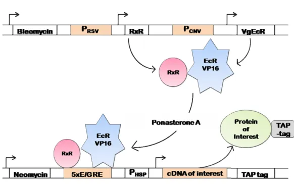
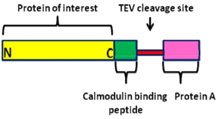
Tandem affinity purification (TAP) is a technique that allows for the purification of protein complexes under native conditions (Rigault et al., 1999). This approach requires the fusion of a TAP-tag to the protein of interest either at the C- or N-termini. The tag consists of two IgG-binding units of protein A of the Staphylococcus aureus, a cleavage site for tobacco etch virus (TEV) protease, and a calmodulin binding peptide (Drakas et al., 2005). Cell lines, carrying the tagged protein of interest, have to be created, from which macromolecular complexes can be isolated (Puig et al., 2001). The tagged protein must be expressed, in these cell lines, near physiological levels to minimize the rate of false positives (non-specific protein-protein interactions). This could be achieved by the use of an ecdysone-inducible expression system.
The protein purification occurs in two steps using two different affinity columns (the IgG- and calmodulin-binding columns) under conditions that leave