O
pen
A
rchive
T
OULOUSE
A
rchive
O
uverte (
OATAO
)
OATAO is an open access repository that collects the work of Toulouse researchers and
makes it freely available over the web where possible.
This is an author-deposited version published in :
http://oatao.univ-toulouse.fr/
Eprints ID : 18536
To link to this article : DOI:10.1007/s00264-015-2687-9
URL :
http://dx.doi.org/10.1007/s00264-015-2687-9
To cite this version : Pailhé, Régis and Cavaignac, Étienne and
Murgier, Jérôme and Cahuzac, Jean-Philippe and Sales de Gauzy,
Jérôme and Accadbled, Franck Triple osteotomy of the pelvis for
Legg-Calve-Perthes disease: a mean fifteen year follow-up. (2016)
International Orthopaedics, vol. 40 (n° 1). pp. 115-122. ISSN
0341-2695
Any correspondence concerning this service should be sent to the repository
administrator:
staff-oatao@listes-diff.inp-toulouse.fr
Triple
osteotomy of the pelvis for Legg-Calve-Perthes disease:
a
mean fifteen year follow-up
Régis Pailhé & Etienne Cavaignac & Jérôme Murgier &
Jean Philippe Cahuzac & Jerôme Sales de Gauzy &
Franck Accadbled
Abstract
Purpose This study presents the results of a prospective con-secutive cohort of patients with Legg-Calvé-Perthes disease (LCPD) operated with triple osteotomy of the pelvis (TOP) between 1989 and 2005. We attempted to determine whether the results of TOP remain stable with time and consequently lower the risk of subsequent osteoarthritis. The primary study aims were to determine the maintenance of head coverage and joint congruity, and functional outcomes of this surgery. Methods Forty-five patients with a mean follow-up of 15.2 years (range eight to 24) were included.
Results At latest up, two patients were lost to follow-up, and two required a surgical reoperation. Cumulative main-tenance of head coverage and joint congruity rate for all TOP was 84.6 % (95 % CI: 82.3–90.6 %) at 15 years. Factors significantly associated with poor long-term results were the age at diagnosis and Greene index.
Conclusion TOP in LCPD provides satisfactory and repro-ducible long-term clinical results.
Keywords Legg Calve Perthes . Osteotomy . Hip . Pediatrics
Introduction
The aim of the treatment of Legg-Calve-Perthes disease (LCPD) is to prevent hip osteoarthritis [1]. Many treatments
have been described in order to prevent or limit femoral head deformation by containing the head within the acetabulum, using it as a mould for guiding its development. Some of the various procedures described (varisation osteotomy of the fe-mur, Salter osteotomy) have sometimes resulted in a prolonged limp or inadequate containment [2]. Triple osteotomy of the pelvis (TOP) is now well documented [3–6]. Most of the available studies report very good results although with only short or medium-term follow-up.
We therefore addressed the following question:Do the re-sults of TOP remain stable with time and consequently lower the risk of subsequent osteoarthritis?
The primary aims of this study were to determine the long-term radiographic results and functional outcomes and their determining factors
Methods
This study presents the results of a prospectively and consec-utively accrued series of LCPD patients treated with TOP between 1989 and 2005. The indications for triple pelvic osteotomies included at least two of the following criteria: (1) six to ten years of age at clinical onset [7], (2) Catterall [8] groups III and IV, and Herring [9] lateral pillar groups B, B/C border, and C (Herring [9] type was considered after 1992), (3) loss of femoral head containment (subluxation) on the anteroposterior radiograph, (4) Catterall [8] signs of «head at risk». Exclusion criteria were: (1) joint stiffness, especially in abduction, (2) hinge abduction as systematically assessed by a hip arthrography under general anaesthesia.
All patients were operated upon by two surgeons (JSDG and JPC). The surgical technique has been previously described in literature [10, 11]. TOP combines a Salter innominate osteotomy and osteotomies of the iliopubic and ischiopubic rami. The surgery is performed under general anaesthesia on
R. Pailhé
:
E. Cavaignac:
J. Murgier:
J. P. Cahuzac:
J. S. de Gauzy
:
F. AccadbledService de Chirurgie Orthopédique et de Traumatologie, Hôpital des Enfants, Toulouse, France
R. Pailhé (*)
Service de Chirurgie Orthopédique, Hôpital Sud, Av Kimberley, 38130 Grenoble Cedex, France
a standard surgical table. The child is installed in the three-quarter supine position, and the position is maintained by a sheet rolled against the child’s back. The intervention begins with iliopubic and ischiopubic osteotomies via a genitofemoral approach. The innominate osteotomy follows, which is identi-cal to a Salter osteotomy. The incision is a bikini-type incision located 1.5-cm below the iliac crest. It extends from the middle part of the iliac crest to the middle of the groin crease. The intermuscular space between the sartorius and tensor fascia lata is approached next. The osteotomy line is perpendicular to the wing of the ilium. It starts at the ischiatic notch and ends just above the antero-inferior iliac spine. A bicortical graft is harvested on the anterior and superior part of the wing of the ilium. The upper fragment is maintained, while the lower fragment is mobilized downward, outward, and forward. Dis-placement should be assisted with the Salter manoeuver, consisting of placing the heel on the knee of the contralateral knee. The graft is placed at the anterior opening and then fixed using two K-wires (diameter 15—18 depending on the child’s age).
Baseline and perioperative information
Baseline information was collected from consenting patients and parents at the time of their pre-operative visit. Collected data included age, sex, age at diagnosis, delay of the diagno-sis, duration of symptoms, Herring Score, Catterall score and Green Index [12]. This is the percentage of the width of the femoral head that is lateral to Perkins’ line as determined from an AP radiograph of the pelvis. The amount of the ossific nucleus that is lateral to Perkins’ line is measured in millimetres along a line perpendicular to Perkins’ line. This distance is divided by the width of the opposite/normal fem-oral head as measured in millimetres along the epiphyseal plate. The quotient so obtained is multiplied by 100 to obtain the percentage of the femoral head that has been extruded from the acetabulum. Data were stored in a central database with the patients’ and parents’ consent.
Follow-up
Follow-up consisted of a review at baseline, three months, one year and two years postoperatively. Thereafter patients were followed yearly for ten years. This study reports latest follow-up outcomes.
Outcome measures
Patients were evaluated by an independent observer (RP). Complications and radiographic findings were recorded. The end point of the study was the latest follow-up or re-operation for any cause related to the hip. Clinical results were assessed by Oxford hip score (OHS) [13]. Plain anteroposterior pelvic
radiographs and a false lateral view according to Lequesne and de Sèze [14] were analysed. Lateral and anterior coverage were measured using the angles described by Wiberg [15] (lateral centre-edge angle) and Lequesne and de Sèze [14] (anterior centre-edge angle). The inclination of the acetabular roof was measured using the technique described by Tönnis [16] (acetabular index). The neck shaft angle (NSA) and the lateral extrusion index of Green [6,12] were also assessed. The deformity index at two years described by Nelson et al. [17] was assessed. The severity of osteoarthritis at the last follow-up was graded from 0 to 4 according to Kellgren and Lawrence classification [18]. Heterotopic ossification was graded from 0 to 4 according to Brooker et al. [19]. Femoral head deformity and hip congruity were assessed by Stulberg classification [20].
Statistical analysis
Statistical analysis was carried out using EXCEL® (Microsoft Inc., Redmond, Wash.) and SPSS software® (SPSS Inc., Chi-cago, Ill). Descriptive data analysis was performed using Stu-dent t test. The difference between the preoperative and follow-up data was analysed with paired Student t tests. A Cox-proportional hazards model was used to compare the differences in TOP survival distributions for each of the co-variates recorded (patient age, gender, radiological parame-ters). A multivariate model was constructed and then covari-ates that were not significantly influential were systematically removed from the model to identify those having the greatest influence on survival. Significance was determined to be p<0.05. Three Kaplan and Meier survivorship [21] analyses were performed using two different end points: an Oxford hip score less than 40 and Stulberg grade IVand Vand/or Kellgren and Lawrence grade greater than 2.
Results
Descriptive analysis and factors affecting survival
After a mean follow-up of 15.2 years (range, 8.0–24.0) 45 patients were included with two patients lost to follow-up. There were 32 males and 11 females. The mean age at time of diagnosis was 6.1 years (range, three to 12) and delay of diagnosis was 3.4 months (range, 1.0–12.0). The mean dura-tion of symptoms before surgical treatment was 6.6 months (range, 1.0–24.0). Mean age at surgery was 7.3 years (range, four to 12.4). The disease was bilateral in one case. Four patients (9.3 %) were rated Catterall 2, 25 (58.1 %) were Catterall 3 and 14 (32.6 %) were Catterall 4 (Table1). Three patients (6.9 %) were rated Herring A, 26 (60.5 %) Herring B and 14 (32.6 %) Herring C.
Two patients, after a mean seven years from the index procedure, underwent a revision (JPC, JSDG). The first one was a male patient with a 2.5-cm symptomatic leg length discrepancy. The patient was treated with contra-lateral distal femoral physiodesis. The second was a male patient with a symptomatic retroverted femoral neck and coxa valga of 150°. The patient initially underwent a femoral osteotomy, then four years later a total hip arthroplasty (THA).
A Cox-proportional hazard model demonstrated a sig-nificantly increased risk of re-operation with a high Green index (risk of revision 0.147 for each increased percent-age of Green index; p = 0.043). Male gender was associat-ed with a 0.048 increasassociat-ed risk of revision comparassociat-ed to female gender, although this was not statistically signifi-cant (p = 0.127). Age of onset was associated with a higher risk of re-operation (0.417), but was not statistically sig-nificant (p = 0.183).
Functional outcomes
The mean OHS at latest follow-up was 46.5 ± 4.5 (range, 24– 48) (Fig.1). There was no covariate influence on OHS except the age of onset which was associated with a 2.28 increased risk of OHS score <40 for each increased year of age (p= 0.035). Only two patients (excluding the patient who underwent a total hip replacement) presented bad functional results with OHS <40. Thus survival rate for all TOP was 95.3 % (95 % CI, 90.3–98.6 %) at 15 years (Fig.2) consider-ing OHS <40 as an end point.
Radiological analysis
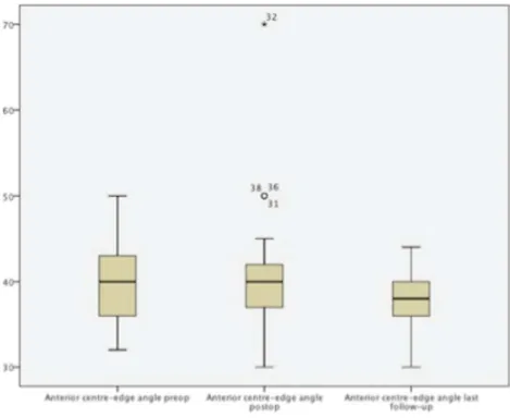
Coxometry parameters are presented in Table 2 at pre-operative stage, postpre-operative stage, end of growth and at final follow-up (Figs.3,4,5and6). There were neither statistical differences between postoperative and end of growth stage nor between end of growth stage and final follow-up. Only two patients presented osteoarthritis: one patient who required a THA was Kellgren and Lawrence 4 and the second was asymptomatic and rated Kellgren and Lawrence 3. Six pa-tients (14 %) were Stulberg I, 19 (44 %) Stulberg II, 11 (25.5 %) Stulberg III, six (14 %) Stulberg IV and one (2.5 %) Stulberg V. Oxford hip score at latest follow-up was correlated with Stulberg classification with r=0.446 and p= 0.04. Deformity index >0.3 was correlated with Stulberg grade III/IV with r=0.786 and p=0.01.There was no correla-tion between the preoperative Catterall score and the Stulberg score at the latest follow-up. Good radiological outcomes rate for all TOP was 84.6 % (95 % CI 82.3–90.6 %) at 15 years
Table 1 Descriptive analysis of potential risk factors
Characteristic Minimum Maximum Mean Standard deviation Age at diagnosis (years) 2.5 12.0 6.14 2.12 Catterall 2.0 4.0 3.05 0.79 Green index 0 50.0 12.97 10.69 Deformity index 0.05 0.66 0.21 0.09 Duration of symptoms (months) 1.0 12.0 3.42 3.45 Delay of diagnosis (months) 1.0 24.0 6.65 4.38
Fig. 1 Distribution of Oxford hip score
(Fig.1) considering Stulberg IV and V and/or Kellgren Law-rence grade superior to 2, as an end point.
Discussion
Few studies to date have reported on the outcomes of triple osteotomy of the pelvis in LCPD with long-term follow-up. To the best of our knowledge, the present series represents the longest follow-up cohort described in literature. This study demonstrates very good long-term survival after TOP with a rate of good radiological outcomes (Stulberg 1 to 3 and/or Kellgren Lawrence<2) of 84.6 % (95 % CI 82.3–90.6 %) at 15 years.
The present study has some limitations. First, two patients (4.4 %) were lost to follow-up. Second, the Cox proportional hazard modelling did not take into account the learning curve of the surgeon, which may have skewed failure rates. Overall, the power of the study is limited by the small number of patients (43 patients) and may not allow a significant risk factors analysis. Finally, this study because of the limited number of patients presents a risk of type 2 error, and many prognostic factors considered important in other Perthes’ stud-ies [7] may have been overlooked.
In the present series, absence of hinge abduction was sys-tematically assessed by hip arthrography under general
anaesthesia before any procedure since it was considered as a contra-indication to TOP. Indeed, established hinge abduc-tion due to enlarged, crushed femoral head may lead to an irreducible hip, and further attempts to contain the femoral head may be more harmful than helpful [22–25]. Furthermore, in this study all the procedures were performed at fragmenta-tion stage. Herring et al. [26] demonstrated that femoral head was all the more likely to undergo progressive flattening in older patients, with severe lateral pillar involvement, and in those with delayed reossification. Joseph et al. [27] concluded that containment surgery aiming at preventing femoral head deformation in LCPD should be performed before the ad-vanced stage of fragmentation.
Thus, time to surgery is a prognostic factor of an utmost importance for TOP outcomes. Considering risk factors asso-ciated with functional outcomes, only Green Index and age of the patient at time of diagnosis seemed to be significant in our study. Green et al. [12] initially developed the femoral epiph-yseal extrusion index as a prognostic indicator in LCPD. Au-thors noted 20 % and less protrusion as a good prognostic indicator in LCPD cases. Tannast et al. [28] alluded to an extrusion index of less than 25 % as normal, although they emphasized no study has defined a minimum extrusion. Hosalkar et al. [6] also observed that extrusion index of less than 20 % was a good prognostic indicator. As Nelson et al. [17], we found that deformity index>0.3 at two years postop-erative was also a good prognosis factor of bad radiological
Fig. 2 Kaplan-Meier survivorship considering different endpoints
Table 2 Descriptive analysis of radiographic parameters
Measure Preoperative Postoperative End of growth Last follow up
Mean Standard deviation Mean Standard deviation Mean Standard deviation Mean Standard deviation Lateral centre-edge angle 41.0 9.05 46.9 12.05 45.0 9.17 37.5 6.66
Acetabular index 10.6 4.42 8.9 4.59 9.4 4.38 13.6 4.55 Anterior centre-edge angle 39.1 5.54 40.5 7.15 36.5 3.17 37.5 3.15 NSA 135.6 4.44 137.6 5.13 137.7 5.10 136.9 5.43
outcome. More recently, Kwang-Won Park et al. pointed out that premature physeal closure of the proximal femoral physis in patients with LCPD occurred 3.5 years earlier than normal hips and could be an adjunct diagnostic tool in the prognosti-cation of LCPD outcomes [29].
Although age of onset was not significant here, Wenger et al. reported in a series of 39 patients, that 17 % of lateral pillar C patients below the age of eight had a poor outcome compared with 50 % above the age of eight [30].
Our study is consistent with the good results of series with short to medium-term follow-up using the same surgical tech-nique. Wenger et al. reported 89 % good results after TOP in a series of 21 patients [30]. Conroy et al. performed TOP in eight patients with a minimum follow-up of four years [4]. The modified Harris hip score (HHS) ranged from 38 to 60 pre-operatively and from 77 to 92 postoperatively. O’Hara et al. reviewed 21 patients after TOP with a 30-month fol-low-up (one to five years) and found very good results [31].
Fig. 3 Box-plots of pre-operative, postoperative and last follow-up acetabular index
Fig. 4 Box-plots of pre-operative, postoperative and last follow-up lateral centre-edge angle
The average gain in HHS was 34.3 (range four to 55). Vukasinovic et al. also reported very good results in a series of 30 patients with mid-term follow-up [32]. More recently, Hosalkar et al. demonstrated the durability of femoral head containment with TOP in a series of 20 patients with a mini-mum follow-up of three years [6].
Nonoperative treatment remains an alternative for Catterall 1 et 2. Recently, Larson et al. [33] reported on the 20-year follow-up results of a prospective multcentric study of nonoperative treatment and found a high preva-lence of osteoarthritis and low clinical outcome scores. Approximately 20 % of Stulberg type II hips and 60 % of type III, IV, and V hips showed radiographic evidence of osteoarthritis. As in our series, a high Stulberg grade was associated with pain, impingement on physical exam-ination and arthritis. Similarly, McAndrew and Weinstein displayed a 50-year follow-up of the Gower and Johnston historical series [34]. By that time, 15 (39 %) of the 35 patients had required THA, and six (21 %) had an IHS (Iowa hip score) <80.
Other authors have recommended varus femoral osteotomy. Terjesen et al. reported in a prospective study on 70 patients that for children aged six to ten years, in whom the whole femoral head was affected, femoral head sphericity five years after femoral osteotomy was better than for those having undergone physiotherapy [35]. How-ever, Mirovsky et al. [36] reported a 58 % incidence of leg length discrepancy ( LLD). Grzegorzewski et al. [37] stat-ed that varus osteotomy produces more shortening than other methods of treatment. Van Der Geest et al. advocated shelf acetabuloplasty for severe cases of LCPD, based up-on a retrospective series of 30 patients with 12 years follow-up [38]. Bernese’s (Ganz) periacetabular osteotomy with lateral muscle sparing has more recently emerged and gained popularity around the globe [2, 39]. However, to date, there is no high level evidence study to assess out-comes. Salter osteotomy in LCPD is an effective treatment which may alter the natural course of the disease. Similar to TOP, its main advantage is its effect on femoral head remodelling with remaining growth [7,40]. Nonetheless,
Fig. 5 Box-plots of pre-operative, postoperative and last follow-up anterior centre-edge angle
this osteotomy has some limitations in patients above six years old given that appropriate tilt may not be achieved due to the relative stiffness of the pubic symphysis.
In addition to the deformities caused by the disease process, containment procedures may cause femoro acetabular im-pingement (FAI). For instance, Salter osteotomy, shelf proce-dure, or TOP may create pincer-type impingement [41,42] which is a pre-arthritic condition [28,43]. Therefore risk-benefit ratio must be taken into account before considering any surgical containment. Furthermore, it is unclear whether and to what extent the acetabular coverage changes during growth and after TOP. Hosalkar et al. [6] recommended an acetabular coverage after TOP for LCPD (in Catterall Stage III or IV) of 44° or less and an ARA below –6°, as they noted residual remodeling could allow a return to a nonpincer mor-phology with growth. Therefore, in growing patients present-ing asymptomatic pincer morphology, it seems reasonable to carry on with follow-up until completion of skeletal maturity [6,44]. According to Hosalkar et al. [6], aggressive procedure aiming at deimpingement may not be necessary unless the patient has severe symptoms. In our study, one patient pre-sented clinical signs of impingement with a cross-over sign indicating a retroverted acetabulum, possibly related to the pelvic osteotomy.
Although TOP may delay secondary osteoarthritis, the resulting altered anatomy of the pelvis and proximal femur might lead to a technically challenging THA [45]. Controver-sy remains on this point as some investigators have shown a possible worsening of the outcome of THA after a TOP, whereas others found it did not compromise the results of THA if TOP is properly performed [39,46,47]. In a recent study, Tokunaga et al. [48] demonstrated that prior pelvic osteotomy lead neither to a higher peri-operative complication rate, nor a higher revision rate, nor compromised HHS nor shortened survivorship after THA.
TOP resulted in maintenance of head shape in lateral pillar B or C patients with a cumulative survival rate of 95.3 % (95 % CI 90.3–98.6 %) at 15 years in this series This method provides effective containment, which allows prolonged re-modeling while avoiding the limitations of femoral varus osteotomy (limp, short limb) and Salter osteotomy (incom-plete containment).
Conflict of interest Each author certifies that he has no commercial associations (e.g., consultancies, stock ownership, equity interest, patent/licensing arrangements, etc.) that might pose a conflict of interest in connection with the submitted article.
Ethical review committee statement Each author certifies that his institution approved the human protocol for this investigation, that all investigations were conducted in conformity with ethical principles of research, and that informed consent for participation in the study was obtained.
References
1. Herring JA (2011) Legg-Calvé-Perthes disease at 100: a review of evidence-based treatment. J Pediatr Orthop 31:S137–S140. doi:10. 1097/BPO.0b013e318223b52d
2. Santore RF, Turgeon TR, Phillips WF, Kantor SR (2006) Pelvic and femoral osteotomy in the treatment of hip disease in the young adult. Instr Course Lect 55:131–144
3. Poul J, Vejrostová M (2001) Triple osteotomy of the pelvis in chil-dren and adolescents. Acta Chir Orthop Traumatol Cech 68:93–98 4. Conroy E, Sheehan E, O’Connor P et al (2010) Triple pelvic
osteotomy in Legg-Calve-Perthes disease using a single anterolateral incision: a 4-year review. J Pediatr Orthop B 19:323–326. doi:10. 1097/BPB.0b013e32833822a4
5. O’Connor PA, Mulhall KJ, Kearns SR et al (2003) Triple pelvic osteotomy in Legg-Calvé-Perthes disease using a single anterolateral incision. J Pediatr Orthop B 12:387–389. doi:10.1097/01.bpb. 0000049565.52224.68
6. Hosalkar H, Munhoz da Cunha AL, Baldwin K et al (2012) Triple innominate osteotomy for Legg-Calvé-Perthes disease in children: does the lateral coverage change with time? Clin Orthop Relat Res 470:2402–2410. doi:10.1007/s11999-011-2189-z
7. Saran N, Varghese R, Mulpuri K (2012) Do femoral or salter innom-inate osteotomies improve femoral head sphericity in Legg-Calvé-Perthes disease? A meta-analysis. Clin Orthop Relat Res 470:2383– 2393. doi:10.1007/s11999-012-2326-3
8. Catterall A, Pringle J, Byers PD et al (1982) A review of the mor-phology of Perthes’ disease. J Bone Joint Surg (Br) 64:269–275 9. Herring JA, Neustadt JB, Williams JJ et al (1992) The lateral pillar
classification of Legg-Calvé-Perthes disease. J Pediatr Orthop 12: 143–150
10. Sales de Gauzy J (2010) Pelvic reorientation osteotomies and acetabuloplasties in children. Surgical technique. Orthop Traumatol Surg Res 96:793–799. doi:10.1016/j.otsr.2010.07.004
11. Le Coeur P (1965) Correction des défauts d’orientation de l’articulation coxofémorale par ostéotomie de l’isthme iliaque. Orthop Rev Chir 51:211–212
12. Green NE, Beauchamp RD, Griffin PP (1981) Epiphyseal extrusion as a prognostic index in Legg-Calvé-Perthes disease. J Bone Joint Surg Am 63:900–905
13. Dawson J, Fitzpatrick R, Carr A, Murray D (1996) Questionnaire on the perceptions of patients about total hip replacement. J Bone Joint Surg (Br) 78:185–190
14. Lequesne M, de Seze (1961) False profile of the pelvis. A new ra-diographic incidence for the study of the hip. Its use in dysplasias and different coxopathies. Rev Rhum Mal Osteoartic 28:643–652 15. Wiberg G (1939) Relation between congenital subluxation of the hip
and arthritis deformans. Acta Orthop Scand 10:351–371. doi:10. 3109/17453673909149515
16. Tönnis D, Arning A, Bloch M et al (1994) Triple pelvic osteotomy. J Pediatr Orthop B 3:54
17. Nelson D, Zenios M, Ward K et al (2007) The deformity index as a predictor of final radiological outcome in Perthes’ disease. J Bone Joint Surg (Br) 89:1369–1374. doi:10.1302/0301-620X.89B10. 18747
18. Kellgren JH, Lawrence JS (1957) Radiological assessment of osteo-arthrosis. Ann Rheum Dis 16:494–502
19. Brooker AF, Bowerman JW, Robinson RA, Riley LH (1973) Ectopic ossification following total hip replacement. Incidence and a method of classification. J Bone Joint Surg Am 55:1629–1632
20. Stulberg SD, Cooperman DR, Wallensten R (1981) The natural his-tory of Legg-Calvé-Perthes disease. J Bone Joint Surg Am 63:1095– 1108
21. Kaplan EL, Meier P (1958) Nonparametric estimation from incom-plete observations. J Am Stat Assoc 53:457–481
22. Reinker KA (1996) Early diagnosis and treatment of hinge abduction in Legg-Perthes disease. J Pediatr Orthop 16:3–9
23. Joseph B, Mulpuri K, Varghese G (2001) Perthes’ disease in the adolescent. J Bone Joint Surg (Br) 83:715–720
24. Bankes MJ, Catterall A, Hashemi-Nejad A (2000) Valgus extension osteotomy for ‘hinge abduction’ in Perthes’ disease. Results at ma-turity and factors influencing the radiological outcome. J Bone Joint Surg (Br) 82:548–554
25. Yoo WJ, Choi IH, Moon HJ et al (2013) Valgus femoral osteotomy for noncontainable Perthes hips: prognostic factors of remodeling. J Pediatr Orthop 33:650–655. doi:10.1097/BPO.0b013e31829569c8
26. Herring JA, Williams JJ, Neustadt JN, Early JS (1993) Evolution of femoral head deformity during the healing phase of Legg-Calvé-Perthes disease. J Pediatr Orthop 13:41–45
27. Joseph B, Nair NS, Narasimha Rao KL et al (2003) Optimal timing for containment surgery for Perthes disease. J Pediatr Orthop 23:601–606 28. Tannast M, Siebenrock KA, Anderson SE (2007) Femoroacetabular impingement: radiographic diagnosis–what the radiologist should know. AJR Am J Roentgenol 188:1540–1552. doi:10.2214/AJR.06. 0921
29. Park K-W, Jang K-S, Song H-R (2013) Can residual leg shortening be predicted in patients with Legg-Calvé-Perthes’ disease? Clin Orthop Relat Res 471:2570–2577. doi:10.1007/s11999-013-3009-4
30. Wenger DR, Pring ME, Hosalkar HS et al (2010) Advanced contain-ment methods for Legg-Calvé-Perthes disease: results of triple pelvic osteotomy. J Pediatr Orthop 30:749–757. doi:10.1097/BPO. 0b013e3181f5a0de
31. Kumar D, Bache CE, O’Hara JN (2002) Interlocking triple pelvic osteotomy in severe Legg-Calvé-Perthes disease. J Pediatr Orthop 22:464–470
32. Vukasinovic Z, Spasovski D, Vucetic C et al (2009) Triple pelvic osteotomy in the treatment of Legg-Calve-Perthes disease. Int Orthop 33:1377–1383. doi:10.1007/s00264-009-0745-x
33. Larson AN, Sucato DJ, Herring JA et al (2012) A prospective mul-ticenter study of Legg-Calvé-Perthes disease: functional and radio-graphic outcomes of nonoperative treatment at a mean follow-up of twenty years. J Bone Joint Surg Am 94:584–592. doi:10.2106/JBJS. J.01073
34. McAndrew MP, Weinstein SL (1984) A long-term follow-up of Legg-Calvé-Perthes disease. J Bone Joint Surg Am 66:860–869 35. Terjesen T, Wiig O, Svenningsen S (2012) Varus femoral osteotomy
improves sphericity of the femoral head in older children with severe
form of Legg-Calvé-Perthes disease. Clin Orthop Relat Res 470: 2394–2401. doi:10.1007/s11999-011-2181-7
36. Mirovsky Y, Axer A, Hendel D (1984) Residual shortening after osteotomy for Perthes’ disease. A comparative study. J Bone Joint Surg (Br) 66:184–188
37. Grzegorzewski A, Synder M, Kozłowski P et al (2005) Leg length discrepancy in Legg-Calve-Perthes disease. J Pediatr Orthop 25:206– 209
38. Van Der Geest IC, Kooijman MA, Spruit M et al (2001) Shelf acetabuloplasty for Perthes’ disease: 12-year follow-up. Acta Orthop Belg 67:126–131
39. Parvizi J, Burmeister H, Ganz R (2004) Previous Bernese periacetabular osteotomy does not compromise the results of total hip arthroplasty. Clin Orthop Relat Res 423:118–122
40. Thompson GH (2011) Salter osteotomy in Legg-Calvé-Perthes dis-ease. J Pediatr Orthop 31:S192–S197. doi:10.1097/BPO. 0b013e318223b59d
41. Novais EN, Clohisy J, Siebenrock K et al (2011) Treatment of the symptomatic healed Perthes hip. Orthop Clin North Am 42:401–417. doi:10.1016/j.ocl.2011.05.003, viii
42. Dora C, Mascard E, Mladenov K, Seringe R (2002) Retroversion of the acetabular dome after Salter and triple pelvic osteotomy for con-genital dislocation of the hip. J Pediatr Orthop B 11:34–40 43. Ganz R, Parvizi J, Beck M et al (2003) Femoroacetabular
impinge-ment: a cause for osteoarthritis of the hip. Clin Orthop Relat Res 417: 112–120. doi:10.1097/01.blo.0000096804.78689.c2
44. Tannast M, Macintyre N, Steppacher SD et al (2013) A systematic approach to analyse the sequelae of LCPD. Hip Int 23(Suppl 9):S61– S70. doi:10.5301/hipint.5000071
45. Scher MAM, Jakim II (1991) Combined intertrochanteric and Chiari pelvic osteotomies for hip dysplasia. J Bone Joint Surg (Br) 73:626– 631
46. Hashemi-Nejad AA, Haddad FSF, Tong KMK et al (2002) Does Chiari osteotomy compromise subsequent total hip arthroplasty? J Arthroplasty 17:731–739
47. Peters CLC, Beck MM, Dunn HKH (2001) Total hip arthroplasty in young adults after failed triple innominate osteotomy. J Arthroplasty 16:8–8. doi:10.1054/arth.2001.20903
48. Tokunaga K, Aslam N, Zdero R et al (2011) Effect of prior Salter or Chiari osteotomy on THA with developmental hip dysplasia. Clin Orthop Relat Res 469:237–243. doi:10.1007/s11999-010-1375-8