OATAO is an open access repository that collects the work of Toulouse
researchers and makes it freely available over the web where possible
Any correspondence concerning this service should be sent
to the repository administrator:
[email protected]
This is an author’s version published in:
http://oatao.univ-toulouse.fr/27602
To cite this version:
Evariste, Lauris
and Flahaut, Emmanuel
and Baratange, Clément
and
Barret, Maialen
and Mouchet, Florence
and Pinelli, Eric
and Galibert,
Anne-Marie
and Soula, Brigitte
and Gauthier, Laury
Ecotoxicological
assessment of commercial boron nitride nanotubes toward Xenopus laevis
tadpoles and host-associated gut microbiota. (2021) Nanotoxicology, 15 (1).
35-51. ISSN 1743-5390
https://doi.org/10.1080/17435390.2020.1839137
Ecotoxicological assessment of commercial boron nitride nanotubes toward
Xenopus /aevis
tadpoles and host-associated gut microbiota
Lauris Evaristea G), Emmanuel Flahautb G), Clément Baratangea, Maialen Barreta, Florence Moucheta,
Eric Pinellia, Anne Marie Galibertb, Brigitte Soulab and Laury Gauthie�
•Laboratoire Ecologie Fonctionnelle et Environnement, Université de Toulouse, CNRS, INPT, UPS, Toulouse, France; bCIRIMAT, Université de Toulouse, CNRS, INPT, UPS, UMR CNRS-UPS-INP N°5085, Université Toulouse 3 Paul Sabatier, Bât. CIRIMAT, Toulouse, France
ABSTRACT
Despite the growing interest for boron nitride nanotubes (BNNT) due to their unique properties, data on the evaluation of the environmental risk potential of this emerging engineered nanoma terial are currently lacking. Therefore, the ecotoxicity of a commercial form of BNNT (containing tubes, hexagonal-baron nitride, and boron) was assessed in vivo toward larvae of the amphibian Xenopus laevis. Following the exposure, multiple endpoints were measured in the tadpoles as well as in bacterial communities associated to the host gut. Exposure to BNNT led to boron accumulation in host tissues and was not associated to genotoxic effects. However, the growth of the tadpoles increased due to BNNT exposure. This parameter was associated to remodeling of gut microbiome, benefiting to taxa from the phylum Bacteroidetes. Changes in relative abun dance of this phylum were positively correlated to larval growth. The obtained results support the finding that BNNT are biocompatible as indicated by the absence of toxic effect from the tested nanomaterials. ln addition, byproducts, especially free boron present in the tested prod uct, were overall beneficial for the metabolism of the tadpoles.
KEYWORDS
Boron n itrid e nanotubes; Xenopus; genotoxicity; gut microbiota; 16S
1. Introduction
Boron nitride nanotubes (BNNTI are nanoparticles structurally analogous to carbon nanotubes (CNTs) in which baron and nitrogen replace carbon atoms (Chopra et al. 1995). lncreasing attention is being paid to these BNNT due to their unique properties such as high thermo-mechanical stability (Suryavanshi et al. 2004; Chen et al. 2004), electrical insulation (Radosavljevié et al. 2003) and thermal conductivity (Chang et al. 2006). As high quality materials are increasingly available due to the improvement of synthesis techniques, BNNT are triggering great interest for the development of applications in a wide range of area such as com posite improvement, medicine, hydrogen storage as well as aquatic pollution remediation (Kim et al. 2018; Kalay et al. 2015; Zhi et al. 2008; Merlo et al. 2018; Laie, Bernard, and Demirci 2018; Yu et al. 2018). Such keen interest for this material predicts a large-scale production in the near future. As any
engineered nanomaterial, BNNT could be released into the environment during the whole material life cycle, from the production, the use and the waste disposai or recycling to reach significant levels in aquatic ecosystems (Yu et al. 2018; Mottier et al. 2017). For these reasons, this implies to evaluate its biocompatibility and its potential hazard for the environment before considering the mass use of this nanomaterial.
Studies related to BNNT toxicity remain scarce and inconsistent. lndeed, although many of the published works indicate biocompatibility of raw and functionalized BNNT in vitro as well as in vivo (Chen et al. 2009; �en, Emanet, and Çulha 2016; Ciofani et al. 2010; Ciofani et al. 2008; Ciofani et al. 2014; Fernandez-Yague et al. 2015; Rocca et al. 2016; Salvetti et al. 2015), some studies highlighted cell type dependent cytotoxic and genotoxic effects (Augustine et al. 2019; Horvath et al. 2011; Çal and Bucurgat 2019). Ecotoxicological data are lacking CONTACT Lauris Evariste
e
[email protected]Q
Laboratoire Ecologie Fonctionnelle et Environnement, Université de Toulouse, CNRS , INPT, UPS, Toulouse, Francewhile it is necessary for the sustainable develop ment of BNNT-based products. lndeed, as for other nanoparticles, a large scale usage of BNNT would lead to releases into the environment at different stages of their life-cycle (Mottier et al. 2017; Sun et al. 2016; Keller and Lazareva 2014; Bundschuh et al. 2018). Thus, there is a need to fill knowledge gaps concerning the ecotoxicity of BNNT, particu larly toward aquatic ecosystems as they constitute a receptacle for most contaminants. For this purpose, the amphibian Xenopus /aevis was chosen as bio logical mode! to assess the ecotoxic potential of BNNT as larval stages were shown to be sensitive to contaminants, including nanoparticles (Evariste, Barret, et al. 2019; Mouchet et al. 201 O; Bourdiol et al. 2013; Muzi et al. 2016; Saria et al. 2014; Colombo et al. 2017; Marfn-Barba et al. 2018). Parameters related to the larval growth were shown to constitute endpoints of interests as they reflect the overall health status of the organisms. ln a con text of exposure to nanoparticles of carbon allo tropes such as CNTs, toxicity leading to larval
growth inhibition was described by the specific sur face area of the tested nanoparticles (Mottier et al. 2016; Lagier et al. 2017). ln addition, genotoxic effects could lead to non-negligible consequences as damaged DNA may cause cellular dysfunctions (Jackson and Bartek 2009) leading to the death of organisms and further impact the ability of popula tions to maintain (Sukumaran and Grant 2013). For this reasons, biomarkers related to genotoxicity are considered as a pertinent ecological endpoint at the population, community and ecosystem level (Anderson et al. 1994). Thus, alterations of genetic materials in amphibians would contribute to the decline of populations while this class contains many endangered species (Oertli et al. 2005).
lt is now widely accepted that the growth of organisms is dependent from its capacity to acquire energy from trophic resources while this energy metabolism is can be strongly influenced by the metabolic capacities of the gut microbiota (Mithieux 2018; Mussa, Gambino, and Cassader 2011; Cani 2014). ln addition, the gut microbiome is emerging as a central target in environmental toxi cology studies due to its contribution in the regula tion of multiple physiological processes of the hast but also in the metabolization of environmental pollutants (Claus, Guillou, and Ellero-Simatos 2016;
Evariste, Barret, et al. 2019; Adamovsky et al. 2018). Alterations of the gut microbiome composition may dysregulate the normal physiological functioning of the hast and lead to various diseases (Durack and Lynch 2019). Furthermore, in the amphibians, changes in the gut microbiota composition during earty stages of life were shown to influence later life resistance to infections (Knutie et al. 2017). Environmental pollution constitutes a major factor influencing the composition of the gut flora and previous studies reported that a wide range of con taminants were able to induce gut dysbiosis in association to deleterious issues on hast (Evariste, Barret, et al. 2019; Jin et al. 2017). Thus, the present work aims to investigate the consequences of an in vivo exposure of X. /aevis tadpoles to BNNT com bining monitoring of hast physiology and gut bac terial community composition. The former endpoint constitutes an integrator of the overall host-metab olism while the latter one would allow to better understand the causes of potential metabolic changes induced by BNNT exposure. Overall, the results obtained in this study suggest that BNNT are rather biocompatible. lndeed, exposure to the nanoparticles did not induced irreversible genotoxic effects and stimulated the growth of the tadpoles. This increased growth was associated to remodeling of the host-associated gut microbiota benefiting to the phylum Bacteroidetes. Measurement of baron released in the exposure media and accumulated in the organisms suggest that the free baron associ ated to the nanoparticles are responsible for the beneficial effects observed.
2. Materials and methods
2. 1. Synthesis and characterization of boron nitride nanotubes
Boron nitride nanotubes were obtained from BNNT LLC (USA). They correspond to the 'BNNT Pl-Beta Products' range of materials as classified on the website of the company (http://www.bnnt.com/ products). This material is described as containing hexagonal baron nitride (h-BN) and baron as well as being catalyst free (elemental B and N > 99.90/2) with ca. 25% of Boron in addition to BNNT. According to the manufacturer, the specific surface area is >200 m2/g. Sample was used as-received,
after simple grinding (agate mortar and pestle) in order to make it suitable for the experiments and characterization. The sample was characterized by X-ray diffraction (XRD), Brunauer-Emmet-Teller (BET), TGA, TEM, Raman and chemical elemental analysis.
X-ray diffraction analysis was performed using a Bruker D4 ENDEAVOR X-ray Diffractometer (Cu.Ket
=
1.5406 Â). Specific surface area was meas ured with a Micrometrics Flow Sorb Il 2300 by N2 adsorption using the BET theory. Dry BNNT powder sample was first degassed at 100 °C under N2 atmosphere for 2 h before being cooled down to 77 K (liquid nitrogen temperature) for adsorption of a monolayer of N2. After heating the sample back to room temperature, the desorption peak was recorded. A calibration was done by injecting a known quantity of N2 (in the same conditions as for the sample measurement). The uncertainty of the measurement was 3%. Thermogravimetric analysis was performed in air atmosphere at a heating rate of 1 °C/min using a SETARAM TAG 16. Transmission Electron Microscopy observations were performed using a TEM JEOL 1400 with an acceleration voltage of 120 kV after deposition of a few drops of a dilute suspension in ethanol on Copper TEM grids (Lacey carbon). Outer diameter of the BNNTs was meas ured from HRTEM images using lmageJ software. For chemical elemental analysis of Boron, the sam ple was digested in an open system with a mixture of sulfuric and nitric acids du ring 8 h. Elemental analysis was performed on an ICP-AES ICAP 6500 from Thermofisher Scientific, Bremen. Both the digestion and analysis were performed by CREALINS, 6Napse group (uncertainty of the meas urement: 3%). Nitrogen and Oxygen elemental anal yses were performed by ISA (CNRS, Lyon, France) by organic micro-analysis after total combustion at 1050 °C under helium/oxygen flux for nitrogen; total pyrolysis at 1080°C under nitrogen flux for Oxygen. Raman signature was analyzed to get information on the structural quality of the nanotubes (Labram HR800 Horiba Yvon Jobin, À= 532 nm). X-Ray photoelectron spectroscopy (XPS) was used to determine the quantitative atomic composition of the BNNT (XPS Kalpha ThermoScientific). IR analysis was performed using a Perkin Elmer Spectrum One FT-IR spectrometer and inclusion of the sample in a dry KBr pellet.The stability of the BNNT suspension was eval uated using a Turbiscan LAB (Formulation) equip ment, at room temperature. Suspensions were prepared at 10 mg L 1 in exposure media before
performing analysis. The suspension was placed in a glass vial and both transmission and reflection were monitored vs time for 24 h (1 scan per minute).
2.2. Xenopus laevis rearing, breeding and exposure conditions
Xenopus laevis originated from our certified breed ing facilities (under the approval number A31113002) and the experimental procedure was approved by an ethic committee (CEEA-073). Rearing and breeding conditions of Xenopus were performed as previously described (Mouchet et al. 2008; Mouchet et al. 2010). Pregnant mare's gonadotropin was injected in sexually mature indi viduals to induce spawning. Fecundated eggs obtained were bred in active charcoal filtered tap water at 22 ± 2 °C and fed with ground aquarium fish food (TetraPhyll®) until they reach stage 50 according to Nieuwkoop & Faber development table (Nieuwkoop and Faber 1958). Following the international standard ISO 21427-1 procedure (ISO/ FDIS 21427-1.1, 2006), 20 larvae per experimental condition were exposed for 12 days under semi static conditions with daily exposure media renewal. Larvae were fed daily ad libitum with ground aquar ium fish food (TetraPhylr®, Tetra, Melle, Germany). Food was added twice a day, directly after media renewal and at the end of the day. Unconsumed food was removed daily during the media renewal process. Negative control (NC) condition was com posed of reconstituted water devoid of contaminant (294 mg L 1 CaCl
2.2H2O; 123.25 mg L 1 MgSO4.7H2O; 64.75 mg L 1 NaHCO
3; 5.75 mg L 1 KCI) (Evariste, Barret, et al. 2019; Mouchet et al. 2008) while posi tive control (PC) for genotoxicity assessment con tained cyclophosphamide monohydrate ([6055-19- 2), Sigma, France) at 40 mg L 1
• BNNT were dis persed by sonication before contamination of the exposure medium. Tested concentrations of BNNT were 0, 0.1, 1, and 10 mg L 1
2.3. Analysis of boron clearance in the water and accumulation in tadpoles
Boron concentration was measured in reconstituted 1
water from the contrai and the 10 mg L of BNNT conditions after 24 h of incubation in the presence and absence of tadpoles in the media. Water sam ples were centrifuged twice at 1500 x g during 20 min and filtered at 0.22 µm to remove nanotubes from the samples before baron analysis. After 12 days of exposure, baron concentration was measured in larvae from the contrai and the 10 mg L 1 of BNNT groups. Guts of tadpoles were removed before lyophilization of organisms, in order to avoid measurement of BNNT-related baron from the gut content. Boron concentration in the water and in the larvae was determined by ICP-AES at CREALINS laboratory (Lyon, France).
2.4. Micronucleus test and ce// cycle analysis The micronucleus assay was performed in accord ance with the ISO 21427-1 guidelines (ISO/FOIS 21427-1.1, 2006). The micronucleus formation con stitutes a good indicator of irreversible genotoxic effects, integrating aneugenic and clastogenic effects occurring after exposure to a genotoxic compound. For this purpose, after 12 days of expos ure, Xenopus larvae were anesthetized by bathing in MS222 solution at 100 mg L 1 before collecting blood samples from cardiac puncture. Blood smears were prepared, fixed in methanol for 10 minutes before performing hematoxylin and eosin staining.
Micronucleated erythrocytes were accounted over a total of 1000 cells (MNE %0) using optical microscopy (oil immersion lens, xl 500). For cell cycle analysis, blood sub-samples were fixed using cold ethanol (70% v/v). Prior to the flow cytometric analysis, cells were rinsed using PBS and labeled with FxCycle™ PI/RNase Staining Solution (Life Technologies SAS) following manufacturer's recom mendations. Propidium iodide fluorescence was measured using Cytoflex (Beckman Coulter, U SA) equipped with a 488-nm excitation laser. For each sample, 10,000 events were acquired in a region corresponding to cells of interest after doublet dis crimination using FSC-H versus FSC-A.
2.5. Larval growth measurement
The total length of larvae was measured at the beginning (d0) and at the end of the 12 days of exposure (d12) using lmageJ 1.49 software. Normalized growth rate was determined as previ ously described (Mottier et al. 2016; Lagier et al.
2017) using the following formula:
. . (Ldl 2-MLd0
)
Normal,zed SIZe (%)
=
MLd0 x 100 X(M:i
1
2)
Ld 12 corresponds to the le n gth of one la rva e at 12 days, MLd0 is the mean length at day 0 of larvae from the exposure condition and MLCdl 2 is the mean length of larvae from the negative contrai at day 12.
2.6. Analysis of sequences from gut microbiota survey
Genomic DNA was extracted from the whole intes tine of larvae using DNeasy PowerSoil kit (QIAGEN) according to manufacturer recommendations with the following adjustments: samples were incubated 10 min at 65 °C after adding the solution Cl and elution buffer C6 was incubated during 10 min before performing the last centrifugation. The V4-V5 region of the 16S rRNA gene was amplified using 515 F (5'-GTGYCAGCMGCCGCGGTA-3')/928R (5'-CCCCGYCAATTCMTTTRAGT-3') primer pair (Wang and Qian 2009) and the following PCR protocol: 94 °C for 120 s, 30 cycles of 94 °C for 60 s, 65 °C for 40s, 72°C 30s and 72°C for l0min. Amplicon sequencing was performed using an Illumina MiSeq (2 x 250 pb) by the Get PlaGe platform (Genotoul, Toulouse, France).
Demultiplexed data were processed using FROGS (Find Rapidly OTU with Galaxy Solution) pipeline on Galaxy (Escudié et al. 2018). Contigs with a length between 380 and 500 pb were kept, clustered with Swarm (Mahé et al. 2014) with an aggregation dis tance of 3 and chimeras were removed. Filters were applied to remove singletons and keep for analysis OTUs with a minimum abundance of 0.005% of the sequences (Bokulich et al. 2013). The taxonomy affiliation was performed using Blastn against the Silva 132 database (pintail 80).
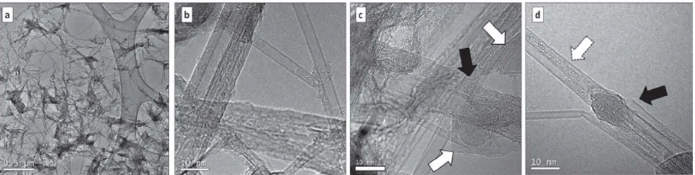
Figure 1. High-resolution TEM images of the BNNT sample. (a) overview at low magnification illustrating the fibrous structure. (b-d) High-resolution images showing on (b) the presence of multlwalled nanotubes (from 2 to 10 concentric walls), (c) damaged walls of some nanotubes (black arrow) and two isolated amorphous particles surrounded by some graphitic-like shells (white arrows), (d) some filling inside some nanotubes (dotted white arrow), a broken and open nanotube (black arrow) as well as an amorphous particle which seems to be at the end of a nanotube. The inset (same scale) of (d) also shows two isolated particles surrounded by some graphitlc-like shells, one being amorphous while the other one (top) is crystallized.
2.7. Statistical analysis
Results from micronucleus assay were analyzed using McGill non-parametric test (Mcgill, Tukey, and Larsen 1978) on median values of each group of larvae. This test consists in comparing medians of samples of size n (where n 2: 7) and in determining their 95% confidence intervals (95% Cl). 95% Cl are expressed by M ± 1.57 x IQR/
✓
n, where M is the median and IQR is the inter-quartile range (Mcgill, Tukey, and Larsen 1978). The difference between the medians of the test groups and the median of the NC group is significant with 95% certainty if there is no overlap. For cell-cycle data, normality was assessed with Kolmogorov-Smirnov test and homogeneity of variances with Levene's test. One way analysis of variance (ANOVA) followed by Tukey test were used to compare cell-cycle phase distribution among conditions.For data manipulations of gut microbiota survey, OTUs counts, alpha diversity indexes and Weighted Unifrac Distances calculations as well as multidi mentional scaling (MDS) plot were carried out using 'Phyloseq' R package (McMurdie and Holmes 2013). Graph visualization of OTUs relative abundances was performed using 'ggplot2' package (Wickham 2016) while differential abundance of bacterial gen era between exposed conditions compared to the control group was performed using 'Deseq2' R package (Love, Huber, and Anders 2014). For multi variate analysis of variance between groups, PERMANOVA was performed using Adonis function from the 'vegan' R package (Oksanen et al. 2015).
3. Results and discussion
3.7. Bnnt characteristics
BET measurement indicated a specific surface area of 163 m2/g, a little bit lower than the > 200 m2/g claimed by the provider. TEM images (Figure 1 (a)) shows that the sample is composed of nanotubes (2-10 walls, Figure l(b)) and small nanoparticles encapsulated in graphitic-like h-BN shells (size ca.
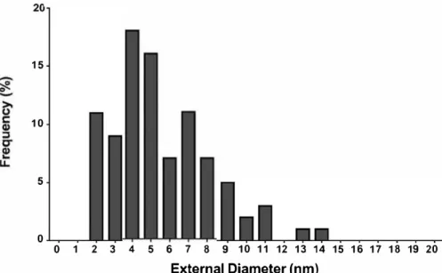
10-15nm) (Figure l(c,d)) as well as some more disor ganized material. Part of the nanotubes are damaged or even broken (Figure 1 (c,d), black arrows), which may be related to the grinding treatment that we have applied. On Figure 1 (c,d), it is possible to see that while most nanopartides look amorphous, a few are crystalized. Ali observed nanoparticles were coated with similar polyhedral graphitic-like h-BN shells. Outer diameter of the BNNT ranged from 2 to 14 nm with a mean outer diameter of 6 ± 2.6 nm (Figure 2).
We obtained through measurement of elemental analysis a total boron content between 38.6 and 42.5 wt. % (ca. 50.5 at. % using the highest weight value), 46.3 wt. % of Nitrogen (ca. 42.5 at. %) and 8.7 wt. % of Oxygen (ca. 7 at. %). If we make a first hypothesis that all the Nitrogen in the sample is in BN, then the excess of Boron is of ca. 8 at. %. Assuming that all the oxygen is involved in B203
(the most stable boron oxide), then the excess of Boron (corresponding to elemental boron) would be ca. 3.3 wt.%. This is rather far from the informa tion from the provider, although this explains that variations are likely between different batches.
15 10 5 0 0 1 2 3 4 5 6 7 8 9 10 11 12 13 14 15 16 17 18 19 20 External Diameter (nm)
Figure 2. BNNT outer diameter distributions evaluated from the HRTEM images (n = 91 ). (A)
C..-t,l!IIWI-·
10 9 8 7 6 � 5 l:i 4 3 2 1J
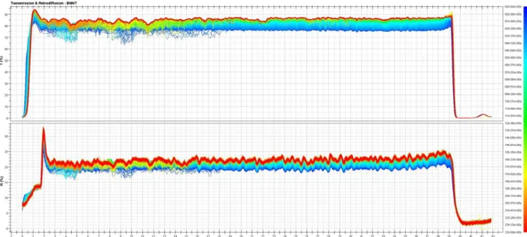
0 ·1,0 OOh 02h 04h 06h 08h 10h 12h 14h 16h 18h 20h 22h 24h Time(hl (B) o.. ... � .. , 0 ·2' -4' ·6, � ·8• l:i ·10 ·12, •14, ·16, ·18 OOh 02h 04h 06h 08h 10h 12h 14h 16h 18h 20h 22h 24h Time(hlFigure 3. Variation of the transmission vs time for suspe nsions of BNNT at 10 mg L -1 in exposure media measured at the top of the vial {A) and at the bottom of the vial {B).
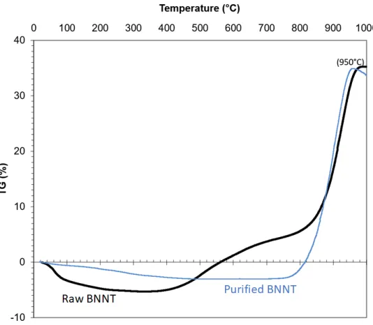
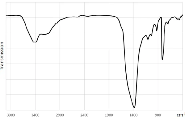
Details results from the XRD, XPS, TGA, Raman and IR analysis are detailed in the Supplementary information.
The variation of the transmission data (compared to t0) vs time at the top and at the bottom of the vial is presented in Figure 3(A,B) respectively. At the top of the vial, transmission progressively increased as the BNNTs were settling down. On the opposite, the trans mission progressively decreased at the bottom of the vial as the sediment was forming. ln both cases, it is obvious that most of the clarification/sedimentation took place in 8-10 h, at a rather constant speed (before slowing down) (Supplementary Figure SS). 3.2. Boron concentration in the exposure media and larvae
Analysis of boron concentration in the water indicated that reconstituted water from the control condition was devoid of boron. ln the 10 mg L 1 of BNNT con dition, 0.32 mg L 1 of boron were measured after 24 h of incubation in the media in absence of Xenopus tadpoles while after 24 h in presence of larvae, the concentration of boron decreased below 0.1 mg L 1 (quantification limit). During the exposure, larvae pro duced BNNT-containing feces according to the color differences with the control group. At the end of the 12 days of exposure, 2 ± 4 µg g 1 of boron were meas ured in the whole tissues of the larvae from the con trai group. Boron concentration in larvae exposed to 10 mg L 1 of BNNT reached 183 ± 83 µg g 1 of tissue.
BNNT were shown to be structurally very stable (Chen et al. 2017; Song et al. 2014) and there is few chance for the digestive tract of tadpoles to consti tute drastic enough conditions allowing BNNT deg radation as strong acidic conditions are needed to dissolve this material (Kleinerman et al. 2017). ln addition, carbon-based structural homologues of BNNT were shown to accumulate in the lumen of Xenopus tadpoles during the exposure while a transfer across the intestinal wall was not evidenced (Mouchet et al. 2011). Thus, crossing of intestinal barrier is not likely to occur in the case of BNNT exposure and it is highly probable that the boron concentration measured in the water as well as in the tissues of the larvae originated from free boron as byproduct found in the commercial form of the tested BNNT. However to confirm this hypothesis, the use of imaging technics would be needed to
determine if BNNT are present or not in the bio logical tissues. lndeed, amphibians were previously shown to accumulate free boron in polluted sites and are able to tolerate boron concentration in the water up to 10 mg L 1 for extended period without adverse effects (Eisler 1990; Emiroglu et al. 2010). Measurement performed under the experimental conditions indicated a full clearance of the water column by the larvae over 24 hours, leading to rejection of BNNT in the animal feces.
3.3. Micronucleus assay and ce// cycle analysis Xenopus larvae exposed to cyclophosphamide at 40 mg L 1 (positive control
=
PC) exhibited significantly higher MNE compared to the control group (negative control
=
NC), validating results obtained from the micronucleus assay. Among experimental conditions containing BNNT, although a trend to an increasing number of micronucleated cells was observed after exposure to 10 mg L 1 of the nano particle, no statistically significant increase of MNE %0 was accounted (Figure 4(A)). Analysis of erythro cyte cell cycle highlighted a significant decrease in S-phase cells in conditions containing BNNT com pared to the negative control (ANOVA, S-phase: p < 0.001 ), while the percentage of G0/G 1 ce lis increased and G2/M decreased only after exposure to 1 mg L 1 of BNNT (ANOVA, G0/G 1: p < 0.001; G2/ M: p < 0.001) (Figure 4(B)).The results obtained indicated that the commer cial form of BNNT tested was not leading to irre versible genotoxic effects at the tested concentrations as no significant induction of micro nucleated erythrocytes was observed. Thus, even if we cannot exclude that reversible DNA damages could occur through single strand break DNA, the obtained results indicated that BNNT exposure do not lead to permanent DNA alteration. This obser vation is contrary to in vitro studies indicating dis turbances of cell proliferation and genotoxicity of BNNT (Fernandez-Yague et al. 2015; Horvath et al.
2011; Çal and Bucurgat 2019; Emanet et al. 2015), but consistent with the few available works per formed in vivo. lndeed, no genotoxicity was observed in the planaria (Salvetti et al. 2015) as well as in drosophila (Demir and Marcos 2018) after BNNT exposure. ln the latter study, BNNT showed antioxidant and antigenotoxic properties against a
(A) w
z
::::e 10*
(B) 0 20 15 OG01G1 Os •G2IM A A A A BFigure 4. Micronucleus induction (A) and cell cycle (B) measured in erythrocytes of Xenopus tadpoles exposed for 12 days to increasing concentrations of BNNT. MNE: micronucleated erythrocytes; NC: negative control; PC: positive control; *significant differ ence compared to the NC (McGill test). For cell cycle analysis, N
=
12, A NOVA p < 0.001 followed by Tukey test. Letters indicate significant differences between concentrations tested for each phase of the cell cycle.(A) 200 .c 150 0 � 100
-�
ê
50 0z
CFigure S. Normalized growth of X. laevis larvae measured after 12 days of exposure to increasing concentrations of BNNT. ANOVA p < 0.001 followed by Tukey test. Letters indicate significant differences between concentrations.
known genotoxic compound. ln addition, similar effects were observed in Xenopus laevis exposed to other non-oxidized 1 D carbon counterparts of BNNT (Mouchet et al. 2008; Mouchet et al. 2010). lt was previously indicated that baron exert protective effects against genotoxic compounds in other bio logical models (Turkez 2008; Ince et al. 2014; Sankaya et al. 2016; Tepedelen, Soya, and Korkmaz 2016; Alak et al. 2019). For this reason, it is not pos sible yet to determine in these conditions whether free baron associated to the commercial mixture is protective against the genotoxic potential of the different types of BN nanoparticles or if BN are devoid of genotoxic potential.
3.4. Larval growth rate
Exposure to BNNT led to a significant increase of Xenopus tadpole growth in a dose-dependent man ner (ANOVA,
p
< 0.001) (Figure S(A)). Growth rate increased from 20.54 ± 9.53% at 0.1 mg L 1 to 44 ± 13.97% at the highest tested concentration compared to negative contrai. However, mean weight/length ratio remained unchanged in larvae exposed to the tested conditions (ANOVA,p
=
0.143) (Figure S(B)).Exposure to BNNT led to a dose-dependent increase of larval growth that is counterintuitive regarding to the literature indicating that no matter the number of dimension of the carbon-based
nanomaterial (CBN) considered, growth inhibition of Xenopus tadpoles was described by the surface area of exposure (Mottier et al. 2016; Lagier et al. 2017). As the surface area of the tested BNNT is compar able to those of the CBNs used in these studies and the observed effects are opposite, we can empha size that this growth inhibition model is specific to CBNs and is thus not applicable for BNNT. The main hypothesis for mechanisms underlying growth inhibition following CBNs exposure was associated to nutrient depletion and reduction of nutrient intake due to the presence of agglomerated CBNs in the gut (Lagier et al. 2017). The accumulation of BNNT in the tadpole gut was not as clear as previ ously observed with carbon-based nanomaterials (CBNs) that is consistent with the absence of growth alteration measured. Thus, despite the cap acity of BNNT to adsorb nutrients (Farmanzadeh and Ghazanfary 2014), the hypothesis of nutrient intake limitation is not likely to occur in the case of BNNT exposure.
Previous studies demonstrated that a deficiency (< 0.003 mg L 1) as well as too high concentrations of boron (<50 mg L 1) were detrimental for the development of amphibian embryo and impaired the reproduction of adults (Fort et al. 1998; Fort et al. 1999; Laposata and Dunson 1998; Fort et al. 2002). According to these studies, the boron con centrations measured in the exposure media of this work (0.32 mg L 1) do not represent a critical con centration impairing tadpole's physiology that is consistent. Despite the lack of literature regarding the enhanced growth of amphibian larvae exposed to boron, these effects were observed in fishes with no explanation of the mechanism involved in such growth increase (Eckhert 1998; Rowe et al. 1998; Oz, lnanan, and Dikel 2018). However, it was sug gested that boron was playing a role in the thyroid axis of X. laevis, probably in the synthesis of T3 (Fort et al. 2002). Thus, among the possible path ways involved in this observed growth stimulation, it may in part occur from a stimulation of the meta morphosis by the boron.
3.5. Gut microbiota survey
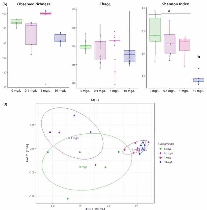
Among the different tested conditions, richness was not affected (Observed species: ANOVA p
=
0.505; Chao 1: ANOVA p=
0.826). However, evennesscalculated with Shannon index was shown to be significantly decreased after 12 days of exposure to 10 mg L 1 of BNNT (ANOVA p < 0.001) (Figure 6(A)). Bacterial communities were shown to be signifi cantly affected by the BNNT concentration as revealed by MDS performed with Weighted-Unifrac distances and PERMANOVA analysis (F
=
14.146; r2=
0.726, p=
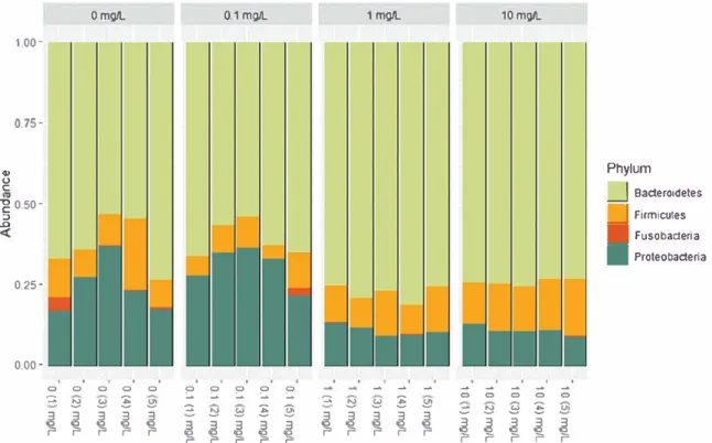
0.001) (Figure 6(8)). Pairwise compari sons indicated that gut bacterial communities were similar between the control group and the lowest tested concentration 0.1 mg L 1, while the two other tested concentrations 1 and 10 mg L 1 were different from each other and from any other conditions.Three major phyla composed the gut microbiota of X. laevis tadpoles: Bacteroidetes, Proteobacteria and Firmicutes with relative abundance in the con trol group representing 62 ± 8.5%, 24.7 ± 8.3% and
12.2 ± 5.5% of the whole microbial communities respectively (Figure 7). Exposure to BNNT led to a significant decrease of Proteobacteria relative abun dance at 1 and 10 mg L 1 of BNNT (ANOVA p < 0.001 ). On the opposite, a significant increase of Bacteroidetes is noticed at these concentrations (ANOVA p < 0.001 ), leading to a phylum relative abundance of 74± 1.17% at the highest tested concentration.
At the family level of the phylum Bacteroidetes, Bacteroidaceae were shown to significantly increase (ANOVA p< 0.001) at 1 and l0 mgL 1 reaching 97.36 ± 1.16% of the overall Bacteroidetes in the higher BNNT concentration compared to the 82.24 ± So/4 observed in the control group (Supplementary Figure S6A). On the contrary, other families such as Spirosomaceae, Flavobacteriaceae, Crodnitomicaceae or Weeksellaceae were shown to significantly decrease (ANOVA p < 0.001; p < 0.001; p
=
0.0356; p=
0.0023 respectively). Similarly, inside the phylum Proteobacteria, taxa from the family Magnetospirillaceae were shown to significantly increase after exposure to 10 mg L 1 of BNNT (ANOVA p=
0.00116) (Supplementary Figure S6B), while Moraxellaceae and Burkholderiaceae were shown to significantly decrease at this concentration (ANOVA p < 0.001 and p=
0.00811 respectively).At the genus scale, 12, 27, and 39 genera were differentially observed between the control group and the BNNT concentration of 0.1, 1, and 10 mg L 1 respectively (excluding multi-affiliated and
(A) Observed richness Chaol Shannon Index 200 30
S=3
160 1509
180+
25 140 �+
160 2.0 130 b 140 120 1.5 1209
110 0 mg/L 0.1 mg/L 1 mg/L 10 mg/L 0 mg/L 0.1 mg/L 1 mg/L 10 mg/L 0 mg/L 0.1 mg/L 1 mg/L 10 mg/L MDS (8)•
0.05• •
•
il: Contaminant � 000 + 0mg/L !5!.•
•
"'
... 0.1 mg/L .,; ... 1 mg/L ·;. ... 10mg/L OmgL•
•
--005 --010 .o 2 --01 0 0 0 1 Axis.1 [85.5%]Figure 6. Effects of 12 days of exposure to BNNT on the structure of gut bacterial communities of X. laevis t adpoles. Observed richness, estimated richness (Chao1 index) and evenness (Shannon index) are compared between the exposure conditions (0, 0.1, 1 and 10 mg L _,) (A). MDS plot of bacterial communities based on Unweighted unifrac distances (B).
unknown genera). Thus, even if the Firmicutes phylum was not globally affected, some genera were shown to be differentially observed between the contrai and the 1 and 10 mg L 1 of BNNT conditions (Supplementary Figure 57). Genera which relative abundance was shown to be unaffected, decreased or increased were clas sified as resistant, sensitive or opportunistic respectively. Thus, each category represented about 58.2% of the overall taxa for the resistant,
21.5 and 20.3% for the sensitive and opportunis tic respectively.
Even if it is difficult to differentiate the part of the effects associated to indirect or direct effects of BNNT and/or to byproducts exposure, several possi bilities can be considered concerning the effects observed on gut bacterial communities after the exposure of tadpoles to this commercial BNNT.
Considering the potential indirect effects, as pre viously mentioned, baron could play a role in the
Q) 0 C 1.00 -0. 75-"g 0.50 -:, .Ll 0.25 -0.00 -0 -::. 3 � 0mg/1.. 0 0 0 !2 !:l � 3 � � 3 � 3 0.1 mg/1.. 0 0 :.. 0
-
0 :.. 0 :.. ::!) -::. � 3 !2 !:l � � � 3 3 � � 3 3 1 mg/1.. 0-
-:.. -::. � :§ 3 !2 3 !:l 3 3 3 � � � � � 10 mg/1..-
;; ;; ;; ;; ::!) !2 !:l :è 3 � � � r � r r � � r ;; ::!) � r Phylum_J
BacteroldetesD
■
Firmicutes Fusobacteria■
ProteobacteriaFigure 7. Phylum composing the gut microbiota after 12 days of exposure to BNNT concentrations ranging from O to 10 mg L _, _ thyroid axis that is involved in the development
and maturation of the gut (Chalmers and Slack 1998; lshizuya-Oka 2011 ). lt was also demonstrated that gut bacterial communities were changing dur ing the metamorphosis of amphibians, notably through an increase of the phyla Bacteroidetes, a decrease of Proteobacteria and decrease of diversity (Kohl et al. 2013; Chai et al. 2018) which is consist ent with our data. Thus, we cannot exclude that the changes occurring in the gut microbial communities could be in part associated to the stimulation of thyroid axis leading to the acceleration of gut maturation.
These changes in bacterial consortium could also be associated to direct effects of BNNT. Surprisingly, antibacterial properties of BN nanoparticles have not been thoroughly investigated yet (Merlo et al.
2018) and results available from the literature regarding the antibacterial activity are contradictory. lndeed, despite its potential to interact with bio logical membranes and to form heteroaggregates with bacteria (Thomas, Enciso, and Hilder 2015; Wang et al. 2015), it was indicated that raw BNNT
at concentrations reaching 1 mg L 1 were not
inducing E.coli and S.aureus growth inhibition (Nithya and Pandurangan 2014). ln addition, expos ure of P. aeruginosa strain to boron nitride
nanosheets at a concentration of 10 mg L 1 was associated to a weak transcriptomic response com pared to other tested nanoparticles and was not leading to growth inhibition (Mortimer et al. 2018) while another study observed bacteriostatic activ ities at 0.4 mg L 1 in multiple gram positive bacter ial strains (K1vanç et al. 2018). On the opposite, coating of copper surfaces with similar hexagonal boron nitride led to protective effects against bac tericidal Cu (Parra et al., 2015). Moreover, exposure
to 2 D BN at high concentration (100 mg L 1
) induced damages to bacterial membranes in E.coli (Zhang et al. 2019). These studies were performed through exposure of single bacterial species in sus pension in a liquid medium that is very different from the exposure conditions found in the gut. However, few data emphasizing the effects of BN
nanoparticles on complex bacterial communities are available. Strong antibiofilm activities of 2 D BN were observed in established biofilm composed of single bacterial species (K1vanç et al. 2018).
As boron compounds were shown to exert weak toxicity against multiple bacterial strains (Minimum inhibitory concentration over 1 g L 1
) that possess a
good tolerance to this element (Ahmed and Fujiwara 201 0; Yilmaz 2012; Sayin, Ucan, and Sakmanoglu 2016), few direct effects from free
baron could be expected on gut microbial com munities. Boron was shown to constitute an essen tial trace element for bacteria, contributing to physiological and metabolic activities (Kabu and
Akosman 2013; Uluisik, Karakaya, and Kac 2018).
Furthermore, baron was shown to be implied in bacterial communication through the activation of an extracellular signaling molecule (autoinducer Al-2), involved in quorum sensing (Chen et al. 2002). This process allows the regulation of gene expres sion and diverse physiological activities in response to fluctuations in bacterial density (Miller and
Bassler 2001; Federle and Bassler 2003; Waters and
Bassler 2005; Papenfort and Bassler 2016). Exposure
to nanoparticles were previously shown to influence
quorum sensing process (Singh et al. 2017; Xiao
et al. 2016; Hayat et al. 2019). This could also be possible for the BN nanoparticle exposure. Thus, disturbances of quorum sensing from the gut bac teria could lead to changes in microbial commun ities (Thompson et al. 2015).
3.6. Statistical correlations
Correlation analysis indicated strong positive corre lations among tadpole biometric parameters. Significant negative correlation between the phy lum Proteobacteria and Bacteroidetes are noticed
(Pearson, r -0.91 p < 0.001). Firmicutes/
Bacteroidetes ratio was shown not to be correlated with any of the larvae biometric parameters or growth rate. However, phylum Bacteroidetes was shown to be significantly and positively correlated
with larval length (r = 0.62, p = 0.0034), growth rate
(r= 0.51, p= 0.0211). On the contrary, phylum
Proteobacteria was shown to be significantly nega tively correlated with larval length (r = -0.69,
p = 0.0008) and growth (r = -0.55, p = 0.012).
Changes in the Firmicutes/Bacteroidetes ratio of gut microbiota was shown to be associated to metabolic disorders in several species (Ley et al.
2005; Ley, Peterson, and Gordon 2006; Haiser and Turnbaugh 2013; Li et al. 2013). ln this study, the F/ B ratio was not significantly affected (ANOVA,
p = 0.2) and was not correlated to growth parame ters following exposure to BN. Thus, we can suggest that the growth stimulation observed is not associ ated to the potential induction of a metabolic disorder in hast On the contrary, phylum
Bacteroidetes was positivety correlated with growth parameters of the tadpoles. Nevertheless, the most known biological function of members of the phy lum Bacteroidetes from the gut is the degradation of biopolymers such as polysaccharides to produce carbohydrates (Thomas et al. 2011; Johnson et al. 2017). Thus, this increase of Bacteroidetes relative abundance can be associated to improved produc tion of carbohydrates and production of energy, leading to an improved fitness for the hast. The use of other amie techniques such as transcriptomic would be needed in order to determine the links between the changes in gut flora and modifications of functional capacities.
4. Conclusion
The aim of this study is to fill lacking data concern ing the ecotoxicity of a commercial form of baron nitride nanotubes toward the aquatic compartment. We used an original approach based on the meas urement of toxicological endpoints including geno toxicity and growth parameters of an amphibian species as well as bacterial communities associated to the hast gut. The obtained results indicate an overall biocompatibility of the tested BN mixture toward X. /aevis tadpoles. Significant induction of larval growth was shown to be correlated with changes in the gut microbial communities of the hast These changes in hast physiology are most probably due to indirect effects of byproducts, especially free baron that could stimulate the mat uration of the gut, benefiting to bacteria favoring the hast metabolism. Thus, BNNT alone represent a minor threat for amphibians in aquatic environ ments. However, due to its sorption capacities, interactive effects with other common contaminants such as PAHs, heavy metals or pesticides are remaining to be assessed to fully characterize its ecotoxic potential.
Acknowledgments
The authors thank BNNT, LLC for providing us with the material used in this study. J. Esvan is acknowledged for his help with the acquisition and analysis of XPS data.
Disclosure statement
No potential conflict of interest was reported by the author(s).
ORCID
Lauris Evariste
8
http://orcid.org/0000-0001-8718-7776Emmanuel Flahaut
8
http://orcid.org/0000-0001-8344-6902 ReferencesAdamovsky, O., A. N. Buerger, A. M. Wormington, N. Ector, R. J. Griffitt, J. H. Bisesi, Jr., and C. J. Martyniuk. 2018. "The Gut Microbiome and Aquatic Toxicology: An Emerging Concept for Environmental Health." Environmental
Toxicology and Chemistry 37 (11): 2758-2775. doi:10.1002/
etc.4249.
Ahmed, 1., and T. Fujiwara. 2010. "Mechanism of Boron Tolerance in Soil Bacteria." Canadian Journal of Microbiology 56 (1): 22-26. doi:10.1139/w09-106.
Alak, G., V. Parlak, M. E. Aslan, A. Ucar, M. Atamanalp, and H.
Turkez. 2019. "Borax Supplementation Alleviates Hematotoxicity and DNA Damage in Rainbow Trout (Oncorhynchus mykiss) Exposed to Copper." Biological Trace Element Research 187 (2): 536-542. doi:10.1007/ s12011-018-1399-6.
Anderson, Susan, Walter Sadinski, Lee Shugart, Peter Brussard, Michael Depledge, Tim Ford, JoEllen Hose, et al. 1994. "Genetie and Molecular Ecotoxicology: A Research Framework." Environmental Health Perspectives. 102 (suppl 12): 3-8. doi:10.1289/ehp.94102s123.
Augustine, J., T. Cheung, V. Gies, J. Boughton, M. Chen, Z. J.
Jakubek, S. Walker, Y. Martinez-Rubi, B. Simard, and S. Zou. 2019. "Assessing Size-Dependent Cytotoxicity of Boron Nitride Nanotubes Using a Novel Cardiomyocyte AFM Assay." Nanoscale Advances 1 (5): 1914-1923. doi:10. 1039/C9NA001 048.
Bokulich, N. A., S. Subramanian, J. J. Faith, D. Gevers, J. 1.
Gordon, R. Knight, D. A. Mills, and J. G. Caporaso. 2013. "Quality-Filtering Vastly lmproves Diversity Estimates from Illumina Amplicon Sequencing." Nature Methods 10 (1):
57-59. doi: 10.103 8/nmeth.227 6.
Bourdiol, F., F. Mouchet, A. Perrault, 1. Fourquaux, L. Datas, C. Gancet, J.-C. Boutonnet, E. Pinelli, L. Gauthier, and E. Flahaut 2013. "Biocompatible Polymer-Assisted Dispersion of Multi Walled Carbon Nanotubes in Water, Application to the Investigation of Their Ecotoxicity Using Xenopus
laevis Amphibian Larvae." Carbon 54: 175-191. doi:10.
1016/j.carbon.2012.11.024.
Bundschuh, M., J. Filser, S. Lüderwald, M. S. McKee, G.
Metreveli, G. E. Schaumann, R. Schulz, and S. Wagner. 2018. "Nanoparticles in the Environment: Where Do We Come From, Where Do We Go to?" Environmental Sdences. 30: 6. doi:10.1186/sl 2302-018-0132�.
Çal, T., and Ü. Ü. Bucurgat. 2019. "ln Vitro Investigation of the Effects of Boron Nitride Nanotubes and Curcumin on
DNA Damage." DARU Journal of Pharmaceutical Sciences
27 (1): 203-218. doi:10.1007/s40199-019-00263-6.
Cani, P. D. 2014. "Metabolism in 2013: The Gut Microbiota
Manages Host Metabolism." Nature Reviews. Endoainology
10 (2): 74-76. doi:10.1038/nrendo.2013.240.
Chai, L, Z. Dong, A. Chen, and H. Wang. 2018. "Changes in Intestinal Microbiota of Bufo Gargarizans and lts Association with Body Weight During Metamorphosis."
Archives of Microbiology 200 (7): 1087-1099. doi:10.1007/
s00203-018-1523-1.
Chalmers, A. D., and J. M. Slack. 1998. "Development of the Gut in Xenopus laevis." Developmental Dynamics 212: 509-521. doi:10.1002/(SICl)l 097-0177(199808)212:4
< 509::AID-AJA4
>
3.0.CO;2-LChang, C. W., A. M. Fennimore, A. Afanasiev, D. Okawa, T. lkuno, H. Garcia, D. Li, A. Majumdar, and A. Zettl. 2006. "Isotope Effect on the Thermal Conductivity of Boron Nitride Nanotubes." Physical Review Letters 97 (8): 085901. doi:10.1103/PhysRevLett.97.085901.
Chen, X., C. M. Dmuchowski, C. Park, C. C. Fay, and C. Ke. 2017. "Quantitative Characterization of Structural and Mechanical Properties of Boron Nitride Nanotubes in High Temperature Environments." Scientific Reports 7 (1 ): 11388. doi:10.1038/s41598-017-11795-9.
Chen, X., S. Schauder, N. Potier, A. Van Dorsselaer, 1. Pelczer, B. L. Bassler, and F. M. Hughson. 2002. "Structural Identification of a Bacterial Quorum-Sensing Signal Containing Boron." Nature 415 (6871 ): 545-549. doi:1 O. 1038/415545a.
Chen, X., P. Wu, M. Rousseas, D. Okawa, Z. Gartner, A. Zettl, and C. R. Bertozzi. 2009. "Boron Nitride Nanotubes are Noncytotoxic and Can be Functionalized for Interaction with Proteins and Cells". Journal of the American Chemical Society 131 (3): 890-891. doi:10.1021/ja807334b.
Chen, Y., J. Zou, S. J. Campbell, and G. Le Caer. 2004. "Boron Nitride Nanotubes: Pronounced Resistance to Oxidation."
Applied Physics Letters 84 (13): 2430-2432. doi:10.1063/1.
1667278.
Chopra, N. G., R. J. Luyken, K Cherrey, V. H. Crespi, M. L Cohen, S. G. Louie, and A. Zettl. 1995. "Boron Nitride Nanotubes." Sdence (New York, N. Y.) 269 (5226): 966-967. doi:10.1126/science.269.5226.966.
Ciofani, Gianni, Serena Danti, Delfo D'Alessandro, Stefania Moscato, and Arianna Menciassi. 2010. "Assessing Cytotoxicity of Boron Nitride Nanotubes: lnterference with the MTT Assay." Biochemical and Biophysical Research Communications 394 (2): 405-411. doi:10.1016/j.bbrc.201 O.
03.035.
Ciofani, G., S. Del Turco, A. Rocca, G. de Vito, V. Cappello, M.
Yamaguchi, X. Li, et al. 2014. "Cytocompatibility Evaluation of Gum Arabic-Coated Ultra-Pure Boron Nitride Nanotubes on Human Cells." Nanomedicine (London, England) 9 (6): 773-788. doi:10.2217/nnm.14.25.
Ciofani, G., V. Raffa, A. Menciassi, and A. Cuschieri. 2008.
"Cytocompatibility, Interactions, and Uptake of Polyethyleneimine-Coated Boron Nitride Nanotubes by Living Cells: Confirmation of Their Potential for Biomedical
Applications." Biotechnology and Bioengineering 101 (4):
850-858. doi:10.1002/bit.21952.
Claus, S. P., H. Guillou, and S. Ellero-Simatos. 2016. "The Gut Microbiota: A Major Player in the Toxicity of Environmental Pollutants?" Npj Biofilms and Microbiomes 2
(1 ): 16003.
Colombo, A., M. Saibene, E. Moschini, P. Bonfanti, M. Collini, K. Kasemets, and P. Mantecca. 2017. "Teratogenic Hazard of BPEI-Coated Silver Nanoparticles to Xenopus /aevis." Nanotoxicology 11 (3): 405-418. doi:10.1080/17435390. 2017.1309703.
Demir, E., and R. Marcos. 2018. "Antigenotoxic Potential of Boron Nitride Nanotubes." Nanotoxicology 12 (8): 868-884. doi: 1 O .1 080/1 7 435390.2018.1482379.
Durack, J., and S. V. Lynch. 2019. "The Gut Microbiome:
Relationships with Disease and Opportunities for Therapy." Journal of Experimenta/ Medicine 216 (1 ): 20-40. doi:10.1084/jem.20180448.
Eckhert, C. D. 1998. "Boron Stimulates Embryonic Trout Growth." Journal of Nutrition 128 (12): 2488-2493. doi:1 O. 1093/jn/128.12.2488.
Eisler, R. 1990. "Boron hazards to fish, wildlife, and inverte brates: a synoptic review." Accessed 26 July 2019. https:// pubs.er.usgs.gov/publication/5200086.
Emanet, M., Ô. �en, Z. Çobandede, and M. Çulha. 2015. "Interaction of Carbohydrate Modified Boron Nitride Nanotubes with Living Cells." Co/laids and Surfaces B Biointerfaces 134: 440-446. doi:10.1016/j.colsurfb.2015.07. 036.
Emiroglu, O., A. Ciçek, N. Arslan, S. Aksan, and M. Rüzgar. 2010. "Boron Concentration in Water, Sediment and Different Organisms around Large Borate Deposits of Turkey." Bulletin of Environmenta/ Contamination and Toxicology 84 (4): 427-431. doi:10. 1007/s00128-010-9961-8.
Escudié, F., L. Auer, M. Bernard, M. Mariadassou, L Cauquil, K. Vidal, S. Maman, G. Hernandez-Raquet, S. Combes, and G. Pascal. 2018. "FROGS: Find, Rapidly, OTUs with Galaxy Solution." Bio informatics (Oxford, England) 34 (8): 1287-1294. doi:10.1093/bioinformatics/btx791.
Evariste, L., M. Barret, A. Mottier, F. Mouchet, L. Gauthier, and E. Pinelli. 2019. "Gut Microbiota of Aquatic Organisms: A Key Endpoint for Ecotoxicological Studies."
Environmenta/ Pollution (Barking, Essex) 248: 989-999. doi:
10.1016/j.envpol.2019.02.101.
Evariste, L, L. Lagier, P. Gonzalez, A. Mottier, F. Mouchet, S.
Cadarsi, P. Lonchambon, et al. 2019. "Thermal Reduction of Graphene Oxide Mitigates lts in Vivo Genotoxicity toward Xenopus /aevis Tadpoles." Nanomateria/s 9 (4): 584. doi: 10 .3 390/na no9040584.
Farmanzadeh, D., and S. Ghazanfary. 2014. "Interaction of Vitamins A, 81, C, 83 and D with Zigzag and Armchair Boron Nitride Nanotubes: A DFT Study." Comptes Rendus Chim 17 (10): 985-993. doi:10.1016/j.crci.2013.11.012.
Federle, M. J., and B. L Bassler. 2003. "lnterspecies
Communication in Bacteria." Journal of Clinicat
Investigation 112
JCl200320195. (9): 1291-1299. doi:10.1172/
Fernandez-Yague, M. A., A. Larranaga, O. Gladkovskaya, A.
Stanley, G. Tadayyon, Y. Guo, J.-R. Sarasua, et al. 2015. "Effects of Polydopamine Functionalization on Boron
Nitride Nanotube Dispersion and
Cytocompatibility.• Bioconjugate Chemistry 26 (10): 2025-2037. doi:10.1021/acs.bioconjchem.Sb00257.
Fort, D. J., T. L Propst, E. L. Stover, P. L Strong, and F. J. Murray. 1998. • Adverse Reproductive and Developmental
Effects in Xenopus from lnsufficient Boron." Biologica/
Trace Element Research 66 (1-3): 237-259. doi:10.1007/ BF02783141.
Fort, D. J., R. L Rogers, D. W. McLaughlin, C. M. Sellers, and C. L Schlekat. 2002. "Impact of Boron Deficiency on
Xenopus /aevis." Biologica/ Trace Element Research 90 (1-3):
117-142. doi:10.1385/BTER.
Fort, D. J., E. L Stover, P. L. Strong, F. J. Murray, and C. L Keen. 1999. "Chronic Feeding of a Low Boron Diet Adversely Affects Reproduction and Development in Xenopus /aevis." The Journal of Nutrition 129 (11 ):
2055-2060. doi:10.1093/jn/129.11.2055.
Haiser, H. J., and P. J. Turnbaugh. 2013. "Developing a Metagenomic View of Xenobiotic Metabolism." Pharmacologica/ Research 69 (1 ): 21-31. doi:10.1016/j.phrs. 2012.07.009.
Hayat, S., S. Muzammil, null Shabana, B. Aslam, M. H.
Siddique, M. Saqalein, and M. A. Nisar. 2019. "Quorum Quenching: Role of Nanoparticles as Signal Jammers in Gram-Negative Bacteria." Future Microbio/ 14: 61-72. doi:
10.2217/fmb-2018-0257.
Horvath, L., A. Magrez, D. Golberg, C. Zhi, Y. Bando, R.
Smajda, E. Horvath, L. Forr6, and B. Schwaller. 2011. "ln Vitro Investigation of the Cellular Toxicity of Boron
Nitride Nanotubes." ACS Nono 5 (5): 38002-3810. doi:10.
1021/nn200139h.
Ince, S., 1. Kucukkurt, H. H. Demirel, D. A. Acaroz, E. Akbel, and 1. H. Cigerci. 2014. "Protective Effects of Boron on Cyclophosphamide lnduced Lipid Peroxidation and Genotoxicity in Rats." Chemosphere 108: 197-204. doi:10. 1016/j.chemosphere.2014.01 .038.
lshizuya-Oka, A. 2011. "Amphibian Organ Remodeling during Metamorphosis: insight into Thyroid Hormone-lnduced
Apoptosis." Deve/opment, Growth & Differentiation 53 (2):
202-212. doi:10.1111/j.1440-169X.2010.01222.x.
ISO/FDIS 21427-1. 2006. Water Qualit y-Eva/uation of
Genotoxidty b y Measurement of the Induction of Micronuclei-Part 7: Evaluation of Genotoxidty Using Amphibian Larvae. Geneva: International Organization for Standardization.
Jackson, S. P., and J. Bartek. 2009. "The DNA-Damage Response in Human Biology and Disease." Nature 461 (7267): 1071-1078. doi:10.1038/nature08467.
Jin, Y., S. Wu, Z. Zeng, and Z. Fu. 2017. "Effects of
Environ mental Pollutants on Gut Microbiota."
Environmenta/ Pollution (Barking, Essex) 222: 1-9. doi:10. 1016/j.envpol.2016.11.045.
Johnson, E. L., S. L. Heaver, W. A. Walters, and R. E. Ley. 2017. "Microbiome and Metabolic Disease: revisiting the Bacterial Phylum Bacteroidetes." Journal of Molecular Medicine (Berl) 95 (1): 1-8. doi:10.1007/s00109-016-1492-2. Kabu, M., and M. S. Akosman. 2013. "Biological Effects of Boron." Reviews of Environmental Contamination and
Toxîcology 225: 57-75. doi:10.1007/978-1-4614-6470-9 2.
Kalay, S., Z. Yilmaz, O. Sen, M. Emanet, E. Kazanc, and M. Çulha. 2015. "Synthesis of Boron Nitride Nanotubes and Their applications." Beilstein Journal of Nanotechnology 6: 84-102. doi:10.3762/bjnano.6.9.
Keller, A. A., and A. Lazareva. 2014. "Predicted Releases of Engineered Nanomaterials: From Global to Regional to Local." Environmental Science & Technology Letters 1 (1): 65-70. doi:10.1021/ez400106t.
Kim, J. H., T. V. Pham, J. H. Hwang, C. S. Kim, and M. J. Kim. 2018. "Boron Nitride Nanotubes: Synthesis and Applications." Nano Convergence 5 (1): 17. doi:10.1186/
s40580-018-0149-y.
K1vanç, M., B. Barutca, A. T. Koparal, Y. Gêincü, S. H. Bostanc1, and N. Ay. 2018. "Effects of Hexagonal Boron Nitride Nanoparticles on Antimicrobial and Antibiofilm Activities, Cell Viability." Materials Science & Engineering C Materials for Biological Applications 91: 115-124. doi:10.1016/j.msec. 2018.05.028.
Kleinerman, O., M. Adnan, D. M. Marincel, A. W. K. Ma, E. A. Bengio, C. Park, S.-H. Chu, M. Pasquali, and Y. Talmon. 2017. "Dissolution and Characterization of Boron Nitride Nanotubes in Superacid." Langmuir: The ACS Journal of
Surfaces and Colloids 33 (50): 14340-14346. doi:10.1021/
acs.langmuir.7b03461.
Knutie, S. A. C. L. Wilkinson, K. D. Kohl, and J. R. Rohr. 2017.
"Early-Life Disruption of Amphibian Microbiota Decreases Later-Life Resistance to Parasites." Nature Communications. 8: 86. doi:10.1038/s41467-017-00119-0.
Kohl, K. D. T. L. Cary, W. H. Karasov, and M. D. Dearing. 2013. "Restructuring of the Amphibian Gut Microbiota through Metamorphosis: The Amphibian Gut Microbiota."
Environmental Microbiology Reports 5 (6): 899-903. doi:10.
1111/1758-2229.12092.
Lagier, L,. F. Mouchet, C. Laplanche, A. Mottier, S. Cadarsi, L Evariste, C. Sarrieu, et al. 2017. "Surface Area of Carbon Based Nanoparticles Prevails on Dispersion for Growth Inhibition in Amphibians." Carbon 119: 72-81. doi:10.1016/ j.carbon.2017.04.016.
Laie, A., S. Bernard, and U. B. Demirci. 2018. "Boron Nitride for Hydrogen Storage." ChemPlusChem 83 (10): 893-903. doi:10.1002/cplu.201800168.
Laposata, M. M., and W. A. Dunson. 1998. "Effects of Boron and Nitrate on Hatching Success of Amphibian Eggs." Archives of Environmental Contamination and Toxicology 35 (4): 615-619. doi:10.1007/s002449900423.
Ley, R. E., F. Backhed, P. Turnbaugh, C. A. Lozupone, R. D. Knight, and J. 1. Gordon. 2005. "Obesity Alters Gut Microbial Ecology." Proceedings of the National Academy of Sdences of the United States of America 102 (31): 11070-11075. doi:10.1073/pnas.0504978102.
Ley, R. E. D. A. Peterson, and J. 1. Gordon. 2006. "Ecological and Evolutionary Forces Shaping Microbial Diversity in the Human Intestine." Cel/ 124 (4): 837-848. doi:10.1016/j.cell. 2006.02.017.
Li, X., Q. Yan, S. Xie, W. Hu, Y. Yu, and Z. Hu. 2013. "Gut Microbiota Contributes to the Growth of Fast-Growing Transgenic Cornmon Carp (Cyprinus carpio L.)." PLoS One 8 (5): e64577. doi:10.1371/joumal.pone.0064577.
Love, M. 1., W. Huber, and S. Anders. 2014. "Moderated Estimation of Fold Change and Dispersion for RNA-seq data with DESeq2 ." Genome Biology 15 (12): 550. doi:10. 1186/sl 3059-014-0550-8.
Mahé, F., T. Rognes, C. Quince, C. de Vargas, and M. Dunthorn. 2014. "Swarm: robust and Fast Clustering Method for Amplicon-Based Studies." Peerj. 2: e593. doi:
10.7717 /peerj.593.
Marin-Barba, M.,. H. Gavilân, L Gutiérrez, E. Lozano-Velasco, 1. Rodriguez-Ramiro, G. N. Wheeler, C. J. Morris, M. P. Morales, and A. Ruiz. 2018. "Unravelling the Mechanisms That Determine the Uptake and Metabolism of Magnetic Single and Multicore Nanoparticles in a Xenopus laevis
Model." Nanoscale 10 (2): 690-704. doi:10.1039/ c7nr06020c.
Mcgill, R. J. W. Tukey, and W. A. Larsen. 1978. "Variations of Box Plots." AMSTAT 32 (1): 12-16. doi:10.1080/00031305. 1978.10479236.
McMurdie, P. J., and S. Holmes. 2013. "Phyloseq: An R Package for Reproducible Interactive Analysis and Graphies of Microbiome Census Data." PLoS One 8 (4): e61217. doi:10.1371/journal.pone.0061217.
Merlo, A., V. R. S. S. Mokkapati, S. Pandit, and 1. Mijakovic. 2018. "Boron Nitride Nanomaterials: Biocompatibility and Bio-Applications." Biomaterials Sdence 6 (9): 2298--2311. doi:10.1039/C8BM00516H.
Miller, M. B., and B. L. Bassler. 2001. "Quorum Sensing in Bacteria." Annual Review of Microbiology 55: 165-199. doi:
10.1146/annurev.micro.55.1.165.
Mithieux, G. 2018. "Gut Microbiota and Host Metabolism: What Relationship." Neuroendoainology 106 (4): 352-356. doi:10.1159/000484526.
Mortimer, M.,. N. Devarajan, D. Li, and P. A. Holden. 2018.
"Multiwall Carbon Nanotubes lnduce More Pronounced Transcriptomic Responses in Pseudomonas aeruginosa PG201 than Graphene, Exfoliated Boron Nitride, or Carbon Black." ACS Nano 12 (3): 2728--2740. doi:10.1021/acsnano.
7b08977.
Mottier, A. F. Mouchet, C. Laplanche, S. Cadarsi, L Lagier, J. C. Arnault, H. A. Girard, et al. 2016. "Surface Area of Carbon Nanoparticles: A Dose Metric for a More Realistic Ecotoxicological Assessment." Nano Letters 16 (6): 3514-3518. doi:10.1021 /acs.nanolett.6b00348.
Mottier, A. F. Mouchet, É. Pinelli, L Gauthier, and E. Flahaut. 2017. "Environmental Impact of Engineered Carbon Nanoparticles: From Releases to Effects on the Aquatic Biota." Current Opinion in Biotechnology 46: 1-6. doi:10. 1016/j.copbio.2016.11.024.