OATAO is an open access repository that collects the work of Toulouse
researchers and makes it freely available over the web where possible
Any correspondence concerning this service should be sent
to the repository administrator:
[email protected]
This is a Publisher’s version published in:
http://oatao.univ-toulouse.fr/23651
To cite this version:
El Jabbar, Youssef and El Ouatib, Rachida and Er-Rakho, Lahcen and Durand,
Bernard
Influence of temperature and pH on the morphology and the color of
the CoAl2O4 prepared by sol gel method. (2015) Journal of Materials and
Environmental Science - JMES, 6 (12). 3452-3456. ISSN 2028-2508
Official URL:
3452
Influence of temperature and pH on the morphology and the color of the
CoAl
2O
4prepared by sol gel method
Y. El Jabbar
1, R. El Ouatib
1*, L. Er-Rakho
1and B. Durand
21Laboratoire de Physico-Chimie des Matériaux Inorganiques, Département de chimie, Faculté des Sciences Ain Chock, Université Hassan II Casablanca, Maroc.
2
Institut Carnot CIRIMAT, CNRS Université de Toulouse, 118 route de Narbonne, 31062 Toulouse Cedex 9, France.
*
Corresponding Author. E-mail: [email protected] ; Tel: (+212661170710)
Abstract
The aim of this study consists in the synthesis of CoAl2O4 spinel by sol gel method using polyacrylic acid as
chelating agent, with focusing on the influence of calcination temperature and pH on the morphology and on the color of the powders obtained. The prepared powders have been characterized by X- ray diffraction (XRD), fourier transform infrared spectroscopy (FTIR), transmission electron microscopy (TEM), Brunauer-Emmett-Teller (BET) and colorimetry (L*a*b*). The results obtained show that the spinel phase appears from 500 °C.
FTIR spectroscopy reveals oxygen-metal bands characteristic of the spinel structure. The TEM micrographs of
powders, synthesized at the temperatures 800°C, 900°C and 1100°C at pH=2,7 and 10, show that these powders are formed of nanometric particle size with a distribution between 20 nm and 90 nm. The colorimetric parameters, measured on powders calcined at 1100 ° C and regardless of the pH values, show an important blueness.
Keywords: Cobalt aluminate, Spinel, Sol-gel, X-ray, Pigment, Polyacrylic acid.
Introduction
The heat-resistant inorganic pigments have many industrial applications, such as decorative coloring ceramics. That requires certain specific characteristics of the pigments like brightness, opacity and chemical resistance towards the alkaline fluxes [1].
The CoAl2O4 spinel is a blue inorganic pigment. In order to elaborate this spinel, a lot of methods have been
involved [2-4].The solid state reaction, based on the treatment between Co3O4 and Al2O3 at a high temperature,
generally leads to a bad homogeneity of the powders and a very low surface area [5].
Other methods of synthesis have been developed for preparing more homogeneous powders with a large specific surface area, for example: the sol-gel process [6,7], the co-precipitation [8], the molten salt method [9] and the hydrothermal pathway [10,11]. The sol-gel process is known to produce materials of high homogeneity and of good purity at low temperatures. Within this framework, several factors were considered (type of precursors, complexing agent, pH of the medium, calcination temperature ...), to study their influence on the structure and morphology of the powders obtained [12-16].
In our work, we have studied the influence of calcination temperature and pH on the morphology and color of
CoAl2O4 spinel elaborated by polymeric method with the polyacrylic acid as chelating agent.
2. Materials and methods
2.1. PreparationThe synthesis is achieved by dissolving the nitrates of cobalt and aluminum with a stoichiometric ratio. In the
3453
pre-calcined at 300°C under air for 12h. A thermic treatment of powders under air gives the spinel phase. According to the calcination temperature, different color shades have been observed. The synthesis protocol is given in the figure 1.
Figure 1: Preparation flowchart of CoAl2O4 powders
2.2. Characterization
The powders obtained at different temperatures have been characterized by X-ray Diffraction (DRX) (Bruker D8 Advance X-ray Diffractometer equipped with Lynx Eye detector), Fourier transformed infrared spectroscopy (FTIR) (IR AFFINITY-1S SHIMIADZU). The morphology and the particle size have been observed by a
transmission electron microscopy (TEM) (model Tecnai G2 series). The specific surface area of powders has
been determined by BET (Micromeritics Desorb 2300A). The colorimetric parameters (L*, a* and b*) have been measured in the CIE system (CHROMA METER CR-400/410, KONICA MINOLTA).
3. Results and discussion
3.1. X-ray diffraction
The X-ray diffractograms of the powders obtained at different temperatures, show the spinel phase CoAl2O4
(JCPDS 44-0160) at 300°C,whatever the value of the pH. X-ray diffraction patterns are presented in figure 2.
Figure 2: Powder X-ray diffraction pattern of CoAl2O4 prepared at pH=7 and calcined for 5h at different
3454
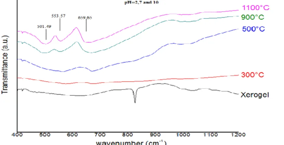
3.2. Infrared spectroscopy analysis (FTIR)
The Infrared spectra of powders achieved at different pH and at different calcination temperatures are given in the figure 3.
Figure 3: Infrared spectra of CoAl2O4 at different pH and different calcinations temperatures.
The bands (501.49, 553.57, 659.65 cm-1) characteristic of a normal spinel phase [17,18] are observed from 500°C
regardless the pH value. This corroborates the results achieved by XRD.
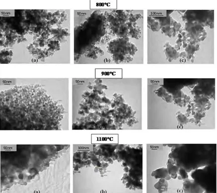
3.3. Powder morphology
The obtained micrographs show that these powders are formed by nanometric grains with a more or less narrow
distribution size.The agglomeration rate of these grains and their sizes seem to vary with pH and temperature
respectively (figure 4). The crystallite sizes calculated by Scherrer’s equation [19] has the same order of magnitude as that determined by TEM. The specific surface area of these powders decreases with increasing temperature and this confirms the evolution of grain size. The table 1 shows the different results.
Table 1: Crystallite size and specific surface area in function of calcination temperature and pH.
The results show that development of crystallite size is due to the pH variation and while augmenting temperature, it is also remarked that the specific area decreases.
3.4. The effect of the temperature of calcination and pH on the color evolution
Thermal treatement of gel dried at 120°C under air leads to powders with different colors. The gel presents a pink coloration which characterizes octahedral coordination of ions, the heating at 300°C is accompanied with a gel deshydratation and color variation due to the partial oxydation of Cobalt II at Cobalt III. A calcination temperature higher than 500°C leads to a green then blue coloration. This variation of colors can be explained by modification of the cobalt coordination.
The colorimetric parameters (L* a* b*) of the obtained powders change depending on the temperature and independently of pH. Above 800 °C, we see greater clarity (L) whatever the pH. The blue component (-b *) is developing strongly with temperature while the red component (a*) remains low (Figure 5).
Temperature (°C) pH BET (m2/g) TEM (nm) Crystallite size (Scherrer)(nm) 900°C 2 7 20.17 34.26 20-27 21-29 23.60 22.90 10 24.15 19-47 28.50 1100°C 2 7 13.32 23-102 40.18 10 14.69 7.23 24-56 26-90 43.70 33.10
3455
Figure 4 : Transmission electron microscopy micrographs of powder with pH of 2 (a), 7(b), 10(c) calcined at
800°C, 900°C and 1100°C.
3456
Conclusion
The polymeric method used for CoAl2O4 elaboration allows us to obtain nanometric particles. The influence of
temperature is strongly marked on the morphology and color, while the pH is significant. The powders obtained
at 1100 ° C and pH = 7 presented specific surface areas of about 15 m2.g-1 with better blueness which allows to
use them as pigment in the ceramic coloring .
References
1. Anand G.T., Kennedy L. J., Judith Vijaya J., J. Alloys Compd. 581 (2013) 558-566.
2. Gaudon M., Robertson L.C., Lataste E., Duttine M., Ménétrier M., Demourgues A., Ceram. Int. 40 (2014) 5201- 5207.
3. Mindru I., Marinescu G., Gingasu D., Patron L., Ghica C., Giurginca M., Mater. Chem. Phys. 122 (2010) 491-497.
4. Wahba A. M., Imam N.G., Mohamed M. B., J. Mol. Struct. 1105 (2016) 61-69. 5. Meyer F., Hempelmann R., Mathur S., Veith M., J. Mater. Chem. 9 (1999) 1755-1763. 6. Li G., Zhang J., Appl. Surf. Sci. 258 (2012) 7612-7616.
7. kurijica S., Popović J. , Tkalćec E. , Grsžeta B. , Mandić V., Mater. Chem. Phys. 135 (2012) 587-593. 8. Ke S., Wang Y., Pan Z., Dyes and Pigments 118 (2015) 145-151.
9. Ouahdi N., Guillemet S., Durand B., El Ouatib R., Er-Rakho L., Moussa R., Samdi A., J. Eur. Ceram. Soc. 28 (10) (2008) 1987-1994.
10. Kim J., Son B.,Yoon D., Hwang K., Noh H., Cho W., Kima U., Ceram. Int. 38 (2012) 5707–5712. 11. Yu F., Yang J., Ma J., Du J., Zhou Y., J. Alloys Compd. 468 (2009) 443-446.
12. Salem S., jazayeri S.H., allahverdi A., Shirvani M., Bondioli F., Merrari A.M., Int. J. Appl. Ceram. Technol. 9 (5) (2012) 968-978.
13. Li W., Li J., Guo J., J. Eur. Ceram. Soc. 23 (2003) 2289-2295. 14. Ibrahim S. Ahmed, Mater. Res. Bull. 12 (2011) 2548–2553.
15. Prim S. R., García A., Galindo R., Cerro S., Llusar M., Folgueras M.V., Monorós G., Ceram. Int. 39 (2013) 6981-6959.
16. JAFARI M., Hassanzadeh-Tabrizi S.A., Powder Technol. 266 (2014) 236-239. 17. Wang C., Liu S., Liu L., Bai X., Mater. Chem. Phys. 96 (2006) 361–370. 18. Preudhome J., Tarte P., Spectrochim. Acta 27A (1971) 1817-1826.
19. Păcurariu C., Lazău I., Ecsedi Z., Lazău R., Barvinschi P., Mărginean G., J. Eur. Ceram. Soc. 27 (2007) 707-710.