ORIGINAL ARTICLE
Genome-wide analysis implicates microRNAs and their target
genes in the development of bipolar disorder
AJ Forstner
1,2,50, A Hofmann
1,2,50, A Maaser
1,2, S Sumer
3, S Khudayberdiev
3, TW Mühleisen
1,2,4, M Leber
5, TG Schulze
6, J Strohmaier
7,
F Degenhardt
1,2, J Treutlein
7, M Mattheisen
8,9, J Schumacher
1,2, R Breuer
7, S Meier
7,10, S Herms
1,2,11, P Hoffmann
1,2,4,11, A Lacour
12,
SH Witt
7, A Reif
13, B Müller-Myhsok
14,15,16, S Lucae
14, W Maier
17, M Schwarz
18, H Vedder
18, J Kammerer-Ciernioch
19, A Pfennig
20,
M Bauer
20, M Hautzinger
21, S Moebus
22, L Priebe
1,2, S Sivalingam
1,2, A Verhaert
1,2, H Schulz
23, PM Czerski
24, J Hauser
24, J Lissowska
25,
N Szeszenia-Dabrowska
26, P Brennan
27, JD McKay
28, A Wright
29,30, PB Mitchell
29,30, JM Fullerton
31,32, PR Scho
field
31,32,
GW Montgomery
33, SE Medland
33, SD Gordon
33, NG Martin
33, V Krasnov
34, A Chuchalin
35, G Babadjanova
35, G Pantelejeva
36,
LI Abramova
36, AS Tiganov
36, A Polonikov
37, E Khusnutdinova
38,39, M Alda
40,41, C Cruceanu
42,43,44, GA Rouleau
42, G Turecki
43,44,45,
C Laprise
46, F Rivas
47, F Mayoral
47, M Kogevinas
48, M Grigoroiu-Serbanescu
49, P Propping
1, T Becker
5,12, M Rietschel
7, S Cichon
1,2,4,11,
G Schratt
3and MM Nöthen
1,2Bipolar disorder (BD) is a severe and highly heritable neuropsychiatric disorder with a lifetime prevalence of 1%. Molecular genetic
studies have identi
fied the first BD susceptibility genes. However, the disease pathways remain largely unknown. Accumulating
evidence suggests that microRNAs, a class of small noncoding RNAs, contribute to basic mechanisms underlying brain
development and plasticity, suggesting their possible involvement in the pathogenesis of several psychiatric disorders, including
BD. In the present study, gene-based analyses were performed for all known autosomal microRNAs using the largest genome-wide
association data set of BD to date (9747 patients and 14 278 controls). Associated and brain-expressed microRNAs were then
investigated in target gene and pathway analyses. Functional analyses of miR-499 and miR-708 were performed in rat hippocampal
neurons. Ninety-eight of the six hundred nine investigated microRNAs showed nominally signi
ficant P-values, suggesting that
BD-associated microRNAs might be enriched within known microRNA loci. After correction for multiple testing, nine microRNAs
showed a signi
ficant association with BD. The most promising were miR-499, miR-708 and miR-1908. Target gene and pathway
analyses revealed 18 signi
ficant canonical pathways, including brain development and neuron projection. For miR-499, four
Bonferroni-corrected signi
ficant target genes were identified, including the genome-wide risk gene for psychiatric disorder CACNB2.
First results of functional analyses in rat hippocampal neurons neither revealed nor excluded a major contribution of miR-499 or
miR-708 to dendritic spine morphogenesis. The present results suggest that research is warranted to elucidate the precise
involvement of microRNAs and their downstream pathways in BD.
Translational Psychiatry (2015)
5, e678; doi:10.1038/tp.2015.159; published online 10 November 2015
1
Institute of Human Genetics, University of Bonn, Bonn, Germany;2
Department of Genomics, Life and Brain Center, University of Bonn, Bonn, Germany;3
Institute of Physiological Chemistry, Philipps-University Marburg, Marburg, Germany;4Institute of Neuroscience and Medicine, Research Center Juelich, Juelich, Germany;5Institute for Medical Biometry, Informatics and Epidemiology, University of Bonn, Bonn, Germany;6
Institute of Psychiatric Phenomics and Genomics, Ludwig-Maximilians-University Munich, Munich, Germany;
7
Department of Genetic Epidemiology in Psychiatry, Central Institute of Mental Health, Medical Faculty Mannheim/University of Heidelberg, Heidelberg, Germany;8
Department of Biomedicine, Aarhus University, Aarhus, Denmark;9
Institute for Genomics Mathematics, University of Bonn, Bonn, Germany;10
National Center Register-Based Research, Aarhus University, Aarhus, Denmark;11
Division of Medical Genetics, Department of Biomedicine, University of Basel, Basel, Switzerland;12
German Center for Neurodegenerative Diseases, Bonn, Germany;13
Department of Psychiatry, Psychosomatic Medicine and Psychotherapy, University Hospital Frankfurt am Main, Frankfurt, Germany;14
Max Planck Institute of Psychiatry, Munich, Germany;15
Munich Cluster for Systems Neurology (SyNergy), Munich, Germany;16
University of Liverpool, Institute of Translational Medicine, Liverpool, UK;17
Department of Psychiatry, University of Bonn, Bonn, Germany;18
Psychiatric Center Nordbaden, Wiesloch, Germany;19
Center of Psychiatry Weinsberg, Weinsberg, Germany;20
Department of Psychiatry and Psychotherapy, University Hospital Carl Gustav Carus, TU Dresden, Dresden, Germany;21
Department of Psychology, Clinical Psychology and Psychotherapy, Eberhard Karls University Tübingen, Tübingen, Germany;22Institute of Medical Informatics, Biometry and Epidemiology, University Duisburg-Essen, Essen, Germany;23
Cologne Center for Genomics, University of Cologne, Cologne, Germany;24
Department of Psychiatry, Laboratory of Psychiatric Genetics, Poznan University of Medical Sciences, Poznan, Poland;25Department of Cancer Epidemiology and Prevention, Maria Sklodowska-Curie Memorial Cancer Centre and Institute of Oncology Warsaw, Warsaw, Poland;26
Department of Epidemiology, Nofer Institute of Occupational Medicine, Lodz, Poland;27
Genetic Epidemiology Group, International Agency for Research on Cancer, Lyon, France;28
Genetic Cancer Susceptibility Group, International Agency for Research on Cancer, Lyon, France;29
School of Psychiatry, University of New South Wales, Randwick, NSW, Australia;30
Black Dog Institute, Prince of Wales Hospital, Randwick, NSW, Australia;31
Neuroscience Research Australia, Sydney, NSW, Australia;32
School of Medical Sciences, Faculty of Medicine, University of New South Wales, Sydney, NSW, Australia;33
Queensland Institute of Medical Research, Brisbane, QLD, Australia;34
Moscow Research Institute of Psychiatry, Moscow, Russian Federation;35
Institute of Pulmonology, Russian State Medical University, Moscow, Russian Federation;36
Russian Academy of Medical Sciences, Mental Health Research Center, Moscow, Russian Federation;37
Department of Biology, Medical Genetics and Ecology, Kursk State Medical University, Kursk, Russian Federation;
38
Institute of Biochemistry and Genetics, Ufa Scientific Center of Russian Academy of Sciences, Ufa, Russian Federation;39
Department of Genetics and Fundamental Medicine, Bashkir State University, Ufa, Russian Federation;40
Department of Psychiatry, Dalhousie University, Halifax, NS, Canada;41
National Institute of Mental Health, Klecany, Czech Republic;42Montreal Neurological Institute, McGill University, Montreal, QC, Canada;43Department of Human Genetics, McGill University, Montreal, QC, Canada;44McGill Group for Suicide Studies and Douglas Research Institute, Montreal, QC, Canada;45
Department of Psychiatry, McGill University, Montreal, QC, Canada;46
Département des sciences fondamentales, Université du Québec à Chicoutimi (UQAC), Chicoutimi, QC, Canada;47
Department of Psychiatry, Hospital Regional Universitario, Biomedical Institute of Malaga, Malaga, Spain;48
Center for Research in Environmental Epidemiology, Barcelona, Spain and49
Biometric Psychiatric Genetics Research Unit, Alexandru Obregia Clinical Psychiatric Hospital, Bucharest, Romania. Correspondence: Professor MM Nöthen, Institute of Human Genetics, University of Bonn, Sigmund-Freud-Strasse 25, Bonn 53127, Germany. E-mail: markus.noethen@uni-bonn.de
50
These authors contributed equally to this work. Received 20 August 2015; accepted 7 September 2015
INTRODUCTION
Bipolar disorder (BD) is a severe neuropsychiatric disorder with an
estimated lifetime prevalence of 1%.
1BD is characterized by
recurrent episodes of mania and depression, and shows a
herita-bility of ~ 70%.
2Molecular genetic candidate studies and
—more
recently—genome-wide association studies (GWAS) have
identi-fied the first BD susceptibility genes.
3–7However, the disease
pathways and underlying regulatory networks remain largely
unknown.
8Accumulating evidence suggests that microRNAs (miRNAs) are
implicated in the biological pathways that regulate brain
devel-opment and synaptic plasticity.
9,10This in turn suggests their
possible involvement in the pathogenesis of several psychiatric
disorders,
11,12including BD.
13,14Studies of the post-mortem brain
tissue of BD patients have demonstrated altered miRNA
expres-sion profiles in the prefrontal cortex.
13,14The miRNAs are a class of 21–25-nucleotide small noncoding
RNAs. In the nucleus they are transcribed by RNA polymerase II to
primary miRNA (pri-miRNA) transcripts, which are double-stranded
stem loop structures comprising 100
–1000 nucleotides.
15,16Approximately 50% of all vertebrate miRNAs are processed from
the introns of protein-coding genes or from genes encoding other
noncoding RNA classes. However, miRNAs can also be encoded in
intergenic regions.
17The pri-miRNAs are then processed by the Drosha-DGCR8
complex to precursor miRNAs.
18,19These precursor miRNAs are
60–70 nucleotides in length. The precursor miRNAs are exported
to the cytoplasm, where they are cleaved into
∼ 20-base pair (bp)
mature miRNAs by the Dicer enzyme.
16,20The mature miRNAs are
incorporated into the RNA-induced silencing complex, which then
targets distinct sets of messenger RNAs (mRNAs).
21The miRNAs control the expression of their target genes by
binding to target sites within the mRNAs, typically in their 3
′
untranslated regions.
22,23A region of 2
–7 or 2–8 consecutive
nucleotides from the 5
′ end of the mature miRNA forms the seed
region, which is crucial for the recognition of the target genes.
24In
general, each miRNA controls up to several hundred target
mRNAs, whereas one mRNA target can be subjected to synergistic
regulation by multiple miRNAs.
25,26In consequence, miRNAs
integrate different intracellular signals and regulate a number of
signaling pathways.
27,28Interestingly, the miRNA regulatory effect
itself has been shown to be a heritable trait in humans.
29The hypothesis that miRNAs are implicated in BD is also
supported by the results of the largest GWAS of BD to date.
6In
this study, a single-nucleotide polymorphism (SNP) in an
intergenic region
flanking MIR2113 on chromosome 6q16.1 was
the eighth strongest
finding. However, no significant enrichment
of BD-associated genes within the known or predicted targets of
MIR2113 was observed.
6Several studies have investigated the role of single miRNAs in
the development of psychiatric disorder,
30–32including BD.
33However, to our knowledge, no systematic, genome-wide analysis
of miRNA-coding genes has yet been performed. The aim of the
present study was, thus, to determine whether common variants
at any of the known miRNA loci contribute to the
devel-opment of BD.
MATERIALS AND METHODS
Sample description
The gene-based tests were performed using data from our previous GWAS of BD (9747 patients and 14 278 controls).6This GWAS data set combined
data from Canada, Australia and four European countries (MooDS) with the GWAS results of the multinational Psychiatric Genomics Consortium (PGC).3The study was approved by the respective local Ethics Committees. Written informed consent was obtained from all participants.6
Genome-wide miRNA association analysis
For the gene-based analyses, a set-based testing approach adapted from the versatile gene-based test for GWAS34 was used. This algorithm is
obtainable upon request. The chromosomal positions of all miRNAs (n = 718) were obtained from miRBase release 13.0.35This release contains a high confidence set of miRNAs for which detailed information about miRNA function and predicted target genes is available. Using the summary statistics, gene-wide P-values were calculated for all 636 autosomal miRNAs and their ± 20 kilobase (kb) flanking sequences. Twenty-seven of these miRNA loci contained no common SNP. Therefore, gene-wide P-values were obtained for 609 miRNAs.
The applied statistical algorithm is described in more detail in the article by Liu et al.34Briefly, SNPs within these boundaries were grouped together, and a set-based test statistic was calculated as the sum of theχ2one degree of freedom association P-values within the miRNA. The test statistic was compared with simulated test statistics from the multivariate normal distribution. An empirical miRNA-based P-value was calculated as the proportion of simulated test statistics above the observed test statistic. For the purposes of the present study, the 10% most significant SNPs for each miRNA were summarized. The calculated gene-based P-values were Bonferroni-corrected for multiple testing according to the number of investigated miRNAs (n = 609).
As different reference panels were used for the imputation of the MooDS and PGC genotype data (1000 Genomes Project, February 2012 release, and HapMap phase 2 CEU, respectively), we used simulated test statistics on the basis of an intermarker linkage disequilibrium (LD) structure as derived from the HapMap phase 2 population genotypes. However, for miRNAs that showed a significant association with BD after Bonferroni correction, we also calculated gene-based tests based on 1000 Genomes Project phase 3 population genotypes.
Inflation of the observed and expected P-values for different SNP subcategories (SNPs in miRNA loci, SNPs in genes and intergenic SNPs) was defined as the degree of deviation from the expected uniform distribution in the quantile–quantile (Q–Q) plot and tested for significance using Fisher’s exact test (one-sided) for different P-value thresholds. Only LD-pruned SNPs (r2o0.8) were used for the enrichment analysis.
Follow-up of miRNA association results
—regional association plots
A window-based approach that included common variants in miRNAs and flanking sequences was applied. To determine whether the signal was associated with any of the miRNAs of interest, visual inspection of the regional association plots was performed.
Regional association results from our BD GWAS6were plotted for all
associated miRNAs and their ± 500-kbflanking regions using LocusZoom.36 A signal was considered miRNA-associated if the top SNP of the region was located at, or was in high or moderate LD (r240.6) with, the miRNA locus.
Follow-up of miRNA association results
—miRNA brain expression
To investigate expression of the associated miRNAs in the human brain, data from a recent study of miRNA expression patterns in the developing human brain were re-analyzed.37A miRNA was defined as showing brain
expression if it had a total read count of 4120 across all investigated samples.37
In addition, miRNA expression was measured in rat cortical neurons and forebrain. All procedures involving animals followed the guidelines of the German Animal Protection Legislation and the experiments were approved by the Local Committee for Animal Health (RP Gießen). Total RNA was isolated from the postnatal day 15 rat forebrain or synaptosomes, as described elsewhere.38 Briefly, the total RNA from the forebrain of
postnatal day-15 Sprague–Dawley rat pups was extracted using peqGOLD TriFast reagent (Peqlab, Erlangen, Germany) in accordance with the manufacturer’s instructions. Small RNA libraries were constructed and sequenced at the EMBL genomic core facility (Heidelberg, Germany) using the HiSeq platform (Illumina, San Diego, CA, USA). The web-based software MiRanalyzer was used to determine miRNA expression levels (http:// bioinfo2.ugr.es/miRanalyzer/miRanalyzer.php.).39
miRNA target gene analysis
Targets of the associated miRNAs 499, 708 and 1908 were obtained from TargetScan (Release 6.2).40The Allen human brain atlas
(http://www.brain-map.org/)41was consulted to determine whether predicted target genes
are expressed in the human brain. Target genes were considered
expressed if they had shown expression in the hippocampal formation in at least four of the six donor brains. Gene-based P-values for all brain-expressed miRNA targets were calculated using versatile gene-based test for GWAS,34and our BD GWAS data set.6To capture regulatory regions, the
default settings in versatile gene-based test for GWAS were used. Enrichment of associated targets was calculated as follows: the number of associated target genes for each miRNA was compared with the number of associated genes from 100 000 random target sets of brain-expressed genes. Each target gene set comprised the same number of genes as the miRNA target genes itself.
Pathway analysis of target genes
The subsequent analyses were restricted to brain-expressed target genes of miR-499, miR-708 and miR-1908, with a gene-based association P-value of o0.05. If the chromosomal distance between two target genes was below 100 kb or if the top SNPs of two target genes were in strong or moderate LD (D’40.4), only the target gene with the lowest gene-based
P-value was retained in the pathway analysis to ensure the independency of association signals. In total, 107 target genes were included in the pathway analyses (Supplementary Box 1). Gene ontology (GO) and Kyoto Encyclopaedia of Genes and Genomes pathway testing was performed using the WebGestalt (Web-based Gene Set Analysis Toolkit) for the brain-expressed, BD-associated target genes of the three associated miRNAs. Bonferroni correction was used to adjust for multiple testings. Significant pathways werefiltered to achieve a minimum of three genes per set.
Functional analyses of miR-499 and miR-708 in rat hippocampal
neurons
To test the possible involvement of miR-499 or miR-708 in the regulation of synaptic function, experiments were performed to investigate the effect of miR-499 and miR-708 overexpression on dendritic spine morphogenesis in primary rat hippocampal neurons. We initially focused on overexpression, as this can be easily achieved by the transfection of expression plasmids containing pri-miRNA cassettes. miRNA overexpression constructs were generated by inserting the respective pri-miRNA sequences into the 3’-untranslated repeat of the luciferase reporter gene within pmiRGLO (Promega, Madison, WI, USA). Thereby, luciferase reporter assays could be used to monitor the efficiency of pri-miRNA processing. To investigate the potential involvement of miR-499-5p and miR-708-5p in dendritic spine morphogenesis, hippocampal neurons of embryonic day-18 Sprague– Dawley rats (Charles River Laboratories, Sulzfeld, Germany) were trans-fected with miRNA-overexpressing constructs for 6 days beforefixation. Images with a resolution of 1024 × 1024 pixels were obtained using a LSM5 Zeiss Pascal confocal microscope (Jena, Germany) and in a magnification of × 63 /1.4. A maximum projection was reconstructed with the Zeiss LSM 510 Meta software from a z-stack consisting of seven optical slices at 0.45-μm interval. The average intensity of an area of 2180 nm2containing 250–300 spines per cell was measured using the ImageJ 1.48v software (National Institutes of Health, Bethesda, MD, USA), as described elsewhere.38During imaging and analysis, the investigator was blind to the transfection condition.
RESULTS
Overall, the nominal P-values of SNPs at miRNA loci were enriched
with lower values than would be expected with a uniform P-value
distribution (Figure 1). This deviation from the expected normal
Q–Q plot distribution indicates a general enrichment for miRNAs
among BD-associated SNPs. Category testing for different P-value
thresholds revealed a signi
ficant enrichment for BD-associated
SNPs in miRNA loci for P-values
o1 × 10
− 4(Supplementary
Table 1). This deviation was also observed among SNPs in genes
but not for intergenic SNPs.
Gene-based analysis in our BD GWAS data
6generated
nominally signi
ficant P-values for 98 of the 609 miRNAs. These
included miR-2113, which was located at the genome-wide
significant locus on chromosome 6q16.1 in the original BD GWAS
analyses.
6After correction for multiple testing, nine miRNAs
Figure 1. Quantile–quantile (Q–Q) plot of single-nucleotide
poly-morphism (SNP) P-values. The
− log10 of the observed
genome-wide association studies (GWAS) P-values for linkage disequilibrium
(LD)-pruned SNPs (on the y axis) are plotted versus the
− log 10 of
the expected P-values (under null, on the x axis). The solid line
represents expected uniform distribution. Red dots represent the
data distribution of P-values of SNPs at microRNA loci; blue dots
represent SNPs in genes; black dots represent P-values of intergenic
SNPs; and green dots represent the data distribution of all SNPs.
Table 1.
Results of the gene-based tests for the nine microRNAs that withstood Bonferroni correctionmiRNA Chr nSNPs Top SNP p Top SNP p Corr Gene miRNA-assoc. signal Expr. hum. brain
miR-499 20 27 rs3818253 6.58 × 10− 7 0.0012 Yes Yes
miR-640 19 21 rs2965184 7.23 × 10− 7 0.0012 Yes No
miR-708 11 72 rs7108878 3.45 × 10− 7 0.0012 Yes Yes
miR-581 5 36 rs697112 3.61 × 10− 6 0.0073 Yes No
miR-644 20 12 rs7269526 1.22 × 10− 5 0.0104 No No
miR-135a-1 3 20 rs9311474 2.16 × 10− 5 0.0122 No Yes
let-7 g 3 9 rs6445358 2.23 × 10− 5 0.0305 No Yes
miR-1908 11 16 rs174575 2.85 × 10− 5 0.0353 Yes Yes
miR-611 11 23 rs174535 5.03 × 10− 5 0.0457 No No
Abbreviations: Chr, chromosome; expr. hum. brain, expression in the human brain according to Ziats and Rennert;37miRNA, microRNA; miRNA-assoc. signal,
specificity of the associated finding in the regional association plot; p Corr Gene, Bonferroni-corrected gene-based P-value; p Top SNP, P-value of the Top SNP within gene; nSNPs, number of investigated SNPs; SNP, single-nucleotide polymorphism.
showed a signi
ficant association with BD (Table 1). The additional
calculation of gene-based tests for these nine miRNAs on the basis
of 1000 Genomes LD structure generated nominal P-values of
⩽ 7.20 × 10
− 5(Supplementary Table 2).
Visual inspection of the regional association plots revealed a
miRNA-associated signal for
five of the nine miRNAs (Figure 2,
Supplementary Figures 1
–4).
The re-analysis of the expression data from Ziats and Rennert
37revealed that
five of the nine miRNAs were expressed in the
human brain (Table 1).
Three of these (miR-499, miR-708 and miR-135a-1) were also
found to be expressed in the rat forebrain. This method could not
be used to investigate the expression of the other miRNAs, as they
are not expressed in rats.
35The regional association plots and the miRNA expression data in
human brain tissue suggest that the three brain-expressed
miRNAs, that is, miR-499, miR-708 and miR-1908, are the most
promising candidates for further analyses. The three miRNAs had
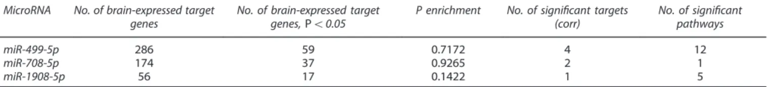
296, 181 and 67 target genes, respectively. Of these 286, 174 and
56 showed brain expression (Table 2).
The target gene enrichment analysis showed no signi
ficant
enrichment of BD-associated genes within the targets of miR-499,
miR-708 or miR-1908 (Table 2). After Bonferroni correction,
miR-1908 had one (KLC2) and miR-708 had two signi
ficant target
genes (NRAS and CREB1), whereas miR-499 had four signi
ficant
target genes (GPC6, C16orf72, WDR82 and CACNB2).
Pathway testing revealed 18 signi
ficant canonical pathways that
are driven by brain-expressed target genes of the three miRNAs
(Table 2). For each miRNA, the results of the GO analysis are
presented as directed acyclic graphs (Supplementary Figure 5).
The target genes that drive a particular pathway are listed in
Supplementary Table 3.
Luciferase assays revealed efficient processing of pri-miR-499,
but not pri-miR-708, upon transfection of the respective constructs
in neurons (Supplementary Figure 6). Overexpression of miR-499
led to a small and statistically nonsigni
ficant increase in spine
volume (Figure 3), but no effect on spine density was observed. As
expected, transfection of the non-effective miR-708 expression
construct had no signi
ficant effect on spine morphological
parameters. Taken together, these results suggest that increasing
levels of the BD-associated miR-499 have no
—or only minimal—
modulatory function during dendritic spine morphogenesis.
DISCUSSION
The present genetic association results for miRNA-coding genes
suggest that miRNAs and their target genes may be implicated in
the development of BD. The nominal P-values of SNPs at miRNA
loci showed early deviation from the expected null line in the Q–Q
plot, and this leftward shift reflects an enrichment of
BD-associated SNPs at miRNA loci.
For the nine miRNAs that withstood Bonferroni correction, we
additionally calculated the gene-based tests on the basis of the
1000 Genomes LD structure. This analysis revealed nominally
gene-based P-values
⩽ 7.20 × 10
− 5for all nine miRNAs, indicating
that the results of gene-based tests on the basis of either HapMap
phase 2 or 1000 Genomes Project data are highly comparable
using our BD GWAS data.
Eight of the nine associated miRNAs were located in a host
gene, including the three brain-expressed miRNAs miR-499,
miR-708 and miR-1908. Recent studies have reported a high
correlation between the expression of a host gene and the
resident miRNA.
15,42Previous authors have hypothesized that this
finding may be because of the fact that miRNAs residing in introns
are likely to share their regulatory elements and primary transcript
with their host gene.
24Some authors point out that host genes
and their resident miRNAs may even have synergistic effects,
which would have important implications for the
fine-tuning of
gene expression patterns in the genome.
43,44On the basis of the
present genetic association results, it is impossible to determine
whether the association was attributable to the host gene, the
miRNA or both. Further analyses are therefore warranted to clarify
this, which was beyond the scope of the present analysis.
However, the general enrichment of BD-associated SNPs at miRNA
loci (Figure 1) and the results of our target gene analyses support
the hypothesis that the majority of the associated miRNAs are
implicated in BD etiology.
Regional association plots and expression data suggest that the
miRNAs miR-499, miR-708 and miR-1908 are the most promising
candidates in terms of the development of BD.
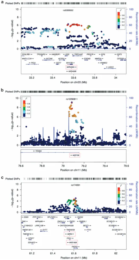
The miRNA miR-499 is located in a region on chromosome
20q11 that showed genome-wide significant association in a
previous GWAS of BD.
45As miR-499 is located in a region of high
LD, which includes the genes GSS, MYH7B and TRPC4AP (Figure 2),
further analyses of this chromosomal region are required to re
fine
the association signal.
45However, miR-499 represents a very
promising candidate in this region.
MiR-499 regulates apoptotic pathways involving the
calcium-dependent protein phosphatase calcineurin.
46A recent study
demonstrated an upregulation of miR-499 in the prefrontal cortex
of patients with depression.
47In a study of exosomal miRNA
expression, miR-499 showed differential expression in the
post-mortem brains of BD patients compared with controls.
48When
considering a possible pathomechanism, it is important to note
that a common SNP (rs3746444) is located in the seed region of
the mature miR-499-3p.
49This seed region is crucial for both the
recognition of the target sites and the binding of the target genes.
The SNP rs3746444 was not among the 2 267 487 SNPs analyzed
in our large BD meta-analysis.
6However, rs3746444 achieved a
nominally significant P-value of 0.0023 (risk allele: rs3746444-G) in
a combined analysis of the seven MooDS samples (2266 patients
and 5028 controls),
6which excluded the PGC data set.
3Furthermore, the allele rs3746444-G has been associated with
hallucinations and lack of motivation in schizophrenia patients.
50This suggests that this SNP may confer susceptibility to BD by
in
fluencing depressive and psychotic endophenotypes. However,
it may only partly explain the association signal at this locus.
Our target gene analysis revealed that miR-499 had four
signi
ficant target genes, including the previously reported
genome-wide signi
ficant risk gene for psychiatric disorders
CACNB2.
51Brain-expressed target genes of miR-499-5p exhibited an
enrichment in biological processes related to cerebral
develop-ment, which might however, at least partly, re
flect the fact that
our pathway analysis was restricted to brain-expressed genes. In
addition, our pathway analysis indicates a potential role of
miR-499 in the regulation of the actin cytoskeleton. Interestingly,
this pathway has been identified in a previous investigation of
differentially and concordantly expressed genes enriched in
association signals for schizophrenia and BD.
52Substantial
research evidence suggests that the rearrangement of the
cytoskeleton is crucial for neuronal cell migration and maturation,
neurite outgrowth and maintenance of synaptic density and
plasticity.
53–56These combined data suggest that miR-499 is an
interesting candidate for BD pathogenesis.
The miRNA miR-708 is located in the
first intron of ODZ4 (odd
Oz/ten-m homolog 4, TENM4), which has been reported as a
genome-wide signi
ficant susceptibility gene for BD.
3A recent study of postpartum psychosis
—a disorder that often
heralds the incipient onset of BD
57—suggested differential
expression of miR-708 in the monocytes of affected patients
compared with controls.
58In another study, Xu et al.
59demon-strated an altered expression pro
file for miR-708 in mouse
hippocampal neurons and showed that this was mediated by
oxidative stress. Another recent study found that miR-708
regulated the expression of neuronatin, which is a membrane
4
Figure 2. Regional association plots of miR-499, miR-708 and miR-1908. Regional association results for the three most promising associated
microRNAs miR-499 (
a), miR-708 (b) and miR-1908 (c), and their ± 500-kb flanking regions were plotted using LocusZoom (Pruim et al.
36). The
plot of miR-1908 (
c) includes miR-611, which is also localized at the depicted chromosomal locus.
protein
in
the
endoplasmic
reticulum.
Interestingly,
the
neuronatin-mediated regulation of intracellular Ca
2+levels has
been implicated in cell migration and neural induction within
embryonic stem cells.
60Our target gene analysis revealed that miR-708 had two
significant target genes. These include CREB1 that has previously
been identified as a susceptibility gene for major depressive
disorder.
61–63In addition, CREB1 was found to be associated with
BD in a recent study of large-scale BD samples
64that included
8403 patients and 11 588 controls of our BD GWAS.
6However, the
present pathway analysis provided no strong evidence for an
enrichment of biological processes of relevance to psychiatric
disorder.
MiR-1908 is located in the
first intron of the fatty acid desaturase
1 (FADS1) gene on chromosome 11. To date, few published studies
have investigated the function of miR-1908. One recent study
implicated miR-1908 as a cancer biomarker.
65A further study
found that miR-1908 belonged to a miRNA cluster that
down-regulates the MARK1 signaling pathway, thus altering cell
proliferation and differentiation.
66Pathway analysis results for miR-1908 indicate a potential role of
the miRNA-regulated target gene network in key neuronal
processes (GO subcategories: neuron projection and nervous
system development). As these pathways showed the strongest
enrichment, further research into miR-1908 and its regulated
network appears to be warranted.
Although initial efforts have been made to elucidate the
regulation of miRNA expression,
67the manner in which miRNA
expression
and
processing
are
regulated
remains
largely
unknown. Given that primiRNAs have a length of 100–1000
-bp,
16the present study investigated common variants at the
miRNA loci and ± 20 kb
flanking sequences in order to capture
possible regulatory regions. However, further analyses of the
regulation of miRNA expression by common variants are required
to determine whether, and how, the presently described
association signals in
fluence the expression levels and function
of the implicated miRNAs. The present approach did not allow
investigation of SNPs with trans-expression quantitative trait loci
(eQTL) effects on miRNAs. As recent studies suggest that ~ 50% of
the identi
fied miRNA eQTLs are trans-eQTLs,
68investigations into
the association between miRNA trans-eQTLs and BD are indicated.
The results of the functional analyses of miR-499 and miR-708 in
rat hippocampal neurons revealed no major contribution of these
miRNAs to the morphogenesis of dendritic spines, which
represent the major sites of synaptic contact. However, only the
results for miR-499 can be considered robust, as the miR-708
expression construct did not increase miR-708 in primary neurons
effectively. Alternative strategies for miR-708 expression, together
Table 2.
Target gene and pathway analysis for miR-499, miR-708 and miR-1908 MicroRNA No. of brain-expressed targetgenes
No. of brain-expressed target genes,Po0.05
P enrichment No. of significant targets (corr)
No. of significant pathways
miR-499-5p 286 59 0.7172 4 12
miR-708-5p 174 37 0.9265 2 1
miR-1908-5p 56 17 0.1422 1 5
Abbreviations: No. of significant pathways, number of significant pathways at P ≤ 0.05; No. of significant targets (corr), number of significant target genes after Bonferroni correction for multiple testing; P enrichment, P-value of the enrichment analysis (Χ2-test). Results of the target gene analysis for the three
brain-expressed microRNAs miR-499, miR-708 and miR-1908 that were associated with bipolar disorder after correction for multiple testing.
Figure 3. Effect of the overexpression of miR-499 and miR-708 on dendritic spine size and density in primary rat hippocampal neurons. DIV14
primary hippocampal neurons were transfected with: (i) empty pmirGLO (250 ng) or (ii) pmirGLO (250 ng) containing pri-miR-499 or pri-miR-708
in the 3
’-untranslated repeat of the Firefly luciferase gene and green fluorescent protein (GFP). The transfected neurons were then cultured
until DIV19 and
fixed for fluorescence microscopy. (a) Representative images for cells transfected with the indicated pmirGLO constructs or
GFP only. A three-dimensional reconstruction was made from seven 45-
μm stacks; scale bars, 5 μm. (b) Spine volume quantification of
hippocampal neurons transfected with the indicated pmirGLO constructs. Values are represented as means
± s.d. (n = 3; 24 neurons per
condition with a 200
–250 spine count per cell). (c) Spine density of hippocampal neurons transfected with the indicated pmirGLO constructs.
Values are represented as means
± s.d. per 10 μm dendritic length (n = 3; 24 neurons per condition). Data are presented as the mean of three
independent transfections normalized to the empty pmirGLO condition
± s.d.
with miR-499/708 loss-of-function approaches, must be tested
before de
finite conclusions regarding the role of these miRNAs in
dendritic spine morphogenesis can be drawn. Moreover, to obtain
more comprehensive insights into the potential effects of these
miRNAs on synaptic function, future experiments should be
complemented by immunocytochemistry analyses of synaptic
marker proteins and electrophysiological recordings. Beyond a
potential involvement in dendritic spine morphogenesis, these
miRNAs could also regulate other aspects of neuronal
morphol-ogy, such as dendrite arborization or axon growth, which could be
tested in future studies.
CONCLUSION
The results of the present miRNA and target gene analyses
suggest that the brain-expressed miRNAs miR-499, miR-708 and
miR-1908 may contribute to the development of BD. Further
research is warranted to elucidate the involvement of these
miRNAs and their downstream pathways in BD.
CONFLICT OF INTEREST
The authors declare no conflict of interest.
ACKNOWLEDGMENTS
We are grateful to all of the patients and control subjects who contributed to this study. The study was supported by the German Federal Ministry of Education and Research (BMBF) through the Integrated Network IntegraMent (Integrated Under-standing of Causes and Mechanisms in Mental Disorders), under the auspices of the e:Med Programme (grant 01ZX1314A to MMN and SC, grant 01ZX1314G to MR, grant 01ZX1314J to BMM), and through e:AtheroSysMed (Systems medicine of myocardial infarction and stroke, grant 01ZX1313B to BMM). MMN is a member of the DFG-funded Excellence-Cluster ImmunoSensation. MMN also received support from the Alfried Krupp von Bohlen und Halbach-Stiftung. The study was supported by the German Research Foundation (DFG; grant FOR2107; RI908/11-1 to MR; SCHR1136/3-1 to GS; NO246/10-1 to MMN). MG-S received the grant no. 89/2012 from UEFISCDI, Romania. Canadian patients were genotyped within the ConLiGen project (www. ConLiGen.org), with the support of a grant from the Deutsche Forschungsge-meinschaft to MR, MB and TGS (RI 908/7-1). Controls for Germany II were drawn from the Heinz Nixdorf Recall Study (HNR) cohort, which was established with the support of the Heinz Nixdorf Foundation. Recruitment of the Australian sample was supported by an Australian NHMRC program grant (number 1037196). The recruitment of the Canadian patients was supported by a grant from the Canadian Institutes of Health Research #64410 to MA. The study also used data generated by the GABRIEL consortium (controls for the sample Russia). Funding for the generation of these data was provided by the European Commission as part of GABRIEL contract number 018996 under the Integrated Program LSH-2004-1.2.5-1. Post-genomic approaches to understand the molecular basis of asthma aiming at a preventive or therapeutic control and the Wellcome Trust under award 084703. Canadian controls were drawn from the French Canadian study (SLSJ), which was supported in part by the Canada Research Chair Environment and genetics of respiratory diseases and allergy, the Canadian Institutes of Health Research (Operating grant No. MOP-13506) and the Quebec Respiratory Network of the Fonds de recherche en Santé du Québec (FRQS). Polish controls were recruited by the International Agency for Research on Cancer (IARC)/Centre National de Genotypage (CNG) GWAS Initiative. We thank the Bipolar Disorder Working Group of the PGC (PGC-BD) for providing access to the relevant data.
REFERENCES
1 Craddock N, Sklar P. Genetics of bipolar disorder. Lancet 2013;381: 1654–1662. 2 Nothen MM, Nieratschker V, Cichon S, Rietschel M. Newfindings in the genetics of
major psychoses. Dialogues Clin Neurosci 2010;12: 85–93.
3 Psychiatric GWAS Consortium Bipolar Disorder Working Group. Large-scale gen-ome-wide association analysis of bipolar disorder identifies a new susceptibility locus near ODZ4. Nat Genet 2011;43: 977–983.
4 Baum AE, Akula N, Cabanero M, Cardona I, Corona W, Klemens B et al. A genome-wide association study implicates diacylglycerol kinase eta (DGKH) and several other genes in the etiology of bipolar disorder. Mol Psychiatry 2008;13: 197–207.
5 Ferreira MA, O'Donovan MC, Meng YA, Jones IR, Ruderfer DM, Jones L et al. Collaborative genome-wide association analysis supports a role for ANK3 and CACNA1C in bipolar disorder. Nat Genet 2008;40: 1056–1058.
6 Muhleisen TW, Leber M, Schulze TG, Strohmaier J, Degenhardt F, Treutlein J et al. Genome-wide association study reveals two new risk loci for bipolar disorder. Nat Commun 2014;5: 3339.
7 Sullivan PF, Daly MJ, O'Donovan M. Genetic architectures of psychiatric disorders: the emerging picture and its implications. Nat Rev Genet 2012;13: 537–551. 8 Nurnberger JI Jr., Koller DL, Jung J, Edenberg HJ, Foroud T, Guella I et al.
Identi-fication of pathways for bipolar disorder: a meta-analysis. JAMA Psychiatry 2014; 71: 657–664.
9 Fineberg SK, Kosik KS, Davidson BL. MicroRNAs potentiate neural development. Neuron 2009;64: 303–309.
10 Schratt G. MicroRNAs at the synapse. Nat Rev Neurosci 2009;10: 842–849. 11 Forstner AJ, Degenhardt F, Schratt G, Nöthen MM. MicroRNAs as the cause of
schizophrenia in 22q11.2 deletion carriers, and possible implications for idio-pathic disease: a mini-review. Front Mol Neurosci 2013;6: 47.
12 Xu B, Karayiorgou M, Gogos JA. MicroRNAs in psychiatric and neurodevelop-mental disorders. Brain Res 2010;1338: 78–88.
13 Kim AH, Reimers M, Maher B, Williamson V, McMichael O, McClay JL et al. MicroRNA expression profiling in the prefrontal cortex of individuals affected with schizophrenia and bipolar disorders. Schizophr Res 2010;124: 183–191. 14 Moreau MP, Bruse SE, David-Rus R, Buyske S, Brzustowicz LM. Altered microRNA
expression profiles in postmortem brain samples from individuals with schizo-phrenia and bipolar disorder. Biol Psychiatry 2011;69: 188–193.
15 Godnic I, Zorc M, Jevsinek Skok D, Calin GA, Horvat S, Dovc P et al. Genome-wide and species-wide in silico screening for intragenic MicroRNAs in human, mouse and chicken. PLoS One 2013;8: e65165.
16 Maffioletti E, Tardito D, Gennarelli M, Bocchio-Chiavetto L. Micro spies from the brain to the periphery: new clues from studies on microRNAs in neuropsychiatric disorders. Front Cell Neurosci 2014;8: 75.
17 Rodriguez A, Griffiths-Jones S, Ashurst JL, Bradley A. Identification of mammalian microRNA host genes and transcription units. Genome Res 2004;14: 1902–1910. 18 Han J, Lee Y, Yeom KH, Kim YK, Jin H, Kim VN. The Drosha-DGCR8 complex in
primary microRNA processing. Genes Dev 2004;18: 3016–3027.
19 Lee Y, Ahn C, Han J, Choi H, Kim J, Yim J et al. The nuclear RNase III Drosha initiates microRNA processing. Nature 2003;425: 415–419.
20 Krol J, Loedige I, Filipowicz W. The widespread regulation of microRNA biogen-esis, function and decay. Nat Rev Genet 2010;11: 597–610.
21 Gregory RI, Chendrimada TP, Cooch N, Shiekhattar R. Human RISC couples microRNA biogenesis and posttranscriptional gene silencing. Cell 2005;123: 631–640.
22 He L, Hannon GJ. MicroRNAs: small RNAs with a big role in gene regulation. Nat Rev Genet 2004;5: 522–531.
23 Meola N, Gennarino VA, Banfi S. microRNAs and genetic diseases. Pathogenetics 2009;2: 7.
24 Bartel DP. MicroRNAs: genomics, biogenesis, mechanism, and function. Cell 2004; 116: 281–297.
25 Didiano D, Hobert O. Molecular architecture of a miRNA-regulated 3' UTR. RNA 2008;14: 1297–1317.
26 Sathyan P, Golden HB, Miranda RC. Competing interactions between micro-RNAs determine neural progenitor survival and proliferation after ethanol exposure: evidence from an ex vivo model of the fetal cerebral cortical neuroepithelium. J Neurosci 2007;27: 8546–8557.
27 Choi WY, Giraldez AJ, Schier AF. Target protectors reveal dampening and balancing of Nodal agonist and antagonist by miR-430. Science 2007;318: 271–274.
28 Johnston RJ, Hobert O. A microRNA controlling left/right neuronal asymmetry in Caenorhabditis elegans. Nature 2003;426: 845–849.
29 Geeleher P, Huang SR, Gamazon ER, Golden A, Seoighe C. The regulatory effect of miRNAs is a heritable genetic trait in humans. BMC Genomics 2012;13: 383. 30 Forstner AJ, Basmanav FB, Mattheisen M, Bohmer AC, Hollegaard MV, Janson E
et al. Investigation of the involvement of MIR185 and its target genes in the development of schizophrenia. J Psychiatry Neurosci 2014;39: 386–396. 31 Lopez JP, Lim R, Cruceanu C, Crapper L, Fasano C, Labonte B et al. miR-1202 is a
primate-specific and brain-enriched microRNA involved in major depression and antidepressant treatment. Nat Med 2014;20: 764–768.
32 Strazisar M, Cammaerts S, van der Ven K, Forero DA, Lenaerts AS, Nordin A et al. MIR137 variants identified in psychiatric patients affect synaptogenesis and neuronal transmission gene sets. Mol Psychiatry 2015;20: 472-481.
33 Wang Z, Zhang C, Huang J, Yuan C, Hong W, Chen J et al. MiRNA-206 and BDNF genes interacted in bipolar I disorder. J Affect Disord 2014;162: 116–119. 34 Liu JZ, McRae AF, Nyholt DR, Medland SE, Wray NR, Brown KM et al. A versatile
gene-based test for genome-wide association studies. Am J Hum Genet 2010;87: 139–145.
35 Kozomara A, Griffiths-Jones S. miRBase: annotating high confidence microRNAs using deep sequencing data. Nucleic Acids Res 2014;42: D68–D73.
36 Pruim RJ, Welch RP, Sanna S, Teslovich TM, Chines PS, Gliedt TP et al. LocusZoom: regional visualization of genome-wide association scan results. Bioinformatics 2010;26: 2336–2337.
37 Ziats MN, Rennert OM. Identification of differentially expressed microRNAs across the developing human brain. Mol Psychiatry 2014;19: 848–852.
38 Siegel G, Obernosterer G, Fiore R, Oehmen M, Bicker S, Christensen M et al. A functional screen implicates microRNA-138-dependent regulation of the depalmitoylation enzyme APT1 in dendritic spine morphogenesis. Nat Cell Biol 2009;11: 705–716.
39 Hackenberg M, Rodriguez-Ezpeleta N, Aransay AM. miRanalyzer: an update on the detection and analysis of microRNAs in high-throughput sequencing experi-ments. Nucleic Acids Res 2011;39: W132–W138.
40 Lewis BP, Burge CB, Bartel DP. Conserved seed pairing, oftenflanked by adeno-sines, indicates that thousands of human genes are microRNA targets. Cell 2005; 120: 15–20.
41 Hawrylycz MJ, Lein ES, Guillozet-Bongaarts AL, Shen EH, Ng L, Miller JA et al. An anatomically comprehensive atlas of the adult human brain transcriptome. Nature 2012;489: 391–399.
42 Baskerville S, Bartel DP. Microarray profiling of microRNAs reveals frequent coexpression with neighboring miRNAs and host genes. RNA 2005;11: 241–247. 43 Lutter D, Marr C, Krumsiek J, Lang EW, Theis FJ. Intronic microRNAs support their host genes by mediating synergistic and antagonistic regulatory effects. BMC Genomics 2010;11: 224.
44 Rearick D, Prakash A, McSweeny A, Shepard SS, Fedorova L, Fedorov A. Critical association of ncRNA with introns. Nucleic Acids Res 2011;39: 2357–2366. 45 Green EK, Hamshere M, Forty L, Gordon-Smith K, Fraser C, Russell E et al.
Replication of bipolar disorder susceptibility alleles and identification of two novel genome-wide significant associations in a new bipolar disorder case-control sample. Mol Psychiatry 2013;18: 1302–1307.
46 Wang JX, Jiao JQ, Li Q, Long B, Wang K, Liu JP et al. miR-499 regulates mito-chondrial dynamics by targeting calcineurin and dynamin-related protein-1. Nat Med 2011;17: 71–78.
47 Smalheiser NR, Lugli G, Zhang H, Rizavi H, Cook EH, Dwivedi Y. Expression of microRNAs and other small RNAs in prefrontal cortex in schizophrenia, bipolar disorder and depressed subjects. PLoS One 2014;9: e86469.
48 Banigan MG, Kao PF, Kozubek JA, Winslow AR, Medina J, Costa J et al. Differential expression of exosomal microRNAs in prefrontal cortices of schizophrenia and bipolar disorder patients. PLoS One 2013;8: e48814.
49 Gong J, Tong Y, Zhang HM, Wang K, Hu T, Shan G et al. Genome-wide identi fi-cation of SNPs in microRNA genes and the SNP effects on microRNA target binding and biogenesis. Hum Mutat 2012;33: 254–263.
50 Zou M, Li D, Lv R, Zhou Y, Wang T, Liu J et al. Association between two single nucleotide polymorphisms at corresponding microRNA and schizophrenia in a Chinese population. Mol Biol Rep 2012;39: 3385–3391.
51 Cross-Disorder Group of the Psychiatric Genomics Consortium. Identification of risk loci with shared effects onfive major psychiatric disorders: a genome-wide analysis. Lancet 2013;381: 1371–1379.
52 Zhao Z, Xu J, Chen J, Kim S, Reimers M, Bacanu SA et al. Transcriptome sequencing and genome-wide association analyses reveal lysosomal function and actin cytoskeleton remodeling in schizophrenia and bipolar disorder. Mol Psychiatry 2015;20: 563-572.
53 Auer M, Hausott B, Klimaschewski L. Rho GTPases as regulators of morphological neuroplasticity. Ann Anat 2011;193: 259–266.
54 Bellenchi GC, Gurniak CB, Perlas E, Middei S, Ammassari-Teule M, Witke W. N-cofilin is associated with neuronal migration disorders and cell cycle control in the cerebral cortex. Genes Dev 2007;21: 2347–2357.
55 de Curtis I. Functions of Rac GTPases during neuronal development. Dev Neurosci 2008;30: 47–58.
56 Kreis P, Barnier JV. PAK signalling in neuronal physiology. Cell Signal 2009;21: 384–393.
57 Jones I, Craddock N. Familiality of the puerperal trigger in bipolar disorder: results of a family study. Am J Psychiatry 2001;158: 913–917.
58 Weigelt K, Bergink V, Burgerhout KM, Pescatori M, Wijkhuijs A, Drexhage HA. Down-regulation of inflammation-protective microRNAs 146a and 212 in mono-cytes of patients with postpartum psychosis. Brain Behav Immun 2013; 29: 147–155.
59 Xu S, Zhang R, Niu J, Cui D, Xie B, Zhang B et al. Oxidative stress mediated-alterations of the microRNA expression profile in mouse hippocampal neurons. Int J Mol Sci 2012;13: 16945–16960.
60 Ryu S, McDonnell K, Choi H, Gao D, Hahn M, Joshi N et al. Suppression of miRNA-708 by polycomb group promotes metastases by calcium-induced cell migration. Cancer Cell 2013;23: 63–76.
61 Carlezon WA Jr., Duman RS, Nestler EJ. The many faces of CREB. Trends Neurosci 2005;28: 436–445.
62 Maher BS, Hughes HB 3rd, Zubenko WN, Zubenko GS. Genetic linkage of region containing the CREB1 gene to depressive disorders in families with recurrent, early-onset, major depression: a re-analysis and confirmation of sex-specific effect. Am J Med Genet B Neuropsychiatr Genet 2010; 153B: 10–16. 63 Wallace TL, Stellitano KE, Neve RL, Duman RS. Effects of cyclic adenosine
monophosphate response element binding protein overexpression in the baso-lateral amygdala on behavioral models of depression and anxiety. Biol Psychiatry 2004;56: 151–160.
64 Li M, Luo XJ, Rietschel M, Lewis CM, Mattheisen M, Muller-Myhsok B et al. Allelic differences between Europeans and Chinese for CREB1 SNPs and their implica-tions in gene expression regulation, hippocampal structure and function, and bipolar disorder susceptibility. Mol Psychiatry 2014;19: 452–461.
65 Rawlings-Goss RA, Campbell MC, Tishkoff SA. Global population-specific variation in miRNA associated with cancer risk and clinical biomarkers. BMC Med Genomics 2014;7: 53.
66 Ye SB, Li ZL, Luo DH, Huang BJ, Chen YS, Zhang XS et al. Tumor-derived exosomes promote tumor progression and T-cell dysfunction through the regulation of enriched exosomal microRNAs in human nasopharyngeal carcinoma. Oncotarget 2014;5: 5439–5452.
67 Gamazon ER, Ziliak D, Im HK, LaCroix B, Park DS, Cox NJ et al. Genetic architecture of microRNA expression: implications for the transcriptome and complex traits. Am J Hum Genet 2012;90: 1046–1063.
68 Borel C, Deutsch S, Letourneau A, Migliavacca E, Montgomery SB, Dimas AS et al. Identification of cis- and trans-regulatory variation modulating microRNA expression levels in humanfibroblasts. Genome Res 2011; 21: 68–73.
This work is licensed under a Creative Commons Attribution 4.0 International License. The images or other third party material in this article are included in the article’s Creative Commons license, unless indicated otherwise in the credit line; if the material is not included under the Creative Commons license, users will need to obtain permission from the license holder to reproduce the material. To view a copy of this license, visit http://creativecommons.org/licenses/ by/4.0/