Development of novel aptasensors for
the detection of mycotoxins
Xiaodong GUO
Promotors: Prof. Marie-Laure Fauconnier
Prof. Jiaqi Wang
COMMUNAUTÉ FRANÇAISE DE BELGIQUE UNIVERSITÉ DE LIÈGE – GEMBLOUX AGRO-BIO TECH
DEVELOPMENT OF NOVEL APTASENSORS
FOR THE DETECTION OF MYCOTOXINS
Xiaodong GUO
Dissertation originale présentée en vue de l’obtention du grade de docteur en sciences agronomiques et ingénierie biologique
Promoteurs: Prof. Marie-Laure Fauconnier Prof. Jiaqi Wang
Copyright. ette uvre est sous licence reative o ons ous tes libre de
reproduire de odi ier de distribuer et de co uni uer cette création au public selon les conditions suivantes:
- paternité vous deve citer le no de l auteur original de la anie re indi uée par l auteur de l uvre ou le titulaire des droits ui vous con re cette autorisation ais pas d une ani re ui suggérerait u'ils vous soutiennent ou approuvent votre utilisation de l uvre ;
- pas d utilisation co erciale vous n ave pas le droit d utiliser cette création à des fins commerciales;
- partage des conditions initiales à l'identique (SA): si vous modifiez, transfor e ou adapte cette création vous n ave le droit de distribuer la création ui en résulte ue sous un contrat identi ue celui-ci c a ue réutilisation ou distribution de cette création, vous devez faire apparaitre clairement au public les conditions
Résumé
Xiaodong Guo. (2020). Développement de nouveaux aptasenseurs pour la détection des mycotoxines (Thèse de doctorat en anglais). Gembloux, Belgique,
Gembloux Agro-Bio Tech, Université de Liège, 107 p., 11 tableaux, 29 fig.
Résumé —Les contaminations par des mycotoxines, un défi mondial important,
attirent de plus en plus l'attention du Centre international de recherche sur le cancer (CIRC), de l'Organisation mondiale de la santé (OMS) et des scientifiques en sécurité alimentaire. De nombreux pays et organisations ont établi les limites maximales de contamination des principales mycotoxines à des valeurs très faibles. Les stratégies analytiques traditionnelles sont principalement basées sur des méthodes quantitatives instrumentales et des tests immunologiques. Les aptamères sont un nouvel élément de reconnaissance de molécules similaires aux anticorps mais potentiellement plus performant, ils attirent de plus en plus l'attention des scientifiques. Par conséquent, des aptamères spécifiques pourraient être utilisés pour construire des biocapteurs pour la détection des mycotoxines. L'objectif de cette thèse est de développer de nouveaux biocapteurs à base d'aptamères pour la détermination sensible des traces de mycotoxines et de fournir une application prometteuse de ces aptasenseurs pour plus de facteurs de risque dans les sciences de la sécurité alimentaire.
Le contenu de la thèse et ses principaux résultats sont les suivants:
(1) Biocapteur à base d'aptamères pour la détection des mycotoxines
Les mycotoxines sont une grande famille de métabolites secondaires synthétisés par des champignons, elles présentent un grand danger pour l'homme et les animaux. Une ajorité de pays et d’organis es décisionnels tels ue l Union européenne ont établi une série d'exigences et fixé les niveaux maximaux tolérés. De ce fait, le développement d'une plateforme analytique hautement sensible et spécifique pour les mycotoxines est très demandé. En raison de leur simplicité, de leur coût et de leur rapidité, les biocapteurs à base d'aptamères ont été développés avec succès pour la détection de diverses mycotoxines avec une sensibilité et une sélectivité élevées par rapport aux méthodes instrumentales traditionnelles et aux approches immunologiques. Dans ce travail, nous discutons et analysons le développement d'aptasenseurs pour la détection des mycotoxines dans les produits alimentaires et agricoles au cours des onze dernières années et couvrons la littérature depuis le premier rapport en 2008 jusqu'à aujourd'hui. Sont également résumés les défis et les tendances futures pour la sélection des aptamères spécifique de divers mycotoxines et aptasenseurs pour l'analyse multi-mycotoxines. Compte tenu du développement prometteur et de l'application potentielle d'aptasenseurs, les futures recherches seront le témoin de la grande potentialité du biocapteur à base d'aptamères pour le domaine de la sécurité alimentaire.
(2) Un aptasensor qPCR pour la détection sensible de l'aflatoxine M1
L'aflatoxine M1 (AFM1), l'une des mycotoxines les plus toxiques, présente de graves risques pour la santé. L'AFM1 avait précédemment été classée comme
cancérogène du groupe 2B (CIRC, 1993) et a été classé comme cancérogène du groupe 1 par le Centre international de recherche sur le cancer (CIRC) de l'Organisation Mondiale de la Santé (OMS) (CIRC, 2002). La détection de l'AFM1 joue donc un rôle important pour le contrôle de la qualité et de la sécurité sanitaire des aliments. Dans ce travail, un aptasensor sensible et fiable a été développé pour la détection de l'AFM1. L'immobilisation de l'aptamère par une forte interaction avec la biotine – streptavidine a été utilisée comme élément de reconnaissance moléculaire, et son ADNsb complémentaire a été utilisé comme modèle dans l’utilisation de Real Ti e - Quantitative Polymerase Chain Reaction (RT-qPCR). Dans des conditions d'essai optimisées, une relation linéaire (allant de 1,0 × 10⁻⁴ à 1,0 µg L-1) a été obtenue avec une limite de détection (LOD) jusqu'à 0,03 ng L-1. De plus, l'aptasensor développé ici présente une sélectivité élevée pour l'AFM1 par rapport aux autres mycotoxines et de petits effets de réaction croisée avec des analogues structuraux. La méthode proposée ici a été appliquée avec succès à la détection quantitative de l'AFM1 dans des échantillons de céréales de riz pour nourrissons et de lait en poudre pour nourrissons. Les résultats ont démontrés que l'approche actuelle est potentiellement utile pour la sécurité sanitaire des aliments et qu'elle pourrait être étendue à un grand nombre de produits cibles.
(3) Un nouvel aptasenseur à base d'oxyde de graphène pour la détection par fluorescence de l'aflatoxine M1 dans le lait en poudre
Dans cet article, un aptasensor fluorescent rapide et sensible pour la détection de l'aflatoxine M1 (AFM1) dans le lait en poudre a été développé. De l'oxyde de graphène (GO) a été utilisé pour inhiber la fluorescence de l'aptamère marqué à la carboxyfluorescéine et empêcher le clivage de l'aptamère par la nucléase. Lors de l'ajout d'AFM1, une formation de complexe AFM1 / aptamère entraine le détachement de celle-ci de la surface du GO, l'aptamère est ensuite clivé par la DNase I et l'AFM1 cible est libéré pour un nouveau cycle, ce qui a conduit à une grande amplification du signal et une haute sensibilité. Dans des conditions optimisées, la détection basée sur le GO de l'aptasensor présente une réponse linéaire AFM1 dans une plage dyna i ue de 0 2 10 μg / kg avec une li ite de détection LOD de 0 05 μg / kg De plus l aptasensor développé a ontré une aute spécificité envers AFM1 sans interférence avec d'autres mycotoxines. En outre, la technique a été appliquée avec succès pour la détection de l'AFM1 dans des échantillons de lait en poudre pour nourrissons. Cet aptasensor proposé ici offre une technologie prometteuse pour la sécurité alimentaire et peut être étendu à différent produits cibles.
(4) Aptasensor fluorescent entraîné par oxyde de graphène pour la détection
de la fumonisine B1
La fumonisine B1 (FB1), est également une toxine très toxique, elle a été désignée comme cancérogène possible du groupe 2B par le Centre international de recherche sur le cancer (CIRC) en 2002. Par conséquent, la demande pour des approches
extincteur de fluorescence contre les aptamères modifié par ROX et comme protecteur de l'aptamère contre le clivage par la D ase I ainsi u’en a ont des cycles permettant la détection d'amplification de signal. Cette stratégie de détection proposée a montré une bonne linéarité pour la détermination de FB1 dans la gamme dynamique de 0,5 à 20 ng mL-1 avec une bonne corrélation de R2 = 0,995. Sa détection de limite a été établie à 0,15 ng mL-1 (S / N = 3). L'analyse spécifique a indiqué que cet aptasenseur était sélectif pour FB1 ainsi que pour d'autres mycotoxines. De plus, l'application pratique dans des échantillons réels de cet aptasenseur pour la détection de FB1 a été étudiée. La plateforme de détection proposée ici sera utile pour une application dans le domaine de la sécurité alimentaire en vue l'analyse des mycotoxines.
(5) Articles faits saillants et perspectives d'avenir
En utilisant les nouveaux biocapteurs à base d'aptamères, nous avons développé plusieurs approches afin de détecter les mycotoxines les plus toxiques pour la sécurité alimentaire. De plus, cette stratégie de détection pourrait être appliquée pour une plus grande détermination des composés toxiques par simple remplacement des aptamères spécifiques. Bien que l'applicabilité, la faisabilité et la précision de ces aptasenseurs ai ait l’objet d’études sur des éc antillons artificiellement contaminés, des recherches complémentaires étaient nécessaires pour une validation de ces aptasenseurs en conditions réelles afin de déterminer les performances telles que limite de détection et de quantification, la précision, la justesse l’exactitude etc Grâce la validation de cette méthode, ces aptasenseurs pourront être largement utilisés pour la détection des mycotoxines. Les perspectives se concentreront sur la simplification du principe et des dispositifs analytiques et sur la combinaison de nouveaux aptamères avec de nouveaux matériaux et techniques pour améliorer les performances analytiques et l'aspect pratique du marché des aptasenseurs.
Mots-clés: mycotoxines, aflatoxine, fumonisine, aptamère, biocapteur, RT-qPCR,
Abstract
Xiaodong Guo. (2020). Development of novel aptasensors for the detection of mycotoxins (PhD Dissertation in English). Gembloux, Belgium, Gembloux
Agro-Bio Tech, University of Liège, 107 p., 11 table, 29 fig.
Abstract — Mycotoxins contaminants, one of the most serious global challenges,
have been attracted more and more attention from International Agency for Research on Cancer (IARC) of the World Health Organization (WHO) and scientists in food safety sciences. Many countries and organizations have established the maximum contamination level of the main mycotoxins at very low values. Traditional analytic strategies are mainly based on instrumental quantitative method and immunoassays approaches. Aptamer, a novel molecules recognition element like or even superior to antibodies, has attracted more and more attentions for scientists. Therefore, specific aptamers could be employed to construct biosensors for the detection of mycotoxins. The objective of this thesis is to develop novel aptamer-based biosensors for sensitive determination of trace levels of mycotoxins and to provide a promising application of these aptasensors for more hazard factors in food safety sciences.
The main contents and results are as follows:
(1) Aptamer-based biosensor for detection of mycotoxins
Mycotoxins are a large types of secondary metabolites appeared by fungi, they pose a great hazard and toxic reactions to human and animals. A majority of countries and regulators, such as European Union, have established series of requirements and set the maximum tolerated levels. The development of high sensitive and specific analytical platform for mycotoxins is much in demand to address new challenges for food safety in worldwide. Due to the superiority of simple, rapid, and low-cost characteristics, aptamer-based biosensors are successfully developed for the detection of various mycotoxins with high sensitivity and selectivity compared with traditional instrumental methods and immunological approaches. In this article, we discuss and analyze the development of aptasensors for mycotoxins determination in food and agricultural products during the last eleven years and cover the literatures from the first report in 2008 until today. In addition, challenges and future trends for the selection of aptamers towards various mycotoxins and aptasensors for multi-mycotoxins analysis are summarized. Given the promising development and potential application of aptasensors, the future researches will witness the great practicability of aptamer-based biosensor for food safety field.
(2) A qPCR aptasensor for sensitive detection of aflatoxin M1
Aflatoxin M1 (AFM1), one of the most toxic mycotoxins, imposes serious health a ards AFM1 ad previously been classified as a group 2 carcinogen (IARC,
food safety. In this work, a sensitive and reliable aptasensor was developed for the detection of AFM1. The immobilization of aptamer through a strong interaction with biotin–streptavidin was used as a molecular recognition element, and its complementary ssDNA was employed as the template for a real-time quantitative polymerase chain reaction (RT-qPCR) amplification. Under optimized assay conditions, a linear relationship (ranging from 1.0×10⁻⁴ to 1.0 µg L-1) was achieved with a limit of detection (LOD) down to 0.03 ng L-1. In addition, the aptasensor developed here exhibits high selectivity for AFM1 over other mycotoxins and small effects from cross-reaction with structural analogs. The method proposed here has been successfully applied to quantitative determination of AFM1 in infant rice cereal and infant milk powder samples. Results demonstrated that the current approach is potentially useful for food safety analysis, and it could be extended to a large number of targets.
(3) A novel graphene oxide-based aptasensor for amplified fluorescent detection of aflatoxin M1 in milk powder
In this paper, a rapid and sensitive fluorescent aptasensor for the detection of aflatoxin M1 (AFM1) in milk powder has been developed. Graphene oxide (GO) was employed to quench the fluorescence of carboxyfluorescein-labelled aptamer and protect the aptamer from nuclease cleavage. Upon the addition of AFM1, a formation of AFM1/aptamer complex resulted in the aptamer detached from the surface of GO, then the aptamer was cleaved by DNase I and the target AFM1 was released for a new cycle, which led to a great signal amplification and high sensitivity. Under optimized conditions, the GO-based detection of the aptasensor exhibited a linear response to AFM1 in a dynamic range from 0.2 to 10 μg/kg wit a li it o detection LOD o 0 05 μg/kg Moreover t e developed aptasensor showed a high specificity towards AFM1 without interference from other mycotoxins. In addition, the technique has been successfully applied for detection of AFM1 in infant milk powder samples. This aptasensor proposed here offers a promising technology for food safety and can be extended to various targets.
(4) Graphene oxide driven fluorescent aptasensor for the detection of fumonisin B1
Fumonisin B1 (FB1), one of the most toxic mycotoxins, has been designated as possible 2B group carcinogen by the International Agency for Research on Cancer (IARC) in 2002. Therefore, simple, sensitive and specific approaches for the detection of FB1 are much in demand. In this study, a novel aptasensor was introduced for FB1 analysis based on graphene oxide (GO) and DNase I signal amplification. GO was adopted as a fluorescence quencher against ROX-modified aptamer and a protectant for the aptamer from cleavaging by DNase I for subsequent target cycling and signal amplification detection. This proposed sensing strategy exhibited a good linearity for FB1 determination in the dynamic range from 0.5 to 20 ng mL-1 with a good correlation of R2 = 0.995. Its detection of limit was established at 0.15 ng mL-1 (S/N = 3). The specific analysis indicated that this aptasensor was selective for FB1 other than other mycotoxins. In addition, the
practical application in real samples of this aptasensor for the detection of FB1 was investigated. The sensing platform proposed here was useful for a potential application in the field of food safety for mycotoxins analysis.
(5) Articles highlights and future perspective
By using the novel aptamer-based biosensors, we developed several approaches for the detection of the most toxic mycotoxins for food safety. In addition, these sensing strategies could be applied for more hazard factors determination by simple replacement of the specific aptamers. More importantly, though the practical applicability, feasibility, and accuracy of these proposed aptasensors were investigated and evaluated through the analysis of the spiked samples experiments, the future’s researches were needed for a validation of these aptasensors with real contaminated samples to determine the performances such as limit of detection and quantification, precision, trueness, accuracy, etc. Through the method validation, these aptasensors will be widely used for the detection of mycotoxins. In addition, future direction will focus on the simplification of analytic principle and devices and the combination of novel aptamers with new materials and techniques to improve the analytical performance and market practicality of aptasensors.
Keywords: mycotoxins, aflatoxin, fumonisin, aptamer, biosensor, RT-qPCR,
Acknowledgments
I wish to express my sincere gratitude and appreciation to all the members who contributed to the achievements of the PhD dissertation. These achievements were supported by the innovation groups and excellent profs and doctors in Ulg and CAAS.I wish to thank the Special Fund for Agroscientific Research in the Public Interest (201403071), the National Natural Science Foundation of China (No. 21305158), the Agricultural Science and Technology Innovation Program (ASTIP-IAS12), Modern Agro Industry Technology Research System of the PR China (CARS-36), and the Project of risk assessment on raw milk (GJFP2019026). We specially thank the University of Liège-Gembloux Agro-Bio Tech and more specifically the research platform AgricultureIsLife for the funding of the scientific stay in Belgium that made this thesis possible.
Time flies, an intense and fulfilling doctoral life is near to end. During the doctoral study, many thanks to many teachers, classmates and friends for their kind cares and helps. Both happiness and challenges are present in my life and I have made some achievements. The harvest is amazing, such as increasing of abundant knowledge, improving the thinking of innovation ideas, expression potential and broad vision, cultivated in reading and practice. From the beginning of the project to the successful completion of the thesis, I have always received the support, help and encouragement from my teachers, classmates, and friends. I would like to express my sincere THANKS to them since the thesis is about to be completed.
First of all, I wish to express my deepest gratitude to my promotor, Prof. Jiaqi WANG, for his guidance and concern in my study and life in the past doctoral years. His research spirit and academic achievements are very admirable. He provided me the opportunity to meet this fascinating and challenging work. The research ideas and thesis writing are carried out under the guidance of Prof. Wang. He has devoted a lot of hard work to the process of the overall design and implementation of the experiment, as well as writing and revision of my papers. It was a kindness to provide me possibilities to attend various scientific conferences, meetings and workshops. I am indebted to them for their professional, social and moral support throughout my PhD. Here, I would like to extend my heartfelt thanks to Prof. Wang, hoping he will have a smooth life and work in the future.
Secondly, I sincere gratitude the co-promotor, Prof. Marie-Laure FAUCONNIER, for her guidance, encouragement and academic advice in the experimental process and thesis writing. Prof. Fauconnier is a rigorous, careful, open-minded teacher with innovative spirit, which is the direction of my future study. Her guidance and insights have been reflected in many parts of this thesis and will be useful for my future career as well.
Third, my sincere thanks to Prof. Fang WEN and Prof. Nan ZHENG for their selfless help in my experiment and life. These two teachers are conscientious and dedicated in their work. They put forward valuable suggestions and provided a guarantee for the smooth progress of my experiment. Their selfless and approachable
attitude to life also affected me and made me grow up in the doctoral life.
Many thanks to Prof. Li Min for his help on data analysis and useful academic instruction for my experiment, and thanks to Dr Wei FANG and Dr. Matthew SAIVE for their carefully editing the English and scientific language for my papers.
Thanks also to the large group of milk research team in CAAS who have enjoyed doctoral life together. This group makes my doctoral life monotonous but not boring, plain but not dull. Special thanks to Dr. Fang WEN and Dr. Ming LI for their guidance and help in my experimental techniques. At the same time, I would also like to thank Songli LI, Shengguo ZHAO, Yangdong ZHANG, Bingyao DU, Chuanyou SU, Huaigu YANG, Jinhui YANG, Xuewei ZHOU, Meixia CHEN, Hao ZHANG, Muchen ZHANG, Xu ZHOU, Guodong LI, Lei XING, for their supports and helps, and international students THOMAS and JOSSELIN for guiding me in English. Thanks to Xingwen WANG and Lu CHEN in the laboratory for their selfless help and concern.
I wish to express my sincerest thanks to Yifeng GUO, Peipei ZHANG, Hejun LU, Yu CHEN, Lin LI, Bowen HU, Hui HU, Jingwang CHEN, Mingchao MA, Xiaomei YANG, Yingying ZHU, Lei ZHANG, Baoqing CHEN, Yong ZHANG, Peng LI, and Li CHEN for their constant support and help during my study in Gembloux. And thanks to my friends and classmates, who gave me understanding and support in learning and life.
I am extremely grateful to all extensive support from my dear colleagues in the Gembloux Agro-Bio Tech, University of Liege (GABT-ULg). It is a pleasure to experience doctoral work at Gembloux Agro-bio Tech and having friendships and scientific discussion with my colleges, namely, Josselin, Davin, Thomas, Danny and Matthew, as well as Prof.FAUCONNIER and office secretary Kenne in the laboratory of chemistry.
I would also like to send my warmest thanks to Mrs. Joëlle HAINE: Thank you for all the help for arranging my accommodation and allowance.
I would like to express my sincere thanks to THESIS COMMITTEE MEMBERS, Prof. Patrick du JARDIN, Prof. Marianne SINDIC, Prof. Hélène SOYEURT, Prof. ML SCIPPO, Prof. M. DEBOEVER and Prof. Jiaqi WANG for their guidance, comments and suggestions for my thesis.
Finally, special thanks to my parents, my brother, my sister, my nieces, my wife Yunjing ZENG and my young daughter Shuxin GUO for their continuous support and understanding for my studies. They share my pain and joy and make me happy and power to improve. Hereby, I extend my sincere thanks and best wishes to them! I love you forever.
Xiaodong Guo 2020
Tables of Contents
Résumé ... I Abstract ... IV Acknowledgments ... VII Tables of Contents ... IX List of Figures ... XII List of Tables ... XV List of Abbreviations ... XVI Chapter 1 General introduction: Aptamer-based biosensor for detection of
mycotoxins ... 1
1 Introduction ... 4
2 Aptamers selection ... 7
3 Aptasensor for the analysis of ochratoxin A ... 10
3.1 Fluorescent aptasensor for OTA ... 13
3.2 Colorimetric aptasensor for OTA ... 15
3.3 Electrochemical aptasensor for OTA ... 16
4 Aptasensor for the analysis of aflatoxins ... 20
4.1 Aptasensor for AFB1 ... 23
4.2 Aptasensor for AFM1 ... 25
5 Aptasensors for other mycotoxins ... 27
6 Aptasensors for simultaneous multi-mycotoxins analysis ... 28
7 Microbial volatile organic compounds (mVOCs)-based biosensors for potential application of mycotoxins determination ... 30
8 Challenges and limitations of aptasensors ... 31
9 Conclusions and future trends ... 31
Author contributions ... 32
Acknowledgements ... 32
References ... 32
Chapter 2 Objectives and thesis structure ... 43
Chapter 3 A qPCR aptasensor for sensitive detection of aflatoxin M1 ... 46
1 Introduction ... 49
2 Materials and methods ... 50
2.1 Methods ... 50
2.2 Materials and reagents ... 51
2.3 Immobilization of the aptamer ... 52
2.4 RT-qPCR measurements for AFM1 ... 52
2.5 Specificity analysis ... 53
2.6 Method validation ... 53
2.7 Statistical analysis ... 53
3 Results and discussion ... 53
3.1 Optimization of the amplification of complementary ssDNA ... 53
3.2 Optimization of streptavidin and the biotinylated aptamer ... 55
3.3 AFM1 determination ... 56
3.5 Repeatability analysis ... 61
3.6 Method validation ... 62
4 Conclusion ... 63
Acknowledgements ... 63
References ... 63
Chapter 4 A novel graphene oxide-based aptasensor for amplified fluorescent detection of aflatoxin M1 in milk powder ... 67
1 Introduction ... 70
2 Experimental ... 71
2.1 Materials and reagents ... 71
2.2 Fluorescent response of the amplified aptasensor for AFM1 ... 72
2.3 Specificity analysis ... 72
2.4 Method validation ... 72
2.5 Statistical analysis ... 72
3 Results and discussion ... 73
3.1 Design strategy for AFM1 detection based on graphene oxide sensing platform ... 73
3.2 Optimization of experimental conditions ... 74
3.3 Analytical performance of the aptasensor ... 76
3.4 The specificity of the aptsensor ... 77
3.5 Method validation ... 78
4 Conclusion ... 78
Author contributions ... 78
Acknowledgements ... 78
References ... 79
Chapter 5 Graphene oxide driven fluorescent aptasensor for the detection of fumonisin B1 ... 83
1 Introduction ... 86
2 Experimental ... 87
2.1 Materials and reagents ... 87
2.2 Fluorescent aptasensing towards FB1 ... 87
2.3 Specificity analysis ... 88
2.4 Practicability of the sensing platform ... 88
2.5 Statistical analysis ... 88
3 Results and discussion ... 88
3.1 Sensing strategy for FB1 detection ... 88
3.2 Amplified detection of FB1 with DNase I ... 89
3.3 Analytical performance of this sensing platform ... 90
3.4 The specificity of this sensing platform ... 92
3.5 Method validation ... 92
4 Conclusion ... 94
Reference ... 103
List of Figures
Figure 1-1: Chemical structures of the important mycotoxins. ... 6 Figure 1-2: Principle illustration of fluorescence, colorimetric and electrochemical
aptasensor for the detection of small molecule mycotoxins. ... 8
Figure 1-3: Schematic representation of the fluorescent aptasensor for OTA
determination based on the conformational of aptmer. ... 13
Figure 1-4: Principle illustration of colorimetric aptasensor for detection of OTA via
AuNPs encapsulated DNA hydrogel. Reprinted from Liu et al. (2015) with permission. ... 16
Figure 1-5: Principle diagram of electrochemical aptasensor for OTA analysis based
on RCA signal amplification. Reprinted from Huang et al. (2013) with permission. ... 18
Figure 1-6: Sensing strategy of electrochemical aptasensor for detection of OTA
based on exonuclease-assistant signal amplification. ... 20
Figure 1-7: Schematic illustration of chemiluminescent aptasensor for AFB1
determination by using HCR signal amplification. Reprinted from Yao et al. (2019) with permission. ... 24
Figure 1-8: Sensing illustration of electrochemical aptasensor for detection of AFB1
based on the conformational change of aptamer. ... 25
Figure 1-9: Schematic illustration of fluorescent aptasensor for detection of AFM1
by using graphene oxide signal amplification. Reprinted from Guo et al. (2019) with permission. ... 26
Figure 1-10: (A) Diagram of the construction of 3 ds DNA-PtNi@Co-MOF networks.
(B) Schematic illustration of the proposed aptasensor for the detection of ZEN. Reprinted from He et al. (2020). ... 28
Figure 1-11: (A) Principle diagram of SPR aptasensor platform. (B) Principle
diagram of sensor chip and optical setup in SPR. Reprinted from Wei et al. (2019) with permission. ... 29
Figure 3-1: Schematic to illustrate the aptasensor for detection of Aflatoxin M1.... 50
Figure S3-1: (A) The amplification curves at different concentrations of the
complementary ssDNA in the range of 1×10⁻³ to 10 nM. (B) The standard curve relating and the Ct value to the complementary ssDNA concentration. ... 54
Figure S3-2: Five melting curves corresponding to the amplification curves in Fig.
S3-1A for the detection of AFM1. An obvious single peak was shown at 80°C.
... 55
Figure S3-3: (A) Ct values change upon different concentrations of aptamer in the
complementary ssDNA (10 nM) and AFM1 (1 ng mL-1). Values shown are means of triplicate analyses and standard deviations are also plotted. ... 56
Figure 3-2: (A) The amplification curves at different concentrations of AFM1 in the
range of 1×10⁻⁴ to 1 µg L-1 for the determination of AFM1 DNA1, including the negative control without AFM1. (B) The standard curves between the AFM1 concentration and the Ct value in the range of 1×10⁻⁴ to 1 µg L-1. ... 57
Figure 3-3: The amplification curves at different concentrations of AFM1 in the
range of 1×10⁻⁴ to 1 µg L-1 for the determination of different AFM1 DNA, including the negative control without AFM1. (A) AFM1 DNA2; (B) AFM1 DNA3; (C) AFM1 DNA4; (D) AFM1 DNA5; (E) AFM1 DNA6. ... 60
Figure 3-4: The Ct values in the absence and presence of 1 ng mL-1 mycotoxins
including AFM1, OTA, ZEA, AFB1, AFB2, FB1, Mix1 (OTA, ZEA, AFB1, AFB2, FB1) and Mix2 (OTA, ZEA, AFB1, AFB2, FB1, and AFM1). The experiment conditions are as following: complementary ssDNA 10 nM, aptamer 10 nM, and streptavidin 2.5 ng mL-1. Means and standard deviations are shown with three replicates for each treatment. ... 61
Figure S3-4: Repeatability of the proposed aptasensor for AFM1 detection. The
experiment conditions are as following: AFM1 1.0 ng mL-1, complementary ssDNA 10 nM, aptamer 10 nM, and streptavidin 2.5 ng mL-1. Means and standard deviations are shown with three replicates at each time. ... 62
Figure 4-1: Illustration of the aptasensor for detection of aflatoxin M1. ... 73 Figure 4-2: Fluorescence emission spectra of the aptasensor in the absence (0) of
AFM1, presence of 10 ng mL−1 AFM1, and 10 ng mL−1 AFM1 and 200 U D ase I Excitation wavelengt λex is set at 480 n onditions 200 nM AFM1 apta er 20 μg L−1 GO in Tris buffer (10 mM Tris, 120 mM NaCl, 5 mM KCl, 20 mM CaCl2, pH 7.0). ... 74
Figure S4-1: Fluorescence emission spectra of the aptasensor in the addition of GO
at various concentrations T e experi ent conditions are as ollowing λex = 480nm, 200 nM AFM1 aptamer. ... 75
Figure S4-2: Fluorescence intensity with the addition of DNase I at various
concentrations. The experiment conditions are as follows: Excitation and e ission wavelengt are at λex/λe = 480/520 n 200 nM AFM1 apta er 20 μg L-1
GO, 10 ng mL-1 AFM1. ... 75
Figure 4-3: (A) Fluorescence emission spectra of the aptasensor in the addition of
AFM1 at various concentrations. (B) Linear relationship between the fluorescence intensity and AFM1 concentrations in the range of 0.2 to 10 ng mL−1. ... 76
Figure 4-4: Fluorescence intensity in the absence (control) and presence of 4 ng
mL−1 ycotoxins AFM1 AF 1 OTA ZEA and α-ZOL. The experiment conditions are as ollows Excitation wavelengt λex is set at 480 n 200nM AFM1 apta er 20 μg L−1 GO, 200 U DNase I. Every data point is the mean
of three replicates. ... 77
Figure 5-2: Fluorescence emission spectra of the aptasensor in the absence (0) of
FB1, presence of 10 ng mL-1 FB1, and 10 ng mL-1 FB1 and 100 U DNase I. Excitation wavelengt λex is set at 585 n onditions 100 nM F 1 apta er 20 μg L-1
GO in Tris buffer (10 mM Tris, 120 mM NaCl, 5 mM KCl, 20 mM CaCl2, pH 7.0). ... 90
Figure 5-3: (up) Fluorescence emission spectra of the aptasensor in the addition of
FB1 at various concentrations. (down) Linear relationship between the fluorescence intensity and FB1 concentrations in the range of 0.5 to 20 ng mL-1. ... 91
Figure 5-4: Fluorescence intensity in the absence (control) and presence of 5 ng
mL-1 mycotoxins: FB1, AFM1, AFB1, OTA. The experiment conditions are as ollows Excitation wavelengt λex is set at 585 n 100 nM F 1 apta er 20 μg L-1
GO, 100 U DNase I. Every data point is the mean of three replicates. ... 93
List of Tables
Table 1-1: Summary of the international legislation on mycotoxins (Anfossi et al.,
2016). ... 5
Table 1-2: Comparison of the properties between antibody and aptamer (Zhuo et al.,
2017). ... 7
Table 1-3: Comparison of the current SELEX-based aptamer selection techniques
(Zhang et al., 2019). ... 9
Table 1-4: Summary of aptasensor for the analysis of ochratoxin A. ... 11 Table 1-5: Summary of aptasensor for aflatoxins analysis and fumonisins and
zearalenone. ... 20
Table 3-1: Comparison of the sensitivity of currently available methods for the
detection of AFM1. ... 60
Table 3-2. Determination of AFM1 spiked into infant rice cereal and milk powder
samples. ... 62
Table 4-1: Comparison of the sensitivity of currently available methods for the
detection of AFM1. ... 77
Table 4-2: Determination of AFM1 spiked into infant milk powder samples. ... 78
Table 5-1: Comparison of the sensitivity of currently available methods for the
detection of FB1. ... 92
List of Abbreviations
AF aflatoxins AFB1 aflatoxin B1 AFB2 aflatoxin B2 AFG1 aflatoxin G1 AFG2 aflatoxin G2 AFM1 aflatoxin M1 AFM2 aflatoxin M2AFM atomic force microscopy
AEGIS artificially expanded genetic information system
AgNCs silver-nanocluster
AuNPs gold nanoparticles
ADANRs AuNR@DNTB@Ag nanorods
CV Cyclic Voltammetry
CRET Chemiluminescence resonance energy transfer
CaCl2 anhydrous calcium chloride
CE capillary electrophoresis
C6H5Na3O7 sodium citrate
Ct cycle threshold number
DPV Differential Pulse Voltammetry
ds DNA double-stranded DNA
ds RNA double-stranded RNA
EC European Commission
ELISA enzyme-linked immune sorbent assay
EXO exonuclease
EIS Electrochemical Impedance Spectroscopy
ECL electrochemiluminescence
EDTA ethylenediaminetetraacetic acid
EU European Union
FAM carboxyfluorescein
FB1 Fumonisin B1
FB2 Fumonisin B2
FB3 Fumonisin B3
Fe3O4/PANi Fe3O4 incorporated polyaniline
FLD fluorescence detection
FDA Food and Drug Administration
GC–MS gas chromatography–mass spectrometry
GO graphene oxide
HPLC high-performance liquid chromatography
HRPzyme HRP-mimicking DNAzyme
HT high-throughput
HCR hybridization chain reaction
IARC International Agency for Research on Cancer
IP immunoprecipitation
Ir iridium
Kd dissociation constant
KCl potassium chloride
LC–MS liquid chromatography–mass spectrometry
LSV Linear Sweep Voltammetry
LOD limit of detection
MBs magnetic beads
MB methylene blue
MRR microring resonators
MRLs maximum residue levels
NA Nucleic acid
NaCl sodium chloride
NaHCO3 sodium bicarbonate
Na2CO3 sodium carbonate
OT ochratoxins
OTA Ochratoxin A
OTB Ochratoxin B
PDDA Poly diallyldimethylammonium chloride
pg pictogram
RCA rolling circle amplification
ROX carboxyl-X-rhodamine
RSD relative standard deviation
RT-qPCR real-time quantitative polymerase chain reaction
SWV Square Wave Voltammetry
SiON Silicon oxynitride
SAMs self-assembled monolayers
SERS Surface Enhance Raman Scattering
SELEX Systematic Evolution of Ligands by Exponential Enrichment
ss DNA single-stranded DNA
ss RNA single-stranded RNA
SWNTs single-walled carbon nanotubes
SDs Standard deviations
S/N signal/noise
TLC thin layer chromatography
Tris 2-amino-2-(hydroxymethyl)-1,3-propanediol
USA United States of America
WHO World Health Organization
ZEN zearalenone
α-ZOL α-zearalenin
VOC volatile organic compound
Development of novel aptasensor for the detection of mycotoxins
1
General introduction: Aptamer-based
biosensor for detection of mycotoxins
In this article, we discuss and analyze the development of aptasensors for mycotoxins determination in food and agricultural products during the last eleven years and cover the literatures from the first report in 2008 until today. In addition, challenges and future trends for the selection of aptamers towards various mycotoxins and aptasensors for multi-mycotoxins analysis are summarized. Given the promising development and potential application of aptasensors, the future researches will witness the great practicability of aptamer-based biosensor for food safety field.
1. General introduction: Aptamer-based biosensor for detection of mycotoxins
Abstract: Mycotoxins are a large types of secondary metabolites appeared by
fungi, they pose a great hazard and toxic reactions to human and animals. A majority of countries and regulators, such as European Union, have established series of requirements and set the maximum tolerated levels. The development of high sensitive and specific analytical platform for mycotoxins is much in demand to address new challenges for food safety in worldwide. Due to the superiority of simple, rapid, and low-cost characteristics, aptamer-based biosensors are successfully developed for the detection of various mycotoxins with high sensitivity and selectivity compared with traditional instrumental methods and immunological approaches. In this article, we discuss and analyze the development of aptasensors for mycotoxins determination in food and agricultural products during the last eleven years and cover the literatures from the first report in 2008 until today. In addition, challenges and future trends for the selection of aptamers towards various mycotoxins and aptasensors for multi-mycotoxins analysis are summarized. Given the promising development and potential application of aptasensors, the future researches will witness the great practicability of aptamer-based biosensor for food safety field.
1 Introduction
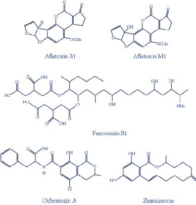
There are about 25% of crops in worldwide that are contaminated with mycotoxins (FAO, 2004). Mycotoxins contaminants, one of the most serious global challenges, have been attracted more and more attention from International Agency for Research on Cancer (IARC) of the World Health Organization (WHO) and scientists in food safety sciences. Mycotoxins, one of the most important and toxic contaminants in food and agricultural products, are secondary metabolites produced mainly by various molds (Atar et al., 2015; Mata et al., 2015; Zhu et al., 2015). There are hundreds of mycotoxins that were identified mainly aflatoxins (AF), ochratoxins (OT), fumonisins (F), and zearalenone (ZEN) as seen in Figure 1-1.
Mycotoxins, a kind of great hazard contaminants for human and animals health, have been a part of food chain. High exposure of mycotoxins in food and feed can lead to mycotoxicosis on human and animals with acute and chronic effects, which mainly has an effect on the kidney, liver, endocrine, nervous and immune systems (Cigic and Prosen, 2009).
Aflatoxin B1 is present in a variety of feed and food, including cereals, maize, nuts, and fruits (Chen et al., 2014; Iqbal et al., 2014; Zhang et al., 2016). AFB1 is the most toxic mycotoxin which mainly causes liver cancer. Its metabolite aflatoxin M1 can appear in milk when dairy cows consumed the feed contaminated with AFB1 (Guo et al., 2014; Guo et al., 2016). AFM1 occurs mainly in dairy products such as raw milk, heat-treated milk, processing milk, and milk-based products. Therefore, mycotoxins pose a common threat in both feed and food products industry and their processing products industry. Ochratoxin A is responsible for the mutagenic, teratogenic and immunosuppressive effects, which occurs in various feed and food including cereals, meats, milk, grape, coffee, beer and wine, etc. Fumonisin B1, produced by Fusarium moniliforme, can cause oesophagal cancer, liver tumor, and kidney tumor via the contamination of maize and maize products, as well as animal feeds (Scott, 2012).
The certain mycotoxin toxicity and the general exposure of the population determins the severity and attention of this kind of mycotoxin. In industrialized countries, the chronic mycotoxicosis effects and diseases are largely more than that of the acute issues as a result of low level exposure of the population. In contrast, in developing countries, it is common that the control of mycotoxin exposure is difficult in both agricultural activities and regulations. Mycotoxin exposure of the population is generally high in these areas (Sanzani and Ippolito, 2014). China is an developing country, which is also a great agriculture-based country. In addition, it has posed an important role in food supply chain worldwide. Therefore, China government and Ministry of Health have established the maximum limits against mycotoxins in food and agricultural products for food safety. The aflatoxins levels
1. General introduction: Aptamer-based biosensor for detection of mycotoxins
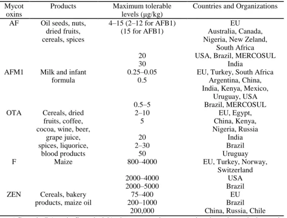
No AFM1 appeared in infant milk powder as a result of the strict control and requirement of China for human and infants health. The levels of ochratoxin A occurred in wine were below the permissible limits established by European Union. In addition, no permissible limits for fumonisins have been regulated in China. However, due to the common occurrence of fumonisins in maize and maize products, the contamination state of fumonisins should be attentioned, and the permissible limits should be set in the future (Selvaraj et al., 2015). As seen in table 1-1, international legislation on mycotoxins was summarized with maximum admissible levels in certain commodities and products.
Table 1-1: Summary of the international legislation on mycotoxins (Anfossi et al., 2016).
Mycot oxins
Products Maximum tolerable levels (μg/kg)
Countries and Organizations AF Oil seeds, nuts,
dried fruits, cereals, spices 4–15 (2–12 for AFB1) (15 for AFB1) 20 30 EU Australia, Canada, Nigeria, New Zeland,
South Africa USA, Brazil, MERCOSUL
India AFM1 Milk and infant
formula
0.25–0.05 0.5
0.5–5
EU, Turkey, South Africa Argentina, China, India, Kenya, Mexico,
Uruguay, USA Brazil, MERCOSUL OTA Cereals, dried
fruits, coffee, cocoa, wine, beer,
grape juice, spices, liquorice, blood products 2–10 5 20 2–30 50 EU, Egypt, China, Kenya, Nigeria, Russia India Brazil Uruguay F Maize 800–4000 2000–4000 2000–5000
EU, Turkey, Norway, Switzerland
USA Brazil ZEN Cereals, bakery
products, maize oil
75–400 200–1000
200,000
EU Brazil China, Russia, Chile Aflatoxin B1 and aflatoxin M1, the most toxic mycotoxins, have been designated as group 1 carcinogen by the International Agency for Research on Cancer (IARC) of the World Health Organization (WHO), and ochratoxin A is classified as group 2 carcinogen by IAR IAR 1993; IAR 2002; O’ rien and Dietrich, 2005). Given of the serious toxicity effect of mycotoxins on animals and human, the European o ission set t e axi u conta ination AF 1 level to 2 μg kg-1
for all cereals and cereal-derived products for food safety (Commission, 2010). In
addition, European Union has regulated a maximum tolerated level for AFM1 to 0 050 μg kg-1
or adult and lower level or AFM1 to 0 025 μg kg-1 for children and infants consumption (Commission, 2006). Taking the high toxicity and low permissible limits into consideration, rapid, low-cost, sensitive analytical strategies for the detection of mycotoxins are vitally important and required.
Figure 1-1: Chemical structures of the important mycotoxins.
Confirmatory and quantitative approaches for detection of mycotoxins are mainly thin layer chromatography (TLC) (Var et al., 2007), high-performance liquid chromatography (HPLC) (Mao et al., 2015; Wang et al., 2012; Lee et al., 2015; Pietri et al., 2016; Herzallah, 2009; Yazdanpanah et al., 2013), and liquid chromatography coupled with mass spectrometry (LC–MS) (Corcuera et al., 2011; Abia et al., 2013; Warth et al., 2013). However, expensive and special instruments, complicated pretreatment and professional personnel are required in these typical equipment methods (Shim et al., 2007). In the meantime, antibody-based immunoassays were developed for mycotoxin detection, including enzyme-linked immune sorbent assay (ELISA) and immunosensors methods (Li et al., 2009; Kav et al., 2011; Anfossi et al., 2015; Parker et al., 2009; Bacher et al., 2012; Vdovenko et
1. General introduction: Aptamer-based biosensor for detection of mycotoxins
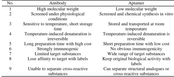
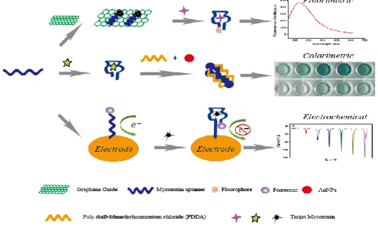
high specificity, the high-cost and storage stability of antibody limits the application of these rapid analysis procedures. Aptamers, an alternative molecule recognition element to antibodies, are single-stranded (ss) DNA or RNA oligonucleotides that can form aptamer/target complex with very strong affinity and specificity via the conformational change. The advantages of aptamer were compared to antibody in table 1-2. With these advantages, aptamer-based biosensors were widely introduced for the detection of mycotoxins like AFB1 (Castillo et al., 2015; Evtugyn et al., 2013; Seok et al., 2015; Shim et al., 2014; Wang et al., 2016), AFM1 (Nguyen et al., 2013; Istamboulie et al., 2016), OTA (Guo et al., 2011; Kuang et al., 2010; Yang et al., 2013), FB1 (Wu et al., 2012; Wu et al., 2013), especially based on fluorescent, colorimetric and electrochemical aptasensors (Figure 1-2). However, the ultrasensitive approaches are difficult to develop via a simple aptasensor recognition. Therefore, a series of novel aptasensors with signal amplification and enhancement have been introduced for mycotoxins (Yang et al., 2007; Weizmann et al., 2006; Patolsky et al., 2002; Wu et al., 2017; Deng et al., 2009; Pavlov et al., 2004; Guo et al., 2014), which can meet the requirement of the low maximum contamination level set by many countries and organizations.
Table 1-2: Comparison of the properties between antibody and aptamer (Zhuo et al., 2017).
No. Antibody Aptamer
1 High molecular weight Low molecular weight
2 Screened under physiological conditions
Screened and chemical synthesis in vitro 3 Sensitive to temperature, short storage
time
Stored and transported at room temperature
4 Temperature-induced denaturation is irreversible
Temperature-induced denaturation is reversible
5 Long preparation time with high cost Short preparation time with low cost
6 Strongly immunogenic No obvious immunogenicity
7 Limited target substances Wide range of target substances 8 Lose affinity to target with labels Keep original biological activity with
labels 9 Unable to separate cross-reactive
substances
Can separate structural analogues or cross-reactive substances
2 Aptamers selection
Aptamers, an alternative target recognition probe to antibodies, are ssDNA or ssRNA oligonucleotides that can bind to targets with high affinity and specificity through the dimensional structure change after the formation of target/aptamer complex. At the first time, systematic evolution of ligands by exponential enrichment (SELEX) was introduced to obtain RNA sequence against T4 DNA
polymerase, which is performed in a random sequences pool. In this study, nitrocellulose filter was adopted to obtain RNA sequence, and filter binding methods was employed to recognize the process of SELEX selection. It was predicted that this SELEX strategy could obtain strong affinity and specificity ligands for any target (Tuerk and Gold, 1990).
Figure 1-2: Principle illustration of fluorescence, colorimetric and electrochemical aptasensor
for the detection of small molecule mycotoxins.
In the meantime, another RNA sequence specific to organic dyes was first selected based on affinity chromatography column. There is a ligand binding site of this RNA sequence towards organic dyes. This RNA sequence was defined as aptamers (Ellington and Szostak, 1990), which indicated that the aptamer was the first time successfully selected. Two years later, ssDNA aptamer of human thrombin was successfully obtained with the dissociation constant from 25 to 200 nM (Bock et al., 1992). From then on, the RNA and ssDNA sequences libraries were simultaneously employed for the process of aptamers selection (Darmostuk et al., 2015). Aptamers have many advantages, such as small molecular, screened and chemical synthesis in vitro, stored and transported at room temperature, reversible temperature-induced denaturation, short preparation time with low cost, no obvious immunogenicity, wide range of target substances, keep original biological activity with labels,
1. General introduction: Aptamer-based biosensor for detection of mycotoxins
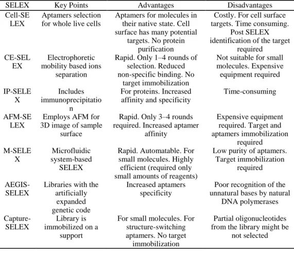
In the beginning 18 years from the finding of first aptamer, the selected aptamers and novel aptamer-based biosensors were mainly used in the field of medicine, biology, chemistry and bioinformatics (Wang et al., 2019; Tan et al., 2019; Zhang et al., 2019; Xiong et al., 2019). For food safety sciences, especially in mycotoxins detection for food safety, the first aptamer specific to OTA has been reported by Cruz-Aguado in 2008 (Cruz-Aguado and Penner, 2008).In recent years, a series of SELEX-based techniques have been developed for the selection of aptamers against various targets, mainly including cell-SELEX, capillary electrophoresis-SELEX (CE-SELEX), immunoprecipitation-coupled SELEX (IP-SELEX), atomic force microscopy SELEX (AFM-SELEX), and artificially expanded genetic information system-SELEX (AEGIS-SELEX). As shown in table 1-3, the characteristics, advantages and disadvantages are summarized and discussed.
Table 1-3: Comparison of the current SELEX-based aptamer selection techniques (Zhang et
al., 2019).
SELEX Key Points Advantages Disadvantages
Cell-SE LEX
Aptamers selection for whole live cells
Aptamers for molecules in their native state. Cell surface has many potential
targets. No protein purification
Costly. For cell surface targets. Time consuming.
Post SELEX identification of the target
required CE-SEL
EX
Electrophoretic mobility based ions
separation
Rapid. Only 1–4 rounds of selection. Reduced non-specific binding. No
target immobilization
Not suitable for small molecules. Expensive equipment required IP-SELE X Includes immunoprecipitatio n
For proteins. Increased affinity and specificity
Time-consuming
AFM-SE LEX
Employs AFM for 3D image of sample
surface
Rapid. Only 3–4 rounds required. Increased aptamer
affinity
Expensive equipment required. Target and aptamers immobilization required M-SELE X Microfluidic system-based SELEX
Rapid. Automatable. For small molecules. Highly efficient (required only small amounts of reagents)
Low purity of aptamers. Target immobilization
required
AEGIS-SELEX
Libraries with the artificially
expanded genetic code
Increased aptamers specificity
Poor recognition of the unnatural bases by natural
DNA polymerases Capture-SELEX Library is immobilized on a support
For small molecules. For structure-switching aptamers. No target immobilization
Partial oligonucleotides from the library might be
3 Aptasensor for the analysis of ochratoxin A
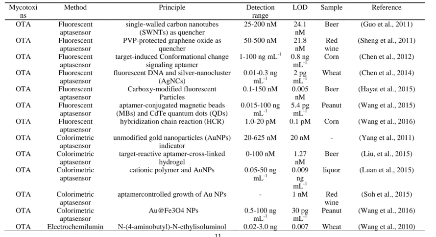
Ochratoxins are one important type of mycotoxins, which are mainly produced by several fungi, such as Aspergillus ochraceus and Penicillium verrucosum (Liu et al., 2015). Of the several subtypes of ochratoxins, ochratoxin A (OTA) is the very common one, and is designated as a possible human carcinogen by IARC (Lv et al., 2016). Researches have paid much attention to the studies on OTA in recent years owing to its widespread occurrence and extraordinary toxic reactions on animals and human. The first aptamer, the minimal one of the selected sequences, has the highest affinity to OTA. The dissociation constant is 200 nM. Since this aptamer specific to OTA has been reported by Cruz-Aguado in 2008 (Cruz-Aguado and Penner, 2008), large numbers of novel aptasensors were developed for OTA analysis in various food products, including fluorescenct, colorimetric and electrochemical aptasensors, as well as some methods based on nanomaterials. The recent literatures of aptasensors for the analysis of ochratoxin A for food safety are illustrated in table 1-4. In addition, these articles are analyzed in more details for each group of the targets.
Development of novel aptasensor for the detection of mycotoxins
Table 1-4: Summary of aptasensor for the analysis of ochratoxin A.
Mycotoxi ns
Method Principle Detection
range
LOD Sample Reference
OTA Fluorescent aptasensor
single-walled carbon nanotubes (SWNTs) as quencher
25-200 nM 24.1 nM
Beer (Guo et al., 2011) OTA Fluorescent
aptasensor
PVP-protected graphene oxide as quencher 50-500 nM 21.8 nM Red wine (Sheng et al., 2011) OTA Fluorescent aptasensor
target-induced Conformational change signaling aptamer
1-100 ng mL-1 0.8 ng mL-1
Corn (Chen et al., 2012) OTA Fluorescent
aptasensor
fluorescent DNA and silver-nanocluster (AgNCs)
0.01-0.3 ng mL-1
2 pg mL-1
Wheat (Chen et al., 2014) OTA Fluorescent aptasensor Carboxy-modified fluorescent Particles 0.1-150 nM 0.005 nM
Beer (Hayat et al., 2015) OTA Fluorescent
aptasensor
aptamer-conjugated magnetic beads (MBs) and CdTe quantum dots (QDs)
0.015-100 ng mL-1
5.4 pg mL-1
Peanut (Wang et al., 2015) OTA Fluorescent
aptasensor
hybridization chain reaction (HCR) 1.0-20 pM 0.1 pM Corn (Wang et al., 2016) OTA Colorimetric
aptasensor
unmodified gold nanoparticles (AuNPs) indicator 20-625 nM 20 nM - (Yang et al., 2011) OTA Colorimetric aptasensor target-reactive aptamer-cross-linked hydrogel 0-100 nM 1.27 nM
Beer (Liu, et al., 2015) OTA Colorimetric
aptasensor
cationic polymer and AuNPs 0.05-50 ng mL-1
0.009 ng mL-1
liquor (Luan et al., 2015)
OTA Colorimetric aptasensor
aptamercontrolled growth of Au NPs - 1 nM Red wine (Soh et al., 2015) OTA Colorimetric aptasensor Au@Fe3O4 NPs 0.5-100 ng mL-1 30 pg mL-1
Peanut (Wang et al., 2016) OTA Electrochemilumin N-(4-aminobutyl)-N-ethylisoluminol 0.02-3.0 ng 0.007 Wheat (Wang et al., 2010)
escent biosensor and AuNP-modifiedgold electrode mL-1 ng mL-1 Mycotoxi
ns
Method Principle Detection
range
LOD Sample Reference
OTA Electrochemical aptasensor
aptamer modified gold electrode 0.1-1000 pg mL-1 0.095 pg mL-1 Red wine (Wu et al., 2012) OTA Electrochemical aptasensor
gold electrode coupled with silver nanoparticles
0.3-30 nM 50 pM Beer (Evtugyn et al., 2013) OTA Electrochemical
aptasensor
rolling circle amplification (RCA) 0.1-5000 pg mL-1
0.065 pg mL-1
Wine (Huang et al., 2013)
OTA Electrochemical aptasensor
Au NPs and methylene blue 2.5-2500 pM 0.75 pM Red wine (Yang et al., 2014) OTA Electrochemical aptasensor exonuclease-induced recycling amplification 0.01-1.0 ng mL-1 0.004 ng mL-1 Corn and Oat (Tan et al., 2015) OTA Electrochemical aptasensor nanocomposites of AuNPs 0.2-4000 pg mL-1 0.07 pg mL-1 - (Hao et al., 2016) OTA Chemiluminescenc e aptasensor HRP-mimicking DNAzyme (HRPzyme) 0.1-100 ng mL-1 0.22 ng mL-1 Coffee beans (Jo et al., 2016) OTA Electrochemical aptasensor
exonuclease (Exo) III-assisted recycling amplification 0.001-0.5 ng mL-1 0.58 pg mL-1
Development of novel aptasensor for the detection of mycotoxins
3.1 Fluorescent aptasensor for OTA
First, Guo et al, developed a sensitive and selective aptasensor for fluorescent detection of OTA. The single-walled carbon nanotubes (SWNTs) were employed to quench the fluorescence signal produced by carboxyfluorescein-labelled aptamer. Upon the presence of OTA, the spatial structure change of the specific aptamer leads to the separation of the SWNTs with the aptamer, the fluorescence is therefore detected. The fluorescence signal has a good linear relationship with concentrations in a range from 25 to 200 nM, with a detection limit of 24.1 nM (Guo et al., 2011). In addition, this aptasensor is successfully used for OTA determination in real beer samples. In the same year, another fluorescent aptasensor was introduced by the same research team via the graphene oxide as a fluorescence quencher (Sheng et al., 2011). In this sensing platform, a linear response of 50-500 nM was obtained between the fluorescence intensity and OTA levels, with the detection limit of 21.8 nM. More importantly, the limit detection could be lowered by two orders of magnitude by using PVP-coated graphene oxide. Similarly, this current aptasensor was also validated for OTA detection in red wine samples.
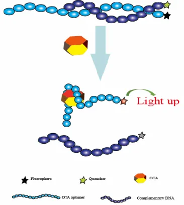
Figure 1-3: Schematic representation of the fluorescent aptasensor for OTA determination
Based on the similar fluorescence response and fluorescence quench schemes, Chen et al, reported a rapid and feasible aptasensor for OTA determination in corn samples (Chen et al., 2012). In this sensing platform (Figure 1-3), the fluorophore was used to label the aptamer while the quencher moiety was employed to label the complementary DNA to quench the fluorescence. When there was no OTA, the hybridization reaction between the complementary DNA and the aptamer led to the close distance of these two moieties, the fluorescence signal was effectively quenched. Upon OTA addition, the produce of aptamer/OTA complex resulted in the separation of the complementary DNA, and the fluorescence was thus recovered. A good linear response between the fluorescent change and OTA levels was obtained ranged from 1 to 100 ng mL-1, with the detection limit of 0.8 ng mL-1. More importantly, the whole analysis process of this method was within 1 min. Therefore, this was another fluorescent aptasensor for rapid determination of OTA with high efficiency.
Nanomaterials were also used to construct biosensors with several modifications to produce fluorescence. One of those aptasensors was introduced for the detection of OTA. In this research, magnetic beads, the fluorescence characterization of DNA silver nanocluster, and spatial conformational change of the aptamer was adopted. An ultrasensitive method was achieved with the limit of detection (2 pg mL-1). Additionally, the practical analysis was successfully completed on wheat samples for OTA detection. This aptasensor might provide a promising sensing platform for OTA due to its unique advantages of low-cost, rapid, portable, selective, and ultrasensitive (Chen et al., 2014). Based on the same magnetic beads separation method, Akhtar et al, reported a generic fluorescent aptasensor to detect OTA. In this sensing strategy, carboxy-labelled aptamer was adopted to signal produce element, the magnetic beads were employed to separate unbound portions. A high sensitivity of this aptasensor was determined with a limit of detection (0.005 nM). Moreover, this proposed method was successfully applied on beer samples for the analysis of OTA (Hayat et al., 2015).
Additionally, another one-step fluorescent aptasensor was illustrated for OTA determination. The specific aptamer for OTA was employed as a target recognition probe, CdTe quantum dots (QDs) was used as a label, and magnetic beads were acted as the separation support. Upon OTA addition, the produce of aptamer/OTA complex and magnetic separation resulted in a significant fluorescence intensity enhancement. More importantly, a wide range response between the fluorescence signal and OTA concentrations (from 15 pg mL-1 to 100 ng mL-1) was achieved, and the limit of detection was 5.4 pg mL-1. Therefore, this developed sensing platform might represent a potential strategy for OTA routine controls for food safety(Wang et al., 2015). Finally, hybridization Chain Reaction (HCR), an important signal enhancement strategy, has been widespreadly applied for the analysis of DNA, proteins, metal ions, virus, and cancer cells, as well as the mycotoxins. An
1. General introduction: Aptamer-based biosensor for detection of mycotoxins
which caused the aggregation of perylenediimide probe and subsequent signal enhancement. Under the optimized conditions, fluorescence signal was established to have a good linear relationship with the targeted OTA concentrations. It ranged from 1.0 to 20 pM with a detection limit of 0.1 pM, indicating that this current aptasensor was highly sensitive for the analysis of OTA. Moreover, the practicality of this sensing strategy was validated through the successful detection of OTA levels in corn samples (Wang et al., 2016).
3.2 Colorimetric aptasensor for OTA
In addition to fluorescent aptasensors, colorimetric aptasensors are also widespread techniques for the analysis of mycotoxins because of their simple and rapid uses, without the requirement of complicated instruments. A color change was produced as a result of the aggregation of gold nanoparticals (AuNPs) in the salt presence. Yang et al. developed a colorimetric aptasensor for OTA determination, which employed the advantages of the specific aptamer and AuNPs (Yang et al., 2011). Upon the OTA addition, structural change to G-quadruplex of the aptamer from random coil to G-quadruplex resulted in the separation of the aptamer to AuNPs, leading to AuNPs aggregation under the addition of salt and subsequent color change. The absorbance values presented a good linear relationship with OTA concentrations ranged from 20 to 625 nM. Its detection limit is 20 nM. Using the same AuNPs colorimetric indicators, a novel visual aptasensor was introduced for visual analysis of OTA via the synthesis of DNA hydrogels. This technology has been widely applied for the analysis of proteins (Zhang et al., 2013), ions (Dave et al., 2010; Guo et al., 2014; Lin et al., 2011) and nucleic acids (Sun et al., 2014), as well as small molecules (Yan et al., 2013; Zhu et al., 2010). In this design, as depicted in Figure 1-4, the DNA hydrogels were obtained through linkage between the aptamer of OTA and two polymer single-stranded DNAs. With the addition of OTA, the aptamer/OTA complex formation induced dissociation of the DNA hydrogels. Subsequently, the AuNPs were released for the colorimetric detection of OTA levels. Its detection limit was determined to be 1.27 nM by signal amplification strategy. Therefore, this proposed sensing strategy represented a novel and portable method for the analysis of OTA to ensure food safety (Liu, et al., 2015).
Moreover, Luan et al. reported a sensitive and selective aptasensor for OTA determination via aggregation of AuNPs as the colorimetric generator induced by poly diallyldimethylammonium chloride (PDDA). Upon the optimal conditions, its detection limit was obtained down to 0.009 ng mL-1, which demonstrated that this sensing platform provided a promising simple and sensitive platform for rapid analysis of OTA (Luan, et al., 2015).
Under the normal circumstances, the coexistence of multiple mycotoxins is a very common phenomenon in food and agricultural products. High-throughput screening and analysis of multiple mycotoxins will play an important role for food
safety. Based on the recognition reaction of aptamer and the colorimetric response of AuNPs, a rapid and sensitive aptasensor for the analysis of multiple small molecules including OTA was introduced. Upon the addition of target, the interactions between aptamer and targets caused the dissociation of the aptamer from the surface of AuNPs, leading to the color change of AuNPs. the limits of detection were 1 nM for OTA, 0.2 nM for 17β-estradiol, and 1 nM for cocaine, respectively. Therefore, the novel aptasensor became a potential strategy for high-throughput application for multiple mycotoxins detection by simple replacement of aptamer for different mycotoxins (Soh, et al., 2015).
Figure 1-4: Principle illustration of colorimetric aptasensor for detection of OTA via AuNPs
encapsulated DNA hydrogel. Reprinted from Liu et al. (2015) with permission. In addition, Au@Fe3O4 NPs were obtained through AuNPs being
functionalized by Fe3O4 NPs, and the Au@Fe3O4 NPs activity was thus increased.
The aptamer of OTA was labelled to the magnetic beads while its complementary single-stranded DNA was modified on Au@Fe3O4 NPs. Upon the addition of OTA,
the recognition between the aptamer and OTA caused the release of ssDNA-labelled Au@Fe3O4 NPs, which could exert the color change to a blue solution. Its detection
limit was down to 30 pg mL-1, demonstrating that this sensing strategy was a novel and sensitive approach for OTA determination for food safety via the application of peroxidase-like activity of AuNPs (Wang et al., 2016).
3.3 Electrochemical aptasensor for OTA
The immobilization of aptamers on the surface of transducer substrates exerted an important function on the construction of electrochemical aptasensors. The transducer substrates mainly consisted of gold and carbon-based electrodes. A series of electrochemical aptasensors were developed for OTA analysis in food and agricultural products, including Electrochemical Impedance Spectroscopy (EIS), Differential Pulse Voltammetry (DPV), Cyclic Voltammetry (CV), Linear Sweep Voltammetry (LSV), Square Wave Voltammetry (SWV), Field Effect Transistor and