REPRODUCTION
RESEARCHPorcine embryo development and fragmentation and their
relation to apoptotic markers: a cinematographic and confocal
laser scanning microscopic study
Bart Mateusen, Ann Van Soom, Dominiek G D Maes, Isabelle Donnay
1, Luc Duchateau
2and Anne-Sophie Lequarre
1Department of Reproduction, Obstetrics and Herd Health, Faculty of Veterinary Medicine, Ghent University, Salisburylaan 133, B-9820 Merelbeke, Belgium,1Institut des Sciences de la Vie, Unite´ des Sciences Ve´te´rinaires, Universite´ catholique de Louvain, B-1348 Louvain-La-Neuve, Belgium and2Department of Physiology, Faculty of Veterinary Medicine, Ghent University, B-9820 Merelbeke, Belgium
Correspondence should be addressed to B Mateusen; Email: bart.mateusen@ugent.be
Abstract
Porcine embryo selection prior to transfer is mainly influenced by morphological criteria. However, the relationship between embryonic morphology, developmental potential and cell death by apoptosis in porcine embryos is still unclear. The aim of this study was to establish embryo quality parameters for in vivo fertilised porcine embryos based on timing of development in vitro, embryo morphology and the presence of apoptosis. The kinetics of development and morphological parameters were investigated in a time-lapse cinematographic experiment. Possible links between embryo morphology and apoptosis were examined via a confocal laser scanning experiment, analysing nuclear changes, annexin V and terminal dUTP nick-end label-ling. The timing of early cleavages was firmly linked to embryo developmental competence in vitro. Attainment of at least the 5-cell stage before 77 h post insemination and attainment of the morula stage before 102 h post insemination significantly increased the odds for reaching the early blastocyst stage. Overall, a negative effect of fragmentation percentage and fragmen-tation pattern on subsequent embryonic development was observed, but the developmental potential of embryos experiencing slight fragmentation (0–5%) was not different from embryos without fragmentation. Correlations detected between develop-mental arrest and fragmentation, and fragmentation and apoptosis were 0.60 and 0.87 (P < 0.05) respectively. Only a minority of the embryos arrested between the 1- and 4-cell stage displayed biochemical characteristics of apoptosis. Consequently, a significant correlation (0.57) between developmental arrest and apoptosis could only be established for embryos arrested after embryonic genome activation.
Reproduction (2005) 129 443–452
Introduction
Quality assessment of preimplantation embryos relies heavily on non-invasive techniques based on morphologi-cal criteria. One of these techniques is timing of develop-ment which has been linked to in vitro blastocyst formation for hamster (Gonzales et al. 1995) and bovine (Van Soom et al. 1992, Grisart et al. 1994, Holm et al. 1998, Lonergan et al. 1999) embryos. Furthermore, it has been shown in hamster (McKiernan & Bavister 1994, bovine (Hasler 1998) and human (Racowsky et al. 2000) embryos that slow-cleaving embryos are less viable, i.e. have a lower foetal development ratio after transplantation, in comparison to fast-cleaving embryos. Despite the relative abundance of kinetics data in other species, data on timing of develop-ment have not been established yet for porcine embryos.
Another important parameter for mammalian embryo quality is the assessment of blastomere fragmentation (Lindner & Wright 1983, Antczak & Van Blerkom 1999). Cellular fragmentation is a common feature during early development of mammalian embryos (Van Blerkom et al. 2001), but is generally considered as indicative of poor embryo quality both in bovine (Lindner & Wright 1983) and human (Puissant et al. 1987). Moreover, human embryos with a substantial amount of cellular fragmenta-tion have a markedly reduced implantafragmenta-tion rate (Ziebe et al. 1997, Ebner et al. 2001). For human in vitro ferti-lised (IVF) embryos, not only degrees (in percentage) but also distinct patterns of fragmentation, which are corre-lated with in vitro development, have been defined (Warner et al. 1998a, Alikani et al. 1999). Similar data in pig embryos are presently lacking.
Despite the importance of fragmentation as a morpho-logical indicator of embryonic viability, the origin of frag-mentation is still unclear. Since fragments resemble apoptotic bodies seen in other cell types (Hardy 1999), it seems obvious to investigate whether fragmentation can be used as a non-invasive marker of the occurrence of apoptosis in the embryo. However, the relationship between fragmentation and apoptosis in embryos is not clear. In human embryology, Jurisicova et al. (1996) pro-posed that these fragments represented apoptotic bodies, but failure of the majority of the fragmented embryos to show either in situ TUNEL or annexin V labelling led Antczak and Van Blerkom (1999) to postulate that frag-mentation was not related to apoptosis. Apoptotic nuclei have been detected in embryos from many mammalian species including the pig (Long et al. 1998, Hao et al. 2003, Rubio Pomar et al. 2004) Apoptosis is a natural pro-cess during mammalian preimplantation development which could involve elimination of unwanted or damaged cells, but its role in preimplantation embryonic develop-ment is not well characterized.
The aim of this study was to investigate the develop-mental competence of porcine embryos by looking for associations between timing of development, embryo mor-phology characteristics and occurrence of apoptosis. We chose to use in vivo fertilised pig embryos because such embryos have been produced under optimal conditions and their development may be considered as a gold stan-dard for in vitro embryo development in pigs.
Materials and Methods
Collection of in vivo fertilised porcine embryos
A total of 29 multiparous sows, Sus scrofa (Rattlerow-Seghers) were used. The sows were superovulated using equine chorionic gonadotrophin (eCG) (Folligon 1500 IU i.m. Intervet, Boxmeer, The Netherlands) three days after weaning, followed by human CG (hCG) (Chorulon 1500 IU i.m.) 72 h later. They were fixed-time inseminated with boar semen of proven fertility 24 h after hCG adminis-tration and were slaughtered 45 h post insemination. The reproductive tracts were removed and transported to the laboratory in a pre-warmed box (39 8C) within 30 min after slaughter. Each oviduct was flushed with 15 ml of pre-warmed Hepes-buffered North Carolina State University-23 (NCSU-University-23) medium to collect the embryos. After ten washings with Hepes-buffered NCSU-23, embryos were cultured in NCSU-23 (Petters & Wells 1993) at 39 8C.
Cinematography of pig embryos
To perform time-lapse cinematography, in vivo derived pig embryos were incubated in a culture dish placed in a small chamber on the plate of an inverted microscope (Carl Zeiss NV-SA, Zaventem, Belgium). The chamber was regularly flushed with a humidified and warmed gas mixture consist-ing of 5% CO2, 5% O2, and 90% N2. To maintain a
constant temperature of 39 8C, a plexiglas box was adapted to fit onto the microscope and connected to a heating sys-tem controlled by a sys-temperature probe. The recording equipment consisted of a colour video camera KY-F55E (JVC) and two computers. The first computer synchronized the lighting of the lamp and the shooting. The second digi-tized and recorded the frames with the program Perception Video Recorder (Alpha M). One image was recorded every 240 seconds (4 min) (Lequarre et al. 2003). The develop-ment of the embryos was filmed starting from 48 h post insemination (hpi) (3 h after collection) for 8 consecutive days at magnification £ 100. Only embryos visible in the camera field throughout the complete culture period were examined.
Evaluation of embryonic morphology
Morphology of cleaving embryos was evaluated every 4 min starting from 48 hpi in experiment 1 and at day 7 post insemination in experiment 2. The degree of fragmen-tation was first expressed as a percentage and defined as the embryonic volume occupied by anucleate cytoplasmic fragments (Puissant et al. 1987). In addition, a fragmenta-tion pattern was defined based on the spatial distribufragmenta-tion and relative size of the fragments as described previously for human embryos (Alikani et al. 1999) (Fig. 1). Embryos exhibiting minimal fragments, usually in association with a single blastomere were designated as fragmentation pat-tern 1 (FP1). FP2 was used to characterize highly localized
Figure 1 Four distinct patterns of fragmentation in porcine morulae: (A) fragmentation pattern 1 (FP1) is minimal in volume and fragments are associated with only one blastomere, (B) FP2 is characterized by highly localized fragments which often appear in a cluster or a col-umn, (C) FP3 with many small fragments seen throughout the clea-vage cavity and perivitelline space, (D) FP4 embryos have many large fragments which are randomly distributed and always associated with uneven cells.
fragments which often appear in a cluster or a column. In FP3, many small fragments could be seen throughout the cleavage cavity and perivitelline space. FP4 also had many large fragments which were randomly distributed and always associated with uneven cells.
Evaluation of apoptosis by means of propidium iodide, annexin V and TUNEL assay
Propidium iodide (PI) and annexin V staining (Vybrant Apoptosis Assay kit #3, Molecular Probes, Eugene, OR, USA) of living embryos was performed to determine the cell membrane integrity and the presence of phosphatidyl-serine residues on the outer surface of the plasma mem-brane, respectively. Positive day 7 control embryos were incubated for 12 h with 1 mM staurosporine to induce apoptosis. Day 7 embryos were first washed for 5 min in annexin binding buffer at 37 8C and incubated for 15 min in the presence of FITC conjugate of annexin V (25 ml/ml) and PI solution (3 mg/ml) according to the manufacturer’s recommendations for the assay kit. Then embryos were washed for 5 min in PBS and transferred to a drop of pre-warmed PBS (37 8C) on a microscopic slide and examined by scanning laser confocal microscopy. Positive labelling for annexin V on the outer surface membrane was observed as bright yellow staining.
In preparation for TUNEL, the embryos analyzed for annexin V labelling were fixed in 4% paraformaldehyde in PBS and washed for at least 12 h in polyvinyl pyrrolidone (PVP) solution (1 mg PVP/ml PBS). After wash-ing, they were permeabilized with 0.5% Triton X-100 in PBS for 20 min and washed again in PVP solution. Positive and negative controls were treated with DNase (50 Units/ml in PBS) for 1 h at 37 8C to ensure detection of strand breaks by TUNEL (In Situ Cell Death Detection kit, Boehringer, Mannheim, Germany). After three washings in PVP solution, positive controls and samples were incu-bated in fluorescein-dUTP and terminal deoxynucleotidyl transferase for 1 h at 37 8C in the dark. Negative controls were incubated in nucleotide mixture only in the absence of transferase. After three more washings in PVP solution, controls and samples were incubated in RNase A (50 mg/ml in PBS) for 1 h at room temperature. The nuclei were then counterstained with 0.5% PI for 1 h at room temperature. Subsequently, the slides were washed three times with PVP solution and embryos were mounted in glycerol with 1,4-diazabicyclo (2.2.2) octane (25 mg/ml). Samples were examined by laser scanning confocal microscopy. TUNEL-positive nuclei appeared bright yellowish-green, and the PI staining allowed a rapid identification, localization and quantification of normal, fragmented or condensed nuclei as defined previously by Gjørret et al. (2003).
Confocal laser scanning microscopy
Double stained samples were examined with a Leica TCS SP2 laser scanning spectral confocal system (Leica Microsystems GmbH, Heidelberg, Germany) linked to a
Leica DM IRB inverted microscope (Leica Microsystems GmbH, Wetzlar, Germany). An Argon laser was used to excite FITC (488 nm) and PI (586 nm) fluorochromes. Total embryo height was evaluated and sections were made at 3 mm intervals. Analysis of the images was performed with Leica confocal software.
Experimental design
Experiment 1: cinematographic analysis of porcine embryonic morphology and development
In vivo fertilised pig embryos (n ¼ 86) obtained 48 hpi were cultured in groups of 25 in 50 ml NCSU23 under oil at 39 8C in 5% CO2, 5% O2, and 90% N2. These embryos were subjected to cinematographic analysis. Embryos of the same donors that were simultaneously cultured in a classic incubator served as a control (n ¼ 103). The blas-tocyst yield of these control embryos was 64.1% after 8 days of in vitro culture.
The length of the first cell cycle could not be measured as the exact timing of conception was unknown. Movies were analyzed by assessing, for each embryo, the time of appearance of 3-, 4-, 5-, 6-, 7- and 8-cell, early morula, compacted morula, early blastocyst, expanded blastocyst and hatched blastocyst stages. Beyond the 8-cell stage, it was not possible to count or extrapolate cell numbers accurately on the basis of cleavage observations. An embryo was defined as an early morula after it contained a ‘cell ball’ of small distinguishable blastomeres (Lindner & Wright 1983). A compacted morula was defined as the stage at which blastomeres had coalesced to form a smooth, compact cell mass. Cavitation was estimated by the first appearance of a stable confluent blastocoel. Blas-tocyst expansion was defined by the increase in diameter of the zona pellucida (ZP). Cracking of the ZP of an expanded blastocyst was described as hatching.
Experiment 2: evaluation of the relationship between embryonic morphology and apoptotic markers
In vivo fertilised porcine embryos (n ¼ 132) obtained 48 hpi were cultured in groups of 25 in 500 ml NCSU23 at 39 8C in 5% CO2 in air. At day 7 post insemination, the embryonic cleavage stage, degree and pattern of fragmen-tation were assessed by differential interference contrast microscopy (Olympus IX70 Olympus Belgium NV, Aartse-laar, Belgium) at a total magnification of £ 300. Embryos that had not reached the blastocyst stage at day 7 post inse-mination were defined as arrested embryos. Such embryos were divided in two groups, embryos that stopped cleaving before transition of maternal to the embryonic genome (at the 4-cell stage in porcine embryos) (Jarrell et al. 1991, Schoenbeck et al. 1992, Viuff et al. 2002) and embryos that arrested during the 5-cell to the morula stage period.
Subsequently, all embryos were stained with annexin V, and TUNEL assay performed then analyzed using confocal laser scanning microscopy. An embryonic cell was categorized as apoptotic if: (i) the cell had nuclear
morphological characteristics of apoptosis such as frag-mentation or condensation; (ii) the cell membrane was annexin V positive and, (iii) the nucleus of the cell was TUNEL labelled. Based on this definition, an apoptotic cell ratio (ACR) was determined as the percentage of apoptotic cells per embryo.
Statistical analysis
The time that an embryo remained in a particular cell stage was defined as the time from appearance of that stage to the first appearance of one or more additional cells. To study the 4-cell lag phase phenomenon, the dur-ation of each cell stage was compared with the durdur-ation of the 4-cell stage by the general linear model, using only those embryos for which the two relevant duration times were available.
The effect of asynchronous cleavage and fragmentation on further development of the embryos was studied by the Cox model with last stage attained as response variable and asynchronous cleavage as time-varying covariate. Asynchronous cleavage was introduced as a binary vari-able (yes/no), whereas for fragmentation different aspects were considered. First, fragmentation was investigated as a binary variable (yes/no) and next as a categorical vari-able with four categories: no fragmentation, 0–5% frag-mentation, 5–15% and .15%. Additionally, the fragmentation pattern was investigated as a categorical variable with the categories: no fragmentation, and frag-mentation pattern 1 (FP1), FP2, FP3 and FP4. Time points at which embryos attained a specified cell stage with the highest significant odds of becoming a blastocyst in later embryonic development were calculated using Chi-square analysis. The standard statistical analysis for binary data is based on the odds ratio. The analysis of such data is based on odds and not on probabilities (Hosmer & Lemeshow 1989). The standard statistical analysis for time-to-event or survival data is based on the proportional hazards model and the corresponding measure – the hazard ratio. The hazard is a conditional probability: given the event did not take place up to a certain moment, what is the prob-ability it occurs that moment (Collette 1994).
Differences in ratios of fragmented or apoptotic embryos were analyzed by Chi-square tests or, when small numbers were involved, Fisher’s exact tests. Logistic regression with the embryo as a random factor was used to compare average fragmentation %, and average ACR. Correlation analysis between embryonic arrest, fragmenta-tion and apoptosis was performed by Spearman’s rank test. Statistical significance was assumed at P , 0.05. The statistical analyses were performed using SAS version 8.
Results
Experiment 1: cinematographic analysis of porcine embryonic morphology and development
Five movies were analyzed including a total of 86 embryos. At recovery, 3 one-cell, 28 two-cell, 10 three-cell, 42
four-cell and three more than five-cell embryos were retrieved. The descriptive developmental kinetics of the cell stages are given in Table 1. The developmental capacity of embryos cultured in the time-lapse culture sys-tem was comparable to that in a classic incubator (n ¼ 103) since the blastocyst rate was 67.4% and 64.1% in the two systems, respectively.
Cinematographic analysis revealed that the 4-cell stage (average duration 38.20 h) lasted longer (P , 0.01) than the 8-cell, early morula, compacted morula and early blas-tocyst stage, although all embryos resumed division regardless of the duration of the 4-cell stage. Three out of the 32 (9.4%) embryos with measurable third cell cycle (from 4-cell to 5-cell stage) were passing through the 4-cell stage in less than 15 h. All of these reached the early blas-tocyst stage, while the early blasblas-tocyst percentage of embryos with a 4-cell lag phase was 93.1% (27/29).
The asynchrony between blastomere cleavages increased at the third cell cycle. The time interval between the appearance of 3-cell and 4-cell stage was on average 2.3 ^ 5.4 h (n ¼ 25), whereas the blastomeres of 4-cell stage embryos cleaved asynchronously with an average interval between the first and the last blastomere cleavage of 9.2 ^ 10.8 h (n ¼ 55). No significant effects of these asynchronous cleavages on further development or blasto-cyst formation were detected (P ¼ 0.43).
Extrusion of blastomeres was recorded in 24.4% (21/86) of the embryos. The highest frequency was observed at the morula stages (11/21, 52.4%) followed by the 5-cell to the 8-cell stages (6/21, 28.6%). Extrusion of blastomeres also had no negative influence on further embryonic development. In 76.2% (16/21) of the cases, this asymme-try of the embryo disappeared during a later cell cycle due to remerging of the extruded blastomeres with the embryonic mass.
In general, fast cleaving embryos reached the blastocyst stage at higher frequencies than slower cleaving embryos. In Fig. 2, a difference in time between insemination and first appearance of the 5- to 8-cell stages was detected for embryos that stopped developing at the morula stage unlike embryos that reached the blastocyst stage
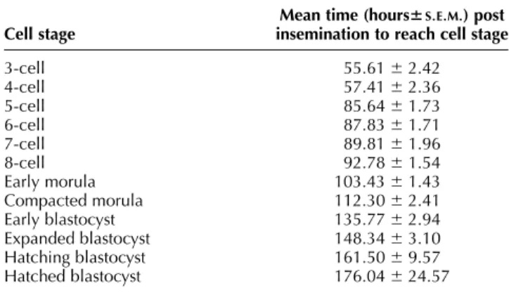
Table 1 Descriptive developmental kinetics of porcine embryos in a cinematographic time lapse system.
Cell stage
Mean time (hours6S.E.M.) post insemination to reach cell stage 3-cell 55.61 ^ 2.42 4-cell 57.41 ^ 2.36 5-cell 85.64 ^ 1.73 6-cell 87.83 ^ 1.71 7-cell 89.81 ^ 1.96 8-cell 92.78 ^ 1.54 Early morula 103.43 ^ 1.43 Compacted morula 112.30 ^ 2.41 Early blastocyst 135.77 ^ 2.94 Expanded blastocyst 148.34 ^ 3.10 Hatching blastocyst 161.50 ^ 9.57 Hatched blastocyst 176.04 ^ 24.57
(P ¼ 0.019). A retrospective analysis of the cleavage data revealed an optimal cleavage pattern for embryos with in vitro blastulation capacity at a given cell stage. Embryos attaining at least the 5-cell stage before 77 hpi, had better odds of reaching the blastocyst stage (estimated odds ratio (OR) ¼ 9.95, P ¼ 0.031) than embryos that reached these cell stages at a later time point (P , 0.05). Another selec-tion criterion for blastocyst formaselec-tion was attainment of the early morula stage before 102 hpi (OR ¼ 4.29) (P ¼ 0.019).
Time-lapse recordings showed two different kinds of fragmentation: static fragments detached from blastomeres (n ¼ 2) and fragments that moved in concert with adjacent blastomeres (n ¼ 54). These latter fragments often changed location and size during further development. An apparent disappearance and reoccurrence of fragmentation was also a common feature of these embryos.
Overall, a negative effect of fragmentation percentage on subsequent embryonic development was detected. The hazard for not reaching the next embryonic stage was 11.8 times higher for embryos with .15% fragmentation (n ¼ 10) compared with embryos with a fragmentation percentage of #15% (n ¼ 76) (P , 0.0001). The effect on subsequent embryonic development of embryos
experiencing slight fragmentation (0–5%; n ¼ 34) was not different from embryos without fragmentation (n ¼ 30; hazard ratio ¼ 0.89, P ¼ 0.891).
The pattern of fragmentation was also associated with subsequent embryonic development. FP1 (n ¼ 45) and FP2 or 3 (n ¼ 7) had detrimental effects on subsequent embryo cleavage since they had a hazard ratio of 3.1 (P ¼ 0.011) and 20.5 (P , 0.0001) respectively, in comparison to embryos without fragmentation. Further-more, FP2 or 3 embryos had a lower developmental potential than FP1 embryos with an estimated hazard ratio of 6.5 (P ¼ 0.007).
Experiment 2: evaluation of the relationship between embryonic morphology and apoptotic markers
Of the 132 embryos included in this experiment, 61 (46%) arrested during the in vitro culture period, and 71 (54%) embryos reached the blastocyst stage at 7 days post insemination (dpi). A small proportion (8/61, 13%) of the arrested embryos stopped cleaving before or at the 4-cell stage. More arrested embryos were fragmented compared with embryos that reached the blastocyst stage at 7 dpi (P , 0.05). Also, the average fragmentation percentage was higher for arrested embryos compared with blastocyst stage embryos at day 7 post insemination (P , 0.05) (Table 2). The correlation detected between developmen-tal arrest and fragmentation was 0.60 (P , 0.05).
None of the embryos without fragmentation had cells categorized as apoptotic, whereas 50 out of 55 embryos with fragmentation possessed apoptotic cells. The percen-tage of embryos with apoptotic cells was higher for embryos arrested during the 5-cell to the morula stage compared with embryos that arrested before or at the 4-cell stage and embryos with blastocyst development at day 7 post insemination (P , 0.05). The average ACR of embryos arrested at the 5-cell to the morula stage was higher compared with the average ACR of blastocysts at 7 dpi (P , 0.05) (Table 2). The correlation detected between the developmental arrest during the 5-cell to the morula stage period and apoptosis was 0.57 (P , 0.01).
As shown in Tables 3 and 4, the percentage as well as the pattern of embryo fragmentation were both associated with the apoptotic cell ratio (P , 0.05). For both fragmen-tation assessments a significant difference in apoptosis was detected between embryos without and with fragmentation. Embryos experiencing slight fragmentation (0–5%) had a lower average ACR than embryos with a
Figure 2 The developmental kinetics of embryos that arrested at the morula stage in comparison with the development of embryos that reached the blastocyst stage. *Significant difference within a stage of development (P , 0.05). Stages of development: 3–4c, 3- to 4-cell stage; 5–8c, 5- to 8-cell stage; EM, early morula; CM, compacted morula; EB, early blastocyst; ExB, expanded blastocyst; HchB, hatch-ing blastocyst; HB, hatched blastocyst.V, embryos that stopped
cleav-ing at the morula stage;B, embryos reaching the blastocyst stage.
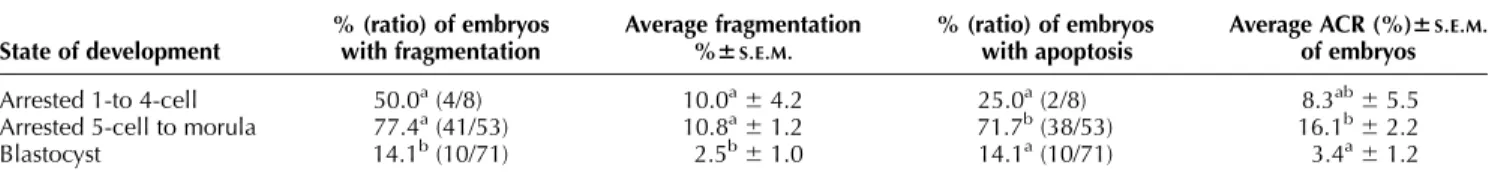
Table 2 Assessment of fragmentation % and apoptosis in arrested and blastocyst stage embryos (n ¼ 132) by annexin V. TUNEL assay and nuclear morphology analysis at day 7 post insemination.
State of development % (ratio) of embryos with fragmentation Average fragmentation %6S.E.M. % (ratio) of embryos with apoptosis
Average ACR (%)6S.E.M.
of embryos Arrested 1-to 4-cell 50.0a(4/8) 10.0a^4.2 25.0a(2/8) 8.3ab^5.5 Arrested 5-cell to morula 77.4a(41/53) 10.8a^1.2 71.7b(38/53) 16.1b^2.2
Blastocyst 14.1b(10/71) 2.5b^1.0 14.1a(10/71) 3.4a^1.2
fragmentation percentage of .5% (P , 0.05) (Table 3). The correlation detected between fragmentation and apoptosis was 0.87 (P , 0.05). Occurrences of biochemi-cal cell changes indicative for apoptosis of fragmented embryos are presented in Fig. 3.
Discussion
The present study was performed to establish guidelines and standards for evaluating the quality of porcine embryos. The in vitro developmental kinetics of in vivo fertilised porcine embryos, as defined in the first exper-iment, can be used as a reference for further embryologi-cal cellular and molecular studies of porcine embryos. At the same time, the kinetics of embryonic fragmentation in developing pig embryos was described. Timing of embryo development is a rapid, simple, accurate and non-invasive way to evaluate embryos. Using time-lapse cinematography, a high degree of precision on the measurements of development timing is established and it captures all morphological characteristics at a light micro-scope level. The ability to form a blastocoel cavity is prob-ably the best morphological indicator of the developmental competence of a preimplantation embryo (Bavister 1995) but the relationship between developmen-tal competence and viability is more complex and can only be established using embryo transfer experiments.
In the present study, high developmental rates to the blastocyst stage (67.4% and 64.1%) were obtained after in vitro culture in contrast to the blastocyst rates (about
25%) which are usually found after culture of IVF pig embryos (Abeydeera 2002). This is not surprising, since in other species such as cattle, substantial differences in mor-ula –blastocyst rates have been described after culture of in vivo vs in vitro produced embryos, which are probably due to the inferior conditions of maturation and fertilisa-tion to which IVF embryos have been exposed (Van Soom & de Kruif 1992).
In our study, pig embryos showed a 4-cell lag phase which lasted on average 38.2 h, which is comparable to findings by Anderson et al. (1999) who found an average 4-cell stage length between 38 and 44 h for in vitro cul-tured porcine embryos. For in vivo developing pig embryos, the 4-cell stage lasts between 20 to 24 h (Hunter 1974, Flint 1981) which is characteristically shorter than findings of in vitro cultured embryos (Bavister 1995). The 4-cell lag phase is likely attributed to imperfections of in vitro culture conditions. Possible causes for this delay are transition of maternal to zygotic control of embryonic development which takes place at the 4-cell stage for por-cine embryos (Jarrell et al. 1991, Schoenbeck et al. 1992, Viuff et al. 2002), change in metabolism and needs of embryos (Schultz et al. 1993), inadequate energy supply by the medium and/or effects of the production of free radicals (Jarrell et al. 1991).
The average time needed to cleave from the 3 to the 4-cell stage was 2.3 h, but increased to 9.2 h for the 5- to 8-cell stage. For bovine embryos, a comparable time inter-val of 9.2 h was detected between the 9- and 16-cell stage (Holm et al. 1998). An increase in asynchrony at these two species specific cleavage stages can be related to the transition of maternal to zygotic control that takes place at the cell stage prior to these cleavage stages (4-cell stage for porcine and 8-cell stage for bovine embryos). Human embryos with unevenly sized blastomeres have a lower pregnancy and implantation rate (Hardarson et al. 2001). Also, asymmetry in bovine early embryonic stages is regarded as a characteristic of poor embryo quality (Lind-ner & Wright 1983), but its impact on viability is uncer-tain. In the present study, no effect of asynchronous cleavage or blastomere extrusion on blastulation was detected. Cleavage of extruded blastomeres ceased, but in most of the embryos (76%), the asymmetry disappeared by reabsorbing the extruded blastomeres in the embryonic mass during later cleavage divisions.
We could demonstrate that also in pig embryos, the time needed to reach the third cell cycle and the early morula stage was inversely correlated with the probability of blastulation (P , 0.05). Embryos which failed to reach the blastocyst stage needed on average 6.25 h and 5.44 h more to reach the third cell cycle and early morula stage respectively, compared with embryos that completed blas-tocyst development. These findings are in agreement with earlier studies that correlate cleavage kinetics with blasto-cyst development in bovine (Van Soom et al. 1992, Grisart et al. 1994, Holm et al. 1998, Lonergan et al. 1999) and in hamster embryos (McKiernan & Bavister 1994, Gonzales
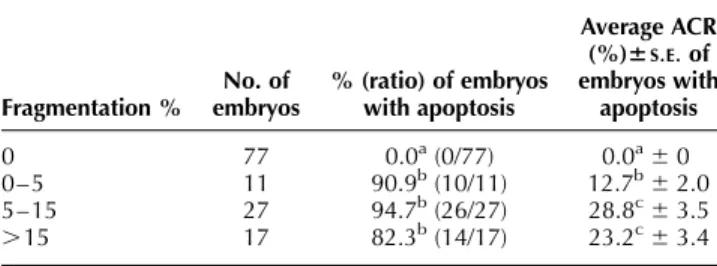
Table 3 The relationship between fragmentation % and average apoptotic cell ratio (ACR) in day 7 cultured pig embryos (n ¼ 132)
Fragmentation % No. of embryos % (ratio) of embryos with apoptosis Average ACR (%)6S.E.of embryos with apoptosis 0 77 0.0a(0/77) 0.0a^0 0–5 11 90.9b(10/11) 12.7b^2.0 5–15 27 94.7b(26/27) 28.8c^3.5 .15 17 82.3b(14/17) 23.2c^3.4 a,b,cWithin a column, values with a different superscript differ
signifi-cantly (P , 0.05).
Table 4 The relationship between fragmentation pattern and average apoptotic cell ratio (ACR) in day 7 cultured pig embryos (n ¼ 132).
Fragmentation No. of embryos % (ratio) of embryos with apoptosis Average ACR (%)6S.E.of embryos with apoptosis No fragmentation 77 0.0a(0/77) 0.0a^0 FP1 31 96.8b(30/31) 22.3b^2.9 FP2 10 80.0b(8/10) 17.8b^4.2 FP3 13 84.6b(11/13) 19.6b^4.3 FP4 1 100.0b(1/1) 37.9b^0 a,bWithin a column, values with a different superscript differ
et al. 1995). By analogy with studies in other species, reference time points for in vitro development in hpi were calculated. For the specific culture system used, ment of at least the 5-cell stage before 77 hpi and attain-ment of the early morula stage before 102 hpi increased the odds for reaching the early blastocyst stage to 995% and 429% respectively, compared with embryos that reached these cell stages at a later time point (P , 0.05). The reason why faster cleaving embryos are more capable of developing is not known, but there are a number of fac-tors that can influence in vitro cleavage rate such as ooplasm quality, culture medium, environment and sev-eral genetic factors. The preimplantation development (PED) gene in mouse embryos has a remarkable regulatory function on the timing of embryo development (Warner
et al. 1998b) and potential human and bovine homol-ogues of this PED gene have been identified (Cao et al. 1999, Fair et al. 2004). Further are paternal influences on the S- and G1-phase of zygotes (Eid et al. 1994, Comiz-zoli et al. 2000), aberrant maternal inherited cytoplasm (Liu & Keefe 2000, Meirelles et al. 2004) and sex differ-ences of mouse and bovine embryos related to cleavage rate (Tsunoda et al. 1985, Mittwoch 1989, Yadav et al. 1993). Chromosome abnormalities have also been shown to influence early development (Kawarsky et al. 1996, Viuff et al. 2001) but more research is necessary to clarify their impact.
Embryonic fragmentation was next to the timing of development as the most important morphological par-ameters analyzed in the cinematographic experiment.
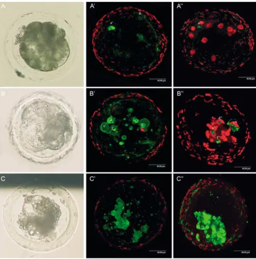
Figure 3 Differential interference contrast microscopic and confocal laser scanning images of fragmented in vivo fertilised and in vitro cultured porcine embryos stained with propidium iodide, annexin V and TUNEL (bar ¼ 40.00 mm). (A, B and C) Embryos under differential interference contrast microscopy. (A0, B0and C0) Confocal images of live embryos stained with propidium iodide and annexin V. (A00, B00and C00) Confocal
images of fixed embryos stained with propidium iodide and TUNEL. A-A00: (A) Image of an arrested morula stage embryo with ,5%
fragmenta-tion. (A0) Annexin V labelling surrounding one blastomere (a0), also annexin V positive labelling was detected surrounding fragmentation (b).
(A00) The nucleus of the blastomere in A0is condensed and displays TUNEL labelling (a00). (B) Image of an arrested morula stage embryo with
5–15% fragmentation. (B0) One blastomere (c0) is labeled with annexin V but the nucleus is also propidium iodide positive, indicating that the
blastomere is undergoing necrotic cell death. Three other blastomeres (d0, e0, f0) have annexin V labelling without a propidium positive nucleus.
(B00) Notice in blastomere c00that TUNEL labelling is not only confined to the nucleus, but diffusely stains the blastomere, indicating necrosis of
that blastomere. The nuclei of the blastomeres with annexin V labelling in B0are condensed and display TUNEL labelling (d00, e00, f00). (C) Image
of a highly fragmented embryo (.15%) with a blastocoele. (C0and C00) Many of the blastomeres show simultaneous annexin V and TUNEL
The time-lapse recordings clearly showed that porcine embryo fragmentation is a dynamic feature in which the location and size of fragmentation can vary in time. These findings are in accordance with observations of fragmentation in human IVF embryos (Van Blerkom et al. 2001). Overall, a negative effect of fragmentation on sub-sequent embryonic development was observed, but frag-mentation per se is not an absolute determinant of developmental incompetence. As described for human IVF embryos (Alikani et al. 2000), the results of the time-lapse experiment show that the developmental potential of slightly fragmented (0–5%) embryos was not different from embryos without fragmentation, indicating that minor fragmentation in porcine embryos may be normal. However, fragmentation exceeding 15% of the embryonic volume had a significant adverse effect on subsequent development with an estimated hazard ratio of 11.8. The distribution and relative size of the fragments also had a significant impact on embryo developmental potential. The presence of localized fragments which appeared to have resulted from complete fragmentation of one or more blastomeres (FP2) or small, scattered fragments dis-tributed all over the embryonic cell mass (FP3) posed the most serious threat on further embryonic development. There are several possible explanations for the negative effects of marked fragmentation on embryonic viability. In human IVF embryos, extensive fragmentation has been associated with a higher incidence of chromosomal abnormalities in less viable embryos (Pellestor et al. 1994). Furthermore, according to Antczak & Van Blerkom (1999) fragmentation can result in a depletion of cortically positioned regulatory proteins resulting in a compromising effect on embryo cleavage. Fragments may also interfere with normal cell-to-cell contact between blastomeres, or induce degenerative processes in adjacent blastomeres (Alikani et al. 1999).
Because fragmentation is one of the hallmarks of pro-grammed cell death or apoptosis (Hardy 1999), it could also be used as a non-invasive marker of embryonic apop-tosis. Nevertheless, in (human) embryology the relation-ship between fragmentation and apoptosis has been the subject of controversy. In studies using arrested fragmen-ted early cleavage embryos, morphological and biochemi-cal (TUNEL and annexin V staining) characteristics of apoptosis were detected (Jurisicova et al. 1996, Levy et al. 1998). However, in a study by Antczak and Van Blerkom (1999), a majority of fragmented developing embryos did not show either TUNEL or annexin V labelling, leading to their conclusion that fragmentation was not correlated with apoptosis.
In the present study, only cells that concurrently dis-played nuclear fragmentation or condensation, positive annexin V staining of the cell membrane and TUNEL posi-tive nuclei were categorized as apoptotic. Nuclear frag-mentation and condensation are key morphological elements of apoptosis and are necessary to confirm bio-chemical assessments of apoptosis (Hardy 1999, Gjørret
et al. 2003). Annexin V has a specific and high affinity for phosphatidylserine that redistributes to the outer leaflet of the cell membrane in an apoptotic cell (Martin et al. 1995). By using propidium iodide staining to assess mem-brane permeability in addition to the annexin V, it is poss-ible to distinguish apoptosis from necrosis (Levy et al. 1998, van den Eijnde et al. 1997). TUNEL allows the assessment of another classic feature of apoptosis namely nuclear DNA fragmentation (Gavrieli et al. 1992). Using these conservative criteria for apoptosis, the incidence of false positive results due to necrosis, misinterpretation of prophase nuclei or nuclear fragmentation independent of apoptosis should be reduced to a minimum. Because the annexin V staining is characteristic for early apoptosis (Martin et al. 1995), false negative results could have occurred only in 2 embryos where nuclei were fragmen-ted and the annexin V staining was positive, but where no TUNEL signal was detected. The average ACR of day 7 blastocysts (3.4%) in our study was higher than the aver-age ACR estimated for in vivo embryos flushed at day 4 (0.4%) (Rubio Pomar et al. 2004) but numerically lower than the average ACR of in vitro produced day 7 blasto-cysts (4.9%) (Hao et al. 2003). These results indicate that apoptosis is a natural process of porcine preimplantation embryo development that is increased by suboptimal in vitro culture conditions.
Following an analysis for apoptosis of arrested (n ¼ 61) and non-arrested (n ¼ 71) porcine preimplantation stage embryos, significant correlations between developmental arrest, fragmentation and apoptosis were detected. Sev-enty-two percent of the arrested embryos showed signs of cytoplasmic fragmentation, which is comparable to the 89% found in arrested, in vitro produced human embryos (Jurisicova et al. 1996). A majority (89%) of these frag-mented, arrested embryos showed biochemical evidence of apoptosis. This is in accordance with the finding of Hardy (1999) that a prolonged culture of arrested embryos can trigger the apoptotic machinery. In the present study, a correlation between arrested development and apoptosis was only detected for embryos arrested between the 5-cell and morula stage, but not for embryos arrested before or at the 4-cell stage. Only two out of eight embryos arrested at the 1- to 4-cell stage showed biochemical character-istics of apoptosis. This indicates that embryonic arrest is associated with apoptosis in a stage-specific manner in which the apoptotic cascade for embryos arrested before embryonic genome activation is induced at a lesser extent.
In the current study, a strong direct correlation of 0.87 between fragmentation and apoptosis was detected fol-lowing the analysis of in vivo fertilised, in vitro cultured embryos with biochemical apoptotic markers. This is in accordance with a recent study by Hao et al. (2003) suggesting that cytoplasmic fragmentation is a typical mor-phological feature of porcine IVF and nuclear transfer embryos undergoing apoptosis. However, not all fragmented embryos displayed positive apoptotic markers,
indicating that not all fragmentation is related to apopto-sis. As stated by others, fragmentation can also be caused by instability of the microfilament network (Antczak & Van Blerkom 1999), low levels of ATP (Van Blerkom et al. 1995), chromosomal abnormalities (Munne´ & Cohen 1993) and necrotic processes (Jurisicova et al. 1996). The percentage of embryos with .5% fragmentation (33.3%) was comparable to the percentage detected in the study of Hao et al. (2003) using in vitro cultured IVF embryos (35.2%), suggesting that in vitro culture conditions have a major influence on embryo fragmentation. The result of the time-lapse experiment showed that slight fragmenta-tion (0–5%) did not affect subsequent embryonic develop-ment. Furthermore, the average ACR of embryos with slight fragmentation was significantly lower than the aver-age ACR of embryos with .5% fragmentation. It seems therefore that embryonic cell death by apoptosis affects the developmental potential of porcine embryos only when it crosses a certain threshold ACR value. Further research will focus on determining this threshold value for porcine embryos.
In summary, using the in vitro time-lapse system it was shown that in vivo fertilised porcine embryos that reached the blastocyst stage cleaved faster than embryos whose development ceased at the morula stages. On this basis, kinetic selection criteria for porcine embryos with blastu-lation capacity were defined under the specific culture conditions used. In addition, a negative effect of fragmen-tation on subsequent embryonic development was detected. Strong significant correlations between develop-mental arrest and fragmentation, and fragmentation and apoptosis were observed, whereas a significant correlation between developmental arrest and apoptosis could only be established after embryonic genome activation.
Acknowledgements
The authors would like to thank Bart Buysse and Nadine Buys of Rattlerow-Seghers for providing the donor sows; Griet Spaepen, Johanna Mestach and Tom Van Holder for their help with flushing of the sows; Jean Feugang for techni-cal assistance with the cinematographic experiment and Leen Vandaele for technical assistance with the annexin V and TUNEL assay.
References
Abeydeera LR 2002 In vitro production of embryos in swine. Ther-iogenology 57 257–273.
Alikani M, Cohen J, Tomkin G, Garrisi GJ, Mack C & Scott RT 1999 Human embryo fragmentation in vitro and its implications for pregnancy and implantation. Fertility and Sterility 71 836–842. Alikani M, Calderon G, Tomkin G, Garrisi J, Kokot M & Cohen J
2000 Cleavage anomalies in early human embryos and survival after prolonged culture in vitro. Human Reproduction 15 2634–2643.
Anderson JE, Matteri RL, Abeydeera LR, Day BN & Prather RS 1999 Cyclin B1 transcript quantitation over the maternal to zygotic tran-sition in both in vivo- and in vitro-derived 4-cell porcine embryos. Biology of Reproduction 61 1460–1467.
Antczak M & Van Blerkom J 1999 Temporal and spatial aspects of fragmentation in early human embryos: possible effects on devel-opmental competence and association with the differential elimin-ation of regulatory proteins from polarized domains. Human Reproduction 14 429–447.
Bavister BD 1995 Culture of preimplantation embryos: facts and arti-facts. Human Reproduction Update 1 91– 148.
Cao W, Brenner CA, Alikani M, Cohen J & Warner CM 1999 Search for a human homologue of the mouse Ped gene. Molecualr Human Reproduction 5 541–547.
Collette D 1994 Modelling survival data. In Modelling survival data in medical research, 1st edn, pp 55. Ed. B Raton. London: CRC Press LLC.
Comizzoli P, Marquant-Le Guienne B, Heyman Y & Renard JP 2000 Onset of the first S-phase is determined by a paternal effect during the G1-phase in bovine zygotes. Biology of Reproduction 62 1677–1684.
Ebner T, Yaman C, Moser M, Sommergruber M, Polz W & Tews G 2001 Embryo fragmentation in vitro and its impact on treatment and pregnancy outcome. Fertility and Sterility 76 281–285. Eid LN, Lorton SP & Parrish JJ 1994 Paternal influence on S-phase in
the first cell cycle of the bovine embryo. Biology of Reproduction 51 1232–1237.
van den Eijnde SM, Luijsterburg AJ, Boshart L, De Zeeuw CI, van Dierendonck JH, Reutelingsperger CP & Vermeij-Keers C 1997 In situ detection of apoptosis during embryogenesis with annexin V: from whole mount to ultrastructure. Cytometry 29 313–320.
Fair T, Gutierrez-Adan A, Murphy M, Rizos D, Martin F, Boland MP & P 2004 Search for the bovine homolog of the murine Ped gene and characterization of its messenger RNA expression during bovine preimplantation development. Biology of Reproduction 70 488–494.
Flint APF 1981 A unifying hypothesis for the control of blastocyst growth based on observations on the pig. Journal of Reproduction and Fertility Supplement 29 215–227.
Gavrieli Y, Sherman Y & Ben-Sasson SA 1992 Identification of pro-grammed cell death in situ via specific labelling of nuclear DNA fragmentation. Journal of Cell Biology 119 493–501.
Gjørret JO, Knijn HM, Dieleman SJ, Avery B, Larsson LI & Maddox-Hyttel P 2003 Chronology of apoptosis in bovine embryos pro-duced in vivo and in vitro. Biology of Reproduction 69 1193–1200.
Gonzales DS, Pinheiro JC & Bavister BD 1995 Prediction of the developmental potential of hamster embryos in vitro by precise timing of the third cell cycle. J Reprod Fertil 105 1–8.
Grisart B, Massip A & Dessy F 1994 Cinematographic analysis of bovine embryo development in serum-free oviduct-conditioned medium. Journal of Reproduction and Fertility 101 257–264. Hao Y, Lai L, Mao J, Im GS, Bonk A & Prather RS 2003 Apoptosis
and in vitro development of preimplantation porcine embryos de-rived in vitro or by nuclear transfer. Biology of Reproduction 69 501–507.
Hardarson T, Hanson C, Sjogren A & Lundin K 2001 Human embryos with unevenly sized blastomeres have lower pregnancy and implantation rates: indications for aneuploidy and multi-nucleation. Human Reproduction 16 313–318.
Hardy K 1999 Apoptosis in the human embryo. Reviews of Repro-duction 4 125–134.
Hasler JF 1998 The current status of oocyte recovery, in vitro embryo production, and embryo transfer in domestic animals, with an emphasis on the bovine. Journal of Animal Science 76 52– 74. Holm P, Shukri NN, Vajta G, Booth P, Bendixen C & Callesen H
1998 Developmental kinetics of the first cell cycles of bovine in vitro produced embryos in relation to their in vitro viability and sex. Theriogenology 50 1285–1299.
Hosmer DW & Lemeshow S 1989 Interpretation of the coefficients of the logistic regression model. In Applied Logistic Regression, 1st edn, pp 39–47. Eds V Barnett, RA Bradley, JS Hunter, SB Kadane,
DG Kendall, RG Miller, AFM Smith, SM Stigler & GS Watson. New York: John Wiley & Sons.
Hunter RHF 1974 Chronological and cytological details of fertiliza-tion and early embryonic development in the domestic pig, Sus scrofa. Anatomical Record 178 169– 186.
Jarrell VL, Day BN & Prather RS 1991 The transition from maternal to zygotic control of development occurs during the 4-cell stage in the domestic pig. Sus scrofa: quantitative and qualitative aspects of protein synthesis. Biology of Reproduction 44 62–68.
Jurisicova A, Varmuza S & Casper RF 1996 Programmed cell death and human embryo fragmentation. Molecular Human Reproduc-tion 2 93–98.
Kawarsky SJ, Basrur PK, Stubbings RB, Hansen PJ & King WA 1996 Chromosomal abnormalities in bovine embryos and their influence on development. Biology of Reproduction 54 53–59.
Lequarre AS, Marchandise J, Moreau B, Massip A & Donnay I 2003 Cell cycle duration at the time of maternal zygotic transition for in vitro produced bovine embryos: effect of oxygen tension and transcription inhibition. Biology of Reproduction 69 1707–1713.
Levy R, Benchaib M, Cordonier H, Souchier C & Guerin JF 1998 Annexin V labelling and terminal transferase-mediated DNA end labelling (TUNEL) assay in human arrested embryos. Molecular Human Reproduction 4 775–783.
Lindner GM & Wright RW 1983 Bovine embryo morphology and evaluation. Theriogenology 46 711–718.
Liu L & Keefe DL 2000 Cytoplasm mediates both development and oxidation-induced apoptotic cell death in mouse zygotes. Biology of Reproduction 62 1828–1834.
Lonergan P, Khatir H, Piumi F, Rieger D, Humblot P & Boland MP 1999 Effect of time interval from insemination to first cleavage on the developmental characteristics, sex ratio and pregnancy rate after transfer of bovine embryos. Journal of Reproduction and Ferti-lity 117 159–167.
Long CR, Dobrinsky JR, Garrett WM & Johnson LA 1998 Dual label-ling of the cytoskeleton and DNA strand breaks in porcine embryos produced in vivo and in vitro. Molecular Reproduction and Development 51 59–65.
Martin SJ, Reutelingsberger CP, McGahon AJ, Rader JA, van Schie RC, LaFace DM & Green DR 1995 Early redistribution of plasma membrane phosphatidylserine is a general feature of apoptosis regardless of the initiating stimulus: inhibition by over expression of Bcl-2 and Abl. Journal of Experimental Medicine 182 1545–1556.
McKiernan SH & Bavister BD 1994 Timing of development is a criti-cal parameter for predicting successful embryogenesis. Human Reproduction 9 2123–2129.
Meirelles FV, Caetano AR, Watanabe YF, Ripamonte P, Carambula SF, Merighe GK & Garcia SM 2004 Genome activation and devel-opmental block in bovine embryos. Animal Reproduction Science 82 13– 20.
Mittwoch U 1989 Sex differentiation in mammals and tempo of growth: probability vs. switches. Journal of Theoretical Biology 137 445–455.
Munne´ S & Cohen J 1993 Unsuitability of multinucleated human blastomeres for preimplantation genetic diagnosis. Human Repro-duction 8 1120–1125.
Pellestor F, Dufour MC, Arnal F & Humeau C 1994 Direct assess-ment of the rate of chromosomal abnormalities in grade IV human embryos produced by in vitro fertilization procedure. Human Reproduction 9 293–302.
Petters RM & Wells KD 1993 Culture of porcine embryos. Journal of Reproduction and Fertility Supplement 48 61–73.
Puissant F, Van Rysselberge M, Barlow P, Deweze J & Leroy F 1987 Embryo scoring as a prognostic tool in IVF treatment. Human Reproduction 2 705–708.
Racowsky C, Jackson KV, Cekleniak NA, Fox JH, Hornstein MD & Ginsburg ES 2000 The number of eight-cell embryos is a key determinant for selecting day 3 or 5 transfer. Fertility and Sterility 73 558–564.
Rubio Pomar FJ, Ducro-Steverink DWB, Hazeleger W, Teerds KJ, Colenbrander B & Bevers MM 2004 Development, DNA fragmen-tation and cell death in porcine embryos after 24 h storage under different conditions. Theriogenology 61 147–158.
Schoenbeck RA, Peters MS, Rickords LF, Stumpf TT & Prather RS 1992 Characterization of deoxyribonucleic acid synthesis and the transition from maternal to embryonic control in the 4-cell porcine embryo. Biology of Reproduction 47 1118–1125.
Schultz GA, Hahnel A, Arcellana-Panlilio M, Wang L, Goubau S, Watson A & Harvey M 1993 Expression of IGF ligand and receptor genes during preimplantation mammalian development. Molecular Reproduction and Development 35 414–420.
Tsunoda Y, Tokunaga T & Sugie T 1985 Altered sex ratio of live young after transfer of fast and slow developing mouse embryos. Gamete Research 12 301–304.
Van Blerkom J, Davis P & Lee J 1995 ATP content of human oocytes and developmental potential and outcome after in vitro fertiliza-tion and embryo transfer. Human Reproducfertiliza-tion 10 415– 454. Van Blerkom J, Davis P & Alexander S 2001 A microscopic and
bio-chemical study of fragmentation phenotypes in stage-appropriate human embryos. Human Reproduction 16 719– 729.
Van Soom A & de Kruif A 1992 A comparative study of in vivo and in vitro derived bovine embryos. 12th International Congress on Animal Reproduction and Artificial Insemination 3 1365–1367. Van Soom A, Van Vlaanderen I, Mahmoudzadeh AR, Deluyker H &
de Kruif A 1992 Compactation rate of in vitro fertilized bovine embryos related to the interval from insemination to first cleavage. Theriogenology 38 905– 920.
Viuff D, Hendriksen PJ, Vos PL, Dieleman SJ, Bibby BM, Greve T, Hyttel P & Thomsen PD 2001 Chromosomal abnormalities and developmental kinetics in in vivo-developed cattle embryos at days 2 to 5 after ovulation. Biology of Reproduction 65 204–208. Viuff D, Greve T, Holm P, Callesen H, Hyttel P & Thomsen PD 2002
Activation of the ribosomal RNA genes late in the third cell cycle of porcine embryos. Biology of Reproduction 66 629–634. Warner CM, Cao W & Exley GE 1998a Genetic regulation of egg and
embryo survival. Human Reproduction 13 178–190.
Warner CM, McElhinny AS, Wu L, Cieluch C, Ke X, Cao W, Tang C & Exley GE 1998b Role of the Ped gene and apoptosis genes in control of preimplantation development. Journal of Assisted Repro-duction and Genetics 15 331–337.
Yadav BR, King WA & Betteridge KJ 1993 Relationships between the completion of first cleavage and the chromosomal complement, sex, and developmental rates of bovine embryos generated in vitro. Molecular Reproduction and Development 36 434–439. Ziebe S, Petersen K, Lindenberg S, Andersen AG, Gabrielsen A &
Andersen AN 1997 Embryo morphology or cleavage stage: how to select the best embryos for transfer after in vitro fertilization. Human Reproduction 12 1545–1549.
Received 17 October 2004 First decision 22 November 2004
Revised manuscript received 30 December 2004 Accepted 14 January 2005