J
OURNAL OFC
LINICALM
ICROBIOLOGY, Feb. 2005, p. 862–869
Vol. 43, No. 2
0095-1137/05/$08.00
⫹0 doi:10.1128/JCM.43.2.862–869.2005
Copyright © 2005, American Society for Microbiology. All Rights Reserved.
Population-Level Retrospective Study of Neurologically Expressed
Disorders in Ruminants before the Onset of Bovine Spongiform
Encephalopathy (BSE) in Belgium, a BSE Risk III Country
C. Saegerman,
1D. Berkvens,
2L. Claes,
3A. Dewaele,
4F. Coignoul,
4R. Ducatelle,
5D. Cassart,
4B. Brochier,
6F. Costy,
7S. Roels,
8H. Deluyker,
9† E. Vanopdenbosch,
8and E. Thiry
10*
Federal Agency for the Safety of the Food Chain, Administration of Control Policy, Secretariat of the Scientific
Committee,
1Department of Microbiology, Section of Virology, Scientific Institute of Public Health,
6Institut
Pasteur, Rabies Service,
7and Veterinary and Agrochemical Research Center (CODA/CERVA), National
Reference Laboratory for Veterinary TSE,
8Brussels, Prince Leopold Institute of Tropical Medicine,
Department of Animal Health, Antwerp,
2Dierengezondheidszorg Vlaanderen, Leefdaal,
3Department of Pathology,
4and Department of Infectious and Parasitic Diseases, Virology,
Epidemiology, and Viral Diseases,
10Faculty of Veterinary Medicine, University of Lie`ge,
Lie`ge, and Vakgroep Pathologie, Bacteriologie en Pluimveeziekten
5and Vakgroep
Verloskunde, Voortplantig en Bedrijfsdiergeneeskunde,
9Faculteit
Diergeneeskunde, Universiteit Ghent, Merelbeke, Belgium
Received 25 June 2004/Returned for modification 12 September 2004/Accepted 17 October 2004
A retrospective epidemiological study (n
ⴝ 7,875) of neurologically expressed disorders (NED) in ruminants
before the onset of the bovine spongiform encephalopathy epidemic (years studied, 1980 to 1997) was carried
out in Belgium. The archives of all veterinary laboratories and rabies and transmissible spongiform
enceph-alopathy (TSE) epidemiosurveillance networks were consulted. For all species, a significantly higher number
of NED with virological causes (rabies) was reported south of the Sambre-Meuse Valley. During the period
1992 to 1997, for which the data were complete, (i) the predicted annual incidence of NED varied significantly
as a function of species and area (higher numbers in areas where rabies was present) but was always above 100
cases per million, and (ii) the mean incidence of suspected TSE cases and, among them, those investigated by
histopathological examination varied significantly as a function of species and area. The positive predictive
value of a presumptive clinical diagnosis of NED ranged from 0.13 (game) to 0.63 (sheep). Knowledge of the
positive predictive value permits the definition of a reference point before certain actions (e.g., awareness and
training campaigns) are undertaken. It also shows the usefulness of a systematic necropsy or complementary
laboratory tests to establish an etiological diagnosis. TSE analysis of a small, targeted historical sampling
(n
ⴝ 48) permitted the confirmation of one case and uncovered another case of scrapie. The results of the
present study help to develop and maintain the quality of the worldwide clinical epidemiological networks for
TSE, especially in countries that in the past imported live animals, animal products, and feedstuffs from
countries with TSE cases.
In affected ruminants, transmissible spongiform
encephalop-athies (TSE) cause neurological signs that can be classified into
three categories: disturbances in behavior, sensitivity, and
lo-comotion. In addition, some general clinical signs are also
observed (4, 8, 44, 71, 78, 80). The course of the disease is
progressive and always fatal. The lesions are restricted to the
central nervous system, although the pathogenesis of infection
implies a primary replication step of TSE agents in the
lym-phoid organs, followed by a neuroinvasive phase (32, 45, 64,
68).
Scrapie occurs in sheep, goats (49), and mouflons (80).
Scrapie in sheep has been an endemic disease for more than
250 years (18, 49). Scrapie affects adult animals, with a peak
age at onset of 2 to 3 years (8, 15). Not a single clinical case of
scrapie has been diagnosed in animals younger than 6 months
(52). The first description of the natural disease in goats dates
back to 1942 (12). Subsequently, only a few cases of scrapie
have been reported for this species (8, 18). In France, a
clini-cally suspected case of spongiform encephalopathy in a cow
was described in 1883, but no brain block was conserved (57).
This case is still an enigma. Currently, scrapie is considered an
infectious disease with maternal and horizontal contagious
transmission, where host genetic factors play a central role (1,
19, 62).
Chronic wasting disease (CWD) has emerged as an
impor-tant wildlife disease in North America since the 1970s (79), and
over the past 5 years, the known distribution has expanded to
free-ranging cervids and to game-farming industries (77).
CWD is horizontally transmitted, and environmental
contam-ination may play an important role in local maintenance of the
disease (78).
Bovine spongiform encephalopathy (BSE) was first
recog-nized and defined as a pathological entity in the United
King-dom in November 1986 (70). Initial epidemiological
investiga-* Corresponding author. Mailing address: Department of Infectious
and Parasitic Diseases, Virology, Epidemiology, and Viral Diseases,
Faculty of Veterinary Medicine, University of Lie
`ge, Boulevard de
Colonster, 20, B43b, B-4000 Lie
`ge, Belgium. Phone: 32/4 366-42-50.
Fax: 32/4 366-42-61. E-mail: etienne.thiry@ulg.ac.be.
† Present address: European Food Safety Authority, B-1140
Brus-sels, Belgium.
862
at UNIV DE LIEGE on May 28, 2010
jcm.asm.org
clinical cases occurred around April 1985 (44). The disease
affects adult animals, with a peak age at onset of 4 to 5 years (2,
30, 35, 71). The age range of clinically confirmed cases is very
wide (from 20 months to almost 20 years) (16), although BSE
is rarely confirmed in animals younger than 30 months (54).
The specific origin is not clear, but the marker of the disease is
the prion protein PrP
Sc(PrP scrapie) (50). In the late 1970s, a
reduction in the use of hydrocarbon solvents in the production
of meat and bone meal coincided with the accepted start of
exposure of the cattle population in Great Britain (2, 3, 72, 73,
74). The first cattle with confirmed BSE cases were born at the
time of this change (17). Most BSE cases resulted from the
recycling within the cattle population of infected cattle tissues
via meat and bone meal (71, 74). The duration of clinical signs
is 1 to 2 months on average, but it can be less than 2 weeks (44,
48, 56, 75) or as long as 1 year (44). Currently BSE can be
confirmed only postmortem, by pathological examination of
brain tissue. The histological changes are typical: microscopic
lesions in the central nervous system consist of bilaterally
sym-metrical, noninflammatory vacuolization of neuronal perikarya
and grey-matter neuropil (71). The new variant of
Creutzfeldt-Jakob disease was first identified in the United Kingdom in
1996 (76). Subsequently, several investigations have indicated
a link with BSE (5, 37, 61).
Before the onset of BSE during the second half of the 1980s,
there were no specific surveillance programs for TSE in
rumi-nants in most countries. In Belgium, notification of rabies has
been compulsory since 1967 and notification of ruminant TSE
has been compulsory since 1990. The first scrapie case in
Bel-gium was recognized in 1963, i.e., before compulsory
notifica-tion. It appeared in a Suffolk ram, which was imported from
the United Kingdom (40). The first BSE case was diagnosed in
October 1997 (66). No case of CWD has ever been reported in
Europe.
In the literature little information has been available on the
definition of reported neurologically expressed disorder
(NED) and on those NED cases that were suspected of TSE.
Almost no information on the incidence of NED in wild
ru-minants has been found. In the United States, the National
Animal Health Monitoring System (NAHMS) has registered
an incidence of 4,000 per million sheep above the age of 1 year
that were culled or died with “behavioral faults” in 1995 (10).
For cattle, some information is available but comparison of the
different studies is not feasible. According to Heim et al. (36),
neurological pathologies have a preferential distribution in
bovines under the age of 1 year (29.3%) and in those aged 4 to
5 years (18.9%). The VIALINE network (Vigilance, Alerte,
Intervention, et E
´ valuation) in Haute Normandie (France)
mentions a total mortality rate of 5.1% in 1993, and 13.5% of
these animals were above the age of 2 years. Of these, 8% had
neurological diseases (23). Combining these figures yields a
mortality rate from neurological diseases of 550 per million
cattle above the age of 2 years. Between 1992 and 1999, an
annual average of 90 clinically suspected BSE cases per million
cattle above the age of 2 years was reported to the veterinary
authorities in Switzerland (passive surveillance); 43% of these
cases were confirmed (20). In the United States, NAHMS
recorded a mortality rate of 1,000 dairy cows per million due to
“lack of coordination or severe depression” in 1995 and a
morbidity rate in beef herds of 1,000 breeding females with
“neurological problems” per million in 1996 (9, 11). In beef
species of ruminants in Belgium, 1980 to 1997
Cause
No. of reported cases Cattleb Gamec Sheepd Goatsd
Total reported cases 5,261 446 1,642 528
Reported cases with determination of causee
3,080 104 1,005 314 Biological causes Parasitological Cenurosis 2 Coccidiosis 2 5 4 Estrosis 3 Bacteriological
Abscess at the base of the brain 18 5
Borulism 20 Enterotoxemia 225 53 332 95 Haemophilus somnus 27 Keratitis 1 1 Listeriosis 67 1 66 46 Otitis 2 Purulent meningoencephalitis 55 2 30 12 Sinusitis 3 2 Tetanus 17 4 3 Virological Aujeszky’s disease 45 9
Bovine enzootic leukemia 4 1 1
Bovine viral diarrhea 1
Caprine arthritis encephalitis virus 4
Malignant catarrhal fever 1
Maedi-visna 2 1
Rabies 1,088 22 305 21
Unconventional transmissible agent
Bovine spongiform encephalopathy 1
Scrapie 9 1
Unspecified biological causes 98 8 50 47
Nonbiological causes Mechanical causes
Cerebral or cerebellar tumor 7 1
Compression of the medulla 10 2 5 2
Foreign body 5
Fracture 4 1 3
Intracranial bleeding 7
Postpartum paraplegia 3
Postpartum paresis or paralysis 6 3 1
Physical causes—electrocution (lightning) 7 Chemical causes Belladonna 1 Copper poisoning 2 Cyanogenetic plants 4 6 8 Ergot 1 Glyphosate 1 Lead 44 1 5 2 Nitrates 77 1 1 Organochlorates 3 1 Organophosphates, carbamates 3
Poisoning by ripe fruit 1 2
Rhododendron, azalea 2 4
Strychnine 1
Taxus baccata 90 1 17 11
Metabolic and nutritional causes
Acute ruminal acidosis 10 2 9 10
Acetonemia 31 5 Cerebrocortical necrosis 3 1 3 5 Hepatic encephalosis 5 5 Milk fever 848 3 Photosensitization 1 Pregnancy toxemia 1 73 18 Tetany 161 1 Uremia 36 3 1 Vitamin A deficiency 1 Genetic causes 2 Immumological causes 1
Unspecified nonbiological causes 46 3 25 11
aInclusion criteria used: (i) laboratory diagnosis; (ii) necropsy diagnosis; (iii)
clinical diagnosis.
bOlder than 12 months. cAll ages.
dOlder than 6 months.
eAccording to the work of Saegerman et al. (54).
863
at UNIV DE LIEGE on May 28, 2010
jcm.asm.org
herds, this rate, expressed as affected bovines per thousand,
doubles when the herd size is below 100 head and is nil when
the herd size is more than 300 head (9). Finally, a limited
retrospective study in New Zealand, based on
histopathologi-cal diagnoses made for 28 cattle suspected of BSE, is available
(38).
Information on ruminant neurological cases is very scarce.
However, it is extremely important to get epidemiological
es-timates of these cases, because most of the neurological
dis-orders enter into the differential diagnosis of TSE in
rumi-nants.
The present study had three goals: first, to determine the
annual incidence of NED in Belgium, a country classified as
level III for geographical BSE risk; second, to determine
whether TSE were present in a country before the first report;
and third, once TSE had been detected, to monitor the
evolu-tion of incidence in space and time.
The present epidemiological retrospective study constitutes
the first validation, at a population level, of the requirements
of the Office International des Epizooties (OIE), which are
based on expert opinion (81).
MATERIALS AND METHODS
Population at risk.The inclusion criteria depended on the species: cattle older than 12 months, small ruminants older than 6 months, and game of all age. Ruminant population size data in Belgium were extracted from the annual registration for the period of 1980 to 1997 (available from the National Institute of Statistics), more specifically called the “15 May census.”
Evolution in time.The period studied started on 1 January 1980 and ended on 31 December 1997 (the year of the first indigenous BSE case in Belgium). The starting date was selected on the basis of the trade of live animals, animal products, and meat and bone meal from the United Kingdom, where BSE occurs, to other European countries (41, 58, 59, 67) and to countries outside Europe (33, 34) and on the basis of an average BSE incubation time of 4 to 5 years (71).
Spatial distribution.Belgium has been free of rabies since July 2001 (83). Rabies was enzootic during the period under investigation. Belgium can be divided into three regions according to previous rabies prevalence. One region, the Sambre-Meuse Valley (SMV), forms a natural border that separates the other two: the area north of the SMV, where rabies was absent, and the area south of the SMV, where rabies was present. The data from the SMV itself are of limited importance and are not presented here.
Definitions.For the purpose of this study, a reported NED case is defined as a case for which either nervous clinical signs were reported to the veterinary laboratory, or a diagnosis of neurological disorder was made at autopsy or through another laboratory test, or, in the absence of the above information, analyses were performed on the central nervous system. A reported NED case with suspected TSE is defined as a reported NED case for which TSE could not be excluded (which could not be explained by any other cause) or a reported NED case, whatever the nature, for which either the survival time was 7 days or more, or the animal was euthanized before 7 days. To meet the objectives of the present study, this definition was deliberately made broader than the definitions
of suspected cases proposed by the European Parliament and Council (28) and by the OIE (81, 82). The annual incidence rate is the ratio of the number of reported NED cases, or of the number of reported NED cases suspected of TSE, to the population at risk.
Database.The rabies and TSE epidemiosurveillance networks, as well the veterinary diagnostic network, report cases of NED. In the first two networks, game of all ages, all cattle above the age of 20 months, and all small ruminants above the age of 12 months, suspected of rabies but testing negative, have been examined for TSE since 1990. The veterinary diagnostic network consists of the provincial veterinary laboratories, the National Reference Laboratory for Vet-erinary TSE (CODA/CERVA), and the VetVet-erinary Medicine Faculties at the University of Lie`ge (UL) and the University of Ghent (UG).
A database with detailed information about all these cases was established. It contained the following information: laboratory, reference number, date of en-try, locality of origin, race, sex, age, clinical signs, results of laboratory tests and/or autopsy, date of result, and indication of whether biological material was forwarded to other laboratories (in order to avoid double entries). The archive search began with those for 1 January 1980. The archives were complete for the period 1992 to 1997. An exhaustive list of NED cases per species was con-structed, with the goal of standardizing the data independently of the source (54). Pathological examination. (i) Targeted samples.Brain blocks have been pro-duced by the TSE networks from 1990 on and have been available in the Department of Pathology of the Faculty of Veterinary Medicine of UL since 1980 and in the corresponding department of UG since 1990. Thirty animals were analyzed at UL, and 18 animals were analyzed at UG. The samples were taken between 1983 and 1997 and came from 17 bovines (1 below, and 16 above, the age of 24 months), 24 sheep (2 below, and 22 above, the age of 12 months), and 7 goats (1 below, and 6 above, the age of 12 months).
(ii) Histology.For histological examination, all tissues were fixed in 4% phos-phate-buffered formalin, routinely processed, embedded in paraffin wax, and sectioned at a thickness of 5m. Sections were stained with hematoxylin and eosin stains.
(iii) Immunohistochemistry.All immunohistochemical staining was carried out on 5-m-thick dewaxed sections. Rehydrated sections were placed in a bath of 98 or 100% formic acid at room temperature (RT) for 30 min. After being rinsed with dematerialized water, sections were sterilized at 125°C for 30 min. Endogenous peroxidase was inactivated by covering the sections with a bath of 0.3% hydrogen peroxide in methanol for 30 min at RT. Incubations with primary antibodies were performed at room temperature for 1 h. The antibody used was R524-7/IDDLOMH-AA7 (ID-Lelystad, Lelystad, The Netherlands), a poly-clonal rabbit anti-PrP peptide serum diluted 1/1,500. As a secondary antibody, a biotinylated goat anti-rabbit antibody (EO432; DAKO, Glostrup, Denmark) was applied for 10 min at RT, followed by a 5-min incubation in peroxidase-conju-gated streptavidin (PO397; DAKO), both diluted 1/500 at RT. 3,3 ⬘-Diaminoben-zidine tetrahydrochloride (DAB) (Sigma, St Louis, Mo.) was used as a chromo-gen in the presence of hydrochromo-gen peroxide. Sections were counterstained with hematoxylin (Gill 3; Prosan N.V., Merelbeke, Belgium) for 30 s, mounted with DPX mountant (BDH Laboratory Supplies, Poole, England), and covered with a coverglass. This immunohistochemistry procedure has been performed in Bel-gium since 1996.
(iv) Transmission electron microscopy.For examination of the fibers associ-ated with TSE (scrapie-associassoci-ated fibers [SAF]), the fresh brain stem material was dissected and discarded, and 1-g aliquots were homogenized in 10% (wt/vol) N-lauroylsarcosine and processed by the Hilmert and Diringer technique (39). After proteinase K digestion, the final pellet was resuspended in 50l of distilled water and negatively stained for electron microscopy as described by Scott et al. (60). Examinations were carried out with a Philips EM 208S transmission elec-tron microscope at a magnification of⫻22,000.
Statistical analysis.Statistical analyses were carried in STATA/SE 8 (63). Unless otherwise indicated, negative binomial regression was used for analysis of the annual incidence rates of reported NED cases, suspected rabies cases, and suspected TSE cases, by considering three independent variables for the first two rates (species, originating area, and year) and four for the suspected TSE cases (species, originating area, year, and number of suspected rabies cases). Predicted values were used for the annual incidence rates of reported NED cases and suspected TSE cases in order to better highlight the results of the statistical analysis in the respective table. This was not done for the incidence rate of suspected or histologically examined TSE cases, in order to allow comparison of the results with the OIE requirements.
The cattle population south of the SMV was taken as the reference popula-tion. The following modification of the formula of Cannon and Roe was used to estimate the number of brains to be examined in order to detect BSE, if present
TABLE 2. Predicted mean incidence rates of reported NEDs in
Belgium during the period 1992 to 1997
Species
Predicted mean incidence rate (per 105
animals) north or south of the SMV
North South
Cattle
a10
40
Game
b70
20
Goats
c390
500
Sheep
c70
160
aAnimals older than 12 months. bAnimals of all ages.
cAnimals older than 6 months.
864
SAEGERMAN ET AL.
J. C
LIN. M
ICROBIOL.
at UNIV DE LIEGE on May 28, 2010
jcm.asm.org
in at least 1% of NED cases, with a 99% probability (7, 22, 46), as recommended by the OIE (81): n⫽
冋
1⫺ 共1 ⫺ a兲 1 D册
䡠冋
N⫺共D ⫺ 1兲 2册
where n is the sample size needed to detect one or more BSE-affected animals in the sample, a is the confidence level of observing at least one affected animal in the sample (in our case, the confidence level is 99%), D is the number of TSE-affected animals in a population with the selected minimum annual inci-dence, and N is the parent population size.
RESULTS
Etiological classification of the reported NED cases.
The
numbers of reported NED cases in Belgium between 1980 and
1997 were 5,261 for cattle, 1,642 for sheep, 528 for goats, and
446 for game. Table 1 shows the distribution by type of etiology
according to Saegerman et al. (54). Laboratory diagnosis was
not always performed because of the cost of investigation.
Among the reported cases with determination of the cause,
80% or more (depending on species) were included on the
basis of a laboratory diagnosis, 7 to 19% (depending on
spe-cies) were based on necropsy diagnosis, and 5% or less
(de-pending on species) were based on clinical diagnosis. In all
species, the biological-cause group was the most common
dur-ing the period studied. Among cattle, a virological cause was
most frequently encountered, followed by a bacteriological
cause. This order was reversed for the other species, especially
for goats and game. A parasitological cause was rarely put in
evidence. Among nonbiological causes, metabolic and
nutri-tional causes were more frequently found for cattle, sheep, and
goats, and a chemical cause was most commonly recorded for
game. An unconventional transmissible agent was diagnosed in
sheep (nine cases of scrapie), goats (one case of scrapie), and
cattle (one case of BSE). Except for cattle (
2⫽ 5.51; df ⫽ 2;
P
⫽ 0.064), the proportions of cause groups in the areas north
and south of the SMV were significantly different (
2⫽ 78;
df
⫽ 2; P ⬍ 0.001). Significantly different proportions of
etio-logical causes were found north and south of the SMV for
cattle (
2⫽ 1,111; df ⫽ 7; P ⬍ 0.001), sheep (
2⫽ 739; df ⫽
7; P
⬍ 0.001), goats (
2⫽ 155; df ⫽ 6; P ⬍ 0.001), and game
(
2⫽ 193; df ⫽ 3; P ⬍ 0.001). A significantly higher number of
cases with virological causes (rabies) was reported south of the
SMV (P
⬍ 0.003 by Fisher’s exact test). No etiological cause
was found in 41.5% (cattle), 39% (sheep), 40.5% (goats), and
77% (game) of the cases.
Predicted annual incidence rate. (i) Predicted annual
inci-dence rate of reported NED.
Table 2 shows the predicted
incidence rate for reported NED (PIR-NED) for the period
1992 to 1997. No difference could be demonstrated between
different years, and the final model included only the variables
“species” and “area of origin.” PIR-NED in cattle was
signif-icantly higher south of the SMV, and PIR-NED was
signifi-cantly higher in sheep and goats than in cattle. Game had a
higher PIR-NED than cattle in the area north of the SMV.
South of the divide, the situation was reversed.
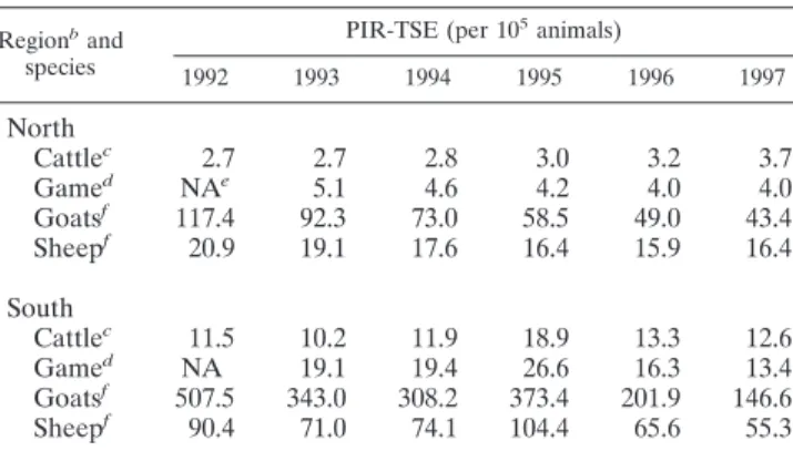
(ii) Predicted annual incidence rate of suspected TSE cases.
The predicted annual incidence rates of suspected TSE cases
(PIR-TSE) are given in Table 3. PIR-TSE was lower north of
the SMV, irrespective of the species. This difference was
con-stant over time. PIR-TSE was lowest in cattle and game, higher
in sheep, and significantly higher in goats. There was a negative
trend over time in all species, except for cattle, where there was
a significant increase. PIR-TSE was furthermore positively
cor-related with the predicted annual incidence rate of suspected
rabies cases (PIR-rabies), again in all species. PIR-rabies for
the period 1992 to 1997 is shown in Table 4. The following
trends were observed: there was no significant effect of time,
except for cattle, for which there was a significant increase in
the number of suspected rabies cases over time both north and
south of the SMV; PIR-rabies was always higher south of the
SMV, irrespective of species; and PIR-rabies was higher for
sheep and goats than for cattle and game, which had similar
rates.
(iii) Incidence rate of suspected and histologically examined
TSE cases.
Table 5 presents the observed annual incidence
rate of histologically examined TSE cases (IR-TSEHE). The
statistical analysis revealed that significantly more records were
obtained for goats, both north and south of the SMV. The
incidence decreased significantly over time in all species, and
there was a correlation between IR-TSEHE and PIR-NED in
both regions, although the relationship was stronger in the
north.
Positive predictive value of presumptive clinical diagnosis.
The level of agreement between the presumptive clinical
diag-nosis (actually made or written in the anamnesis) and the
necropsy findings was calculated for the Faculties of
Veteri-nary Medicine of UL and UG for 224 cattle older than 12
months, 112 sheep and 49 goats older than 6 months, and 8
game animals of all ages. The obtained positive predictive
values of presumptive clinical diagnosis versus necropsy were
as follows: 0.50 for cattle (95% confidence interval [95% CI],
0.43 to 0.56), 0.63 for sheep (95% CI, 0.53 to 0.71), 0.49 for
goats (95% CI, 0.34 to 0.64), and 0.13 for game (95% CI, 0.003
to 0.53).
Pathological examination of targeted samples.
On the basis
of the present study, a supplementary targeting of the available
brain blocks from suspected TSE cases (e.g., presence of
sus-pected clinical signs and/or presence of vacuoles in reports)
was carried out.
Among the samples examined by immunohistochemistry and
histopathology, three were positive for TSE. A first brain block
from a sheep that was presented at UL in March 1983 was
confirmed as a case of scrapie. For this case, the
immunohis-tochemistry confirmed the original histopathological diagnosis
(neuron with pathognomonic cytoplasmic vacuolization). A
second brain block from the TSE network was classified as
negative for scrapie in November 1992, because the result of
histopathology was not conclusive and the SAF result was
negative. For this sample also, the result of
immunohistochem-istry was positive (multiple sites with brownish positive
stain-ing). This sample originated from a 5-year-old Hampshire ewe
imported from the United Kingdom. The third brain block
from UG, from a 3-year-old goat, was histologically diagnosed
as scrapie in August 1992(neuron with pathognomonic
multi-ple cytoplasmic vacuolization), but this diagnosis was not
con-firmed by immunohistochemistry in our study. All other
sam-ples tested remained negative.
at UNIV DE LIEGE on May 28, 2010
jcm.asm.org
DISCUSSION
The present study had three goals: (i) to determine the
annual incidence of NED; (ii) to determine whether TSE were
present in a country before the first report; and (iii) once TSE
had been detected, to monitor the evolution of incidence in
space and time. Our study allowed us (i) to identify the
strengths and weaknesses of the organization of the network,
(ii) to choose the appropriate awareness and training
cam-paigns for the participants in the epidemiological network, and
(iii) to devise appropriate measures, implement them, and
monitor their effectiveness. The geographical TSE risk
assess-ment (25, 26, 27) and TSE risk mapping methodologies would
enable identification of the countries and areas where TSE
emerge and of the animal groups at risk (21, 24, 33, 65).
Targeted passive and active surveillance might be performed in
these geographical areas and for these animal groups at risk.
Etiological classification of the reported NEDs.
Even
though the laboratory archives were incomplete before 1992,
all laboratories processed a sufficient number of samples to
make them representative of the bovine population in their
vicinity. Moreover, the same types of causes were found in
every region before and after 1992. It is therefore assumed that
the percentages of the morbidity causes calculated for every
area during the period 1992 to 1997 were indeed
representa-tive for the population at risk. The fairly high percentage of
cases for which no etiological cause could be established
(around 40% of reported NED cases for domestic ruminants
to 77% for game) is in line with previous observations
else-where: a similar percentage (39%) was found in a Swiss study
of cattle with nervous signs and suspected of rabies (29). This
result could be due partly to a metabolic or toxicological
pa-thology producing no lesion (47). Improvements in
examina-tion techniques and acquisiexamina-tion of more experience could
probably reduce this percentage by half (36).
During the period of this study, the role of the SMV as a
dividing line for the distribution of causes of NED in Belgium
was pivotal, essentially due to the rabies prevalence in the
south. Differences in causes between countries had already
been noted previously (42, 43, 53, 69). Knowledge about the
distribution of NED causes and their associated risk factors
should be improved, because the development of decision
sup-port tools is based on such knowledge (see, e.g., references 13,
14, and 53).
Predicted annual incidence rates of NED and suspected
TSE cases.
The annual incidence rates of NED and suspected
TSE cases are higher south of the SMV than north of the
SMV, and the PIR-TSE is correlated with the annual incidence
rate of suspected rabies. Thus, the main hypothesis to explain
this observation is that south of the SMV, where rabies is
prevalent, farmers and veterinarians have historically been
more aware (56). Moreover, PIR-NED is underestimated
be-cause many NED cases are not reported if the be-cause and/or
therapy is known. In addition, the IR-TSEHE is directly
cor-related with the PIR-NED. Any measure resulting in an
in-crease in NED reporting has a nonspecific knock-on effect on
TABLE 3. Predicted mean incidence rates of reported NED cases
with suspected TSE in Belgium during the period between 1992
and 1997
aRegionb
and species
PIR-TSE (per 105animals)
1992 1993 1994 1995 1996 1997
North
Cattle
c2.7
2.7
2.8
3.0
3.2
3.7
Game
dNA
e5.1
4.6
4.2
4.0
4.0
Goats
f117.4
92.3
73.0
58.5
49.0
43.4
Sheep
f20.9
19.1
17.6
16.4
15.9
16.4
South
Cattle
c11.5
10.2
11.9
18.9
13.3
12.6
Game
dNA
19.1
19.4
26.6
16.3
13.4
Goats
f507.5
343.0
308.2
373.4
201.9
146.6
Sheep
f90.4
71.0
74.1
104.4
65.6
55.3
aThe definition of suspected TSE cases was deliberately broadened (see the
text).
bNorth or south of the SMV. cAnimals older than 12 months. dAnimals of all ages.
eNA, not available.
fAnimals older than 6 months.
TABLE 4. Predicted mean incidence rates of suspected rabies cases
in Belgium during the period 1992 to 1997
Regiona
and species
PIR-rabies (per 105animals)
1992 1993 1994 1995 1996 1997
North
Cattle
b0.1
0.2
0.2
0.3
0.5
0.7
Game
cNA
d0.3
0.3
0.3
0.3
0.3
Goats
e5.7
4.9
4.2
3.6
3.1
2.7
Sheep
e2.5
2.1
1.7
1.4
1.2
1.0
South
Cattle
b6.5
9.1
12.7
17.8
25.0
35.1
Game
cNA
16.9
16.3
15.6
15.1
14.5
Goats
e290.8
249.8
214.6
184.4
158.5
136.1
Sheep
e129.7
107.2
88.6
73.2
60.5
50.0
aNorth and south of the SMV. bAnimals older than 12 months. cAnimals of all ages.
dNA, not available.
eAnimals older than 6 months.
TABLE 5. Mean incidence rates of reported NED cases with
suspected TSE and with complete histological examination in
Belgium during the period between 1992 and 1997
Regiona
and species
IR-TSEHE (per 105animals)
1992 1993 1994 1995 1996 1997
North
Cattle
b2.3
2.5
2.6
2.8
3.0
3.4
Game
cNA
d6.0
5.6
5.3
4.8
4.4
Goats
e127.1
106.3
87.2
68.8
57.3
46.0
Sheep
e22.7
22.3
20.8
19.5
18.6
17.5
South
Cattle
b9.9
12.3
17.0
16.3
13.7
13.4
Game
cNA
21.6
19.9
18.5
17.0
15.5
Goats
e451.7
360.5
292.2
241.2
194.7
159.3
Sheep
e79.7
72.5
69.5
71.4
63.1
57.8
aNorth and south of the SMV. bAnimals older than 12 months. cAnimals of all ages.
dNA, not available.
eAnimals older than 6 months.
866
SAEGERMAN ET AL.
J. C
LIN. M
ICROBIOL.
at UNIV DE LIEGE on May 28, 2010
jcm.asm.org
reporting of suspected and histologically examined TSE cases.
Progressive increases in numbers reported through the specific
TSE network were noted toward the end of the study period,
particularly for cattle (data not shown). Overall, PIR-NED is
higher, and there are more suspected TSE cases, in small
ruminants. Several hypotheses can be put forward to explain
this observation. In Belgium, a considerable number of
live-stock holders have a few small ruminants, and research into the
cause of morbidity is more frequent with hobby farmers,
be-cause they consider their animals to be pets. The “true”
num-ber of registered small ruminants has increased in Europe
following the episode of food-and-mouth disease in Great
Brit-ain (31). Even when these facts are taken into account, the rate
in small ruminants remains higher. In the United States, the
NAHMS network has registered a NED incidence rate around
fourfold higher for sheep than for cattle.
According to the OIE requirement, based on expert
opin-ion, the annual incidence rate of NED for cattle in all
countries is 100 cases per million animals, irrespective of
their BSE status (81, 82). The present retrospective study
provides the first external validation of this requirement at
the population level. Because no clinical sign is
pathogno-monic for BSE (54, 82), laboratory examination of brains is
essential for an efficient BSE epidemiological network. OIE
(81) calculates the minimum number of samples that must
be examined in order to have a probability of 99% to detect
at least one case, if the disease is present in 1% of the cattle
with NED. According to this OIE requirement and the
number of suspected BSE cases for which a complete
his-topathological examination has been carried out, the power
of the epidemiosurveillance effort for the period 1992 to
1997 was on average 59, 28, and 33%, respectively, for the
area south of the SMV, the area north of the SMV, and
Belgium as a whole. If all suspected BSE cases were
ana-lyzed (by using a definition deliberately broader than the
definition of suspected cases proposed by the OIE or the
European Commission [28, 82]), the power would become
99, 28, and 52%, respectively. This suggests the necessity of
organizing awareness and training campaigns to improve
presumptive BSE clinical sign detection for all participants
in the epidemiosurveillance network, especially in countries
that in the past imported live animals, animal products, and
feedstuffs from countries where TSE occurs. This need
par-ticularly is addressed by the methodology for geographical
BSE risk (GBR) assessment, developed by the Scientific
Steering Committee of the European Commission to classify
countries according to their BSE risks (25, 26, 27). Thus, in
Belgium, with the onset of the BSE epidemic and one
in-formation campaign, the power of the BSE
epidemiosurveil-lance network increased to 99% (for Belgium and the area
south of the SMV) and to around 70% (north of the SMV)
in 1998 and 1999 (55, 56).
The mean numbers of histologically investigated cases of
NED during the period 1992 to 1997 (51 and 30 per million
cattle south and north of the SMV, respectively) compare well
with those for other countries at the same GBR level, level III:
10 per million in France (6) and 90 per million in Switzerland
(20) during the same period.
Positive predictive value of presumptive clinical diagnosis.
Determination of the positive predictive value of presumptive
clinical diagnosis permits us to obtain a reference point before
undertaking actions (e.g., awareness and training campaigns).
In fact, it offers the possibility of following up and evaluating
these actions continuously. It also shows the added benefits of
systematically turning to necropsy examinations or
comple-mentary tests to establish an etiological diagnosis and above all
to dispel uncertainty over the identification of suspected TSE
cases (54, 82). The main purpose of a clinical TSE
epidemio-surveillance network is to attain as high a sensitivity as possible
(in order to identify every BSE case). This goal has to be
promoted by permanent awareness and training campaigns for
veterinarians, farmers, and other actors so that a higher
num-ber of nervous-disorder cases are reported and analyzed (28,
81). The low predictive value for game (0.13) can probably be
explained by the low level of clinical observation and
anamne-sis possible for wildlife.
Pathological examination of targeted samples.
The present
study confirms that neuronal vacuolization can occasionally be
observed in cattle in the absence of BSE (2). The additional
importance of immunohistochemical staining in the control of
TSE in ruminants is also proved. In fact, immunohistochemical
staining revealed one extra TSE case and confirmed one
sus-pected TSE case. The negative result upon histopathological
examination is possibly an indication of a (sub)clinical case
without clear signs. However, due to the lack of tonsillar tissue,
confirmation could not be established (51). The absence of
SAF in electron microscopy can be explained by the difficulty
this technique encounters at such a large magnification. The
fact that no BSE case was detected before October 1997 may
be due to the low sample size.
The present retrospective study constitutes the first
valida-tion, at a population level, of the OIE requirements. The
predicted mean incidence rate of NED in a GBR level III
country is sufficiently high to detect confirmed BSE cases only
when a minimal number of samples has been examined. The
results of the present study help to develop and maintain the
quality of the clinical epidemiological TSE networks
world-wide, especially in countries that in the past imported live
animals, animal products, and feedstuffs from countries where
TSE occurs.
Finally, our study also revealed the two main limiting factors
for this type of study: (i) standardization of the definitions and
list of NEDs and (ii) archiving of data and brain blocks over
time. The use of an identification and registration system and
a laboratory information management system would
undoubt-edly make it easier to analyze data.
ACKNOWLEDGMENTS
We thank the members of the Belgian TSE Advisory Group for their
support. This group was founded by the Federal Agency for the Safety
of the Food Chain in Belgium (FASFC). We also thank all our
col-leagues from the private and provincial veterinary investigation
cen-ters, the Pasteur Institute, the National Reference Laboratory for
Veterinary TSE (CODA/CERVA), and the Faculties of Veterinary
Medicine at the Universities of Lie
`ge and Ghent, as well as all the
veterinarians in practice who have provided the necessary information.
We particularly thank P. Dechamps (FASFC) for facilitating access to
data. The calculations of population size were made possible by the
help of R. Hellemans, then of the Agriculture Economics Centre, and
of administrative staff for different regions of Belgium (for the size of
the game population).
at UNIV DE LIEGE on May 28, 2010
jcm.asm.org
REFERENCES
1. Belt, P. B. G. M., I. H. Muileman, B. E. C. Schreuder, J. Bos-De Ruijter, A. L. J. Gielkins, and M. A. Smits.1995. Identification of five allelic variants of the sheep PrP gene and their association with natural scrapie. J. Gen. Virol. 76:509–517.
2. Bradley, R. 1991. Bovine spongiform encephalopathy (BSE): the current situation and research. Eur. J. Epidemiol. 7:532–544.
3. Bradley, R., and J. W. Wilesmith. 1993. Epidemiology and control of bovine spongiform encephalopathy (BSE). Br. Med. Bull. 49:932–959.
4. Braun, U. 2002. Clinical signs and diagnosis of BSE. Schweiz. Arch. Tier-heilk. 144:645–652.
5. Bruce, M. E., R. G. Will, J. W. Ironside, I. McConnell, D. Drummond, A. Suttie, L. McCardle, A. Chree, J. Hope, C. Birkett, S. Cousens, H. Fraser, and C. J. Bostock.1997. Transmissions to mice indicate that “new variant” CJD is caused by the BSE agent. Nature 389:498–501.
6. Calavas, D., and C. Ducrot. 2003. L’ESB en France. Synthe`se sur l’e´volution de l’e´pizootie a` partir des donne´es disponibles au 1erjanvier 2003. Agence
Franc¸aise de Se´curite´ Sanitaire des Aliments, Paris, France.
7. Cannon, R. M., and R. T. Roe. 1982. Livestock disease surveys: a field manual for veterinarians. Australian Bureau of Animal Health, Canberra, Australia.
8. Capucchio, M. T., F. Guarda, N. Pozzato, S. Coppolino, S. Caracappa, and V. Di Marco.2001. Clinical signs and diagnosis of scrapie in Italy: a com-parative study in sheep and goats. J. Vet. Med. 48:23–31.
9. Centers for Epidemiology and Animal Health. 1997. Beef ’97. National Animal Health Monitoring System, U.S. Department of Agriculture, Fort Collins, Colo.
10. Centers for Epidemiology and Animal Health. 1996. Reference of 1996 U.S. sheep health and management practices. National Animal Health Monitor-ing System, U.S. Department of Agriculture, Fort Collins, Colo. 11. Centers for Epidemiology and Animal Health. 1996. Dairy ’96. National
Animal Health Monitoring System, U.S. Department of Agriculture, Fort Collins, Colo.
12. Chelle, P. L. 1942. Un cas de tremblante chez une che`vre. Bull. Acad. Ve´t. Fr. 15:294–295.
13. Cockroft, P. D. 2000. Clinical sign profile likelihood ratios for bovine spon-giform encephalopathy suspects. Res. Vet. Sci. 68:285–290.
14. Cockroft, P. D. 1999. Pattern-matching models for the differential diagnosis of bovine spongiform encephalopathy. Vet. Rec. 144:607–610.
15. Department for Environment, Food and Rural Affairs. 2004. Age distribu-tion of confirmed scrapie cases (in sheep and goats) from 1998 to 2002. [Online.] http://www.defra.gov.uk/animalh/bse/bse-science/scrapie/scrapie_age.PDF. 16. Department for Environment, Food and Rural Affairs. 2004. Bovine
spon-giform encephalopathy in Great Britain: youngest and oldest cases by year of onset (passive surveillance only). [Online.] http://www.defra.gov.uk/animalh/ bse/statistics/bse/yng-old.html.
17. Department for Environment, Food and Rural Affairs. 2004. Bovine spon-giform encephalopathy in Great Britain: confirmed cases by year of birth. [Online.] http://www.defra.gov.uk/animalh/bse/statistics/bse/yrbirth.html. 18. Detwiler, L. A. 1992. Scrapie. Rev. Sci. Tech. Off. Int. Epizoot. 11:491–537. 19. Dickinson, A. G., P. J. T. Stamp, and C. C. Renwick. 1974. Maternal and
lateral transmission of scrapie in sheep. J. Comp. Pathol. 84:19–25. 20. Doherr, M. G., D. Heim, R. Fatzer, C. H. Cohen, M. Vandevelde, and A.
Zurbriggen.2001. Targeted screening of high-risk cattle populations for BSE to augment mandatory reporting of clinical suspects. Prev. Vet. Med. 51:3– 16.
21. Doherr, M. G., A. R. Hett, J. Ru¨fenacht, A. Zurbriggen, and D. Heim.2002. Geographical clustering of cases of bovine spongiform encephalopathy (BSE) born in Switzerland after the feed ban. Vet. Rec. 151:467–472. 22. Durand, B., M. Savey, and F. Moutou. 1998. Etude critique de la surveillance
de l’ence´phalopathie spongiforme bovine dans le monde. Epide´miol. Sante´ Anim. 34:29–39.
23. Durand, F. 1995. Le re´seau VIALINE. Epide´miol. Sante´ Anim. 27:31–43. 24. European Commission Scientific Steering Committee. 2003. Assessment of
the geographical risk of bovine spongiform encephalopathy carried out worldwide by the European Commission’s Scientific Steering Committee. [Online.] http://europa.eu.int/comm/food/fs/sc/ssc/out363_en.pdf. 25. European Commission Scientific Steering Committee. 7–8 November 2002.
Opinion on the geographical BSE risk for sheep and goats (GBR-S): adap-tation of the cattle GBR methodology to small ruminants, in case BSE in small ruminants would become probable or evident under field conditions. [Online.] http://europa.eu.int/comm/food/fs/sc/ssc/out294_en.pdf. 26. European Commission Scientific Steering Committee. 11 January 2002.
Up-date of the opinion on the geographical risk of bovine spongiform enceph-alopathy (GBR). [Online.] http://europa.eu.int/comm/food/fs/sc/ssc/out243_en .pdf.
27. European Commission Scientific Steering Committee. 6 July 2000. Final opinion on the geographical risk of bovine spongiform encephalopathy (GBR). [Online.] http://europa.eu.int/comm/food/fs/sc/ssc/out113_en.pdf. 28. European Parliament and Council of the European Union. 2001. Regulation
(EC) No 999/2001 of the European Parliament and of the Council of 22 May
2001 laying down rules for the prevention, control and eradication of certain transmissible spongiform encephalopathies. Off. J. Eur. Communities L147: 1–40.
29. Fatzer, R., and F. Steck. 1974. Histologische Differentialdiagnose bei toll-wurverda¨chtigen Rindem. Schweiz. Arch. Tierheilkd. 116:347–356. 30. Fergusson, N. M., C. A. Donnely, M. E. I. Woolhouse, and R. M. Anderson.
1997. The epidemiology of BSE in cattle herds in Great Britain. II. Model construction and analysis of transmission dynamics. Philos. Trans. R. Soc. Lond. 352:803–838.
31. Gibbens, J. C., C. E. Sharpe, J. W. Wilesmith, L. M. Mansley, E. Michalo-poulou, J. B. M. Ryan, and M. Hudson.2001. Descriptive epidemiology of the 2001 foot-and-mouth disease epidemic in Great Britain: the first five months. Vet. Rec. 149:729–743.
32. Hadlow, W. J., R. C. Kennedy, and R. E. Race. 1982. Natural infection of Suffolk sheep with scrapie virus. J. Infect. Dis. 146:657–664.
33. Heim, D., and J. Kreysa. 2002. Risk assessment as an indicator for the distribution of BSE in the world. Schweiz. Arch. Tierheilkd. 144:710–715. 34. Heim, D., L. Detwiler, E. Williams, and U. Kihm. 2001. Update on bovine
spongiform encephalopathy, scrapie, and chronic wasting disease. [Online.] Office International des Epizooties, Paris, France. ftp://ftp.oie.int /69SG_2001/A_69_SG_12_CS3C.pdf.
35. Heim, D., and U. Kihm. 1999. Bovine spongiform encephalopathy in Swit-zerland: the past and the present. Rev. Sci. Tech. Off. Int. Epizoot. 18:135– 144.
36. Heim, D., R. Fatzer, B. Hornlimann, and M. Vandevelde. 1997. Ha¨ufigkeit neurologischer Erkrankungen beim Rind. Schweiz. Arch. Tierheilkd. 139: 354–362.
37. Hill, A. F., M. Desbruslais, S. Joiner, K. C. L. Sidle, J. Gowland, L. Collinge, L. J. Doey, and P. Lantos.1997. The same prion strain causes vCJD and BSE. Nature 389:448–450.
38. Hill, F. 1994. Neurological diseases of cattle where BSE has been included in the differential diagnosis. Surveillance 21:25.
39. Hilmert, H., and H. Diringer. 1984. A rapid and efficient method to enrich SAF-protein from scrapie brains of hamsters. Biosci. Rep. 4:165–170. 40. Hoorens, J., and W. Oyaert. 1966. Scrapie bij het shaap. Vlaams
Dier-geneeskd. Tijdschr. 35:313–317.
41. Hornlimann, B., D. Guidon, and C. Griot. 1994. Risikoeinscha¨tzung fu¨r die Einschleppung von BSE. Dtsch. Tiera¨rztl. Wochenschr. 101:295–298. 42. Jeffrey, M., M. M. Simmons, and G. A. H. Wells. 1993. Observations on the
differential diagnosis of bovine spongiform encephalopathy in Great Britain, p. 342–362. In R. Bradley and B. Marchant (ed.), Transmissible spongiform encephalopathies. Proceedings of a Consultation on BSE with the Scientific Veterinary Committee of the Commission of the European Communities. Commission of the European Communities, Brussels, Belgium.
43. Jeffrey, M., and J. W. Wilesmith. 1992. Idiopathic brainstem neuronal chro-matolysis and hippocampal sclerosis: a novel encephalopathy in clinically suspect cases of bovine spongiform encephalopathy. Vet. Rec. 131:359–362. 44. Kimberlin, R. H. 1992. Bovine spongiform encephalopathy. Rev. Sci. Tech.
Off. Int. Epizoot. 11:347–390.
45. Lasme´zas, C. I. 2003. The transmissible spongiform encephalopathies. Rev. Sci. Tech. Off. Int. Epizoot. 22:23–36.
46. Martin, S. W., A. H. Meek, and P. Willeberg. 1987. Veterinary epidemiology. Principles and methods. Iowa State University Press, Ames.
47. Mayhew, I. G. 1989. Large animal neurology: a handbook for veterinary clinicians, p. 3–69. Lea & Febiger, Philadelphia, Pa.
48. McElroy, M. C., and E. D. Weavers. 2001. Clinical presentation of bovine spongiform encephalopathy in the Republic of Ireland. Vet. Rec. 149:747– 748.
49. Parry, H. B., and D. R. Oppenheimer. 1983. Scrapie disease in sheep, p. 31–51. Academic Press, London, United Kingdom.
50. Prusiner, S. B. 1998. Prions. Proc. Natl. Acad. Sci. USA 95:13363–13383. 51. Roels, S., E. Vanopdenbosch, J. P. Langeveld, and B. E. Schreuder. 1999.
Immunohistochemical evaluation of tonsillar tissue for preclinical screening of scrapie based on surveillance in Belgium. Vet. Rec. 145:524–525. 52. Russo, P., C. Ducrot, P. Belli, J.-J. Fontaine, and C. Peyrouse. 1999.
Trem-blante ovine: bilan de six anne´es d’e´pide´miosurveillance dans le Sud de la France (etude de 173 cas). Point Ve´t. 28:667–670.
53. Saegerman, C., N. Speybroeck, S. Roels, E. Vanopdenbosch, E. Thiry, and D. Berkvens.2004. Decision support tools for clinical diagnosis of disease in cows with suspected bovine spongiform encephalopathy. J. Clin. Microbiol. 42:172–178.
54. Saegerman, C., L. Claes, A. Dewaele, D. Desmecht, F. Rollin, J. Hamoir, P. Gustin, G. Czaplicki, J. Bughin, J. Wullepit, J. Laureyns, S. Roels, D. Berkvens, E. Vanopdenbosch, and E. Thiry.2003. Differential diagnosis of neurologically expressed disorders in Western European cattle. Rev. Sci. Tech. Off. Int. Epizoot. 22:83–102.
55. Saegerman, C., P. Dechamps, S. Roels, K. Petroff, R. Geeroms, G. Torck, J. Dufey, R. Fourez, M. Hamerijckx, A. Cormann, P. Viatour, V. De Connick, F. Lomba, J.-P. Vermeersch, L. Hallet, O. Lhost, M. Leemans, A. Vander-sanden, D. Peharpre, B. Brochier, F. Costy, P.-P. Pastoret, E. Thiry, and E. Vanopdenbosch.2001. Epide´miosurveillance de l’ence´phalopathie
spongi-868
SAEGERMAN ET AL.
J. C
LIN. M
ICROBIOL.
at UNIV DE LIEGE on May 28, 2010
jcm.asm.org
forme transmissible chez les bovins en Belgique: bilan de l’anne´e 1999. Ann. Med. Ve´t. 145:47–58.
56. Saegerman, C., P. Dechamps, E. Vanopdenbosch, S. Roels, K. Petroff, J. Dufey, G. Van Caeneghem, D. Devreese, H. Varewyck, H. De Craemere, I. Desmedt, A. Cormann, G. Torck, L. Hallet, M. Hamerijckx, M. Leemans, A. Vandersanden, D. Peharpre, B. Brochier, F. Costy, P. Mullier, E. Thiry, and P.-P. Pastoret. 1999. Epide´miosurveillance de l’ence´phalopathie spongi-forme transmissible chez les bovins en Belgique: bilan de l’anne´e 1998. Ann. Med. Ve´t. 143:423–436.
57. Sarradet, M. 1883. Un cas de tremblante sur un bœuf. Rev. Vet. Toulouse 7:310–312.
58. Savey, M., P. Belli, and M. Coudert. 1993. L’ence´phalopathie spongiforme bovine en Europe. Pre´sent et avenir. Vet. Res. 24:213–225.
59. Schreuder, B. E. C., J. W. Wilesmith, J. B. M. Ryan, and O. C. Straub. 1997. Risk of BSE from the import of cattle from the United Kingdom into countries of the European Union. Vet. Rec. 141:187–190.
60. Scott, A. C., S. H. Done, C. Venables, and M. Dawson. 1987. Detection of scrapie-associated fibrils as an aid to the diagnosis of natural sheep scrapie. Vet. Rec. 120:280–281.
61. Scott, M. R., R. Will, J. Ironside, H.-O.B. Nguyen, P. Tremblay, S. J. DeAr-mond, and S. B. Prusiner.1999. Compelling transgenetic evidence for trans-mission of bovine spongiform encephalopathy prions to humans. Proc. Natl. Acad. Sci. USA 96:15137–15142.
62. Smits, M. A., A. Bossers, and B. E. C. Schreuder. 1997. Prion protein and scrapie susceptibility. Vet. Q. 19:101–105.
63. StataCorp. 2003. Stata statistical software, release 7.1. Stata Corporation, College Station, Tex.
64. Terry, L. A., S. Marsch, S. J. Ryder, S. A. C. Hawkins, G. A. H. Wells, and Y. I. Spencer.2003. Detection of disease-specific PrP in the distal ileum of cattle exposed orally to the agent of bovine spongiform encephalopathy. Vet. Rec. 152:387–392.
65. Vanopdenbosch, E., S. Roels, and C. Saegerman. 2000. Animal TSE epi-demiosurveillance and diagnosis in Belgium and BSE risk assessment, 29–30. In Proceedings of the First Scientific Day on Transmissible Spongiform Encephalopathies: Creutzfeldt-Jakob Disease and Bovine Spongiform En-cephalopathy. Scientific Institute of Public Health, Brussels, Belgium. 66. Vanopdenbosch, E., P. Dechamps, C. Saegerman, J. Dufey, S. Roels, P.
Mullier, L. Hallet, B. Brochier, F. Costy, G. Charlier, R. Fourez, and P.-P. Pastoret.1998. Le premier cas d’ence´phalopathie spongiforme bovine diag-nostique´ en Belgique. Ann. Me´d. Ve´t. 142:111–118.
67. Vicari, A., B. Hornlimann, and L. Audige´. 1996. Appre´ciation du risque de contamination des aliments concentre´s suisses pour bovins par l’agent de l’ence´phalopathie spongiforme bovine. Epide´miol. Sante´ Anim. 30:77–84. 68. Wells, G. A. H., S. A. C. Hawkins, R. B. Green, A. R. Austin, I. Dexter, Y. I.
Spencer, M. J. Chaplin, M. J. Stack, and M. Dawson.1998. Preliminary
observations on the pathogenesis of experimental bovine spongiform en-cephalopathy (BSE): an update. Vet. Rec. 142:103–106.
69. Wells, G. A. H., A. R. Sayers, and J. W. Wilesmith. 1995. Clinical and epidemiological correlates of the neurohistology of cases of histologically unconfirmed, clinically suspect bovine spongiform encephalopathy. Vet. Rec. 136:211–216.
70. Wells, G. A. H., A. C. Scott, C. T. Johnson, R. F. Gunning, R. D. Hancock, M. Jeffrey, M. Dawson, and R. Bradley.1987. A novel progressive spongi-form encephalopathy in cattle. Vet. Rec. 121:419–420.
71. Wilesmith, J. W. 1998. Manual on bovine spongiform encephalopathy. Food and Agriculture Organization of the United Nations, Rome, Italy. 72. Wilesmith, J. W., J. B. M. Ryan, and W. D. Hueston. 1992. Bovine
spongi-form encephalopathy: case-control studies of calf feeding practices and meat and bone meal inclusion in proprietary concentrates. Res. Vet. Sci. 52:325– 331.
73. Wilesmith, J. W., J. B. M. Ryan, W. D. Hueston, and L. J. Hoinville. 1992. Bovine spongiform encephalopathy: epidemiological features 1985-1990. Vet. Rec. 130:90–94.
74. Wilesmith, J. W., J. B. M. Ryan, and M. J. Atkinson. 1991. Bovine spongi-form encephalopathy: epidemiological studies on the origin. Vet. Rec. 128: 199–203.
75. Wilesmith, J. W., G. A. H. Wells, M. P. Cranwell, and J. B. M. Ryan. 1988. Bovine spongiform encephalopathy: epidemiological studies. Vet. Rec. 123: 638–644.
76. Will, R. G., J. W. Ironside, M. Zeidler, S. N. Cousens, K. Estibeiro, A. Alperovitch, S. Poser, M. Pocchiari, A. Hofman, and P. G. Smith.1996. A new variant of Creutzfeldt-Jakob disease in the UK. Lancet 347:264–267. 77. Williams, E. S., and M. W. Miller. 2003. Transmissible spongiform
enceph-alopathies in non-domestic animals: origin, transmission and risk factors. Rev. Sci. Tech. Off. Int. Epizoot. 22:145–156.
78. Williams, E. S., and M. W. Miller. 2002. Chronic wasting disease in deer and elk in North America. Rev. Sci. Tech. Off. Int. Epizoot. 21:305–316. 79. Williams, E. S., and S. Young. 1980. Spongiform encephalopathy of Rocky
Mountain elk. J. Wildl. Dis. 18:465–471.
80. Wood, J. N. L., L. J. Lund, and S. H. Done. 1992. The natural occurrence of scrapie in moufflon. Vet. Rec. 130:25–27.
81. World Animal Health Organization. 2004. Terrestrial animal health code, 12th ed., appendix 3.8.4. Surveillance systems for bovine spongiform encephalopathy. [Online.] http://www.oie.int/eng/normes/mcode/en_chapitre_3.8.4.htm. 82. World Animal Health Organization. 1997. Guidelines for continuous
sur-veillance and monitoring of bovine spongiform encephalopathy. January 1997 Meeting of the International Animal Health Code Commission, ap-pendix VIIIb. Document 65 SG/12/CS 1. Office International des Epizooties, Paris, France.
83. World Health Organization. 2001. Rabies surveillance report. Center for Rabies Surveillance and Research. Rabies Bull. Eur. 3:4–8.