Dosimetry of preclinical and clinical case studies of
18
F-radiopharmaceuticals using Positron Emission
Tomography and Computed Tomography: Methods of
quantification, their improvement and considerations
of critical exposures
Florian Bretin
University of Liège
Maastricht University
A thesis submitted for the degree of
Doctor of Philosophy
Abstract
Computed tomography (CT) and positron emission tomography (PET) are routinely used in (pre)clinical research and practice. The radiation burden inflicted on subjects from both modalities needs to be addressed to keep exposures as low as possible. While the focus in literature is on human dosimetry, few studies investigate the animal dosimetry in preclinical studies. This doctoral thesis aimed to quantify the radiation burden imposed on subjects by CT and PET in clinical and especially preclinical case studies using both experimental data and Monte Carlo simulations.
We derived the biodistribution of three18F-radiopharmaceuticals (18F-UCB-H,
6-[18F]fluoro-L-DOPA and 2-[18F]fluoro-L-Tyrosine) in mice using the gold standard
method organ harvesting, as well as dynamic microPET imaging and the new tech-nique of hybrid imaging, where microPET activities are cross-calibrated with the activity remaining in post-scan harvested organs. The radiation dosimetry in hu-mans was calculated and results of all methods were compared. For the newly
developed tracer18F-UCB-H, a first-in-human clinical study was conducted and the
radiation dosimetry was compared to preclinically obtained data. Additionally, the
preclinically obtained human dosimetry of 6-[18F]fluoro-L-DOPA was compared to
clinical values available in the literature. The derived biodistributions of the three tracers were also applied to mouse S-factors to calculate absorbed doses in mice. Furthermore, the radiation dosimetry of the small animal microCT GE eXplore 120 was experimentally investigated ex vivo in a custom built phantom and in vivo in rats. A model of the imaging system was built using Monte Carlo simulations to further investigate the radiation dosimetry non-invasively.
The preclinical study of the three18F-radiopharmaceuticals in mice showed that
hybrid imaging produces equivalent results compared to labour intensive organ har-vesting while being more efficient from an ethical, economical, and scientific point of view. Sole dynamic microPET imaging produced poor results due to partial vol-ume effects and quantification errors in small volvol-umes. Clinically and preclinically
derived data of18F-UCB-H and 6-[18F]fluoro-L-DOPA agreed roughly regarding
significant amounts of radiation in preclinical studies. Monte Carlo simulations of the GE eXplore 120 were in good agreement with experimental results and provided a deeper insight into the spatial dose distribution.
Based on our analysis, hybrid imaging could serve as a replacement for the cur-rent gold standard of organ harvesting in preclinical dosimetry. Preclinically ob-tained estimates of human dosimetry showed relatively poor accuracy and should be used with caution for the calculation of injection limits in first-in-human studies. We demonstrated that excessive radiation doses are inflicted on small animals in preclinical imaging, especially in dual-modality longitudinal studies. The radiation should be addressed in advance to avoid critical exposures that might compromise the outcome of the study.
List of Publications
This thesis is based on the following papers, which are referred to in the text by their Roman numerals:
I. Preclinical radiation dosimetry for the novel SV2A
radiotracer [18F]UCB-H
Bretin F., Warnock G., Bahri MA., Aerts J., Mestdagh N., Buchanan T., Valade A., Mievies F., Giacomelli F., Lemaire C., Luxen A., Salmon E., Seret A. and Plenevaux A.
EJNMMI Research 3(1):35, 2013
II. Hybrid MicroPET Imaging for Dosimetric Applications in Mice:
Improvement of Activity Quantification in Dynamic MicroPET Imaging
for Accelerated Dosimetry Applied to 6-[18F]Fluoro-l-DOPA
and 2-[18F]Fluoro-l-Tyrosine
Bretin F., Mauxion T., Warnock G., Bahri MA., Libert L., Lemaire C., Luxen A., Bardiés M., Seret A. and Plenevaux A.
Molecular Imaging and Biology 16(3):383-394, 2014
III. Absorbed doses to mice for three [18F]-tracers calculated from experimental data and Monte Carlo simulations
Bretin F., Mauxion T., Bahri MA., Luxen A., Plenevaux A., Bardiés M. and Seret A.
Manuscript, submitted for Marie Curie Award at EANM conference 2013 in Lyon, France; Reached Top 5
IV. Biodistribution and radiation dosimetry for the novel SV2A radiotracer
[18F]UCB-H: First-in-human study
Bretin F., Bahri MA., Bernard C., Warnock G., Aerts J., Mestdagh N., Buchanan T., Otoul C., Koestler F., Mievis F., Giacomelli F., Degueldre C., Hustinx R.,
Luxen A., Seret A., Plenevaux A.∗ and Salmon E.∗
Bretin F., Warnock G., Luxen A., Plenevaux A., Seret A. and Bahri MA. IEEE Transactions on Nuclear Science 5(60):3235-3241, 2013
VI. Monte Carlo Simulations of the dose from imaging with
GE eXplore 120 micro-CT using GATE
Bretin F., Bahri MA., Luxen A., Phillips C., Plenevaux A. and Seret A. Manuscript
Contents
1 Introduction 1
1.1 Motivation. . . 1
1.2 Units of radiation and biological effects. . . 2
1.3 Typical clinical and preclinical exposures . . . 5
1.4 Regulations for new radiopharmaceuticals . . . 6
1.5 Specific aims . . . 6
1.6 Structural outline . . . 8
2 Scientific background 9 2.1 Positron Emission Tomography . . . 9
2.2 Computed Tomography . . . 11
2.3 Radionuclides . . . 13
2.4 Particle interactions with matter. . . 17
2.4.1 Photon interactions. . . 17
2.4.1.1 Rayleigh or Thomson scatter . . . 18
2.4.1.2 Compton Scattering . . . 18
2.4.1.3 Photoelectric absorption . . . 19
2.4.1.4 Attenuation coefficient . . . 19
2.4.2 Charged particle interactions . . . 20
2.5 Monte Carlo simulations in medical physics . . . 23
2.5.1 General concept . . . 23
2.5.2 GATE package. . . 24
2.5.3 PET dosimetry using Monte Carlo simulations . . . 25
2.5.4 CT dosimetry using Monte Carlo simulations. . . 26
2.6 Methods of assessing the kinetic biodistribution of radiopharmaceuticals 26 2.6.1 Organ harvesting . . . 27
2.6.2 Dynamic PET imaging . . . 27
2.6.3 Hybrid imaging . . . 28
2.7 Extrapolation from preclinical to clinical data . . . 28
2.8 MIRD scheme for dosimetry in nuclear imaging . . . 29
2.9 Dosimetry in CT imaging . . . 32
3 Publications & studies 35 3.1 Preclinical radiation dosimetry for [18F]UCB-H . . . . 35
3.1.1 Overview . . . 35
3.1.2 Discussion . . . 37
3.2 Hybrid microPET imaging in mice for accelerated dosimetry . . . 39
3.2.1 Discussion . . . 42
3.3 Absorbed doses to mice for three [18F]-tracers . . . . 44
3.3.1 Overview . . . 44
3.3.2 Discussion . . . 46
3.4 Radiation dosimetry for [18F]UCB-H: First-in-human study . . . 46
3.4.1 Overview . . . 46
3.4.2 Discussion . . . 48
3.5 X-ray dose quantification of the GE explore 120 micro-CT . . . 49
3.5.1 Overview . . . 49
3.5.2 Discussion . . . 51
3.6 Monte Carlo simulations of the dose from the GE eXplore 120 microCT 52 3.6.1 Overview . . . 52
3.6.2 Discussion . . . 54
4 Discussion 57 4.1 Methods of preclinically derived human radiation dosimetry . . . 57
4.2 Comparison of preclinically and clinically derived human dosimetry . 61 4.2.1 Absorbed dose vs. effective dose . . . 63
4.3 Exposure considerations in preclinical longitudinal studies . . . 65
4.4 Future directions . . . 68
5 Conclusions 69
Appendix A Paper I 71
Appendix B Paper II 81
Contents iii
Appendix D Paper IV 105
Appendix E Paper V 113
Appendix F Paper VI 121
List of Figures
2.1 X-ray emission spectra . . . 12
2.2 Mass attenuation coefficient and mass energy-absorption coefficient . 20 2.3 Stopping powers of electrons . . . 22
2.4 Self- and cross-radiation in a cubical arrangement . . . 25
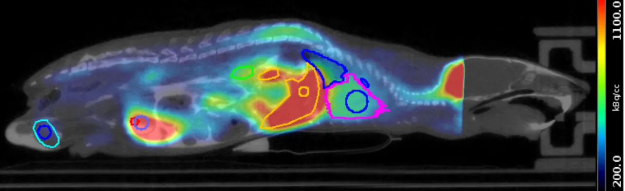
3.1 Representative PET/CT image of 18F-UCB-H . . . 36
3.2 TACs of 18F-UCB-H derived from preclinical data . . . 37
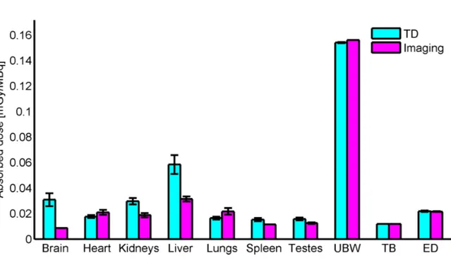
3.3 Absorbed doses of 18F-UCB-H derived from preclinical data . . . 38
3.4 3D PET/CT images of 18F-FDOPA and 18F-FTYR. . . 40
3.5 TACs of 18F-FDOPA and 18F-FTYR . . . 41
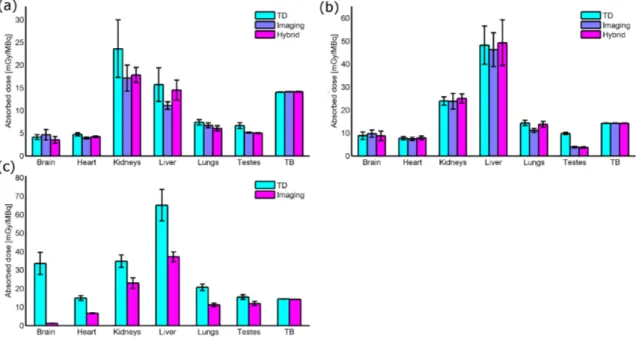
3.6 Absorbed doses of 18F-FDOPA and 18F-FTYR . . . 42
3.7 3D PET/CT images of 18F-FDOPA, 18F-FTYR and 18F-UCB-H. . . 45
3.8 Absorbed doses of 18F-FDOPA, 18F-FTYR and 18F-UCB-H in mice . . 45
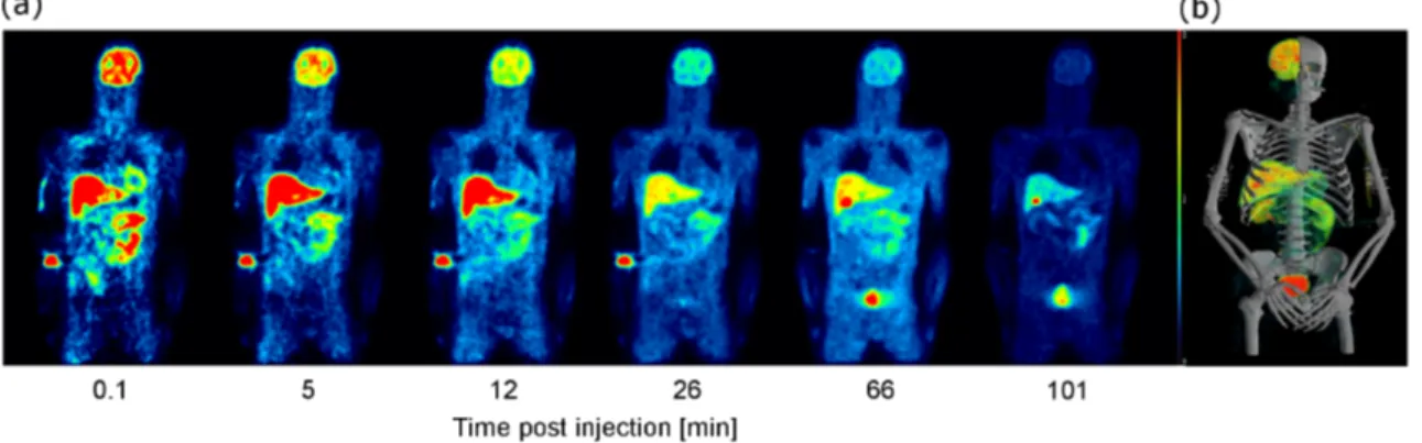
3.9 Sequential whole-body images of 18F-UCB-H . . . 47
3.10 Preclinically and clinically derived absorbed dose estimates of 18F-UCB-H . . . 48
3.11 Experimentally determined doses of microCT . . . 51
3.12 In vivo microCT contrast . . . 51
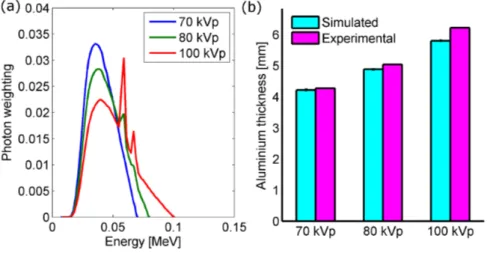
3.13 Simulated X-ray spectra and corresponding HVL . . . 54
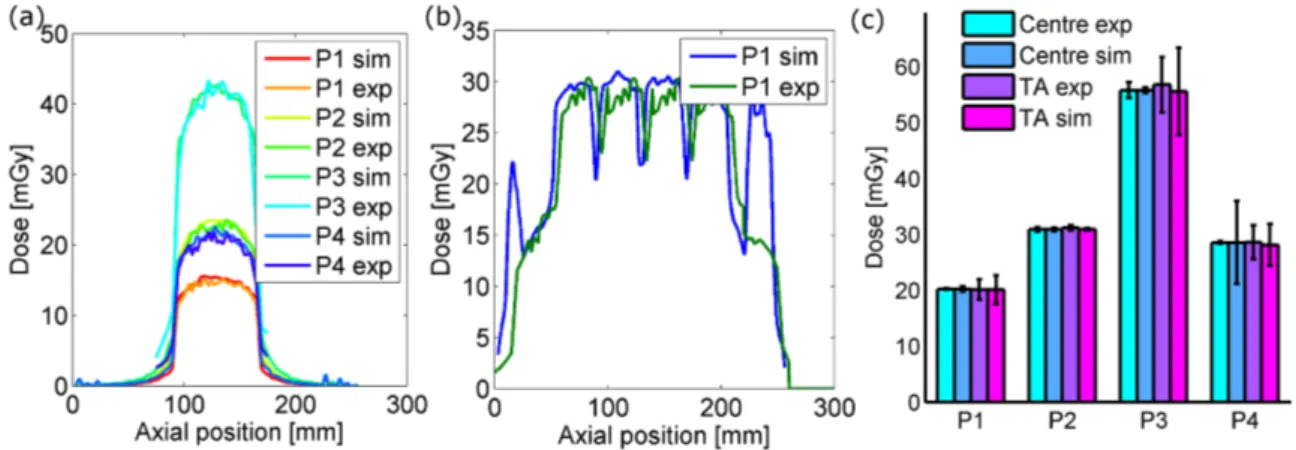
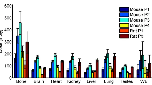
3.14 Comparison of experimentally obtained and simulated dose profiles . 55 3.15 Simulated organ doses from microCT in mice and rats . . . 56
List of Tables
3.1 Investigated microCT protocols . . . 50
3.2 Simulated CTDI100 and MSAD . . . 54
4.1 Pros and Cons of preclinical methods . . . 58
4.2 Longitudinal dual modality scenarios . . . 66
4.3 Cumulative doses in longitudinal studies . . . 67
Glossary
18F-FDG 2-deoxy-2-[18F]fluoro-D-glucose.1
18F-FDOPA 6-[18F]Fluoro-l-DOPA.7
18F-FTYR 2-[18F]Fluoro-l-Tyrosine.7
18F-UCB-H [18F]UCB-H.7
CERN Conseil Européen pour la Recherche Nucléaire.24
CT Computed tomography.1
CTDI Computed tomography dose index.33
FDA Food and Drug Administration.6
FOV Field of view.50
FWHM Full width half maximum.50
GATE Geant4 Application to tomographic emission.8
HVL Half-value layer.53
ICRP International Committee of Radiation Protection.3
MCS Monte Carlo simulation.8
mFOV Multiple field of view.50
microCT Micro computed tomography.1
microPET Micro positron emission tomography.1
microSPECT Micro single photon emission computed tomography.2
MIRD Medical Internal Radiation Dose. 29
MOSFET Metal oxide semiconductor field-effect transistor.33
MRI Magnetic resonance imaging.28
MSAD Multiple scan average dose.33
PDF Probability density function.17
PET Positron emission tomography.1
PMMA Poly(methyl methacrylate).33
PVE Partial volume effect. 11
sFOV Single field of view.50
TAC Time activity curve.26
TLD Thermoluminescent dosimetry chip.33
C
H A P T E R1
Introduction
1.1
Motivation
In the last two to three decades the use ofcomputed tomography (CT)andpositron
emission tomography (PET) increased constantly and they are now essential for routine clinical use, as well as preclinical and clinical research [1, 2], e.g. in
on-cology [3]. Due to advances in technology in the past decade, stand-alone PET
scanners have almost disappeared from modern hospitals and have been substituted
for the dual-imaging modality PET/CT [3]. The combined system exhibits several
advantages, such as intrinsically spatially co-registered images as well as faster and qualitatively better transmission scans for attenuation correction [4]. The main clin-ical applications of PET/CT imaging are staging, restaging and assessment of tumor response in oncological applications [3], with2-deoxy-2-[18F]fluoro-D-glucose
(18F-FDG) being the currently most used radiopharmaceutical [5]. Other applications
consist of the target volume definition for highly accurate radio therapy treatment [6], the assessment of myocardial viability and perfusion in cardiology [7,8], and the diagnosis of neurological pathologies such as Alzheimer’s [9], epilepsy [10], and
Parkinson’s [11]. One common application of preclinical micro positron emission
tomography (microPET) imaging, which is often combined with micro computed tomography (microCT)imaging for anatomical referencing, is in the field of drug development as it allows for dynamic, pharmacological, longitudinal observations
on the same animal [12]. Using suitable radiopharmaceuticals microPET imaging
enables researchers to non-invasively monitor biological processes, disease progres-sion and therapy response in small animals providing a potential translational model
for human medicine [13]. Because of the widespread use of PET/CT and the
ever-increasing number of applications, the radiation exposure of subjects undergoing a single PET or CT imaging session and especially a combined PET/CT scan needs to
be addressed to keep the radiation burden as low as possible. The focus of past dosi-metric considerations in the literature has mainly been human applications. With the emergence of preclinical microPET and microCT of small animals in the last decade, however, there is a need for further investigations into the radiation burden of ani-mals. Since longitudinal studies are commonly conducted in preclinical imaging and
often involve dual-modality microCT/microPET or micro single photon emission
computed tomography (microSPECT)scans [14,15], the radiation exposure needs
to be quantified to rule out any influence on the outcome of the study [16].
1.2
Units of radiation and biological effects
The amount of radiation received by any subject from any type of ionizing radiation in nuclear medicine and radiology is quantified using the absorbed dose (D). It is
defined as the energy absorbed per unit mass of any material [17]
D = dε
dm, (1.1)
where dε is the mean deposited energy induced by the ionizing radiation to the matter in the volume element of mass dm. In radiology the term absorbed dose is used interchangeably with the term dose, however, in nuclear medicine as dose could possibly refer to other quantities D is always referred to as absorbed dose. Physical processes caused by ionizing radiation depositing the energy within matter
will be described in Section2.4. The SI unit of the absorbed dose is gray (Gy) with
1 Gy being equal to 1J
kg. Other units for the same quantity exist but are outdated
and ergg or rad will not be used here. In diagnostic nuclear medicine and radiology it
is under all circumstances the aim to keep the absorbed dose as low as possible to avoid any biological effects in living tissue. Nonetheless, the overall aim is to acquire diagnostically useful images and the subject should not be exposed to radiation unnecessarily.
Radiobiology differentiates between two types of effects caused by ionizing radi-ation: stochastic effects and deterministic effects. Stochastic effects resulting from exposure to ionizing radiation are carcinogenesis and hereditary effects, whose sever-ity are independent of the received absorbed dose and may, or may not, manifest
many years or even decades after exposure [18,19]. However, their probability of
occurrence increases with the absorbed dose. Deterministic (or non-stochastic) ef-fects occur only above a certain threshold of radiation exposure and are usually not of concern in diagnostic imaging opposed to radiation therapy (nuclear medicine
1.2. Units of radiation and biological effects 3
or external beam), which aims at producing deterministic effects in pathological
tis-sue [19]. Below this threshold deterministic effects do not manifest, but beyond the
threshold their severity increases with the absorbed dose. Because of inter-individual differences the threshold may vary. Manifestations of deterministic effects after acute exposure are i.e. erythema (reddening of the skin), epilation (loss of hair), depres-sion of bone marrow cell dividepres-sion, NVD (nausea, vomiting, diarrhea), central nervous system damage, and damage to unborn children. However, in diagnostic imaging patients, whether in nuclear medicine or radiology, only stochastic effects are of concern due to the low absorbed doses.
Since the absorbed dose does not take into account the type of radiation or the type of target material, there is the need for other quantities in medical dosimetry as different materials, especially biological tissues, are more radiosensitive to different
types of radiation than others [17,19]. TheInternational Committee of Radiation
Protection (ICRP)publishes regularly recommendations on radiation protection and
has defined the quantity of the effective dose [20], which is a sex, age, and dose
rate (delivered dose over time) independent quantity. It is a measure of the resulting stochastic risk to a subject by a non-uniform radiation. As the effective dose is ap-plicable to many situations (i.e. medical imaging including patient and practitioner, aviation exposures, etc.) one must account for different types of radiation and bio-logical tissues. Before giving the definition of the effective dose the equivalent dose (or radiation-weighted dose) is to be considered. The equivalent dose is accounting for the different types of radiation (i.e. photons, electrons/positrons, neutrons, pro-tons etc.) and their relative biological effectiveness in producing biological damage. Therefore, the equivalent dose H to a specific tissue or organ T is the weighted sum of all absorbed doses produced by each species of radiation [17,19,20]
HT =X
R
wRDT ,R, (1.2)
where wR is the weighting factor for the radiation type R and DT ,R the absorbed
dose to tissue T from that particular radiation. The weighting factors were
pub-lished first in ICRP publication 26 [21], and were most recently updated in ICRP
publication 103 [20] in 2007. As the weighting factor wR for radiations produced
in diagnostic nuclear medicine and radiology are equal to unity, the equivalent dose can be interpreted as the physical absorbed dose in photons or electrons that has the same biological effect to an organ or tissue. However, since the quantity is committee-defined, the unit was assigned to be sievert (Sv) instead of gray (Gy). Heavier particles like protons, alpha particles or neutrons have higher weighting
factors as they are more potent in producing biological damage. In order to estimate the stochastic risk to a subject from any given ionizing imaging modality, all types of radiation must be known and therefore the equivalent dose H to all tissues. Addi-tionally, the respective radiosensitivity of all tissues must be taken into account, as they vary for different organs and tissues. Their intrinsic radiosensitivity is reflected in the tissue weighting factor wT, which allows for calculation of the effective dose
E (also in sievert) to any organ or tissue T with a known weighting factor as
E =X
T
wTHT. (1.3)
The weighting factors wT were first published in ICRP publication 26 [21] and
were updated twice in ICRP 60 [22] and the most recent publication 103 [20]. The
first weighting factors from ICRP 26 reflected mortality risk only, but due to the advancement in knowledge of radiosensitivity, the weighting factors of ICRP 60 and 103 reflect the following quantities: probability of attributable fatal cancer, weighted probability of attributable non-fatal cancer, weighted probability of severe hereditary effects, and relative amount of time lost [19]. The sum of all tissues / organs (with a known weighting factor) results in the effective dose for the whole body, which is commonly used in diagnostic nuclear medicine to assess the exposure risk. According to the EANM dosimetry committee the most recent weighting factors from ICRP 103 should not be applied yet, as they are phantom specific (male and female, published
in [23]) and specific absorbed fractions for those phantoms were not published at the
moment this manuscript was prepared [24]. Controversially, many studies apply the
tissue weighting factors from ICRP 103 to effective dose calculations based on the
standard hermaphroditic phantom [25–29] as was requested by reviewers during
the publication process of Paper I in AppendixA. The effective dose based on ICRP
103 was also added in Paper II in AppendixB. We were made aware of that widely
spread misconception during the review of Paper IV in AppendixDand therefore the
reader is advised to focus on effective doses based on tissue weighting factors from
ICRP 60, which are displayed exclusively in Chapter3to5. The use of the effective
dose for assessing radiation risk from medical exposures is contentious anyway, as the value is averaged over sex and age, and neglects the impact of absorbed dose
rate [19,30]. Many still recommend the use of absorbed doses for patient specific
1.3. Typical clinical and preclinical exposures 5
1.3
Typical clinical and preclinical exposures
Typical radiation exposures of patients undergoing PET imaging highly depend on the administered radiopharmaceutical, the amount of activity necessary to obtain a diagnostically useful image and partially on the scanners sensitivity towards sig-nal detection. Therefore, every newly developed radiopharmaceutical needs to be investigated regarding its radiation burden to the subject. In CT imaging however, it depends exclusively on the technical properties of the scanner itself, such as beam geometry, collimation, detector size and sensitivity, and the noise reduction
tech-niques applied in image reconstruction [32]. Mettler et al. published in 2008 a
literature review on effective doses in radiology and nuclear medicine [33] based
on over 150 peer-reviewed scientific publications. They reported average effective doses for several types of clinical CT examinations including head CT (2 mSv, range 0.9 to 4 mSv), chest CT (7 mSv, range 4 to 18 mSv), and coronary angiography (16 mSv, range 5 to 32 mSv). For PET imaging using 18F-FDG they stated an average effective dose of 14.1 mSv based on an administered activity of 740 MBq, however, injected activities are less using high sensitivity state of the art PET/CT scanners. Brix et al. studied the radiation exposure of patients undergoing whole-body dual-modality 18F-FDG PET/CT examinations in four different German hospitals using their hospital specific routine protocols and arrived at the conclusion of an average
effective dose of 25 mSv (range of 23.7 to 26.4 mSv) [34]. Another study conducted
by Huang et al. presented the radiation dosimetry of whole-body PET/CT scanning using different CT protocols and an injected activity of 370 MBq of 18-FDG. They reported an effective dose of 13.45 mSv to 31.91 mSv depending on the chosen CT
imaging protocol [35]. There are far fewer studies published about the radiation
dosimetry of small animals in either single PET or CT scans, or in dual-imaging PET/CT and SPECT/CT. Taschereau et al. published two separate studies on
mi-croCT [36] and microPET imaging in mice [37], reporting an average whole body
absorbed dose of 93 mGy from microCT and an average whole body absorbed dose of 106 mGy for an injection of 7.4 MBq 18F-FDG from microPET. Since there are no existing radiation or tissue weighting factors for mice, absorbed doses can only be reported. Absorbed doses from preclinical microCT scanners tend to be much higher than clinically used CT scanners due to the trade-off that has to be made between the resolution (typically 50 to 100 µm) and the X-ray dose to achieve a sufficient signal-to-noise ratio in images [38].
1.4
Regulations for new radiopharmaceuticals
When planning a first-in-human clinical trial with a newly developed radiopharma-ceutical, assessing the dosimetry preclinically in animals first is a prerequisite in order to rule out any overexposure of the subjects [39–41]. First injection limits pro-jected from preclinically derived dosimetry data can be estimated and aim to keep the radiation exposure of subjects in clinical trials in acceptable ranges, while still producing diagnostically useful images. The regulations for the maximum allowed radiation burden to voluntary subjects in clinical trials for new radiopharmaceuticals depend on the country and agency. In Europe, guidelines adopted from ICRP publi-cation 62 [42,43], suggest different radiation exposures depending on the benefit of the radiopharmaceutical used compared with the detriment caused by the involved radiation. In other words, the more substantial the benefit is, the higher the allowed radiation burden to the subject might be, which is summarized in risk categories
[40]. Their radiation exposure limits are based on the effective dose, neglecting any
specific organ absorbed doses [43]. For a radiopharmaceutical with a minor level
of expected societal benefit, which is categorized as simply increasing knowledge of e.g. a disease, an effective dose below 0.1 mSv is allowed with the risk category being trivial (1 in a million have a direct consequence linked to the radiation
bur-den) [42]. When the expected level of societal benefit of the radiopharmaceutical is
substantial, radiations above 10 mSv are allowed and direct consequences (such as cancer) for 1 out of 1000 are considered to be acceptable. However, the radiation must be kept below deterministic thresholds except for therapeutic experiments. In the United States of America the agency describing the radiation limits for research
studies is theFood and Drug Administration (FDA). They follow a different approach
and use specific organ doses limiting the maximum injectable dose based on the equivalent dose and specify a single scan and an annual limit. For the whole body, blood forming organs, lens of the eye, and gonads 30 mSv should not be exceeded per scan with the annual limit being 50 mSv. For all other organs 50 mSv per scan
and an annual limit of 150 mSv should not be exceeded [43,44].
1.5
Specific aims
The primary goal of this doctoral thesis was to assess the radiation burden inflicted on subjects in clinical and preclinical case studies involving newly developed PET radiopharmaceuticals. These case studies involve PET imaging and often CT imaging
1.5. Specific aims 7
for localisation and/or for attenuation correction of PET data. Therefore the focus was on the quantification of the radiation exposure of both imaging modalities. Specifically, several aims were defined:
I. Preclinically derived human radiation dosimetry of newly developed
(or unpublished) radiopharmaceuticals in PET imaging. The well-known
methodology of biodistribution assessment in small animals using organ harvesting and dynamic microPET imaging was experimentally applied
to-wards radiopharmaceuticals. A newly developed compound[18F]UCB-H
(18F-UCB-H)with a presently unknown biodistribution and radiopharmaceuticals, whose preclinically derived radiation dosimetry was not available in
litera-ture (6-[18F]Fluoro-l-DOPA (18F-FDOPA)and2-[18F]Fluoro-l-Tyrosine
(18F-FTYR)) at the time, were studied . A third method of activity quantification, which aims to combine the advantages of organ harvesting and dynamic mi-croPET imaging while possibly eliminating their disadvantages, was applied for the first time to mice experiments. Human radiation dosimetry calcula-tions based on the preclinicially derived data using all three methods were performed and presented for the first time.
II. Clinically derived human radiation dosimetry of 18F-UCB-H and
com-parison to preclinical results. The human radiation dosimetry for the newly
developed radiopharmaceutical 18F-UCB-H was derived for the first time based on a clinical study conducted within the frame of this thesis. The re-sults were compared to preclinically derived rere-sults.
III. Small animal radiation dosimetry of newly developed (or unpublished)
radiopharmaceuticals in microPET imaging. The experimentally obtained
kinetic biodistributions were used to calculate the radiation dosimetry for all three radiopharmaceuticals in mice. The results, presented for the first time for the three specific tracers, and based on the different methods for assessing the biodistribution, were compared and discussed.
IV. Experimental X-ray dose quantification of the GE eXplore 120 microCT.
The radiation dose delivered by several relevant imaging protocols of the GE eXplore 120 micro CT was quantified for the first time. It was assessed ex vivo using a custom built phantom and in vivo in sacrificed rats. The dosimetry was presented in the standard clinical measure of computed tomography dose index and organ doses inflicted on the animal.
V. Simulation of X-ray dose of the GE eXplore 120 microCT using Monte Carlo simulations. The X-ray source and the geometry of the GE eXplore
120 microCT was modelled in the Monte Carlo simulation (MCS) software
packageGeant4 Application to tomographic emission (GATE) and the energy
deposition of the radiation was quantified in the same custom built phantom used in the experiments. Additionally the dose was derived for rats and mice and compared to the experimental results.
1.6
Structural outline
Chapter2provides the basic scientific background for readers unfamiliar with the
topic to fully comprehend the published / unpublished publications this thesis is based on. A brief insight into medical imaging (PET and CT), radiopharmaceuticals, radiation transport in matter, and absorbed dose quantification techniques is given. References to in-depth information on each topic for the interested reader are sug-gested as covering all topics in detail is far beyond the scope of this manuscript. In
Chapter 3 and subsections all performed studies dealing with one of the specific
aims mentioned in Section1.5are introduced, their results are briefly discussed and
links are established between the different studies. The full detailed (published /
unpublished) manuscripts are provided as AppendixA toFfor the interested reader.
They offer in-depth descriptions of all involved methods, results and their discussion.
Chapter4consists of a comprehensive discussion of all results and their impact on
the field of preclinical and clinical dosimetry. Conclusions drawn from the discussion
C
H A P T E R2
Scientific background
In this chapter a basic overview of the involved imaging techniques is presented with the focus on their respective production of ionizing radiation leading to the absorbed dose in matter. A basic physical overview of radionuclides and their radioactive decay, photon interactions with matter, the subsequent energy deposition due to interactions of charged particles with matter and ways of quantifying it, is covered. The mathematical basics of absorbed dose calculations in radiology and nuclear medicine are presented to provide the necessary background to comprehend the journal publications involved in this manuscript.
2.1
Positron Emission Tomography
PET imaging exploits the positron emission for imaging purposes, which is part of the radioactive decay of the isotope bound to a radiopharmaceutical. The subsequent annihilation of the positron with an electron is followed by the emission of two
almost opposite photons [45]. The interactions of all emitted particles with matter
inside the subject cause the radiation burden, which will be described in Section2.4.
The detection of the two opposing photons is called coincidence detection, which
represents a line of response as the photons emitted under approximately 180◦
orig-inate from an annihilation event on the straight line between the opposite detectors. The first coincidence detection technique with positron emitters was reported in
1951 by Wrenn et al. [46] for localization of brain tumors. There are a broad variety
of radiopharmaceuticals available for use in e.g. oncology, neurology or cardiology and an overview of common radiopharmaceuticals, their use and production can be
obtained here [5]. The radiopharmaceutical, which is often an analog to a natural
occurring compound in the human body, is introduced to the blood circulation of the subject by injection (or inhalation for special tracers [47]) and distributes inside
the organism according to its physiological task making PET imaging a functional imaging technique. Since the radioactive isotope used for labelling of the tracer constantly decays, the coincidence detection can be used to spatially quantify the dynamic distribution of the isotope inside the subject as every detected event can be related to a nuclear disintegration. The PET scanner consists of a cylindrical de-tector array located around the subject, which dynamically detects the coincidence events in its field of view. The detected photons are considered a coincidence event, if they fulfil certain requirements. They need to be detected in both detectors within a certain timing window to ensure they originate from the same annihilation event and with an energy corresponding to the energy window of the detectors. There are several events that can lead to a coincidence event detection, i.e. the “true”,
the “scatter”, the “random” and the “multiple” [48]. Only a true event is a correctly
detected line of response, where two photons are detected originating from a single annihilation located on the line of response between the two opposite detectors. The scatter, random and multiple events are falsely detected lines of response due to scattering, absorption of photons or annihilation events at the same time (for details
see [45, 48]). In order to obtain quantitative data from PET imaging, one has to
account for these falsely detected lines of response during data processing. With increasing amount of radioactivity inside the field of view, the dead time of the de-tectors (non-responsiveness during and after detecting an event) play an important role in quantification as well, which needs to be corrected for. Since the annihilation events occur inside matter that attenuates photons, only a fraction of the emitted photons reach the detector. In order to account for the attenuation and to allow for accurate quantification, an attenuation scan is performed before (or sometimes af-ter) the PET emission scan. In older clinical PET stand-alone systems (or preclinical stand-alone systems) the attenuation scan is performed by acquiring a blank scan without the object present and the transmission scan with the object present using
a rotating source of a gamma or positron emitter (57Co or68Ge). The ratio between
blank and transmission scan results in the attenuation correction factors to correct
the emission data [48]. In state of the art PET/CT systems, the attenuation
correc-tion factors are derived from the CT scan [49]. For details on image reconstruction
the reader is referred to [48], with special focus on the filtered back projection [48]
and the iterative list mode time-of-flight algorithm [50], since they were used for
reconstruction in the presented publications. For most applications of PET imaging, especially dosimetry, accurate quantification of the radiopharmaceutical is essential. However, the spatial resolution of the imaging system is limited by physical effects
2.2. Computed Tomography 11
related to the radioactive isotope [51], the physics of the detection process [52] and
the image reconstruction process [53], which impairs the quantification in small
objects [54]. The finite spatial resolution, generally described by the system’s point spread function, results in thepartial volume effect (PVE)altering the intensities of voxels inside the image, which is more apparent in small objects and boundaries be-tween adjacent structures having different intensities [53]. Additionally, due to the image representation in a spatially finite voxel grid, two structures could be present within the same voxel forming an average intensity of the two distinct structures. Accounting for these impairments of quantification is essential in dosimetry.
2.2
Computed Tomography
After the first publication of a clinical head scan using an EMI head scanner by
Hounsfield and Ambrose in 1972 [55], CT gradually became indispensable in
clini-cal routine. It was the first imaging technique to produce images of the inside of the human body without falsification through superposition of anatomical structures as present in radiography, which produces a planar projected 2D image. CT and radiog-raphy both image a structure using X-rays, which are of electromagnetic nature with
a wavelength approximately between 10−8 m and 10−13m (photon energies of 100
eV to 10 MeV). In diagnostic medicine, X-ray quanta with energies between 40 and 140 keV are used. Due to their short wavelength and high energy they penetrate matter as first observed by Wilhelm Conrad Roentgen in 1895, who received the Nobel Prize for Physics in 1901 for his discovery. X-rays are produced inside an X-ray tube. Electrons are accelerated in a vacuum towards a rotating solid metal anode by applying a high voltage between the anode and a wire filament acting as a cathode. After entering the anode the electrons interact with matter and two physical effects lead to the emission of photons in the energy range of X-rays. These effects will be
ex-plained more detailed in following chapters and can be found here [19,56]. Briefly,
in one effect the electrons interact with the Coulomb field of the atoms inside the anode material and consequently are slowed down. The deceleration of the charged particle creates an electric dipole emitting electromagnetic waves in the range of X-rays with a continuous energy spectrum called Bremsstrahlung. The other effect occurs during the direct interaction of the fast electron with an atomic electron of the anode material. If the atomic shell electron is removed from the shell by the collision, the atom is ionized and an electron from a higher energetic shell fills the va-cant position. The energy difference between the shells is emitted as X-ray radiation,
leading to a characteristic energy spectrum with peaks according to the difference in energy of the specific shells, which is singular to the material(s) of the anode. Both spectra superimposed lead to the specific energy spectrum of the X-ray tube.
In Figure 2.1 typical X-ray emission spectra are displayed (all produced with the
X-ray spectrum simulator provided by Siemens,Link to X-ray toolbox). Figure2.1a
represents unfiltered spectra of materials and tube voltages used in mammography,
and Figure2.1b displays filtered spectra of tungsten as used in CT. The sharp peaks
in all spectra can be related to the characteristic energy spectrum of the respective material and the underlying continuous spectrum to Bremsstrahlung.
Figure 2.1: X-ray emission spectra of (a) Molybdenum (Mo), Rhodium (Rh) and
Tungsten (W) at 40 kVp and (b) of W at 80, 110 and 140 kVp
The exposure of the subject to X-ray radiation and the subsequent interaction of the photons with matter of the subject cause the radiation burden as will be described later. In medical imaging the energy spectrum is usually filtered before the exposure of the patient to remove low energy X-ray quanta, which reduces the dose, and artifacts due to the energy dependence of the X-ray attenuation in the image reconstruction. The fact that X-ray absorption inside matter depends on their energy and atomic number Z of the material can be exploited to acquire the image of the inside of an object by exposing it to X-rays. In CT the X-ray source and a detector array at fixed geometry and distance rotate around the subject and collect spatially dissolved X-ray attenuation data of the subject at every angle referred to as projections. These projections plotted over all angles represent the sinogram of the respective axial image slice, which is ultimately the raw data that is used for reconstructing a 2D image of the X-ray attenuation inside the slice of the subject, i.e. a tomogram. Reconstructing an image out of projection data is an inverse problem
2.3. Radionuclides 13
and involves complex physics, mathematics and computer science, which is far out of scope of this manuscript. The basics of image reconstruction in CT imaging can
be found here [56] as well as a more detailed description of the whole CT imaging
technique. All CT images presented in this manuscript have been reconstructed using Feldkamp’s filtered backprojection [57].
2.3
Radionuclides
In this section the physical basics of radioactive nuclei used in diagnostic nuclear medicine, their decay scheme including the emitted particles and radiations are cov-ered. Since every radiopharmaceutical used throughout this manuscript was labelled
with the same isotope, the decay scheme of18F is explained only. Knowledge of the
emitted particles, the resulting radiation and the interaction of both with matter are essential for dosimetric calculations. There are several ways for an unstable nucleus to decay depending on the type of nucleus. For therapeutic and diagnostic nuclear medicine only α, β and γ decay are of particular interest, however, there are also internal conversion, spontaneous emission of a single proton or neutron, and spontaneous fission. As18F is a β particle emitter, specifically a β+ emitter, only
the β+decay scheme will be discussed.18F is a proton enriched isotope of fluorine
having an excessive number of protons and consequently an excessive neutral elec-tric charge for its mass. It will try to reach a more energetically favourable state by making a transition to another nuclear state or nucleus. This transition, i.e. nuclear decay, can take place as two different decays, the positron decay and the electron capture. In case of the positron decay the following takes place on an atomic level [19,48] A ZX → A Z−1Y + 0 1β + + ν + Q(+e-). (2.1)
The proton enriched parent nucleus X decays into the energetically more favourable
daughter nucleus Y by emitting the positron β+ and a neutrino ν, which has a very
small mass and no charge. After the nuclear transition described in2.1, the daughter
nucleus Y has an atomic number Z − 1, which is one less than the parent nucleus X. In order to keep the electrical charge balanced, an orbital electron of the daughter nucleus must be ejected from the atom, which is achieved by a process called internal
conversion. The nucleus supplies energy (Q(+e-))to overcome the binding energy
balance charge. On a subnuclear spatial level the decay can be described as the
conversion of a proton into a neutron n, a positron β+, and a neutrino ν
1 1p → 1 0n + 0 1β ++ ν. (2.2)
Since during the positron decay a positron is ejected from the nucleus and an elec-tron from the atom to keep the electrical charge balanced, the requirement for the positron decay to take place is that the parent atom must be heavier at least
by double the electron/positron rest mass of 2me (1.022 MeV) than the daughter
nucleus.
The competing process to positron decay is electron capture, which is possible due to the spatial overlap between the nuclear volume and the wavefunction of an orbital electron, especially the K-orbital of high-A nuclei due to their larger nuclear radii. The process takes place as follows on an atomic level
A ZX + e
-→ A
Z−1Y + νe. (2.3)
The proton-rich parent nucleus X absorbs an inner atomic electron e−thereby
chang-ing a proton to a neutron and emittchang-ing a neutrino. Since the proton is changed into a neutron, the atomic mass remains unchanged while the number of protons decreases transforming the parent nucleus into the daughter nucleus Y . The missing electron on the K-shell is replaced by an electron from a higher orbital, which results in the emission of the difference in binding energy of both shells in form of X-ray radiation. Electron capture is possible when the parent nucleus is heavier than the daughter
nucleus, however, when the mass difference exceeds 1.022 MeV, β+ decay becomes
possible. For18F electron capture occurs only 3.1% of the time, the remaining decay
is ruled by positron decay. The branching ratio (or branching fraction) represents the fraction of particles which decay by an individual decay mode with respect to the total number of decaying particles. Therefore, the branching ratio of18F is 0.969, since decay by positron emission occurs 96.9% of the time.
After emission of the positron from the nucleus to reach the energetically more favourable nuclear state, the positron will have an initial energy with a dynamic distribution up to a maximum value. The particle will lose kinetic energy along its travelling path by different types of interactions with other nuclei. These types of interactions will be described in detail in a later section. In general, there are four different types of interactions: inelastic collision with atomic electrons, elastic scattering with atomic electrons, inelastic scattering with a nucleus, and elastic scattering with a nucleus. For elastic scattering kinetic energy and momentum are
2.3. Radionuclides 15
conserved, but for inelastic scattering the kinetic energy of the positron is reduced. The particle is deflected by every interaction making an estimation of the travelling range of the positron difficult.
Eventually, after the kinetic energy is sufficiently reduced, the positron combines with an electron when both are essentially at rest leading to the annihilation event. The event sends off electromagnetic radiation in the form of two photons of the energy of 0.511 MeV, which is equal to the rest mass of each particle. The particles
are emitted under an angle of 180◦ to conserve the momentum, however, due to the
momentum being only close to zero and not strictly zero, many pairs are emitted with a slightly smaller angle in accordance to momentum preservation laws.
In PET imaging the measured signal consists of the coincidence detection of the photon pair with opposed directions. Usually one is interested in the distribution of the radiopharmaceutical inside the human body. However, due to the travelling path of the positron outside of the atom, the location of the annihilation process is not equal to the location of the positron emitting atom, which produces an un-certainty between the signal detection in PET imaging and the true localization of the radiopharmaceutical. The positron range increases with the initial energy of the positron as it may take longer for the kinetic energy to be reduced sufficiently for
the annihilation process to take place. The positrons emitted by the β+-decay of18F
have with a mean energy of 249.8 keV (maximum energy of 633.5 keV) a relatively low energy and the isotope has a favourable half-life time of 109.8 min. The trav-elling path of the positron in water has a mean of only 0.6 mm (max. of 2.4 mm
for18F; in contrast11C has max of 4.1 mm and mean of 1.1 mm) and the relatively
long half-life makes an on-site cyclotron redundant. Additionally, the fluorine atom has a similar atomic radius to hydrogen and a high electronegativity, allowing it to closely mimic the behaviour of a carbon-hydrogen bond in an organic molecule
when bound to carbon [5]. These properties make18F the ideal positron emitting
candidate for PET tracer development, handling of the tracer, and resulting image resolution.
The probability of the occurrence of the nuclear transition in form of positron emission with subsequent annihilation in the time interval dt is proportional to λdt. The proportionality constant (or physical decay constant) λ is singular to the specific nuclear species and to the respective decay mode with units of reciprocal time. The probability of the transition occurring is equal to the proportion of radioactive decays in an ensemble of N identical radioactive nuclei
dN
By integrating with initial conditions of N0 as the number of nuclei at time t = 0, the number of remaining un-decayed nuclei at time t can be calculated as
N (t) = N0e−λt. (2.5)
By rearranging 2.5it becomes apparent that the transition rate dN
dt is proportional
to the number of radioactive nuclei
N (t) = λN (t). (2.6)
The absolute value of the transition rate (the number of decays per unit time) is
called activity, A(t), which has the unit of Becquerel (Bq). Substituting2.5into2.6
(with A0 ≡ λN0) yields the function of activity, which lets one calculate the activity
of the nuclide after a time t
A(t) = A0e−λt. (2.7)
The physical decay constant λ is related to the nucleus stability and the half-life T1/2is the time point at which half of the initially available radioactive nuclei have disintegrated T1/2 = ln 2 λ = 0.693 λ . (2.8)
As one is interested in the temporally dynamic distribution of a radiopharmaceutical in living organisms in nuclear dosimetry, the distribution will not be exclusively ruled by the physical half-life of the radioactive nucleus, but also by biological pro-cesses resulting in the washout or the clearance of the radiopharmaceutical from the tissue / organism. Therefore, one can define the effective half-life describing the temporal activity distribution inside an organ, assuming the biological process can be described by an exponential function as well
λef f,i = λphys+ λbiol,i, (2.9)
with λphys being the physical decay constant and λbiol,i being the biological rate
constant of the biological process i.
To be able to calculate the absorbed dose to a certain organ in nuclear dosimetry one is interested in the total number of disintegration in a source region, which is equal to the time-integrated activity in the source region (formerly referred to as
cumulated activity ) [58], and can be calculated from the time-activity distribution
A(t) having the unit of Bq ∗ s as
˜ A =
Z ∞
0
2.4. Particle interactions with matter 17
Although being a pure number, Bq ∗ s is currently used as unit for this quantity. This usage is based on practical considerations when making the various calculations leading to the absorbed dose.
2.4
Particle interactions with matter
The reason for radiation depositing energy inside matter, which is the definition of the absorbed dose as previously described, is the interaction of particles with the matter it is traversing. There are various interaction processes that can take place depending on the type of particle, its energy and the physical properties of the medium. All interactions are of stochastic nature, allowing for the interactions
to be described byprobability density functions (PDFs), the so called cross-sections
of interaction and energy transfer, which are important for radiation transport and energy deposition calculations. The particles to be considered in PET and CT imaging are photons, electrons and positrons. They can be divided into uncharged particles,
i.e. the photon, whose interaction mechanisms are described in Section2.4.1, and
charged particles, i.e. the electron and the positron, whose interactions with matter
are covered in Section2.4.2. A brief summary is provided here, for more detailed
mathematical derivations the reader is referred to [19,59,60].
2.4.1
Photon interactions
When photons travel through matter of any kind, they will discretely interact with atoms and nuclei of the medium. There are elastic interactions and inelastic inter-actions with the former transferring no energy leading only to a deflection of the photon. The latter leads to a partial or full energy transfer to an atomic electron, since the electrons subsequently impart energy to the matter. Since photons are elec-trically neutral, they are far more penetrating than charged particles and can travel a significant distance inside matter. The type of photon-electron interaction depends fully on the photon energy, the polarization and the atomic and nuclear properties of the medium. In this section, the basic physical mechanisms of the interactions will be discussed. Since the photon energies in radiology and diagnostic PET imaging
using 18F are limited to the range between 1 keV and 511 keV, the mechanisms
relevant for image formation and dosimetry in this energy window will be discussed only.
2.4.1.1 Rayleigh or Thomson scatter
Rayleigh or Thomson scatter is an elastic scattering event between an incident pho-ton, considered as an electromagnetic wave, and an electron (Thomson) or an en-semble of electrons (Rayleigh), transferring no energy to the matter. Since there is no energy transferred to the electron, only the direction of the incident electromag-netic wave is changed after the interaction while the wavelength is preserved. It can be observed if the wavelength of the incident radiation is large compared with the diameter of the scattering nucleus. The electric field of the incoming beam sets a strongly bound electron into oscillation creating a dipole. Due to the acceleration during oscillation the electron radiates electromagnetic energy with the same
fre-quency of the incident wave with the angular distribution of an electric dipole [56].
The effect is never dominating throughout the relevant energies in nuclear medicine or radiology but occurs the most in low and high Z materials between 10 keV and 100 keV.
2.4.1.2 Compton Scattering
Compton scattering of a photon is the incoherent scatter of a photon with a loosely bound electron, i.e. bound on an outer shell of the atom. The incident photon is
deflected by an angle θγ with reduced energy (increase in wavelength), while the
electron recoils at an angle θe having a kinetic energy of the difference between
incident and scattered photon following the laws of momentum and energy con-servation. This effect is important in dosimetric calculations, as the recoil electron subsequently deposits energy inside matter. The maximum energy transfer to the
recoil electron occurs for a backscattered photon with an angle θγ = 180◦. The
probability of the occurrence of Compton scattering depends on the energy of the incident photon, the scattering angles and is given by the Klein-Nishina equation
[48]. However, as the formulas were derived under the assumption of the electron
being at rest and free, i.e. no momentum and no binding energy, the formula only yields accurate results for photons with incident energy of above 1 MeV and for low-atomic number materials. In reality electrons have a momentum distribution and are bound (even if loosely) to the atom prior to the interaction with the photon, which alters the scattering angle and the transferred energy. Depending on Z of the traversing medium, Compton scattering becomes the dominant effect in photon attenuation above approximately 40 keV in carbon and 700 keV in lead.
2.4. Particle interactions with matter 19
2.4.1.3 Photoelectric absorption
In the photoelectric absorption process a bound atomic electron absorbs the incident photon and is subsequently ejected. The photons energy k, minus the binding energy and the kinetic energy of the recoil nucleus, is transferred to the kinetic energy of the electron. The photoelectric absorption has a strong dependence on the atomic number of the medium (Z5) and on the incident photon energy (k-7/2). If the incident
photon energy k is below the binding energy (EB)K of the K-orbital, photoelectric
absorption in the K-orbital cannot take place and the energy transfer is essentially negligible except for high Z materials. For low Z materials like carbon photoelectric absorption is the dominant effect to photon energies of up to 40 keV and up to 700 keV in high Z materials like lead. Due to the ejection of the atomic electron from the atom, a vacancy in the specific shell where the absorption took place is created, leaving the atom in an excited state. Thus, an atomic electron from a higher orbital fills the vacancy to remove the excess of energy, while the difference between the electron binding energies of the orbitals is emitted as X-ray radiation. This can entail a cascade of subsequent vacancies including their filling by electrons from higher orbitals and emission of photons if the radiation energy is large enough. This effect is responsible for the characteristic X-ray emission spectrum of any material, as it possesses an X-ray peak at the specific energies corresponding to the difference of binding energies between the orbitals. However, the excess of energy that is produced by the ejection of the electron from the atom can also be directed to eject one or more atomic electrons from outer orbits. This non-radiative process is called Auger effect and the ejected electron is called the Auger electron. Both effects contribute to dosimetry, as the emitted X-ray radiation will be absorbed or the Auger electron will be slowed down along its respective travelling path and therefore both will deposit energy in the medium.
2.4.1.4 Attenuation coefficient
The overall probability, so the combination of all interaction cross-sections, of a photon undergoing an interaction per unit distance travelled by any of the above
processes is expressed by the linear attenuation coefficient µ in 1
cm, which depends
on the energy of the incident photon and the density of the medium. The mass atten-uation coefficient equals the linear attenatten-uation coefficient divided by the mediums
density and its unit is cm2
g . As one is interested in the energy transfer in
absorp-tion are added up to form the total mass energy-transfer coefficient. Some of the energy, transferred from the incident photons to the atomic electrons, is carried away by subsequent radiative relaxation processes. The subtraction of the radia-tive relaxation processes from the total mass energy-transfer coefficient leads to the mass energy-absorption coefficient, which quantifies the total amount of energy transferred to atomic electrons enabling the calculation of the amount of energy
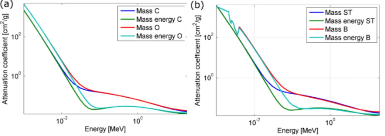
imparted to the matter. In Figure2.2the mass attenuation coefficient and the mass
energy-absorption coefficient are displayed for carbon, oxygen, soft tissue and bone (material definitions from ICRP and tabulated data taken from NIST Standard
Ref-erence Database 126,Link NIST data).
Figure 2.2: Mass attenuation coefficient (Mass) and mass energy-absorption
coeffi-cient (Mass energy) of (a) carbon (C) and oxygen (O) and (b) soft tissue (ST) and bone (B)
2.4.2
Charged particle interactions
Moving charged particles, originating from either the radioactive decay described
in Section2.3or the interaction of photons with matter as described in the previous
Section, are the cause of the absorbed dose. As in PET and CT imaging only light particles like electrons and positrons are of concern, heavy particles like protons and ions will not be discussed. The energy deposition of heavy particles is due to their increased mass slightly different to those of light particles and their path inside matter is different due to less scattering. When charged particles travel through matter, they transfer energy to the medium by coulombic interactions with atomic electrons and nuclei. In contrast to photons, which can travel a significant distance before interacting discretely leading to partial or total energy transfer to an atomic
2.4. Particle interactions with matter 21
electron, charged particles undergo far more discrete interactions and slow down faster due to coulombic interactions making them less penetrating than photons.
The particular way and amount of energy transferred to the medium by charged particles is described by the total stopping power, which is subdivided into collisional
and radiative stopping power with both having the unit of M eV
m . Both describe the
energy loss of charged particles along a unit path length. The quantity of interest
for dosimetry is the mass stopping power in M eV ∗m2
kg , which is equal to the stopping
power divided by the density of the medium. For mathematical descriptions and a more detailed picture the reader is referred to [19,59,60], as only a brief summary will be provided within the manuscript.
The collisional mass stopping power describes the coulombic interaction of the in-cident electron with atomic electrons. It combines the contributions from two types of collisions, which depend on the perpendicular distance b between the trajectory of the charged particle and the radius of the scattering atom. If the distance is large
(b ratom), the particle will interact with the atom as a whole, which is referred
to as a soft collision. The probability of the event is high, however, the net energy transfer per interaction is small leading to a temporal polarization or excitation of an atomic electron into empty quantum states. If the distance is approximately as
small as the radius of the atom (b ≈ ratom), the charged particle will interact with
a single atomic electron, being referred to as a hard collision. The probability is lower than for a soft collision, but the net energy transfer is substantially higher, leading to an approximately equal energy transfer from both, soft and hard collision, from a global point of view. The hard collision with an atomic electron can result in ionization of the atom including subsequent radiative relaxation processes as previ-ously described for photoelectric absorption. The ejected electron, which is called a δ−ray, can carry energy a significant distance away from its origin, making this an important feature for dosimetry considerations. The collisional mass stopping power is below an energy of 0.5 MeV antiproportional to energy, i.e. the slower the incident electron the higher the energy deposition, while it flattens out for higher energies and becomes approximately independent for higher energies. It is largely indepen-dent of Z. However, higher Z materials have greater binding energies for inner shell electrons, leading to an increased excitation energy, which in turn decreases the collisional mass stopping power for higher Z materials slightly.
If the distance is smaller than the atomic radius (b ratom), the charged particle will interact with the nucleus of the atom, leading to a strong deflection of the particle’s path or to complete deceleration. The deceleration of the charged particles
creates an electric dipole emitting electromagnetic waves in the frequency range of X-rays known as Bremsstrahlung. The maximum bremsstrahlung photon energy is equal to the kinetic energy of the incident charged particle resulting from the full stop of the particle and conversion of the complete kinetic energy into radiation. This effect is summarized in the radiative mass stopping power, which is not contributing to the locally deposited energy, as the energy is carried away by radiation. The
radiative mass stopping power is in general proportional to Z2 and the energy of
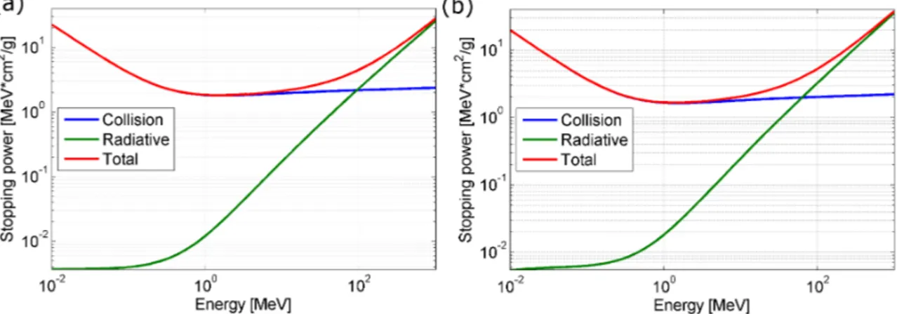
the charged particle. In Figure 2.3 the collision, radiative and total mass stopping
power of electrons in soft tissue and compact bone (material definitions from ICRP
and tabulated data taken from NIST Standard Reference Database 124, Link NIST
data) are displayed.
Figure 2.3: Collision, radiative and total mass stopping power of electrons in (a)
soft tissue and (b) bone
In general, positrons and electrons are assumed to show similar behaviour re-garding collisional and radiative stopping power, since they have the same mass but opposite charge. This however holds only true for high energies. The collisional stop-ping power of positrons is slightly greater below energies of 0.5 MeV and less above that threshold. The radiative stopping power of positrons is significantly different to electrons at low energies. The ratio of the radiative stopping power of electrons and
positrons is unity at about 10 MeV but only 0.5 at 10 keV [19].
In summary, the collisional mass stopping power is the predominant contributor to the total mass stopping power at low energies and becomes essentially indepen-dent of energy at about 0.5 MeV. For high Z materials like lead, the collisional mass stopping power is approximately lower by a factor of 2 compared to water. The ra-diative mass stopping power is negligible at low energies compared to the collisional
2.5. Monte Carlo simulations in medical physics 23
mass stopping power and only becomes the dominant energy transfer mechanism at about 10 MeV in lead and 100 MeV in water, which are energies far outside of the medical diagnostic energy window. However, the radiative mass stopping power
is proportional to Z2 and energy. Another important quantity for dosimetry is the
electron range, i.e. how far electrons travel inside a medium in order to be able to predict the position of the energy deposition. Since the energy loss of a charged particle due to collisions is a stochastic process, the energy distribution of a mo-noenergetic electron beam after traversing a certain depth of medium is varying and consequently the range of the electron. This effect is called energy and range straggling.
2.5
Monte Carlo simulations in medical physics
This section aims to introduce the method of Monte Carlo simulations (MCS), their purpose in medical physics and the specific application of radiation dosimetry will be discussed. The numerical method known as MCS can be described as a statistical simulation method involving sequences of random numbers to solve problems of
stochastic nature [61]. MCS is useful in medical physics because of the stochastic
nature of radiation emission, its transport and detection, since deterministic methods and experimental measurements are infeasible. However, the applications of this method varied greatly and its use for medical physics increased only after the review
paper by Raeside [62] in 1976. The method will be described only briefly, as a
detailed description of the mathematics involved can be obtained elsewhere [62–
64].
2.5.1
General concept
The most common, very simple example to explain the basic idea of MCS is the problem of Buffon’s needle, based on the following question: What is the probability (P ) of a thin needle of length d being randomly thrown onto an array of equally spaced parallel lines, which are separated by a distance D (with d < D), to intersect
with one of the lines? The probability P for the needle hitting a line is equal to 2d
πD, which can be derived by the two probability density functions for the distance of the needle centre to the parallel lines and the orientation angle between lines and the needle. If the needle is randomly thrown N times (N being a large number),
and n intersections are observed, π is approximated by 2D
D N
approximated with a large enough N can serve as the basis for a Monte Carlo compu-tation. When putting that into relation with medical imaging, it becomes clear that three inputs for MCS are essential [61]. One input is the PDFs of all possible particle interactions (cross-sections of interactions) in matter for all involved particles of the particular imaging process. The other important input is the random numbers in the interval [0, 1] to sample from the PDFs. All random number generators (RNG) used in MCS are based on mathematical algorithms. Since they are created deterministi-cally, they are pseudo-random, which is defined as appearing random but exhibiting
a repeatable pattern. As many as 107 to 1012 random numbers are used in a typical
simulation and even slight patterns or correlations could alter the outcome of the
simulation [65]. There are many RNGs that have been suggested for the use in MCS
and will not be discussed further, however, summaries of available pseudorandom
number generators were published [66]. The last important input parameter is the
sampling rules, i.e. how to sample from the pdfs. Therefore, in an oversimplified way, by using a large amount of random numbers (N ) to sample from the PDFs one can reach a solution to a specific physical problem such as energy deposition in a complex system.
2.5.2
GATE package
The dedicated medical imaging Monte Carlo package GATE was first publicly
re-leased in 2004 [67], and combines a wide and accurate physics modelling ability
with a user-friendly way of modelling complex scanner geometry and imaging
con-figurations [68]. It is based on the Geant4 libraries written in C++, which were
developed by Conseil Européen pour la Recherche Nucléaire (CERN) to simulate
interactions of particles with matter over a very wide energy range for i.e. the Large
Hadron Collider [69]. GATE encapsulates the libraries to achieve a modular, widely
applicable and scripted simulation toolkit focused on medical imaging applications. The end-user of GATE does not have to perform any C++ coding, every simulation can be defined and run using script language. The user can freely define the source of radiation, the scanner geometry, the detectors, the phantom (voxellized images are also possible) and can set the various physical phenomena involved. For the physical processes to be simulated, the user can choose several sets of cross-sections in form of PDFs depending on the energy window and the involved physics.
![Figure 3.2: TAC derived by (a) organ harvesting and (b) dynamic microPET imaging (sphere segmentation); adopted from [80]](https://thumb-eu.123doks.com/thumbv2/123doknet/6774740.187612/53.892.147.773.164.394/figure-derived-harvesting-dynamic-micropet-imaging-segmentation-adopted.webp)