THÈSE
En vue de l’obtention du
DOCTORAT DE L’UNIVERSITÉ DE TOULOUSE
Délivré par : l’Université Toulouse 3 - Paul Sabatier
Discipline ou spécialité : Physiopathologie, Biologie et Médecine du Développement et de la Reproduction
Présentée et soutenue par : Mohamed Hadi Mohamed ABDELHAMID Le : 9 juillet 2019
Titre :
Effets de facteurs exogènes sur les gamètes masculins et leur génome: conséquences potentielles d'une élévation modérée de la température
des testicules et des épididymes sur la qualité du gamète.
Jury
Pr. Catherine GUILLEMAIN Rapporteure Pr. Hervé LEJEUNE Rapporteur Pr. Eric HUYGHE Examinateur Dr. Hanaé PONS Examinatrice Pr. Louis BUJAN Directeur de thèse Dr. Safouane HAMDI Co-directeur de thèse
Ecole doctorale : BSB - Biologie, Santé, Biotechnologies
Unité de recherche : Groupe de Recherches en Fertilité Humaine (EA 3694)
Directeur de thèse : Pr. Louis BUJAN Co-directeur de thèse : Dr. Safouane HAMDI Rapporteurs : Pr. Catherine GUILLEMAIN, Pr. Hervé LEJEUNE, Pr. Clément JIMENEZ
لإا لا ق
يعفاشلا مام
....
"
اربص قعلت ىتح دجملا غلبت نل هلكأ تنأ رمت دجملا نبسحت لا
"
لا قو
:
"
ُباسِتكِاَو ٍّمَه ُجُّرَفَت ِدِئاوَف ُسمَخ ِرا فسَلأا يفَف رِفاسَو لاُعلا ِبَلَ ط يف ِناطوَلأا ِنَع بَّرَغَت
ٌّلُذ ِرا فسَلأا يف َليق نِإَو ِدِجام ُةَبحُصَو ٌبادآَو ٌملِعَو ٍةَشيعَم
يفايَفلا ُعطَقَو ٌةَنحِمَو
ِدِساحَو ٍشاو َنيَب ٍناوَه ِرادِب ِهِتايَح نِم ُهَل ٌريَخ ىتَفلا ُتوَمَف ِدِئادَشلا ُباسِتكِاَو
"This thesis is dedicated to,
My parents (Hadi and Laila) who have given me the opportunity of an education from the best institutions and support throughout my life. My wife Najwan Nabil, for her great patience, waiting for me to finish this task, who has always supported me stood by me and dealt with all my absence from many family occasions and with love and smile.
My sisters, (Aml, Lubna, Reem, Fatema, Alaa and Heba) the heart of our spring in our family, they have given me the love, tenderness and happiness in my life.
My uncle (Lutfi Layas) who was my friend, godfather, guide and philosopher.
Finally, to my best friends and soulmates (Saadeddin, Abdul Aziz, Rudwan, and Mohamed) who have always helped me and believed that I could do it.
Acknowledgements
My director,
I wish to thank, first and foremost, my advisor Pr. Louis BUJAN who provided me an opportunity to join his team, for his patience, motivation and immense knowledge, without his guidance this study would not have been possible.
Co-director,
Dr. Safouane Hamdi co-director of my thesis, I express my profound gratitude for his
competitive and vast expertise on the subject which I learned and tried to apply during my PhD work. His critical insight and exceptional command on the project really helped me a lot to think and judge scientifically.
Research group and team,
This thesis would have remained a dream had it not been for Dr. Roger Mieusset who give me the keys to the success of this work, Dr. Myriam Daudin, for her amical attachment and kindness that she really practiced during my study period. I gratefully acknowledge the able guidance of Camille Esquerré, Marie Walschaerts and Dr.Nathalie Moinard to helping me when needed and for her great friendly gesture.
Jury members,
Furthermore, I would like to thank the jury members; Pr. Catherine GUILLEMAIN, Pr.
Hervé LEJEUNE, Pr. Clément JIMENEZ, Pr. Eric HUYGHE and Dr. Hanaé PONS for accepting to evaluate my thesis.
Staff of laboratory,
These acknowledgments would be incomplete if I do not pay thanks to all the members of
EA 3694, whole staff of CECOS, they are helping me to find my way in CHU- Toulouse, improving my French and answering my endless questions and showing me the basics.
Finally, this dissertation is based on a three - year programme of research conducted by me at the Department of Andrology, Hospital Center University Toulouse. My studies there were facilitated by a scholarship awarded by Biotechnology Research Center through the ministry of higher education and scientific research - Libya, I am conscious of the enormous debt I owe these institutions for the opportunity so afforded for the enrichment of my academic career it is with a deeply felt sense of gratitude and thanks.
1
Contents
Résumé ... 10
Abstract ... 12
Introduction ... 14
1. Anatomy and physiology of the male reproductive system... 19
1.1. Testes ... 19
1.1.1. Interstitial compartment ... 20
1.1.2. Tubular compartment ... 20
1.1.3. Different type of testicular cells ... 20
1.2. Epididymis ... 24
1.3. Scrotum ... 27
1.3.1. Skin ... 27
1.3.2. Dartos ... 28
1.3.3. Cremasteric ... 29
1.4. Testicular blood circulation system ... 30
1.4.1. Testicular artery ... 30 1.4.2. Testicular veins ... 30 1.5. Vas deferens ... 31 1.6. Glands ... 32 1.6.1. Seminal Vesicles ... 32 1.6.2. Prostate ... 33
1.6.3. Bulbourethral Glands (Cowper's) ... 34
2.1. Spermatogenesis ... 38
2.1.2. Mitosis ... 39
2.1.3. Meiosis ... 40
2.1.4. Spermiogenesis ... 45
2.1.5. Spermiation ... 50
2.2. Regulation of spermatogenesis process ... 52
2.2.1. Hypothalamic-Pituitary-Testicular Axis ... 52
2.2.2. Thermic regulation ... 55
2.2.2.1. Exchange testicular arterial and venous blood streams ... 56
2.2.2.2. Scrotum and surfaces area heat lost ... 59
2
3.1.1. Sperm (description, function and morphology) ... 64
3.2. Classifications sperm form and sperm morphology ... 67
3.3. Normal and abnormal sperm forms ... 68
3.3.1. Normal sperm form ... 70
3.3.2. Abnormal sperm form ... 74
3.3.3. Sperm form pathology... 74
4.1. Sperm aneuploidy... 80
4.1.1. Testis temperature, lifestyle related to sperm aneuploidy ... 83
5.1 Testis temperature ... 90
5.1.1. Measuring genital temperature ... 90
5.1.1.1. Mercury thermometer ... 91
5.1.1.2. Needle thermistor ... 92
5.1.1.3. Microwave technology ... 92
5.1.1.4. Scrotal skin surface temperature by infrared thermometry ... 93
5.1.1.5. Skin thermocouple ... 93
5.1.2 Testis temperature at various locations ... 96
6.1. Temperature and male fertility ... 103
6.1.1. Factors and situations that could cause an increase of testicular temperature ... 103
6.1.1.1. Sitting and sleeping posture ... 103
6.1.1.2. Environment and seasonal variation ... 105
6.1.1.3. Underwear ... 106
6.1.1.4. Occupation in high temperature ... 107
6.1.1.5. Car driving ... 108
6.1.1.6. Laptop computer users ... 109
6.1.1.7. Hot bath and sauna ... 109
6.1.1.8. Sports ... 111
6.1.1.9. Fever ... 112
6.1.1.10. Cryptorchidism ... 113
6.1.1.11. Varicocele ... 113
6.1.1.12. Obesity ... 116
7.1. Effect of temperature on the physiology of male reproduction ... 124
7.2. Effect of temperature on testis and epididymis ... 124
7.3. Effect of temperature on spermatogenesis ... 126
3
7.3.2. Spermatocyte and Spermatid (Meiosis) ... 128
7.4. Effect of temperature on sperm in epididymis ... 129
7.5. Effect of temperature on sperm quality ... 130
7.5.1. Scrotal exposure to temperatures below core body physiological temperature ... 131
7.5.2. Scrotal exposure to temperatures above core body physiological temperature ... 136
8. What are physiological and pathophysiological mechanisms of testicular heating? What happens to testicular function during and after heating? ... 142
9. Clinical Applications ... 147
9.1. Use of testicular heating as a method for male contraception ... 147
9.2. Infertile situation ... 152
10.1. The main objective ... 157
10.1.1. Method of increase in testicular and epididymal temperature ... 159
10.1.1.1. Study Population ... 159
10.1.1.1.2. Techniques of testicular exposure to heat ... 160
10.1.1.1.3. Part I: A study the effect of a mild testicular and epididymal temperature on human sperm morphology ... 162
Periods of sampling ... 162
Semen collection ... 164
Sperm morphology ... 164
Statistical Analysis ... 165
10.1.1.1.4. Part II: Effect of a mild induced increase in testicular and epididymal temperature on sperm aneuploidy ... 166
Sperm concentration evaluation ... 166
Sperm Fluorescence in situ Hybridization (FISH)... 166
Statistical Analysis ... 169
Articles ... 170
Part I. Mild experimental increase in testis and epididymis temperature in men: Effects on sperm morphology according to spermatogenesis stages. ... 171
Part II. Experimental mild increase in testicular temperature has drastic, but reversible, effect on sperm aneuploidy in men: a pilot study ... 212
11.1. Discussion ... 245
11.1.1. First part of the study previously published by our group (Ahmad et al., 2012) ... 246
11.1.2. Effect of heating on epididymal sperm ... 250
4
11.2. Conclusions and prospective ... 261
11.2.1. Conclusions ... 261
11.2.2. Prospective ... 262
5
List of figures
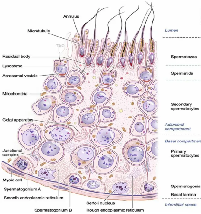
Figure 1 Section of the germinal epithelium in the seminiferous tubule. ... 23
Figure 2 Anatomy of the testis and epididymis ... 26
Figure 3 Anatomy of the male reproductive system ... 35
Figure 4 Schematic overview of meiotic process in spermatogenesis. SC, synaptonemal complex .... 42
Figure 5 Differentiation of a human diploid germ cell into a fully functional spermatozoon ... 44
Figure 6 An extende d diagram of the components in the midpiece of a mamm alian spermatozoon tail. ... 48
Figure 7 Schematic representation of the differentiation of human spermatid ... 49
Figure 8 Schematic representation different steps development of spermatogenesis ... 51
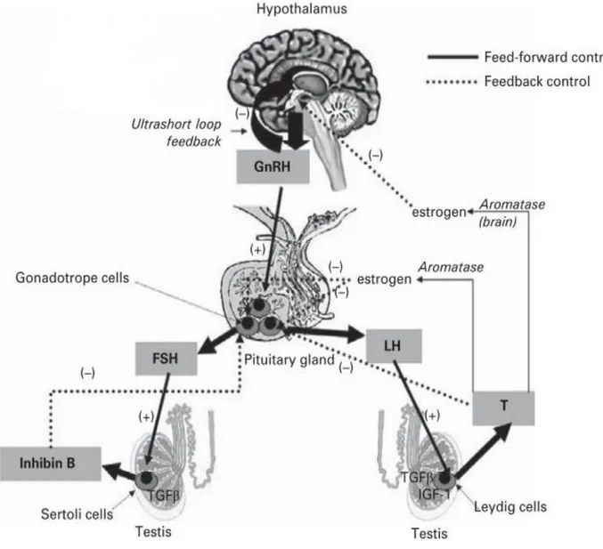
Figure 9 Diagram of the hypothalamic−pituitary−testis axis . ... 54
Figure 10 Model of counter-current transfer of heat or a substance from the venous blood in the pampiniform plexus to the blood in the testicular artery. ... 58
Figure 11 Scrotum and surfaces area heat lost. ... 61
Figure 12 Diagram of a typical mammalian spermatozoon. Cross-sectional insets show the orientation of the internal cell structure. Anatomy and physiology of the male reproductive system by ... 66
Figure 13 Sperm morphology: classifications of normal and abnormal human spermatozoa ... 77
Figure 14 Schematic representation of chromosome non-disjunction ... 82
Figure 15 Testis temperature in a different location. ... 100
Figure 16 Schematic representation of possible changes induced by potential exogenous and endogenous thermic factors on testicular and scrotal temperature in human. ... 120
Figure 17 Minimum and maximum mean scrotal temperature in different situations. ... 121
Figure 18 Two techniques of heating the testis to use for male contraception ... 150
Figure 19 Schematic representation of testicular and epididymal heat increase from 34°C in scrotal to 36°C into the inguinal canals. ... 161
6
Figure 20 Schematic representation of the timing of semen sampling during the three study periods before, during and after heating (phase I, phase II), B) Location and evolutionary stages of sperm during the spermatogenic process at heat induction ... 163 Figure 21 Schematic representation of semen sampling timing during the four study periods; Before D0, During H34, after (PH45 and PH180) ... 168 Figure 22. Percentage of sperm viability, motility, mean (SEM) sperm count (millions per ejaculate) and semen volume (mL per ejaculate) before, during and after a mild induced testicular and epididymal temperature increase in healthy men. ... 248
7
List of tables
Table 1 Values for sperm morphology in the different WHO manuals ... 73 Table 2 presents different methods of measuring genital temperature ... 95 Table 3 Testis temperature in a different location ... 99 Table 4 Factors and situation associated with an increase in scrotal/testicular temperature ... 119 Table 5 Effects of scrotal exposure to temperatures below core body physiological
temperature on sperm parameter ... 135 Table 6 Testicular heating as a method for male contraception ... 151 Table 7 Sperm parameters during and after a mild induced testicular and epididymal
8
Abbreviations
ART Assisted reproductive techniques DAC Diurnal artificial cryptorchidism DFI DNA fragmentation index
DNA Deoxyribonucleic acid
DPBS Dulbecco’s phosphate buffer saline E2 Estradiol
FISH Fluorescence in situ hybridization FSH Follicle stimulating hormone GnRH Gonadotropin releasing hormone hCG Human chorionic gonadotropin HDS High DNA stainability
HOST Hypoosomtic swelling test HSPs Heat shock proteins
ICSI Intracytoplasmic sperm injection INSL3 Insulin-like factor 3
IVF In vitro fertilization LH Luteinizing hormone MAI Multiple anomalies index
9
NAG Neutral α-glucosidase NaOH Sodium hydroxide
NGS Next Generation Sequencing PFA Paraformaldehyde
RNA Ribonucleic acid
ROS Reactive oxygen species RT Rectal temperature
SCSA Sperm chromatin structure assay SDI Sperm deformity index
TSP Testis suprascrotal Position
T Testosterone
TUNEL Terminal deoxynucleotidyl transeferase dUTP nick end labelling assay TZI Teratozoospermia index
10
Résumé
:L'impact des expositions de la vie courante, et en particulier de la température, sur la reproduction masculine est très étudié. Dans ce projet, nous avons étudié deux principaux paramètres liés à l'infertilité après une exposition testiculaire à une augmentation modérée de température (+2°C) chez des hommes sains : la morphologie et l'aneuploïdie du spermatozoïde et exploré leur potentielle réversibilité. Nous avons conçu un protocole expérimental qui a impliqué 5 hommes fertiles et une population témoin de 27 autres hommes fertiles. L'augmentation de température testiculaire et épididymaire a été obtenue en maintenant les testicules dans une position supra-scrotale au moyen de sous-vêtements spécialement conçu, portés 15 +/- 1heure par jour pendant 120 jours consécutifs.
La première partie de ma thèse a été consacrée à l'étude de l'augmentation de température sur la morphologie des spermatozoïdes et l'indice des anomalies multiples (IAM). Nous avons observé un impact significatif sur la morphologie du sperme et l'IAM entre le 20ème et 34ème jour reflétant un effet sur les stades de spermiogenèse et de méiose. Cet effet drastique a été présent pendant toute la période de chauffage et la récupération des valeurs initiales a été observée au 73ème jour.
Dans une deuxième partie, les aneuploïdies des chromosomes X,Y, 18 ont été analysées par hybridation in situ par la fluorescence (FISH) avant, pendant et
11
après l'augmentation modérée de température. Nous avons constaté une augmentation significative de l'aneuploïdie totale du sperme, de la disomie sexuelle et de la nullisomie au 45ème jour après chauffage. Ces effets ont été complétement inversés après deux cycles de spermatogenèse après l'exposition à la chaleur.
Ces résultats confirment l'impact de la température sur la spermatogenèse et peuvent avoir des implications cliniques dans l'infertilité masculine et notamment le concept de réversibilité après au moins deux cycles de spermatogenèse sans exposition à la chaleur.
Mots clés : Température, Testicule, Morphologie, spermocytogramme, Aneuploïdie, hommes, Spermatogenèse, Epididyme.
12
Abstract
:Lifestyle exposures including temperature have been studied in relationship to male reproductive health. In this project, we focused on two main parameters linked to infertility after testicular exposure of temperature. We evaluated the effects of a mild testis temperature increase (+2°C) on sperm morphology and sperm aneuploidy in five healthy men and examined its potential reversibility. We used 27 fertile men for comparison of results (control group), and designed an experimental protocol that induced in five healthy fertile men, an increase of testicular and epididymal temperature by maintained the testes in a supra-scrotal position by means of specially designed underwear worn 15 ± 1 hours daily consecutive days.
The first part of my thesis was dedicated to the study of the effects of a mild testis temperature increase (+2°C) on sperm morphology and multiple anomalies index (MAI). We observed that a significant impact on sperm morphology and MAI as early as days 20 and 34 reflecting an effect on the spermiogenesis and meiosis stages. This drastic effect was present during the entire heated period and recovery of the values before heating was observed at day193.
In the second part, sperm aneuploidies of chromosomes X, Y, 18 were analyzed by fluorescence in situ hybridization (FISH) before, during and after a mild testis temperature increase. We found that a significant increase in total
13
sperm aneuploidy, sex disomy and nullisomy at 45 days post-heating. Moreover, since increased abnormal sex disomy XY18, sex nullisomy and total sperm aneuploidy values were observed at the same time, these effects were completely reversed at least two spermatogenesis cycles after heat exposure. Our results may have clinical implications in male infertility, the effect of a
mild testis temperature increase was reversible but it seems advisable to allow at least one or two cycles of spermatogenesis to pass in order to recover normal exacting function.
Keywords: Temperature, testis, sperm morphology, MAI, aneuploidy, men, spermatogenesis, epididymis
14
Introduction
:Knowledge of the relationship between reproductive biology and the environment or exogenous factors will give us an excellent understanding of factors controlling fertility, reproduction and conservation of species. Sexual reproduction requires two parents and produces genetically distinct offspring. The male sex gamete (sperms) fuses with the female sex gamete (eggs) to create a zygote. The process of the male and female sex cells fusing is called fertilization. However, the medical definition of infertility is the failure to conceive following twelve months of unprotected intercourses.
Male reproductive health has deteriorated in many countries during the last few decades. In the 1990s, declining semen quality has been reported from Belgium, Denmark, France, and Great Britain (Toppari et al., 1996). Furthermore, in the world there is between 8 and 12% infertile couples (WHO 2010), affecting between 50 and 80 million people; both partners (male and female) contribute significantly in infertility disturbances. Moreover, it should be recalled that social and environmental factors, as well as physiological and genetic ones, contribute to the condition. Also, lifestyle conditions like smoking, alcohol consumption and exposure to chemical material are risk factors of infertility (Sharma et al., 2013).
15
What we do know is that spermatogenesis depends on two physiological factors; hypothalamic-pituitary-testicular axis and testicular thermoregulatory systems. The temperature was one of the most important impacts on male fertility; the high-temperature exposure adds an additional challenge to physiological spermatogenesis processes. The temperature, coupled with high relative humidity, can be taxing on the efficiency of sweating and other mechanisms responsible for cooling the testis. Maintenance of a lower testicular temperature relative to core body temperature is achieved by means of the thermoregulatory tissues of the testes; namely, the pampiniform plexus, and scrotum (Wallach et al., 1988).
In many mammals, including human, it is established that testis must be 4–6 °C below core body temperature to sustain physiological production of sperm (Mieusset et Bujan 1995). The testis, epididymal and scrotal are acutely sensitive to an increase of only a very few degrees, and much effort has been devoted to the analysis of this response (Bedford 1991). Dysfunction of thermoregulatory systems or conditions exceeding the efficiency of these systems results in an increase of testis temperature (Candas et al., 1993). Any variation in testicular temperature, high or low, may change the output of testicular functions. Daily life behaviors may be the cause of the increase of testis temperature for example; taking hot baths or sauna, sitting for longer periods, during driving, wearing tight underwear, occupation in higher
16
temperature. Pathological conditions like fever, cryptorchidism and varicocele are also involved in testicular temperature increase.
Moreover, if we look at it we find numerous animal and human studies that described the effect of an increase in testicular temperature on sperm characteristics and spermatogenesis processes. But very few ones have been performed to investigate the effect of temperature on sperm characteristics according to the physiological time of spermatogenesis and epididymis transit in men.
The main objective of this research work was to investigate the effects of the mild increase of testicular temperature on sperm morphology and changes in sperm aneuploidy chromosomes according to the physiological chronology of spermatogenesis in human.
To begin this work, a brief overview of the anatomy and physiology of male reproductive systems, testes and epididymis is presented.
17
18
Anatomy and physiology of the male reproductive
system
(Testes, Epididymis, Different type of testicular cells, Scrotum,
Glands, Testicular blood circulation system)
19
1. Anatomy and physiology of the male reproductive system
1.1. Testes
Testes are glandular organs suspended by a spermatic cord in a cutaneous
pouch-like structure called scrotum. At early fetal life they are contained in the abdominal cavity, before birth each testis moves through the abdominal musculature to descend into the scrotal cavity. In human, testicular size depends on age and stage of sexual development. At birth, the testes measure approximately 1.5 cm in length and 1 cm in width, before the age of 12 years testicular volume is around 1–2 cm3. On average, testes of adults measure 15– 25 cm3 in volume, measure 3.6–5.5 cm in length and 2.1–3.5 cm in width (Leung et al., 1984), and are compartmentalized by septal divisions into individual lobules (Cheng et al., 2016; Niederberger, 2011). They are surrounded by a fibrous capsule, the tunica albuginea, from which septations extend toward the testicular mediastinum, dividing the testis into 200–300 lobules. Each of these lobules contains several highly convoluted seminiferous tubules. These tubules consist of a basement membrane lined by Sertoli cells, interspersed with germ cells at various stages of maturation.
The testes are responsible for the production of sperm cells and the male sex hormone testosterone. The testes are comprised of two compartments:
20
1.1.1. Interstitial compartment
The most important cells of this compartment are the Leydig cells. In the human testis the interstitial compartment represents about 12–15% of the total testicular volume, 10–20% of which is occupied by Leydig cells. These cells are the source of testicular testosterone and of insulin-like factor 3 (INSL3). They also contain immune cells, blood and lymph vessels, nerves, fibroblasts and loose connective tissue (Weinbauer et al., 2010).
1.1.2. Tubular compartment
This compartment represents about 60–80% of the total testicular volume. It contains the germ cells and two different types of somatic cells, the peritubular cells and the Sertoli cells. The testis is divided by connective tissue septa into 250-300 lobules, each containing 1-3 profoundly tangled seminiferous tubules. Each human testis contains around 600 seminiferous tubules. The process of spermatogenesis takes place in the tubular compartment (Niederberger, 2011; Weinbauer et al., 2010) (Fig 1).
1.1.3. Different type of testicular cells
Testicular cells such as; Leydig, peritubular and Sertoli cells, secrete a large variety of factors (proteins, cytokines, growth factors, opioids, steroids,
21
prostaglandins, modulators of cell division) and support the structure of the germinal epithelium (Foster & Lamb, 1988).
1.1.3.1. Leydig Cells
The interstitial compartment contains about 200 × 106 Leydig cells, that produce and secrete the most important male sexual hormone, testosterone. There are the different types of Leydig cells: stem Leydig cells as founder cell, progenitor Leydig cells as a committed stem cell, fetal Leydig cells as a terminally differentiated cell in the fetus, and adult Leydig cells as the terminally differentiated Leydig cell (Christensen, 1975; Kaler et al., 1978).
1.1.3.2. Peritubular Cells
The seminiferous tubules are covered by a lamina propria, which consists of a basal membrane, a layer of collagen and the peritubular cells (myofibroblasts). The role of peritubular cells produce several factors that are involved in cellular contractility: panactin, desmin, gelsolin, smooth muscle myosin and actin (Holstein et al., 1996). These cells also secrete extracellular matrix and factors typically expressed by connective tissue cells: collagen, laminin, vimentin, fibronectin, growth factors, fibroblast protein and adhesion molecules (Albrecht et al., 2006; Schell et al., 2008). An important and accepted role of peritubular cells is the active transport of immotile sperm.
22
1.1.3.3. Sertoli Cells
Human Sertoli cells are located on the basal membrane and extend into the lumen of the tubular seminiferous and supporting structure of the germinal epithelium. Sertoli cells occupy a 30% of the volume of the seminiferous tubules of mammals with active spermatogenesis (Russell et al., 1990) and present elongated or irregularly-shaped nuclei. Testis with complete spermatogenesis contains 800–1200 × 106 Sertoli cells. Along the cell body extending over the entire height of the germinal epithelium, all morphological and physiological differentiation and maturation of the germinal cell up to the mature sperm take place (Weinbauer et al., 2010). Moreover, Sertoli cells assist in the translocation of early meiotic cells from the basal to the adluminal compartment during the epithelial cycle in the seminiferous epithelium. Sertoli cells also play an important secretory function: cytokines, growth factors, opioids, steroids, prostaglandins, modulators of cell division. They also produce testicular fluid, including ABP, a protein that binds to and concentrates androgens, which are essential for the development of the spermatozoa. They finally help for translocating the differentiating cells to the lumen, and phagocytosing degenerating germ cells and surplus cytoplasm remaining from spermiogenesis (Sharpe et al., 2003).
23
24
1.2. Epididymis
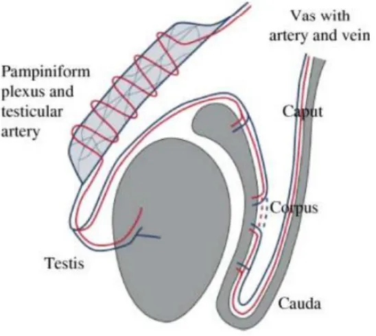
The epididymis is a tubular organ which receives spermatozoa from testis. They are two in number, one on each testis. The major component parts of the epididymis are customarily referred to as head (caput), body (corpus) and tail (cauda) (Fig2). Each epididymidis region supports distinctive functions with the caput and corpus carrying out early and late sperm maturational events, respectively, while the cauda region primarily serves as a storage site for functionally mature spermatozoa. The cauda epididymis is poorly developed in humans (Johnson and Varner, 1988). This correlates with the very limited sperm reservoir capacity in our species as confirmed after performing semen analyses on successive ejaculates from healthy young men (Johnson and Varner, 1988). Moreover, transit along the human epididymis takes 2–4 days (Bedford, 1990; 1991), as spermatozoa passing from the proximal to distal regions of the epididymis, they undergo a series of physiologic, biochemical, and morphologic changes with the end result being spermatozoa that have acquired the function of progressive motility and the ability to fertilize an ovum (Yeung et al., 1993; Soler et al., 2000; Cooper and Yeung, 2006). A distal part of epididymis plays an important role in acquiring the spermatozoa binding capacity (Moore et al., 1992). The chromatin of sperm becomes more condensed during epididymal transit (Haidl et al., 1994; Golan et al., 1996).
25
(i) Producing the epididymal plasma, thus providing the spermatozoa with a fluid environment of a very special composition; where the fixed cells are both resorptive and secretory in nature and sperm can survive for 2 weeks in this environment.
(ii) Promoting the maturation of spermatozoa, which are prerequisite for sperm motility and fertilizing abilities.
(iii) Aiding the disposal of ageing and superfluous spermatozoa.
26
Figure 2 Anatomy of the testis and epididymis A) Longitudunal anatomical section. B) Inner structure of the spermatic route. 1 tunica albuginea, 2 lobules, 3 mediastinum testis, 4 seminiferous tubules, 5, rete testis, 6 head of the epididymis, 7 body of the epididymis, 8 tail of the epididymis, 9 vas deferens. (Liguori et al., 2012).
27
1.3. Scrotum
The testes are located in a skin-covered, highly pigmented, muscular sack called the scrotum that extends from the body behind the penis. This location is very important to keep the testes 2 to 4°C below core body temperature (Mieusset & Bujan, 1995; Liu, 2010). The scrotum divides into two compartments, each housing one testis. In the higher environment temperature or increase in testicular temperature , the scrotum relaxes from the body core with an increase of scrotal surface area, which promotes heat loss. On the contrary, there are the dartos and cremaster muscles to moving are the testes closer to the body and decreas the surface area when testes exposured tocold weather (Shafik, 1973).It varies from 2 to 8 mm in thickness (Leung et al., 1984). The system of different membranes inside the scrotum avoids testes from being injured due to blows or squeezes and acts as a covering and a protection to the testes: the testes lies suspended and loose in their cavity and are surrounded by several different layers in order to allow them a better mobility (Bertolotto et al., 2012).
1.3.1. Skin
The skin of the scrotum is a brownish layer, usually thrown into folds or rugæ, which contains roots of scattered, crisp hairs that cover the scrotum surface. It is very elastic and capable of great distension, and on account of the looseness
28
and amount of subcutaneous tissue, the scrotum becomes greatly enlarged in cases of edema, The skin of scrotum have special characteristics such as ; sweat glands, little subcutaneous fat, connective tissue, an abundance of thermoreceptors (Bertolotto et al., 2012; Wallach et al., 1988).
1.3.2. Dartos
The tunica dartos is a fat-free thin layer of smooth muscular fibers: it is a continuation of Scarpa’s fascia which is a membranous layer of the subcutaneous tissue in the abdominal wall. The dartos divides the scrotum into two cavities, each containing one testis, through an inward septum that extends between the raphe and the under surface of the penis. In older males, the dartos muscle loses its tone, and tends to cause the scrotum to be smoother and to hang downfurther. The tunica dartos’s role is to regulate the temperature of the testicles (Shafik, 1973). It does this by expanding or contracting to wrinkle the scrotal skin. The dartos is closely united to the skin externally, but connected with the subjacent parts by delicate areolar tissue, upon which it glides with the greatest facility.
Moreover, there is other physiological function of this muscle (Shafik et al., 2007), A dartos contraction have a role in testicular temperature elevation. During erection, the recorded dartos muscle (DM) contraction appears to elevate the testicular temperature by two mechanisms: (a) testicular elevation
29
bringing the testicle closer to the warm abdominal wall, and (b) closure of the fenestrae between the DM bundles thus transforming the scrotal compartment into a closed cavity (Shafik, 1973).
1.3.3. Cremasteric
Prolonged down ward around the surface of the cord and testis, the external spermatic fascia is a thin membrane, derived from the aponeurosis of the external oblique muscle. It is separated from the dartos by loose areolar tissue (Redman, 1996). The cremaster muscle consists of scattered bundles of muscular fibers connected together into a continuous covering by intermediate areolar tissue. It is a thin layer of skeletal muscle found in the inguinal canal and scrotum between the external and internal layers of spermatic fascia, surrounding the testis and spermatic cord. The cremaster muscle is a paired structure, there being one on each side of the body. Anatomically, the lateral cremaster muscle originates from the internal oblique muscle, just superior to the inguinal canal, and the middle of the inguinal ligament. The medial cremaster muscle, which sometimes is absent, originates from the pubic tubercle and sometimes the lateral pubic crest. Both insert into the tunica vaginalis underneath the testis (Bertolotto et al., 2012).
30
1.4. Testicular blood circulation system
The testicular arteries and veins play major roles in the thermo-regulation that is essential for the efficient functioning of this organ.
1.4.1. Testicular artery
It originates from the aorta, runs obliquely through the abdominal wall via the inguinal canal (Asala et al., 2001). Then it becomes flexuous from the deep inguinal ring. In all eutherian mammals with scrotal testes, the artery is unique, except for the branches to the epididymis, two in man, superior to the head and inferior to the body and tail (Harrison & Barclay, 1948).
1.4.2. Testicular veins
The testicular vascularization play an important role in reducing scrotal/testicular temperature by acting as a counter-current heat exchange, where the venous return from the testis is described as including three groups of veins (Wishahi, 1992) that intercommunicate and form the spermatic venous plexus “pampiniform plexus” :
1) Testicular vein (internal spermatic), which drains into the left renal vein or inferior vena cava on the right side.
2) Vassal vein (deferential), which follows the vas deferens and ends in the superior or inferior vesical vein.
31
3) Cremasteric vein (external spermatic), which drains into the inferior epigastric vein and then to the external iliac vein.
The testes are drained by a plexus of veins (pampiniform plexus), which continue as the testicular veins. The function of pampiniform plexus is to act as a heat exchanger, cooling the arterial blood before it reaches the testes (See chapter II, Fig 10). The right testicular vein directly drains into the inferior vena cava and the left testicular vein drains into the left renal vein (Labranche et al., 2018).
1.5. Vas deferens
The vas deferens is a thick muscular tube that measures approximately 35 to 40 cm from the cauda epididymis to the point of fusion with the seminal vesicle and ejaculatory ducts. Five portions have been previously described: epididymal, scrotal, inguinal, pelvic, and ampulla. The vas deferens, like the epididymis and seminal vesicle, is derived from the mesonephric duct. The ability to propel sperm forcefully is dependent on a three-layered muscular coat, with an inner and outer longitudinal layer and a middle circular layer. While the vas deferens receives nerve fibers from the sympathetic and parasympathetic nervous system, the rich supply of adrenergic fibers contributes to the efficiency of sperm transport. The vas deferens receives its
32
blood supply from the deferential artery via the inferior vesical artery, and the deferential vein accompanies it (Niederberger, 2011;Goldstein, 2013).
1.6. Glands
The male accessory sex glands vary considerably in mammals with respect to their topographical location, size, morphology, and functions, thus reflecting the diversity of species-specific requirements of these glands for reproduction that may be due to differences in environment and sexual habits (Aumuller & Seitz, 1990). The accessory glands of the male reproductive system are the seminal vesicles, prostate gland, and the bulbourethral glands, this glands secretion that aid in sperm transport, maintenance of stored sperm and fertilization processes (Nieschlag et al., 1992) (Fig 3).
1.6.1. Seminal Vesicles
The paired seminal vesicles are saccular glands posterior to the urinary bladder. Each gland has a short duct that joins with the ductus deferens at the ampulla to form an ejaculatory duct, which then empties into the urethra (Zhang, 1999). While, contributing to approximately 60% of the fluids passed from the human male during ejaculation, this fluid from the seminal vesicles is viscous and contains fructose, which provides an energy source for the sperm, also they secreteacid, inorganic phosphorus, potassium, prostaglandins, which contribute to the mobility and viability of the sperm; and proteins that cause
33
slight coagulation reactions in the semen after ejaculation (Aumuller & Riva, 1992).
1.6.2. Prostate
The prostate gland that is divided into two compartments muscular and glandular structures surround the neck of the male urinary bladder and urethra. The prostate gland in normal adult men weighs 15 to 20 g and length 4 to 6 cm. In human, development of the prostate begins with the growth of prostatic buds from the urogenital sinus at about 10 weeks of fetal development (Hayward & Cunha, 2000). In general, the prostate produces a variety of substances such as zinc (Zn), citric acid (citrate), acid phosphatase and gamma-glutamyltransferase, enzymes, and fibrinolytic. These elements have been considered reliable markers of the prostate gland. Zinc has a tendency to bind with other elements of semen; it can sometimes be bound to the surface of the sperm cells. Zn is an essential trace element for the maintenance of germ cells, the progression of spermatogenesis, and the regulation of sperm motility. (Aumuller & Seitz, 1990). Moreover, the most seminal plasma content of zinc comes from this gland.
34
1.6.3. Bulbourethral Glands (Cowper's)
A bulbourethral gland, also called a Cowper's gland are small, pea-sized glands, located inferior to the prostate gland near the base of the penis between the two layers of the fascia of the urogenital diaphragm in the male reproductive system. A short duct from each gland enters the proximal end of the penile urethra (Schattman et al., 2015). In response to sexual stimulation, the Cowper's glands secrete an alkaline mucus-like fluid. This fluid neutralizes the acidity of the urine residue in the urethra, helps to neutralize the acidity of the vagina, and provides some lubrication for the tip of the penis during intercourse (Chughtai et al., 2005)
35
36
37
Spermatogenic cells
38
2.1. Spermatogenesis
Spermatogenesis is a dynamic process whereby undifferentiated stem cells (spermatogonia) develop into highly specialized spermatozoa. The process can be divided into three distinct phases (Lamb & Foster, 1988, Niederberger, 2011):
A. Spermatogonial proliferation, differentiation, and their division to form
preleptotene spermatocytes.
B. Meiosis of spermatocytes to preleptotene spermatocytes and second meiotic divisions to form spermatids.
C. Spermiogenesis, which is transformation of a round spermatid to a sperm-like mature spermatid.
Spermatogenesis occurs in the seminiferous tubules that form the bulk of each testis. The process begins at puberty, after which time sperm are produced constantly throughout a man’s life. One production cycle, from spermatogonia through formed sperm, takes approximately 74 days (Johnson et al., 1986). A new cycle starts approximately every 16 days, although this timing is not synchronous across the seminiferous tubules. During this process, the number of chromosomes (44,XY) present in spermatogonia is reduced to the haploid number (22,X or 22,Y) characteristic of spermatids.
39
2.1.2. Mitosis
Mitosis involves the proliferation and maintenance of spermatogonia. This phase can also be named spermatogoniogenesis, it is a precise, well-orchestrated sequence of events in which the genetic material (chromosomes) is duplicated, with a breakdown of the nuclear envelope and formation of two daughter cells as a result of the equal division of the chromosomes and cytoplasm. Broadly, spermatogonia divide by mitosis into types A and B. Type Ap spermatogonia function to undergo self-renewing divisions serving as stem cells but also undergo amplification by mitotic proliferation and transition into type B spermatogonia. Type B spermatogonia divide by mitosis and then progress to become primary spermatocytes. A continuous self-renewal process is then required, provided by stem cells that can undergo either mitosis (leading to daughter stem cells) or differentiate. Clermont reported that the stem cells are a subgroup of type A spermatogonia that also comprise Ad (dark) spermatogonia (that may represent the reserve or non-proliferative spermatogonial population, which can give rise to Ap) and Ap (pale) spermatogonia (which are probably the precursors of B spermatogonia) (Clermont, 1963) (Fig 5).
Spermatogonia do not separate completely after mitosis due to incomplete cytokinesis and remain joined by intercellular bridges. These intercellular bridges persist throughout all stages of spermatogenesis and are thought to
40
facilitate biochemical interactions allowing synchrony of germ cell maturation. Division of B spermatogonia leads to the development of preleptotene spermatocytes before the beginning of meiotic division. The B spermatogonia lose their contact with the basement membrane to form preleptotene primary spermatocytes 4n (Niederberger, 2011).
2.1.3. Meiosis
Meiosis is a key process in spermatogenesis, with the final goal of production of gametes with haploid genetic content. A significant number of exhaustive revisions of this process have been published in the specialized literature (Petronczki et al., 2003). The meiosis stage undergoes changes in the nuclear chromatin configuration after the last spermatogonial division. This stage is comprised of two divisions, meiosis I and meiosis II, this division to yield four haploid spermatids from one diploid primary spermatocyte. After the first meiotic division (reduction division), each daughter cell contains one partner of the homologous chromosome pair, and they are called secondary spermatocytes (2n). Each meiotic division is characterized by 4 steps: prophase, metaphase, anaphase, and telophase.
The prophase of first meiotic division lasts 3 weeks and the other phases of first meiotic division and whole of the second meiotic divisions take 1-2 days (Weinbauer et al., 2010). The process starts when type B spermatogonia lose
41
contact with the basement membrane and form preleptotene primary spermatocytes.Prophase itself is subdivided into five phases (Fig 4, 5):
(1) leptotene, wherein thread-type chromosomes can be observed; (2) zygotene, wherein the pairing of homologous chromosomes occurs; (3) pachytene, wherein the paired chromosomes thicken and undertake crossing over; (4) diplotene, wherein paired chromosomes decondense and partially separate; and (5) diakinesis, wherein chromosomes recondense.
Following prophase, spermatocytes enter into metaphase, wherein the highly condensed chromosomes become aligned on a metaphase spindle. During anaphase, the chromosome pairs move to opposite poles of the cell. During telophase, the cells finally separate into daughter cells, termed secondary spermatocytes in the process of cytokinesis. (Sharma & Agarwal, 2011).
42
Figure 4 Schematic overview of meiotic process in spermatogenesis. SC, synaptonemal complex (Simon & Rubio, 2017)
43
The second meiotic division is very short, and in this phase (metaphase -anaphase), the DNA content is reduced to half as the two chromatids of each chromosome separate and move to the opposite poles. At the end of telophase (Fig 4), the spermatids do not separate completely but remain interconnected by fine bridges for synchronous development (Sharma & Agarwal, 2011). Moreover, not surprisingly, errors in the meiotic program have severe consequences for the fertility of an animal. These consequences range from azoospermia to oligospermia, depending on the degree of meiotic failure and the associated germ cell’s death (usually apoptosis), to aneuploidy and elevated fetal loss (Pellestor et al., 2011).
44
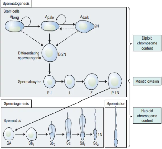
Figure 5 Differentiation of a human diploid germ cell into a fully functional spermatozoon, (Huckins C, 1975).
45
2.1.4. Spermiogenesis
Spermiogenesis is the third stage of spermatogenesis and is the morphological differentiation of spermatids into spermatozoa, which is the process by which spermatids acquire condensation and structural shaping of the nucleus, lose its cytoplasm and develop several organelles and accessory structures such as the acrosome and the flagellum. They begin after spermatocytes complete 2 quick successive meiotic reductive divisions to produce haploid round spermatids.
In humans, six different stages have been described in the process of spermatid maturation; (Sharma & Agarwal, 2011) these are termed as (Sa-1 and Sa-2, Sb-1 and Sb-2, and Sc-1 and Sc-2), each stage can be identified by morphological characteristics over the following phases (see figure 7); Golgi phase, cap phase, acrosomal phase and maturation phase. However, in a recent study reported by Muciaccia et al. (2013) the process of spermatid maturation can be divided into 12 stages on basis of acrosomal development made visible by immunohistochemistry (IHC) for (pro) acrosin. This new classification has been described as has been already done for many other mammals, including monkeys and mice.
At Golgi phase acrosomal bubbles and craniocaudal symmetry appear. While, in the cap phase the spermatids become elongated and the acrosome develops, covering the cranial half to two-thirds of the spermatid. During the fertilization process enzymes are released by the acrosome (Weinbauer et al.,
46
2010).The centrioles start to migrate to a position beneath the nucleus that is opposite the acrosomic vesicle. The proximal centriole (PC) will give rise to the attachment point of the tail, whereas distal centriole (DC) will give rise to the developing axoneme.
In the acrosomal phase the cell nucleus becomes further condensed and elongation of the cell continues. During condensation the majority of histones are lost and gene transcription stops. Nuclear chromatin is now extremely condensed, implying that the proteins necessary for spermiogenesis have to be transcribed before this time point and justifying the finding of RNA species with very long half-life and RNA binding proteins. This is the case for transition proteins and protamine. At the chromosomal level, chromatin condensation begins with the replacement of histones, the predominant chromatin proteins of somatic cells, by the basic low molecular weight transition proteins (TNPs). These proteins are then replaced with protamines (PRMs) in the nuclear matrix producing a tightly compacted nucleus with extensive disulfide bridge crosslinking (Bukowska et al., 2013).
A delicate fibrillary structure appears in the cytoplasm and extends from a close association with the nucleus to surround the flagellum for some distance; the chromatoid body approaches the flagellum near the centrioles and partly surrounds it; a delicate ring appears and closely surrounds the
47
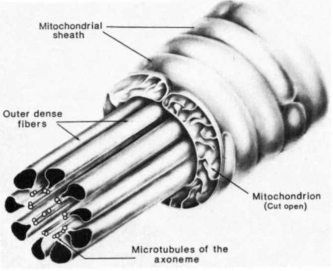
flagellum. Fawcett et al. (1975) clearly demonstrated that the chromatoid body contributed material to the ring. This ring eventually slides down the flagellum for a given distance and comes to rest at the caudal extremity of the middle piece. The middle piece is that portion of the flagellum between the modified centrioles and the ring around which the mitochondria accumulate in a regular covering (Fig 6). The period during which the nucleus and covering acrosomic system undergo the most dramatic morphological transformation was referred to as the acrosome phase.
The principal event during the maturation phase of the spermatids is the extrusion of the rest of the cytoplasm as the so-called residual body. Residual bodies are phagocytosed by Sertoli cells and have a regulatory role. Elongated spermatids and their residual bodies influence the secretory function of Sertoli cells (production of tubular fluid, inhibin, androgen-binding protein and interleukin-1 and 6). In parallel with degradation of the residual bodies, a new spermatogenic cycle begins (Clermont, 1963; Weinbauer et al. 2010).
48
Figure 6 An extende d diagram of the components in the midpiece of a mammalian spermatozoon tail (Fawcett, 1975)
49
Figure 7 Schematic representation of the differentiation of human spermatid 1, Golgi phase; 2–4, cap phase; 5–6, acrosome phase; 7–8, maturation phase; A, acrosome; An, annulus; Ax, axoneme; C, centriole; F, flower-like structures; Fs, flagellar substructures; M, mitochondria; Mp, middle piece; Mt, manchette; Ne, neck; N, nucleus; PP, principal piece; R, ring fibers; Sb, spindle-shaped body (Holdstein & Roosen-Runge 1981).
50
2.1.5. Spermiation
Spermiation is a multi-step process involving changes in both the spermatid and the Sertoli cell that ready the elongated spermatid, at the end of spermatogenesis, for its final Sertoli cells into the seminiferous tubule lumen prior to their passage to the epididymis. It takes place over several days at the apical edge of the seminiferous epithelium and involves several discrete steps including remodeling of the spermatid head and cytoplasm, removal of specialized adhesion structures and the final disengagement of the spermatid from the Sertoli cell (O'Donnell., et al 2011; Russell, 1984). However, the contribution of defects in the spermiation process to abnormal sperm forms in the ejaculate is potentially clinically significant, where variety of environmental exposures in humans could adversely affect spermiation and sperm output, whether it be a transient or long-term effect (O'Donnell., et al 2011).
51
Figure 8 Schematic representation different steps development of spermatogenesis (Sharma & Agarwal, 2014).
52
2.2. Regulation of spermatogenesis process
Spermatogenesis is highly dependent upon optimal conditions for the process to occur correctly. Two important factors have been implicated in the regulation of this process:
Endocrine regulation: Hypothalamic-Pituitary-Testicular Axis
Thermic systems.
2.2.1. Hypothalamic-Pituitary-Testicular Axis
The endocrine control of spermatogenesis is governed by the neuroendocrine activity along the hypothalamic-pituitary-testicular axis. Gonadotropin Releasing Hormone (GnRH) is released in a pulsatile pattern into the pituitary portal blood system from neuroendocrine cells in the basal hypothalamus and acts to stimulate gonadotrope cells in the anterior pituitary to synthesize and release two proteic hormones, follicle-stimulating hormone (FSH) and luteinizing hormone (LH) into the circulation (Fig 9). Once in the bloodstream, these hormones reach the testis, where LH stimulates testosterone production by the Leydig cells in the interstitium while FSH supports spermatogenesis in the seminiferous epithelium by stimulation of the Sertoli cells. A focused network of negative feedback relationships finesses testosterone secretion and sperm production.Both testosterone and estrogens play important roles in the regulation of reproductive function at the cellular and tissue levels. This
53
cascade is maintained by steroid and peptide feedback within the testis as well as the hypothalamic and pituitary gland (see Figure 9) (Dhole & Kumar, 2017).
54
55
2.2.2. Thermic regulation
In scrotal mammals, testicular thermoregulation is achieved by local and central mechanisms that are independent of the temperature regulatory mechanisms of the body core. The scrotum plays this important role in thermoregulation via several structural and functional adaptations, including the contraction of the cremaster and the dartos muscles, the presence of a large number of apocrine sweat glands, the absence of subcutaneous fat and the activity of the counter-current heat exchange system in the pampiniform plexus. The tunica dartos is a smooth muscle found in the subdermal layer of the scrotal skin, Contraction of the dartos reduces the surface area of the scrotum and blood flow to the scrotal skin, preventing heat loss (Shafik, 1974). Dartos relaxation causes excess heat to be removed. Therefore, the dartos plays an important role in the thermoregulation of the testes and congenital absence of this mechanism leads to infertility. The scrotum is not simply a pouch that houses the testis; it plays an active role in providing the necessary thermal environment for the testis. There are five main anatomical features that contribute to the maintenance of the testicular temperature lower than that of the body core. They are:
a) the tunica dartos smooth muscle the striated cremaster muscle
56
C) the absence of a subcutancous fat layer.
d) an abundance of sweat glands.
The physiological lower temperature for human testis results from two thermoregulatory systems (Waites & Moule, 1961). Both systems maintain the testicular temperature lower than core body temperature in a range of 32 and 34.5 C° (i.e. 2- 4 °C below core body temperature) (Mieusset & Bujan, 1995).
2.2.2.1. Exchange testicular arterial and venous blood streams
The first system reduces temperature of the testicular arterial blood which cools then the testis. The venous blood is therefore also cooler than the body temperature. The venous blood cools the arterial, as a result of a special vascular arrangement (the highly coiled pampiniform plexus) in which the artery winds in and out through venous sinuses within the inguinal canal (Fig 10) (Harrison, 1948). Nevertheless, study by Hsiung et al. (Zorgniotti, 1991) reported that any heating of the scrotal skin increases the venous blood temperature, inducing a reduction of the arteriovenous gradient and thus the efficiency of the thermoregulatory system. In cases of very high thermic conditions, this can induce a reversion of the pampiniform heat exchange resulting in a dramatic increase of the testicular temperature. Dahl and Herrick (1959) examined the gradient in temperature of the blood supplying the testis and scrotum: internal spermatic arterial blood temperature was lower than
57
that of the aorta by 3°C and the gradient fell markedly when venous drainage through the pampiniform plexus was interrupted.
58
Figure 10 Model of counter-current transfer of heat or a substance from the venous blood in the pampiniform plexus to the blood in the testicular artery ( Einer-Jensen & Hunter, 2005).
59
2.2.2.2. Scrotum and surfaces area heat lost
The second system induces losing heat outside of the testis through passive convection and radiation by the scrotum (Shafik, 1974; Einer-Jensen & Hunter, 2005; Mieusset & Bujan, 1995; Wallach et al., 1988); the role and function of this system are dependent to changes in body and environment temperature and other factors like varicocele.
Moore and Quick (1923) reported that physiologically the scrotum is a local thermo-regulator for the testes, and they see the possibilities of its having contributed materially to the evolution of the mammalian group. The special characteristics of scrotal skin anatomically such as; sweat glands, little subcutaneous fat, connective tissue, an abundance of thermoreceptors and the presence of the readily responsive dartos and cremasteric muscles to changes in environmental temperature are involved to lower temperature for human testis (Wallach et al., 1988). The musculature and vasculature in the genitals play a role in regulating testicular temperature as well. To maximize heat loss, the cremaster muscle that surrounds the testes and spermatic cords and the dartos muscle that lies beneath the scrotal skin relax, causing the testes to hang away from the abdomen and the scrotal skin to slacken, increasing the total surface area for easy heat dissipation (Fig 11) (Durairajanayagam et al., 2015). Further, vasodilation of scrotal vessels and activation of sweat glands promote heat loss when temperatures increase.If it is colder, these structures
60
pull the testes closer to take advantage of body heat and if it gets hotter muscles relax to allow the testes to get further from the warmth of the body, increasing the exposed surface area for a faster dispersion of heat excess increasing the total surface area for easy heat dissipation (Kleisner et al., 2010; Zorgniotti, 1982; Durairajanayagam et al., 2015).
Moreover, when external temperatures rise and cause the scrotal temperature to increase beyond a threshold value, cutaneous receptors on the scrotal skin are activated, initiating secretions of the scrotal sweat glands and active heat loss occurs through the evaporation of sweat (Waites, 1991; Candas et al.,1993). One or the other half of the scrotal sac hangs at a lower level than the other. The testes, housed within the sacs are also situated, suspended, one slightly lower than it’s other, this form to increase in surfaces area lost temperature (Deralakatte & Nithyanandanagar, 2008). Further, vasodilation of scrotal vessels and activation of sweat glands promote heat loss when temperatures increase.
61
62
63
Sperm morphology
64
3.1. Sperm morphology
3.1.1. Sperm (description, function and morphology)
Spermatozoa, or formed sperm is the end result of the spermatogenesis process, and is smaller than most cells in the body; the human sperm divide to three main parts; head, neck and tail [midpiece (anterior portion), principal piece, and end piece (posterior portion)] is about 60 μm long. A typical human spermatozoon has a distinct structure with an oval-shaped head (3–5 μm length and 2–3 μm width), a midpiece (7–8 μm), and a tail (45 μm) (Sedo et al., 2012; Baccetti, 1984) (Fig 12). In addition, this structure called the acrosome covers most of the head of the sperm cell as a “cap” that is filled with lysosomal enzymes important for preparing sperm to participate in fertilization. Moreover, the head is the most important part of the mature male gamete as it contains a nucleus, which is composed of packed chromosomal paternal genetic material (mostly DNA) containing 23 chromosomes. The nucleus comprises about 65% of the head, but unlike most somatic cells, lacks a large cytoplasm to match. The neck of spermatozoa is the site of articulation between the head and tail, and comprises the sperm centriole and connecting piece. During spermiogenesis, the axoneme of the tail grows from the distal centriole while the proximal centriole migrates to the caudal pole of the nucleus and attaches to it. Around the centriolar pair assembles the complex
65
structure of the connecting piece, a dense protein cylinder formed by nine longitudinal columns closed cranially by the capitulum that articulates with the sperm nucleus. At the final stages of sperm maturation, the distal centriole degenerates and only the proximal one remains at the cranial end of the connecting piece. The tail gives the sperm cell movement and responsible for sperm motility. After spermatogenesis process, the sperm is released into the lumen and is moved along a series of ducts in the testis toward a structure called the epididymis for the next step of sperm maturation, Midpiece contained tightly packed mitochondria for the production of ATP to the power of the flagellum (tail) (Kruger et al., 1986).
66
Figure 12 Diagram of a typical mammalian spermatozoon. Cross-sectional insets show the orientation of the internal cell structure (Holstein & Roosen-Runge, 1981)
67
3.2. Classifications sperm form and sperm morphology
Sperm morphology is described by the size and shape of sperm, and its morphology assessment has been one of the most common tests in evaluation of fertility (Gatimel et al., 2017). Sperm morphology results are reported as the percentage of sperm that appear normal when semen is viewed under a microscope. Moreover, there are three different indices that have been proposed and defined (WHO 1992; 2010; Jouannet et al., 1988; David et al., 1975).
• The multiple abnormalities index (MAI), used in the French modified David classification, is the average number of abnormalities per abnormal spermatozoon.
• The teratozoospermia index (TZI) is similar to the MAI, but a maximum of four abnormalities per abnormal spermatozoon are counted: one each for the head, the midpiece, the principal tail piece, and the residual cytoplasm, regardless of the real number of abnormalities per abnormal spermatozoon.
• The sperm deformity index (SDI) is the number of abnormalities divided by the total number of spermatozoa (normal and abnormal).
68
3.3. Normal and abnormal sperm forms
Sperm morphology is currently examined in semen smears with the main
criteria for normalcy relying on morphometric parameters of the sperm head, mid-piece, and flagellum (Chemes & Rawe, 2003). Morphological analysis of spermatozoa is currently assessed in the stained smears under a light microscope at 1000x magnification (WHO, 2010). Studies on scanning electron microscopy (SEM) and transmission electron microscopy (TEM) facilitated a better understanding of phenotypical abnormalities of human spermatozoa and their cytological details that represented certain functional abilities to an extent (Visco et al., 2010). World Health Organization (WHO) has described the percentage of normal sperm morphology forms through different versions, it was recommended in the last version (5th edition) that the lower reference limit to normal sperm forms is 4% (Table 1). However, this limit is lower than the recommendation in the 4th edition, and the recommended criteria for spermatozoa morphology assessment are also different from that of the 4th edition (see the Table 1). Menkveld (2010) showed that the new proposal of a very low normal value may not provide a strong predictive value for male’s fertility potential, as originally reported for sperm morphology evaluated according to strict criteria.
In an another classification (French modified David classification) the reference value for the percentage of normal forms was first 30% and was then