HAL Id: tel-00691924
https://tel.archives-ouvertes.fr/tel-00691924
Submitted on 27 Apr 2012HAL is a multi-disciplinary open access archive for the deposit and dissemination of sci-entific research documents, whether they are pub-lished or not. The documents may come from teaching and research institutions in France or abroad, or from public or private research centers.
L’archive ouverte pluridisciplinaire HAL, est destinée au dépôt et à la diffusion de documents scientifiques de niveau recherche, publiés ou non, émanant des établissements d’enseignement et de recherche français ou étrangers, des laboratoires publics ou privés.
Molecular and cellular bases for the protective effects of
dopamine D1 receptor antagonist, SCH23390, against
methamphetamine-induced neurotoxicity in the rat
brain
Geneviève Beauvais
To cite this version:
Geneviève Beauvais. Molecular and cellular bases for the protective effects of dopamine D1 receptor antagonist, SCH23390, against methamphetamine-induced neurotoxicity in the rat brain. Human health and pathology. Université René Descartes - Paris V; National institutes of health (Etats-Unis), 2012. English. �NNT : 2012PA05P604�. �tel-00691924�
i
Molecular and cellular bases for the protective effects of dopamine D
1receptor antagonist, SCH23390, against methamphetamine-induced
neurotoxicity in the rat brain
Geneviève Beauvais
Ecole doctorale du médicament
Université Paris Descartes
January 2012
ii
Table of contents
Page Abstract IV Acknowledgements V Abbreviations VI List of Figures IX List of Tables X 1. General introduction 1 1.1 History of methamphetamine 11.2 Physical and pharmacokinetic properties of METH 2
1.3 Methods of administration of METH 4
1.4 Patterns of abuse and clinical effects including addiction 4
1.5 Mechanism of action of METH 6
1.6 Dopamine hypothesis of addiction 6
1.6.1 Dopamine synthesis 6
1.6.2 Dopamine release and stimulation of dopamine receptors 7 1.6.3 Dopamine receptor genes, expression and signaling transduction 8
1.6.4 Dopamine reuptake and metabolism 11
1.6.5 Dopamine pathways in the CNS 12
1.6.6 Dopamine and the neural circuits of the basal ganglia 13
1.7 METH-induced neurotoxicity 15
1.7.1 Human data 15
1.7.2 Animal studies 17
1.7.2.1 Patterns of use of METH 17
iii
1.7.2.3 Hyperthermia 18
1.7.2.4 Neuronal apoptosis 18
1.8 Role of dopamine system in METH neurotoxicity 19
1.8.1 Dopamine overflow and oxidative stress 19
1.8.2 DAT and VMAT2 20
1.8.3 Dopamine receptors 21
1.9 Molecular mechanisms of METH neurotoxicity 22
1.9.1 METH-induced regulation of immediate early genes expression 22 1.9.2 Mechanisms of METH-induced activation of death pathways 24
1.9.2.1 Death receptor pathway 24
1.9.2.2 Mitochondrial dysfunction 25
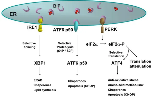
1.9.2.3 Endoplasmic reticulum stress pathway 28
1.10 Methamphetamine and trophic factors 32
1.10.1 Activins and TGF-βs 33
1.10.2 Activin A and TGF-βs in brain injury 34
1.11 Research aims 36
2. Manuscript 1 37
3. Manuscript 2 59
4. Manuscript 3 76
5. Manuscript 4 111
6. Conclusion and future perspectives 132
iv
Abstract
Methamphetamine (METH) is a potent psychostimulant known to cause cognitive abnormalities and neurodegenerative changes in the brains of METH abusers. One approach for developing therapies for METH abuse is to understand the molecular mechanisms of toxicity of the drug. Investigations in our laboratory and elsewhere have shown that single intraperitoneal injections of METH (30-40 mg/kg of body weight) can cause damage to striatal and cortical
monoaminergic systems and induce neuronal apoptosis in the striatum of rodents via activation of endoplasmic reticulum (ER) and mitochondrial death pathways. Hence, the purpose of this thesis was to investigate if toxic binge METH injections can cause ER- and mitochondria-induced stress in the rat striatum. Recent studies have suggested that dopamine (DA) D1 and D2
receptors might mediate neuronal apoptosis in the striatum after single toxic METH doses. We therefore hypothesized that signaling through these two types of DA receptors might activate toxic effects of the binge METH regimen. The role of DA D1 or D2 receptors in METH-induced
cell death pathways was thus examined by using pharmacological inhibitors of these receptors. In this dissertation, I report that binge METH regimen caused differential changes in immediate early genes (IEGs) that are known to influence synaptic changes in the brain. METH-induced changed in the expression of the IEGs were dependent on DA D1 receptor stimulation. The
second study examined the effects of binge METH on the expression of ER stress- and
mitochondrial dysfunction-responsive genes. Pretreatment with the DA D1 receptor antagonist,
SCH23390, caused complete inhibition of METH-induced ER and mitochondrial stresses whereas the DA D2 receptor antagonist, raclopride, provided only partial blockade. SCH23390
also blocked METH-induced hyperthermia whereas raclopride failed to do so. Interestingly, both antagonists attenuated METH-induced dopaminergic and serotonergic deficits in the striatum. Moreover, SCH23390 but not raclopride blocked METH-induced serotonergic deficits in cortical tissues. I also found that METH treatment induced upregulation of activin βA mRNA, increased TGF-β and phosphorylated Smad2 proteins in the rat striatum. SCH23390 pretreatment
completely blocked all these effects whereas raclopride did not block METH-induced increases in TGF-β expression.
In summary, these new data suggest a predominant role of DA D1 over D2 receptors in mediating
v
Acknowledgements
I address my sincere gratitude to my thesis co-directors, Dr. Jean Lud Cadet and Dr. Florence Noble. I thank Dr. Cadet for welcoming me in his team, and I appreciate the opportunity given his confidence and for allowing me to be independent in my work. I thank Dr Noble profoundly for her trust and her permanent support since the beginning of this project.
I deeply thank Dr. Wayne Bowen and Dr. Jesus Angulo to have agreed to judge this thesis. I express here my gratitude for the interest you have consented to bring to this work.
I am especially grateful to Dr. Subramaniam Jayanthi and Dr. Irina Krasnova for their assistance very rewarding, their scientific rigor to their qualities human and for agreeing to evaluate this work and sit on the thesis committee.
I would also like to acknowledge all past and present members of the Cadet lab. First of all, I want to thank Mr Bruce Ladenheim, Mr Mike McCoy and Ms Kenisha Atwell who contributed directly to my work. I express my gratitude to all former and present Cadet lab members and I wish them the best of luck in their careers.
I thank NIH/NIDA for the financial support over the last four years.
My sincere recognition to the “Ecole Doctorale Médicament, Toxicologie, Chimie et
Environnement MTCE” and the University Paris Descartes for making that joint PhD program with NIH/NIDA possible.
vi
Abbreviations
AC Adenylate cyclase
ADHD Attention deficit and hyperactivity disorder AIF Apoptosis inducing factor
AMPH Amphetamine
AP1 Activating protein 1
Apaf1 Apoptosis protease-activating factor 1 ASK Apoptosis signal-regulating kinase ATF Activating transcription factor ATP Adenosine triphosphate
BDNF Brain-derived neurotrophic factor
BH Bcl-2 homology
BIP Immunoglobulin binding protein BMP Bone morphogeneicic protein bZIP Basic region leucine zipper cAMP Cyclic adenosine monophosphate Caspases Cysteine-aspartic proteases CHOP C/EBP-homologous protein CNS Central nervous system COMT Catechol-o-methyltransferase CRE calcium/cAMP responsive element CREB cAMP response-element binding protein SOD Superoxide dismutase
DA Dopamine
DAT Dopamine transporter
DAWN Drug abuse warning network DEA Drug enforcement administration DFF45 DNA fragmentation factor 45
DHBA 2,3- and 2,5-dehydroxybenzoic acid
DIABLO Direct IAP-associated binding protein with low pI DOPA Dihydroxyphenylalanine
DOPAC Dihydroxyphenylacetic acid
EDEM Endoplasmic reticulum-degradation-enhancing-α-mannidose-like protein EGR Early growth response
vii ER Endoplasmic reticulum
ERAD Endoplasmic reticulum associated degradation
ERSE Endoplasmic reticulum stress-response element reporter ETC Electron transport chain
FADD Fas-associated death domain Fas-L Fas ligand
FDA Food and drug administration GABA Gamma aminobutyric acid GAD67 Glutamic acid decarboxylase
GADD34 Growth arrest and DNA damage inducible gene 34
GP Globus pallidus
GPe Globus pallidus external GPi Globus pallidus internal GRP Glucose regulated protein HSP Heat shock protein
HtrA2 High temperature requirement A2 5-HT 5-Hydroxytryptophan or serotonin 5-HTT 5-Hydroxytryptophan transporter HVA Homovanilic acid
IAP Inhibitor of apoptosis protein
ICAD Inhibitor of caspase-activated deoxyribonuclease IEG Immediate early gene
IP3 Inositol 1,4,5-trisphosphate IRE1 Inositol-requiring enzyme 1 JNK Jun-N-terminal kinase
MAO Monoamine oxidase
METH Methamphetamine
mPTP Mitochondrial permeability transition pore NAcc Nucleus Accumbens
NFAT Nuclear factor of activated T cells NMDA N-methyl-D-aspartate
NOS Nitric oxide synthase
NPY Neuropeptide Y
PARP Poly (ADP-ribose) polymerase
PERK Protein kinase R-like endoplasmic reticulum kinase PET Positron emission tomography
viii PKA Protein kinase A
PLC Phospholipase C
ROS Reactive oxygen species
Smac Second mitochondria-derived activator of caspases SN Substantia nigra
SNc Substantia nigra pars compacta SNr Substantia nigra pars reticulata
SP Substance P
SRE Serum response element
SST Somatostatin
TGF-β Transforming growth factor β TH Tyrosine hydroxylase
TRAF2 Tumor necrosis factor receptor associated factor 2
TUNEL Terminal deoxynucleotidyl transferase dUTP nick end labeling TβIR TGF-β type I receptor
TβIIR TGF-β type II receptor UPR Unfolded protein response
VMAT2 Vesicular monoamine transporter 2 VTA Ventral tegmental area
ix
List of Figures
Figure Page
1 Enantiomers of METH 3
2 Structural comparison of METH, AMPH derivatives and catecholamines 3
3 Dopamine synthesis 7
4 Structural features of the DA D1 and D2 receptors
9
5 Metabolism of dopamine 11
6 Illustration of the dopamine pathways within the human brain
12
7 Simplified anatomy of Cortex–Basal Ganglia Circuits 13
8 Representation of the oxidative system 26
9 ER and the unfolded protein response 29
x
List of Tables
Page Table 1 Dopamine receptor agonists and antagonists 8
1
General introduction
1.1 History of methamphetamine
Methamphetamine (METH) is a potent psychostimulant with varied biochemical and molecular effects in the central nervous system (CNS) (Krasnova and Cadet, 2009). The increases in METH abuse throughout the world is a major public health and social concerns (Kulsudjarit, 2004; Rawson et al., 2002). In the United States, the widespread availability of METH makes it a major drug of abuse (Gonzales et al., 2010). Different factors contribute to the expansion of METH use including inexpensive precursor chemicals, the ease to manufacture and sell the drug. The number of recent new users of methamphetamine among persons aged 12 or older was105,000 in 2010 (National Survey on Drug Use and Health, September 2011). Additionally, data from the Drug Abuse Warning Network (DAWN) have indicated a constant high number in METH-related visits to emergency rooms from 2007 to 2009 (approximately 65,000). Moreover, evidence shows that in many western Unites States cities, METH is used extensively by gay males and is frequently associated with high-risk sexual behavior, a major factor in the transmission of HIV and hepatitis (Degenhardt et al., 2010; Gonzales et al., 2006; Gonzales et al., 2008). Epidemiologic data also indicate that METH abuse and trafficking touch different regions in the world including Southeast and East Asia (McKetin et al., 2008), Canada (Callaghan et al., 2007), Australia (Degenhardt et al., 2008), South Africa (i.e., Cape Town) (Kapp, 2008), and parts of Europe (e.g., Czech and Slovak republics) (Griffiths et al., 2008).
From a historical perspective, however, METH has been in existence for many years. Credit for its synthesis is usually given to the Japanese chemist, Akira Ogata (Ogata, 1919). Use of that psychostimulant became popular during World War II, because it was widely distributed to Japanese and German soldiers to help them stay alert and effective (Rasmussen, 2008). After the war, METH became easily available to the public and its abuse rapidly spreads throughout Japan, United Kingdom and different other regions in the world (Matsumoto et al., 2002).
In 1943, Abbott Laboratories requested U.S. Food and Drug Administration (FDA) approval for the use of METH as a treatment for narcolepsy, mild depression, chronic alcoholism and hay fever (Moore, 2010). METH was approved for all these uses and marketed as Desoxyn but later these approvals were removed. METH is currently used to treat attention deficit and hyperactivity
2 disorder (ADHD) and obesity (FDA, 2007). In the United States, METH was widely abused during the 1960’s. The drug Abuse Control Amendments of 1965 were enacted that limited production and distribution of METH. The Control Substances Act of 1970 classified METH as a schedule II substance under the Drug Enforcement Administration (DEA) regulations. Nevertheless, clandestine METH laboratories continue to flourish in the USA and throughout the world.
1.2 Physical and pharmacokinetic properties of METH
When used illicitly, METH (C10H15N) is commonly named “speed”, “meth”, “chalk”, “ice”,
“glass”, and “crystal” (Derlet and Heischober, 1990). METH can be readily prepared from simple chemical precursors and often impurities are found in these preparations (Derlet and Heischober, 1990; Puder et al., 1988). However, during the past years, one common method of synthesis of the METH form known as “ice” is prepared from l-ephedrine or d-pseudoephedrine (Derlet and
Heischober, 1990). The preparation results from the reduction of the β-hydroxyl group on ephedrine with a mixture of iodine and red phosphorous. The product is pure d-METH, which is several times more active than the l form. METH is usually supplied and used as the hydrochloride salt
(C10H16ClN), which is a white crystalline, practically odorless substance (Cook, 1991). METH is easily dissolvable in water, alcohol, chloroform and dimethyl sulfoxide (Freye, 2009).
METH contains a chiral carbon atom, and therefore has two isomers with stereoselective differences in biological action (Figure 1). The drug exists in three forms: dextro (d-METH), levo (l-METH) and the racemic mixture of d and l-methamphetamine (dl-METH). The levorotary isomer of METH is found in inhalers for nasal decongestion such as Vicks Vapor Inhaler and has four times less stimulant activity than d-METH (Mendelson et al., 2008). The d-METH form is a known stimulant (DEA schedule II) and is prescribed to patients as Dexedrine/Desoxyn to treat insomnia, ADHD, narcolepsy as well as obesity. The d-METH form is the most potent but illicit synthesis of METH for abuse results mostly in the racemic mixture. The racemic mixture of the drug was used in this dissertation.
3
Figure 1 Enantiomers of METH
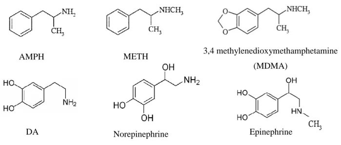
METH and other amphetamine (AMPH) derivatives share a similar phenylethylamine structure with catecholamines, epinephrine, norepinephrine and dopamine (DA) (Figure 2). This similarity is an important determinant of the effects of the drug on monoaminergic systems.
Figure 2 Structural comparison of METH, AMPH derivatives and catecholamines
METH has a half-time of about 12 hours (Schepers et al., 2003), an important factor in the length of its behavioral actions when compared to other psychostimulants such as cocaine. METH is a weak base with a pKa of 9.9 and is highly lipophilic, thus allowing it to cross the brain barrier easily. METH is primarily metabolized in the liver through aromatic hydroxylation and
demethylation, and is excreted mostly in the urine (Cook et al., 1992; Lin et al., 1997). Its
metabolism results in the production of the active compound, AMPH as well as in the production of corresponding para-hydroxylated metabolites, p-OH-METH and p-OH-AMPH. Multiple reports demonstrated that there are variations in METH metabolism between species. In humans, the main
Epinephrine DA
AMPH METH 3,4 methylenedioxymethamphetamine
(MDMA)
4 metabolite is the drug unchanged, while the major urinary metabolite in rats is 4-hydroxy-METH (Caldwell et al., 1972). Minor metabolites are norepinephrin, 4-hydroxy-norepinephrin and benzoic acid (Caldwell et al., 1972).
1.3 Methods of administration of METH
METH is usually administered orally or by nasal inhalation for medical use. In recreational use, ice or crystal, the METH hydrochloride can be taken orally, snorted, swallowed, smoked, injected or inserted anally (Cho and Melega, 2002; Covey, 2006; Keeling, 2008). The effects are different depending on the route of ingestion. When snorted or orally ingested, METH causes euphoria, but not an intense “rush” (Covey, 2006; Keeling, 2008). Smoking or injecting METH causes a “rush” or “high” effect that has been described to be extremely pleasurable (Covey, 2006; Keeling, 2008). This effect is followed by feelings of euphoria. People who take METH by the intravenous route often develop skin rashes, called speed bumps, and infections at the site of injection (Covey, 2006). In addition, share needles also expose users to different diseases like HIV and hepatitis (Degenhardt et al., 2010; Gonzales et al., 2006; Gonzales et al., 2008; Molitor et al., 1999).
1.4 Patterns of abuse and clinical effects including addiction
METH abuse has three patterns of administration of the drug: low, binge and high intensity (Covey, 2006; Keeling, 2008). Ingestion of a high dose of the drug can be fatal to users (Guharoy et al., 1999; Sribanditmongkol et al., 2000). Low-intensity abusers are not psychologically addicted to METH and take the drug occasionally to stay awake long enough to finish a task (Keeling, 2008). Low-intensity use is characterized by the snorting of powered METH or ingestions of pills (Covey, 2006). Binge and high-intensity abusers are addicted to METH and prefer to smoke or inject the drug in order to get its effects faster (Keeling, 2008). The binge pattern of abuse has multiple stages during its cycle. Users continually self-administer increasing doses throughout several days, and experience regular “rush” and euphoria. The total drug consumption during a binge is in the range of 2-4 grams. The crystal users will use the drug over a period of three to fifteen days until no “rush” or “high” is experienced (Cho and Melega, 2002; Covey, 2006). Usually, when the sensation of pleasure wears off, the binge METH user becomes irritable and paranoiac because of the lack of sleep and pharmacological effects of the drug. This state is called “tweaking” when the users experience paranoia, hallucinations and violent behavior (Sexton et al., 2010). The METH binge
5 user eventually crashes because he experiences a drastic drop in mood and energy levels. The individual will subsequently sleep for about one to three days (Keeling, 2008). After crashing, binge users usually return to their premorbid state, with progressive deterioration of both physical and psychological health, accompanied by depression and craving for the drug (Nakama et al., 2008). Withdrawal symptoms lead to repeated cycles of increasing binge ingestion of METH in spite of the negative consequences, well known by the abusers (Kalechstein et al., 2003; McGregor et al., 2005). High-intensity abusers have the impossible goal of never crashing and maintaining a state of euphoria and the perfect rush. The chronic METH users, often called “speed freaks”, consume the drug regularly; they preferentially take regular “hits” of the vaporized compound from pipes as often as every 30 minutes during the day. Estimates of the total drug consumed in a day range from 0.7 to 1gram (Cho and Melega, 2002). This pattern of METH abuse causes less euphoria with repeated use, leading the user to take greater amount of the drug (Covey, 2006; Keeling, 2008). Tweaking for the high-intensity user is the most dangerous time to confront him because of their violent and unpredictable behavior (Keeling, 2008).
METH is a CNS stimulant and is abused for its desirable effects that include euphoria, increased alertness, improved self-confidence, enhanced energy and increased libido (Homer et al., 2008; Meredith et al., 2005). There are also many adverse health effects associated with METH use. METH is a sympathomimetic drug and will cause increased heart rate and blood pressure, leading to damage of heart vessels (Darke et al., 2008). Therefore, administration of large doses of METH can caused cerebrovascular accidents and cardiac arrhythmias, which in addition to hyperthermia can lead to renal failure and death (Albertson et al., 1999; Darke et al., 2008). Chronic therapeutic use of METH has been shown to cause no changes in blood pressure or heart rate, when
administered at the prescribed dosage (about 5 to 10 mg doses) (Mitler et al., 1993). In contrast, illicit chronic use of METH can cause CNS complications such as anxiety, paranoia and chronic psychosis (Brecht et al., 2004; Zweben et al., 2004). Neurological problems include movement disorders, seizures and strokes (Perez et al., 1999). METH addicts can also present with
neuropsychological deficits in tests of attention, working memory, and decision making (Sim et al., 2002; Simon et al., 2002; Verdejo-Garcia et al., 2006).
METH is a highly addictive drug. Long-term use of the drug can lead to tolerance, dependence and/or addiction. Tolerance to METH often happens in chronic and binge METH users.
6 later more sensitive to some effects. This phenomenon is known as reverse tolerance or
sensitization. Chronic use of METH leads to psychological dependence to the drug as evidenced by the compulsive drug use, drug cravings during withdrawal state and inability to stop drug use. Multiple studies have revealed that METH abuse causes plastic changes in the synapses and terminals of the monoamine neurons. These molecular neuroadaptations might constitute physiological components of METH addiction (Barr et al., 2006; Kish, 2008). Although the underlying mechanisms for METH addiction are unknown, it has been suggested that the DA system plays a dominant role, although the 5-hydroxytryptophan (5-HT or serotonin), glutamate and neuropeptides systems have a secondary role.
1.5 Mechanism of action of METH
METH is sympathomimetic drug, which mimics catecholamines functions in the brain. METH produces its euphoric effects by inducing release of monoamines in the brain, especially in the basal ganglia (Kish, 2008; Sulzer et al., 2005). METH-induced monoamine release occurs after its entry into monoaminergic terminals by binding to DA and serotonin (HT) transporters (DAT and 5-HTT) and by passive diffusion. When inside the terminals, METH binds to vesicular monoamine transporter 2 (VMAT2) and enters monoaminergic vesicles where it disrupts the proton charge in the vesicles and allows redistribution of monoamines in the cytosol (Cubells et al., 1994; Sulzer et al., 2005). In addition, METH inhibits the enzyme monoamine oxidase (MAO), which is
responsible for monoamines metabolism, hence resulting in higher availability of the neurotransmitters (Sulzer et al., 2005). METH can also alter the function of the monoamine
transporters which induces reverse transport of monoamines into the synaptic cleft (Haughey et al., 2000; Sulzer et al., 2005). This is accompanied by high levels of monoamines in the synaptic cleft that can activate presynaptic receptors and also postsynaptic receptors that are located in the mesolimbic, mesocortical and nigrostriatal pathways (Berke and Hyman, 2000; Moore and Bloom, 1978)(see below). The present dissertation focuses on the neurotoxic effects of METH on the nigrostriatal pathway.
1.6 Dopamine hypothesis of addiction
7 Accumulated evidence suggests that psychostimulants commonly mediate their effects through activation of the DA system (Berke and Hyman, 2000). DA belongs to a group of neurotransmitters called cathecholamines (see Figure 2). They all contain a nucleus of a catechol (benzene ring with two adjacent hydroxyl groups) and a side chain of ethylamine or one of its derivatives. DA
constitutes about 80% of the catecholamine content of the brain (Vallone et al., 2000). It is mainly synthesized in the substantia nigra (SN) and the ventral tegmental area (VTA), from the amino acid l-tyrosine (Figure 3). The majority of circulating tyrosine originates from dietary sources, but small amounts are derived from hydroxylation of phenylalanine by the liver enzyme phenylalanine hydroxylase. L-tyrosine is transported across the blood-brain barrier of the DA neuron where it is converted to dihydroxyphenylalanine (l-DOPA) by the enzyme tyrosine hydroxylase (TH)
(Daubner et al., 2011). This constitutes the rate-limiting step in the synthesis of DA. Thereafter, l-Dopa is converted to dopamine by the aromatic amino acid decarboxylase enzyme (DOPA decarboxylase). DA enters cytoplasmic vesicles via VMAT2 and gets stored into these vesicles until it gets released.
Figure 3 Dopamine synthesis
1.6.2 Dopamine release and stimulation of dopamine receptors
Upon the arrival of an action potential, there is a change in the vesicular membrane protein conformation that allows the influx of calcium ions. Depolarizing stimuli evoke calcium-dependent DA release mainly from cytoplasmic vesicles but newly synthesized DA can also be released (McMillen et al., 1980; Thomas et al., 2008). DA exerts its functions by interacting with specific membrane receptors. DA receptors are classified in two groups based on physiological and
biochemical properties fall into two families; the D1-like family which includes D1 and D5 receptor
subtypes, and the D2-like family which includes receptor subtypes D2, D3, and D4 (Neve et al.,
2004; Vallone et al., 2000). Use of pharmacological DA receptor ligands allows the differentiation of these receptors. Table 1 lists some of the selective agonists and antagonists of the DA receptors.
Dihydro-biopterin + H2O
8 However, some of the drugs do not clearly differentiate between members of the same subfamily; by example, the DA D2 receptor antagonist, raclopride, has high affinity for both DA D2 and D3
receptors. Furthermore, some of the DA receptor ligands can also have significant affinity for other receptors. As an example, the DA D1 receptor antagonist, SCH23390, inhibited binding of the
serotonergic 5-HT2 receptor antagonist, [3H]spiperone, in rat frontal cortex with an ID50 of 1.5
mg/kg (Bischoff et al., 1986). However, interaction of SCH23390 with 5-HT2 receptors was tissue specific as it was characterized in rat frontal cortex, an area rich in both D1 and 5-HT2 receptors,
but not in the striatum and hippocampus (Bischoff et al., 1986; McQuade et al., 1988). Furthermore, there are contradictory data on the antagonistic properties of SCH23390 indicating that doses of the antagonist up to 5 mg/kg do not affect serotonergic transmission in the cortical tissue (Gandolfi et al., 1988). In this dissertation, rats were administered four doses of 0.5 mg/kg of SCH23390 given at 2-hr intervals in order to antagonize METH-induced DA transmission in the striatum.
Table 1 Dopamine receptor agonists and antagonists, Ki values in nM (Kihigh and Kilow)
1.6.3 Dopamine receptor genes, expression and signaling transduction
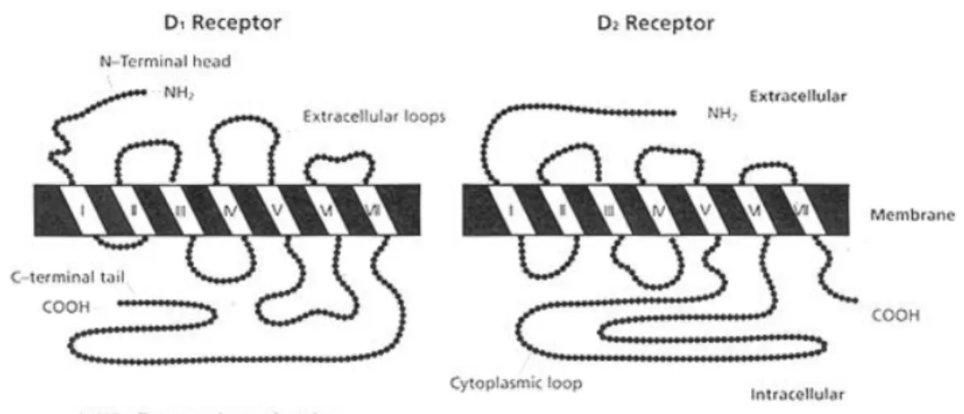
All DA receptors have characteristics of G-protein coupled receptors and contain seven
transmembrane domains linked by protein loops with an extracellular amino terminus (Civelli et al., 1991; Jackson and Westlind-Danielsson, 1994; Missale et al., 1998). D1-like receptors have short
third intracellular loop and long carboxyl terminal tail, whereas, the D2-like receptors contain
D1-like D2-like D1 D5 D2 D3 D4 Agonists Dopamine 0.9–2340 0.9–261 2.8–474 4–27 28–450 Quinpirole 1900 4.8–576 5.1–24 30–46 Apomorphine 0.7–680 122–163 0.7–24 20–32 4 Bromocriptine 440–672 450 5.3–12.6 5–7.4 290–340 SKF-38393 1–150 0.5–100 150–9560 5000 1000–1800 Antagonists SCH23390 0.11–0.35 0.11–0.54 270–1100 314–800 3000–3560 Raclopride 18 000 1–5 1.8–3.5 237–2400 Haloperidol 27–203 33–151 0.6–1.2 2.74–7.8 2.3–5.1 (-)-Sulpiride 20 400–45 000 11 000–77 270 2.5–71 8–206 21–1000 Clozapine 100–261 194–336 56–230 83–620 9–42 Spiperone 99–350 135–4500 0.06–0.37 0.32–0.71 0.05–4
9 introns in their third intracellular loop and have short carboxyl terminal tail (Figure 4). This
characteristic is the source of variants between the receptors.
Figure 4 Structural features of the DA D1 and D2 receptors (Crocker, 1994)
D1-like receptor subfamily
The D1-like receptor genes (D1 and D5) do not contain introns in their coding regions and
therefore do not have any variants (Vallone et al., 2000). The D1 and D5 receptors have their third
intracellular loop and the COOH terminus similar in size but divergent in their sequence. The external loops between transmembrane domains 4 and 5 are different in length (27 and 41 amino acids for D1 and D5 receptors) and sequence (Missale et al., 1998). The expression of these genes is
not uniformly expressed in the brain. The D1 receptor is highly expressed in the projection regions
of DA neurons such as the striatum, nucleus accumbens (NAcc), frontal cortex, olfactory tubercle and amygdale (Jackson and Westlind-Danielsson, 1994; Missale et al., 1998). It is also found in the island of Calleja and the subthalamic nucleus (Jackson and Westlind-Danielsson, 1994). The D5
receptor is expressed at much lower levels and is concentrated in the hippocampus and
hypothalamus, with lower amount found in the striatum and frontal cortex (Jackson and Westlind-Danielsson, 1994; Meador-Woodruff et al., 1992).
D2-like receptor subfamily
Gene structures of the D2-like receptors contain introns within their coding regions; D2, D3
and D4 receptors have six, five and three introns, respectively (Missale et al., 1998). There are short
and long variants of D2 receptor (D2S and D2L), which are generated by alternative splicing of 29
amino acids in the third intracellular loop (Dal Toso et al., 1989). Spliced variants of the D3
10 1991). In addition, variations in the D4 receptor gene were also identified (Van Tol et al., 1992).
The D2L subtype is more common than the D2S subtype (Missale et al., 1998; Vallone et al., 2000).
D2-like receptors are expressed both at the presynaptic and postsynaptic neurons. Presynaptic D2
receptors are expressed in the SN pars compacta (SNc) and in the VTA, while D3 receptor is only
found in the SN (Civelli et al., 1991). These presynaptic neurons called autoreceptors play a role in the modulation of nigrostriatal and mesolimbic systems. Highest levels of the D2 receptors are
found in the projection regions such as the dorsal striatum, NAcc, hypothalamus and pituitary (Jackson and Westlind-Danielsson, 1994; Landwehrmeyer et al., 1993). D3 and D4 receptors have a
lower pattern of expression. D3 receptors are found in the ventral striatum or NAcc, the islands of
calleja, hypothalamus, thalamus and cerebellum (Bouthenet et al., 1991; Jackson and Westlind-Danielsson, 1994). D4 receptors are mostly found in the frontal cortex, amygdala, olfactory bulb
and hippocampus (Jackson and Westlind-Danielsson, 1994). Signaling pathways
It is generally assumed that stimulation of D1-like receptors, which are coupled to Gs or Golf
protein, causes activation of adenylate cyclase (AC) (Herve et al., 1993; Jackson and Westlind-Danielsson, 1994; Monsma et al., 1990; Sunahara et al., 1991). This enzyme causes the conversion of adenosine triphosphate (ATP) to the intracellular second messenger cAMP. The cAMP in turn activates cAMP-dependent protein kinase A (PKA), which phosphorylates numerous substrates such as the transcription factor cAMP response element-binding (Neve et al., 2004). In contrast, D2
-like receptors inhibit AC (Jackson and Westlind-Danielsson, 1994). The D2 receptor was first
characterized as an inhibitor of intracellular cAMP levels in the pituitary gland, and in striatal cells. However, D3 receptor weakly inhibits AC (Missale et al., 1998; Robinson and Caron, 1997). D4
receptor also inhibits cAMP accumulation in the retina and different cell lines (Missale et al., 1998). Stimulation of DA D1-like receptors is also coupled to activation of Gq/phospholipase C (PLC)
and causes increases in levels of inositol 1,4,5-trisphosphate (IP3) (Jin et al., 2003; Wang et al., 1995a). DA D1 receptors mediate release of calcium from intracellular stores through activation of
PLC and opening of voltage-gated calcium channels (Berridge, 2009; Wu et al., 2006). DA D2
receptors coupled to Gi/o protein reduce calcium currents (Jackson and Westlind-Danielsson, 1994;
11 1.6.4 Dopamine reuptake and metabolism
Reuptake is the process, by which DA released in the synaptic cleft, is brought back into presynaptic nerve terminals or is internalized by surrounding glial cells (Giros et al., 1992;
Mannisto and Kaakkola, 1999; Yu and Hertz, 1982). Reuptake of DA is dependent on the presence of external sodium and chloride and mediated by the DAT, which is localized at the plasma
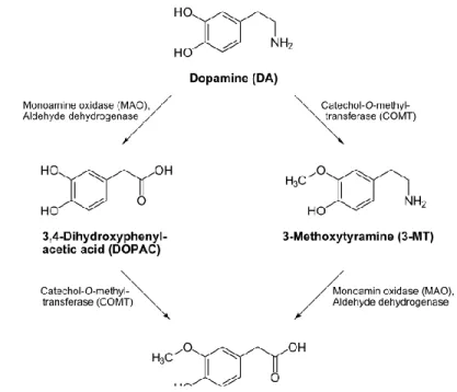
membrane of nerve terminals (Giros et al., 1992). Subsequently, two enzymes metabolize the neurotransmitter: catechol-o-methyltransferase (COMT) localized in the cytoplasm, and MAO expressed on the membrane of mitochondria (Kopin, 1985) (Figure 5). COMT catalyzes the methylation of one group hydroxyl on a DA molecule to produce 3-methoxythyramine.
Deamination of DA by MAO produces dihydroxyphenylacetic acid (DOPAC). The combination of both COMT and MAO effects convert DA in homovanilic acid (HVA) (Kopin, 1985; Kopin, 1994). DA is also removed from the extracellular space and metabolized by astrocytes (Mannisto and Kaakkola, 1999; Yu and Hertz, 1982).
12 1.6.5 Dopamine pathways in the CNS
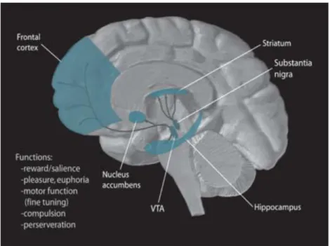
In the CNS, there are four central dopaminergic systems (Figure 6):
(1) The nigrostriatal pathway has 80% of DA neurons in human brain. The cell bodies of the neurons are localized in the SNc. The projections of these neurons end into the dorsal striatum, constituted of caudate and putamen nuclei (Joel and Weiner, 2000; Moore and Bloom, 1978). In rodents, these regions consist of one nucleus named the dorsal striatum or caudate putamen (Joel and Weiner, 2000). This pathway controls motor activity (Gerfen, 1992b).
(2) The mesolimbic pathway begins in the VTA and terminates in different limbic area, such as NAcc, olfactory tubercle, amygdala and hippocampus (Moore and Bloom, 1978; Swanson, 1982). The mesolimbic pathway is responsible for motivation, reward, drug addiction, motor activity (Le Moal and Simon, 1991).
(3) The mesocortical pathway also originates in the VTA and terminates in the cortical structures (Moore and Bloom, 1978; Swanson, 1982). This pathway controls cognitive functions and learning (Le Moal and Simon, 1991).
(4) The tuberoinfundibular pathway goes from the hypothalamus to the hypophysis, regulating neuroendocrine function (Moore and Bloom, 1978).
Figure 6 Illustration of the dopamine pathways within the human brain. Obtained from
13 1.6.6 Dopamine and the neural circuits of the basal ganglia
The striatum is part of the basal ganglia, a complex of nuclei that has important activity in the brain. The basal ganglia contain the dorsal striatum or caudate-putamen, the NAcc, globus pallidus internal and external (GPi and GPe) (Graybiel, 2000). The basal ganglia function in conjunction to other CNS regions including cortex and thalamus; together, they form a sensorimotor circuit “cortico-striato-thalamo-cortico” (Alexander et al., 1990; Brown and Marsden, 1998) (Figure 7). In this neural circuit, the striatum plays a central role; it receives glutamatergic inputs from the cortex and thalamus, dopaminergic inputs from the midbrain (SN pars compacta and VTA), cholinergic inputs from the brainstem and serotonergic inputs from the dorsal raphe nucleus. It also sends gamma aminobutyric acid (GABA) outputs to the GP, SN pars reticulate (SNr), and subsequently to the thalamus and the frontal cortex involved in motor activities. Dysregulations in the function of the basal ganglia and allied nuclei can result in disorders of movement programming and learning processes, and characterize some neurodegenerative diseases such as Parkinson’s disease and drugs addiction (Kreitzer, 2009).
Figure 7 Simplified anatomy of cortex–basal ganglia circuits (Berke and Hyman, 2000)
In the striatum, DA fibers originating from the SNc modulate both projection neurons and interneurons. The striatum is primarily composed of medium spiny neurons which synthesize GABA (90 %) and are projections neurons. Activation of the dopaminergic nigrostriatal pathway
14 stimulates DA D1 and D2 receptors expressed in two different populations of GABAergic neurons
involved in the striatonigral/direct and striatopallidal/indirect pathways, respectively (Figure 6). However, there is evidence that a portion of 10-20% of DA D1 and D2 receptors colocalizes in the
striatal tissue (Surmeier et al., 1992).
The direct pathway originates from GABA spiny medium projection neurons that also express the neuropeptides, substance P (SP) and dynorphin. The membrane of these neurons contains DA D1 receptors coupled to AC. The projections go to two different regions of the basal ganglia, the
GPi (primates)/entopeduncular nucleus (rodents) and SNr. In normal state, these two brain regions inhibit the thalamus by sending GABA outputs. However, when the direct pathway is activated, release of GABA from the striatum inhibits tonic activity of the GABA neurons in the GPi and SNc, deshinibiting the thalamic networks. The activated thalamus releases glutamate in the cortex, then the cortex can stimulate motor activity and send glutamate outputs to the striatum.
The indirect pathway involves GABA medium spiny neurons that are also enkephalin-positive. These neurons express DA D2 receptors at their membrane. Their axons send GABA outputs in the
GPe (primates)/GP (rodents), which subsequently desinhibits glutamatergic neurons in the
subthalamic nucleus. Indeed, this region stimulates the GPi and the SNc, reinforcing their inhibitory effects on the thalamus and causing a decrease in motor functions.
Furthermore, DA neurotransmission also affects GABA and acetylcholine aspiny interneurons in the striatum, which represent 10% of striatal neurons. Despite few number of interneurons, they play an important role in regulating striatal outputs (Kreitzer, 2009). The large cholinergic interneurons (< 5% of striatal neurons) are identified by the presence of the enzyme, choline acetyltransferase. These neurons receive glutamatergic inputs from the thalamus. Cholinergic interneurons express both D2 and D5 receptors and are responsive to their activity (Bergson et al.,
1995; Yan and Surmeier, 1997). Striatal cholinergic interneurons also express the SP receptor and release acetylcholine in response to its activation (Kawaguchi, 1997).
Medium-sized GABA interneurons are divided in three subtypes: parvalbumin-positive;
somatostatin-, neuropeptide Y-, and nitric oxide synthase-positive (SST-NPY-NOS); and calretinin-positive (Kawaguchi et al., 1995). Neurons containing the calcium binding protein parvalbumin, constitute between 3-5% of striatal cells and are slightly larger than the medium-sized spiny GABA-positive cells. They receive powerful excitation from the cerebral cortex. Somatostatin-containing interneurons represent 1-2% of striatal neurons. Somatostatin/NOS-positive cells receive
15 direct cortical inputs, which might be glutamatergic. Calretinin, which is, as noted, expressed in a population of smaller aspiny neurons, is a calcium-binding protein with strong homology with calbindin. These cells express GABA and glutamic acid decarboxylase (GAD67). The modulation of
these interneurons by DA is not clearly characterized, but it is known that the GABAergic aspiny interneurons express DA D5 receptors (Bergson et al., 1995; Centonze et al., 2003). Although the
role of the striatal interneurons is not fully understood, some researchers have investigated their implication in METH toxicity. It has been shown that the intraventricular administration of the neuropeptide NPY or NPY receptor agonist protected the mouse striatum from METH-induced apoptosis, and this effect was dependent on DA D1 receptor (Thiriet et al., 2005). It was suggested
that METH treatment caused an interaction between the axon collaterals of striatonigral projection neurons and NPY/ positive interneurons, knowing that striatonigral neurons express SP and DA D1
receptors, whereas NPY-positive interneurons express the neurokinin-1 receptor for SP (Gerfen, 1992a; Kawaguchi, 1997). In addition, Zhu et al (2006) demonstrated that multiple injections of METH induced apoptosis in about 45% of GABA-parvalbumin-positive neurons in the dorsal striatum, and 29% of cholinergic neurons in the dorsal–medial striatum (Zhu et al., 2006a).
1.7 METH-induced neurotoxicity
1.7.1 Human data
Brain imaging studies of METH abusers showed that METH causes long-term damages to the brain. Magnetic resonance imaging studies gave evidence of brain structural abnormalities
including a significant loss of gray matter in the cortical and limbic systems, reduction in hippocampal volume, significant white-matter hypertrophy, medial temporal lobe damage and striatal enlargement (GP and putamen) (Baicy and London, 2007; Chang et al., 2005; Thompson et al., 2004a). In addition, positron emission tomography (PET) studies showed abnormalities in glucose metabolism in brain of METH users, which have increased metabolism in the parietal cortices but decreased metabolism in the thalamus plus striatum (Volkow et al., 2001c). These structural alterations after chronic use of METH have been linked to motor and cognitive
impairments. By example, METH addicts exhibit deficits in attention, working memory, decision-making and motor activity (Paulus et al., 2002; Thompson et al., 2004a; Volkow et al., 2001d). However, striatal enlargement is thought to be as a compensatory response to repeated METH-induced striatal injury, because METH users with the smallest striatal volumes had the greatest
16 cumulative lifetime of METH use and poorest cognitive performance (Chang et al., 2005). It was suggested that this effect is related to the induction of gliosis and neurotrophic factors, because activated macrophages and microglia after striatal injury caused wouding of the striatum and dopaminergic fibers sprouting (Batchelor et al., 1999).
In addition, analysis of brain of METH users showed neurochemical changes affecting the dopaminergic and serotonergic systems. PET studies revealed that DAT was decreased in the caudate-putamen, the NAcc and the prefrontal cortex (McCann et al., 1998; Sekine et al., 2001). Post mortem analysis also showed a significant decrease in DA, DAT and TH density even after as much as three years of abstinence from the drug (Wilson et al., 1996). Furthermore, the density of 5-HTT was found decreased in the midbrain, striatum, thalamus and cortex of brain of METH abusers (Sekine et al., 2006). Another study confirmed the reduction in 5-HTT levels in
orbitofrontal and occipital cortices (Kish et al., 2009). There is also evidence of a decrease in DA D2 receptors in the striatal tissue (both dorsal and ventral striatum) of METH addicts (Volkow et al.,
2001a). Importantly, there is proof of partial recovery from the structural abnormalities caused by METH abuse in rodents (Cass and Manning, 1999; Friedman et al., 1998), nonhuman primates (Harvey et al., 2000) and humans (Volkow et al., 2001b; Wang et al., 2004) after protracted
abstinence. By example, loss of DAT in METH abusers recovers after abstinence of twelve months or longer in the striatum, without any improvement in slower motor function and decreased
memory (Volkow et al., 2001b). However, abstinence of nine months of longer affects thalamic glucose metabolism (but not striatal glucose metabolism) and correlated with an improvement in neuropsychological performance (Wang et al., 2004). Administration of METH to vervet monkeys also caused long-term but reversible deficits in TH, DAT and VMAT2 in the striatum (Harvey et al., 2000). Studies in rats revealed that METH-induced decreases in presynaptic monoaminergic markers such as 5-HT (Friedman et al., 1998), DA and its metabolites (Cass and Manning, 1999; Friedman et al., 1998) recover to normal levels in the striatum between six and twelve months postinjection. The findings from these studies have implications in the treatment of METH abusers and the elucidation of the mechanisms underlying the apparent reversibility of METH neurotoxicity is needed.
17 1.7.2 Animal studies
1.7.2.1 Patterns of use of METH
Use of animal models is an efficient tool to study the neurotoxic effects of METH abuse (Kita et al., 2003). Different models to study METH neurotoxicity exist, which mimic human METH abuse patterns and include chronic, high-dose and multiple daily (binge) exposure to the drug. In our experiments, we have used a one-day METH dosing regimen that better models the acute overdose pathologies seen in humans. None of the models is perfect; each one can contribute to understand the neuropathological effects of METH. However, it is important to select the most appropriate dosing regimen for a study. Multiple animals were used including guinea pigs, cats, monkeys, mice and rats (Krasnova and Cadet, 2009). The use of these numerous animal models has demonstrated a major role played by dopaminergic system in METH neurotoxicity.
1.7.2.2 Monoamines deficits
Animal studies have demonstrated neurodegenerative effects on the monoaminergic systems in rat and mice brains (Krasnova and Cadet, 2009). Single and multiple high doses of METH caused long-term decreases in DA levels in rat striatum (Cappon et al., 2000; Chapman et al., 2001; Ricaurte et al., 1980). High doses of METH have also been shown to cause lasting depletions of 5-HT in the striatum (Cappon et al., 2000; Ricaurte et al., 1980; Richards et al., 1993), but also in the NAcc, hippocampus and frontal cortex (Friedman et al., 1998; Green et al., 1992; Richards et al., 1993). METH also caused reductions in the activity of TH and TPH, the rate-limiting enzymes for DA and 5-HT synthesis. TH activity was decreased in the striatum (Cappon et al., 2000; Chapman et al., 2001; Kogan et al., 1976), while TPH activity decreased in the striatum, cortex, NAcc and hippocampus (Bakhit et al., 1981; Hotchkiss and Gibb, 1980; Morgan and Gibb, 1980).
Administration of toxic doses of METH to mice resulted in similar dopaminergic nerve
terminals destruction. Studies have shown significant reductions in DA in the striatum (Green et al., 1992; Ladenheim et al., 2000; O'Callaghan and Miller, 1994) and cortex (Fantegrossi et al., 2008; Ladenheim et al., 2000), decreases in DAT proteins levels (Deng et al., 1999; Fumagalli et al., 1999) and binding (Ladenheim et al., 2000; Xu et al., 2005; Zhu et al., 2005) in the striatum, and loss of TH immunoreactivity (Bowyer et al., 2008; Deng et al., 1999) and protein levels
(O'Callaghan and Miller, 1994; Xu et al., 2005; Zhu et al., 2005) in the striatum. However, the serotonergic neurons seem resistant to the effects of METH in mice.
18 METH toxicity also caused terminal damage of monoaminergic neurons characterized by a loss of the functions of DAT, 5-HTT and VMAT2 in rats (Guilarte et al., 2003). It was established that METH causes a decrease in DAT binding in striatum (Guilarte et al., 2003). METH also caused decrease in 5-HTT binding in the striatum, anterior cingulate, NAcc, amygdala, hippocampus, somatosensory cortex, hypothalamus, thalamus and septum (Armstrong and Noguchi, 2004; Guilarte et al., 2003). It have also been reported to cause decreases in VMAT2 binding (Guilarte et al., 2003; Segal et al., 2005) and immunoreactivity (Eyerman and Yamamoto, 2007) in the rat striatum.
1.7.2.3 Hyperthermia
Exposure to METH causes hyperthermia, which can cause mortality. Studies by our lab and others have shown that METH causes an increase of the core body temperature of animals. It has also been established that this effect is modulated by the dose used and the ambient temperature (Bowyer et al., 1992; Brown et al., 2003b). Mice administered a single toxic dose of 45 mg/kg of METH at a room temperature of 22°C exhibited an increase in core temperature up to 39.5°C from a baseline of 38°C; however, at ambient temperature of 6°C, the animals had a decrease in rectal temperature from 37°C to 35.5°C (Xie et al., 2000).
Many reports have suggested that there is a correlation between METH-induced hyperthermic responses and the depletion of monoamine concentrations in the striatum. By example, Bowyer et al (1992, 1994) have shown that the extent of the DA depletion after METH administration is
dependent on the degree of hyperthermia. Rats injected with multiple doses of METH (at an ambient temperature of 23-24°C) exhibited higher depletion in DA content in the brain when their core temperature was more than 41°C (Bowyer et al., 1992; Bowyer et al., 1994). Furthermore, when METH is administered at low ambient temperature of 4°C, there was protection against depletion of striatal monoamines content in mice (Albers and Sonsalla, 1995; Ali et al., 1994) and rats (Bowyer et al., 1992). Mechanisms underlying these thermoregulations are unknown.
1.7.2.4 Neuronal apoptosis
Investigations in our laboratory and elsewhere have shown that the use of doses of METH that cause damage to nigrostriatal and cortical monoaminergic systems can also cause neuronal
19 Terminal deoxynucleotidyl transferase dUTP nick end labeling (TUNEL) immunohistochemistry demonstrated that METH caused neuronal apoptotic in the striatum (Jayanthi et al., 2005), and prefrontal cortex (Kadota and Kadota, 2004) of rats. Fluoro-Jade immunohistochemistry also demonstrated neuronal death in somatosensory cortices (Eisch and Marshall, 1998; O'Dell and Marshall, 2000) of rats. METH can also cause neuronal death in the striatum, frontal and parietal cortices, hippocampus and olfactory bulb of mice (Deng et al., 1999; Deng et al., 2001; Deng et al., 2007; Zhu et al., 2005; Zhu et al., 2006a; Zhu et al., 2006b). METH-induced neuronal apoptosis in the striatum affects populations of medium spiny projection neurons that are enkephalin positive, as well as cholinergic and parvalbumin-positive interneurons (Thiriet et al., 2005; Zhu et al., 2006a). Moreover, there was loss of calbindin interneurons in the cortex and hippocampus of rats 30 days after escalating dose-binge METH regimen (Kuczenski et al., 2007).
1.8 Role of dopamine system in METH neurotoxicity
Many studies have demonstrated an important role of DA in METH-induced neurotoxicity. Combining pharmacological and genetic approaches in animal studies, it has been possible to demonstrate the involvement of DA, its transporter and receptors in response to METH.
1.8.1 Dopamine overflow and oxidative stress
The mechanisms via which METH induced its neurotoxic effects are not fully known, however, many reports suggest that oxidative stress has a principal role in them. Oxidative stress involves the excessive formation of free radicals or reactive oxygen species (ROS) containing unpaired electron, such as the hydroxyl radicals, hydrogen peroxide and superoxide radicals. Oxidative stress has been implicated in a number of neurodegenerative diseases including Alzheimer’s (Sompol et al., 2008) and Parkinson’s (Chin et al., 2008).
After METH enters the brain and displaces DA in the vesicles out in the cytosol and synaptic cleft, the excess of DA can be autooxidized (Kita et al., 2003). The formation of DA quinones and semiquinones can generate toxic radicals like superoxide, hydrogen peroxide and nitrogen radicals (Cadet and Brannock, 1998; Krasnova and Cadet, 2009; Larsen et al., 2002; LaVoie and Hastings, 1999; Stokes et al., 1999). LaVoie and Hastings (1999) found that administration of neurotoxic doses of METH to rats caused formation DA quinones which bind to cysteinyl residues on proteins, leading to an increase in protein cysteinyl-DA levels in the striatum. By example, formation of
20 hydroxyl radicals has been revealed in striatal dialysate and tissue by an increase in 2,3- and 2,5-dehydroxybenzoic acid (DHBA), the stable product of the reaction of salicylate with hydroxyl radicals, after treatment with a neurotoxic dose of METH (Giovanni et al., 1995). In addition, during synthesis and metabolism of DA, TH and MAO produce hydrogen peroxide, which interacts with iron (Cadet et al., 1994b; Melega et al., 2007). It has been shown also that METH induces lipid peroxidation (Gluck et al., 2001; Jayanthi et al., 1998). METH-induced increases in free radicals and lipid peroxidation can damage cell membranes and nuclear DNA, they may be a possible cause of mitochondrial dysfunction and apoptosis in the striatum (this idea will be discussed later).
Parallel to the increase in radicals, METH also causes a decrease in the detoxifying enzymes that play an important role in the metabolism of ROS. For example, METH administration causes decreases in the levels of copper-zinc superoxide dismutase (CuZn SOD), catalase, glutathione, and peroxiredoxins in the brain (Chen et al., 2007; Gluck et al., 2001; Jayanthi et al., 1998; Li et al., 2008). Transgenic mice that overexpressed CuZn SOD, the enzyme that catalyzes the breakdown of superoxide radicals, were protected against METH toxicity (Cadet et al., 1994b; Hirata et al., 1996). In addition, administration of antioxidants such as bromocriptine (Kondo et al., 1994) and
L-carnitine (Virmani et al., 2003) protected against METH-induced neurotoxicity.
1.8.2 DAT and VMAT2
As mentioned earlier, METH binds strongly to DAT and VMAT2 to release DA into the cytosol of DA neurons and in the synaptic cleft (Sulzer et al., 2005). This theory implicates that alteration in VMAT2 functions might also contribute to METH toxicity. In support of that, VMAT2-deficient mice exhibited increased degeneration of DA neurites and accumulation of oxygen radicals in mouse ventral midbrain neuronal cultures (Larsen et al., 2002). Another study showed that impaired VMAT2 function in heterozygote VMAT2 knockout mice potentiates METH-induced neurotoxicity to dopaminergic neurons (Fumagalli et al., 1999). Use of the VMAT2 inhibitor, reserpine,
exacerbated METH neurotoxicity, demonstrated by the fact that striatal DA content in reserpine pretreated mice was significantly lower than in control mice (Albers and Sonsalla, 1995; Kuhn et al., 2008; Thomas et al., 2008).
Furthermore, many studies have demonstrated that the dopaminergic transporters play an important role in METH neurotoxicity. Pretreatment with the DAT inhibitor, amphoneic acid, protected against METH-induced neostriatal DA depletion (Marek et al., 1990) and decrease in
21 striatal TH activity (Schmidt and Gibb, 1985). In addition, posttreatment with the DAT inhibitor, methylphenidate, 1 hour after METH treatment protected also against long-term decreases in vesicular DA uptake, reductions in VMAT2 ligand binding and decreases in VMAT2
immunoreactivity (Sandoval et al., 2003). Furthermore, DAT knockout mice were protected against METH-induced DA depletion (Fumagalli et al., 1998). In contrast, increased DAT function
correlated with intracellular accumulation of METH and increased neurotoxicity in embryonic mesencephalic cells placed at 37°C (Xie et al., 2000). These data also suggest that increased temperature mediates the role of DAT in METH-induced neurotoxicity.
1.8.3 Dopamine receptors
Excess of DA in the synaptic cleft, after METH administration, causes overstimulation of DA receptors. This suggests a role of DA receptors in mediating METH neurotoxicity. Multiple studies have given evidence that DA receptor antagonists can protect against METH-induced toxic effects to the monoamine systems (Angulo et al., 2004; Jayanthi et al., 2005; O'Dell et al., 1993; Sonsalla et al., 1986). For example, pretreatment with the DA D1 receptor antagonist, SCH23390 or D2
receptor antagonist, eticlopride, prior to each injection of binge METH regimen attenuated acute DA overflow and lasting DA depletion in the striatum (O'Dell et al., 1993). In addition,
pretreatment with SCH23390 or the D2 receptor antagonist, raclopride, before a single toxic dose of
METH, attenuated striatal DA deficits (Jayanthi et al., 2005), reductions in DAT binding and depletion in TH protein levels (Xu et al., 2005). SCH23390 also caused complete protection against decreases of TH and DA in the striatum, as well as TPH and 5-HT deficits in mouse striatum and cortex after multiple injections of METH (Sonsalla et al., 1986). However, the D2 receptor
antagonist, sulpiride, blocked the effects of METH on only the dopaminergic system (Sonsalla et al., 1986). Another study showed that SCH23390 and eticlopride attenuated reduction in striatal DA and 5-HT contents after binge METH injections (4x10 mg/kg) at an ambient temperature of 24°C (Broening et al., 2005). However, when the drugs are administered at 33°C, eticlopride failed to prevent 5-HT decreases (Broening et al., 2005). These data suggest that DA D2 receptors-mediated
their effects are dependent on thermoregulation.
Both DA receptor subtypes contribute also to METH-induced neuronal apoptosis. Pretreatment with DA D1 and D2 antagonists SCH23390 and raclopride provided protection against neuronal
22 protection afforded by SCH23390 against METH-mediated cell death may depend on the Fas ligand (Fas L)/Fas death pathway because pretreatment with the antagonist caused significant inhibition of increases in the expression of Fas L and caspase 3 caused by METH treatment in rat striatal cells (Jayanthi et al., 2005). Recently, our laboratory has shown that pretreatment with SCH23390 provided partial protection against activation of ER stress pathways in rats injected with a single toxic dose of METH (Jayanthi et al., 2009).
These reports overall show the involvement of both types of DA receptor in mediating METH-induced monoaminergic damage and neuronal apoptosis. However, METH neurotoxicity activates a mosaic of complex signaling pathways, in which the specific role of each DA receptor subtype has to be elucidated. This dissertation focuses primarily on the effects of SCH23390 on METH-induced regulation of gene expression and activation of cellular stresses.
1.9 Molecular mechanisms of METH neurotoxicity
1.9.1 METH-induced regulation of immediate early genes expression
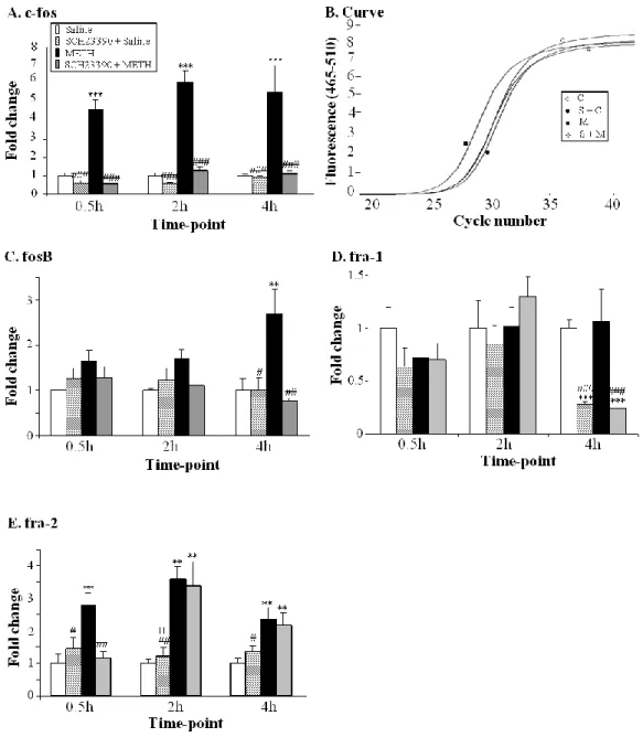
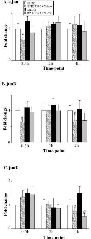
Neuronal responses to psychostimulants involve a regulatory mechanism of gene expression in different brain regions receiving monoamine inputs such as the striatum, cortex and hippocampus. These changes in gene expression represent molecular mechanisms underlying behavioral response, neuroadaptations and addiction to drugs of abuse (Berke and Hyman, 2000). Exposure to METH causes changes in the striatal expression of genes including immediate early genes (IEGs), ER stress-related genes and genes that participate in apoptotic events (Cadet et al., 2010b; Jayanthi et al., 2005; Jayanthi et al., 2009). IEGs include AP1 family of transcription factors that is composed of homodimers and heterodimers of basic region leucine zipper (bZIP) proteins that include cFos, FosB, Fra1, and Fra2, cJun, JunB, and JunD (Hughes and Dragunow, 1995). Several studies have demonstrated that METH-induced changes in these genes are dependent on DA D1
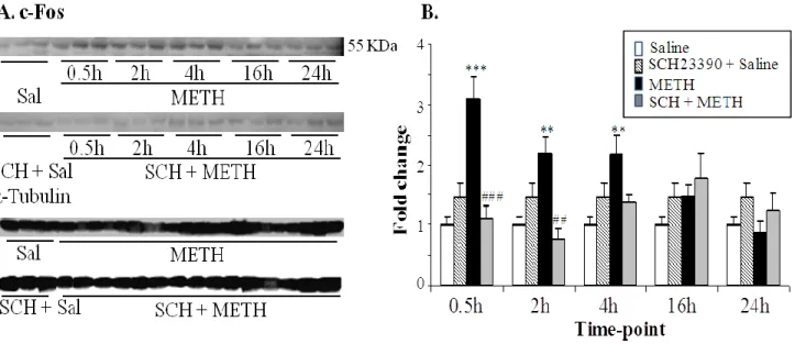
receptor-mediated phosphorylation of the transcription factor cAMP response-element binding protein (CREB). By example, METH caused rapid increases in the IEG cfos in rodent striatum (Cadet et al., 2001; Wang et al., 1995b), increases which were completely blocked by SCH23390 (Cadet et al., 2010b). The role of the transcription factor cFos in METH neurotoxicity has been investigated in different studies. Heterozygote and homozygote cfos knockout mice injected with binge METH regimen, showed exacerbation of DA terminals damage in the striatum characterized by severe decrease in DAT binding, DAT protein levels and TH immunoreactivity (Deng et al., 1999). In
23 addition, both genotypes of cfos knockout mice showed increased apoptotic DNA fragmentation in the striatum and cortex 3 days after METH treatment. Microarray analyses revealed that METH induced increases in the expression of several DNA repair genes such as APEX, PolB, LIG1, nibrin, DDB1 and DNA mismatch repair proteins MSH3 and PMS1, but these increases were absent in cfos knockout mice (Cadet et al., 2002). These reports support a protective role for cfos against METH damage, especially against DNA damage. METH treatment also causes upregulation of fosB in rodent striatum (Cadet et al., 2001; Cadet et al., 2010b). Binge METH injections (4x10 mg/kg, 2 hours intervals) administered to fosB -/- mutant mice caused increased number of
degenerated neurons in the cortex and striatum as determined by Fluoro-Jade B staining (Kuroda et al., 2010). In contrast to results obtained with cfos knockout mice (Deng et al., 1999), there was no excacerbation of striatal dopaminergic neuron degeneration or neuronal apoptosis in fosB mutant mice. This report suggests that fosB mediates neuroprotection against METH neurotoxicity as a negative feedback of calcium signaling and in supporting microglial function. Moreover, single METH injection (40 mg/kg) also caused substantial DA D1-dependent increases in the other
members of the fos family, fra1 and fra2, at 2 and 4 hours posttreatment (Cadet et al., 2010b; Jayanthi et al., 2009).
In contrast to the Fos proteins, the transcription factor cJun seems to mediate activation of pro-death signalings after METH administration. Single injection of METH (40 mg/kg) caused
significant increases in cjun, junB and junD mRNA of rodent striata which peaked at 2 hours after the drug injection (Cadet et al., 2001; Cadet et al., 2010b; Jayanthi et al., 2002). METH-induced increases in these genes were totally blocked by pretreatment with the D1 receptor antagonist,
SCH23390 (Cadet et al., 2010b). The single METH injection was also associated with increases in the protein expression of cJun, phosphorylated cJun at Ser63 and Ser73 and phosphorylated Jun-N-terminal kinases (JNK) at Thr183 and Tyr185 in mice striata (Jayanthi et al., 2002). Deng et al (2002) showed that cjun knockout mice exhibited a reduction in DNA fragmentation, caspase 3 activity and cleaved poly (ADP-ribose) polymerase (PARP) after METH treatment. However, deficit in cjun gene did not attenuate dopaminergic terminal damage (Deng et al., 2002b). These findings suggest that cjun only plays a role in the mediation of neuronal apoptosis in striatal cells. In addition, METH-induced phosphorylated cJun protein colocalized with markers of DNA fragmentation and cell death in mice striata (Deng et al., 2002b). Pretreatment with the JNK-specific inhibitor, SP600125, attenuated METH-induced JNK phosphorylation, activation of
24 caspase 3 and cell death in human neuroblastoma cells, while pretreatment with antioxidant vitamin E prevents METH-associated JNK phosphorylation (Wang et al., 2008). These findings suggest that oxidative stress-activated cJun/JNK signaling pathway is involved in METH-induced cell death.
METH treatment also affects the expression of the early growth response (Egr) family of IEGs, which groups Egr1 (Krox-1, NGF1A, Zif268), Egr2 (Krox20, NGF1B), Egr3 (Pilot), and Egr4 (NGF1C) (Beckmann and Wilce, 1997). Several studies have shown that METH caused induction of egr1 transcript in the striatum (Wang et al., 1995b). METH-induced egr1 transcript was
markedly attenuated in the CuZn SOD transgenic mice (Hirata et al., 1998). This finding suggests that oxidative mechanism mediate the induction of egr1 after METH treatment. Recently, our laboratory has demonstrated that METH also influences the expression of other members of the family. Single dose of 40 mg/kg of METH caused increases in egr1, egr2 and egr3 in rat striatum that were blocked by SCH23390 (Cadet et al., 2010b). It is now necessary to investigate the role of each gene in response to brain injury.
In summary, different regimen doses of METH administered to an animal model will cause specific striatal transcriptional profiles. The pattern of genes induced or repressed would be a molecular hallmark of activated signaling pathways and of synaptic plasticity in response to the drug.
1.9.2 Mechanisms of METH-induced activation of death pathways
Apoptosis plays a critical role in mammalian development; dysregulation of apoptotic processes have been documented in models of cancer formation, in diabetes, and in neurodegenerative
disorders (Cadet et al., 2007; Culmsee and Landshamer, 2006). As reviewed elsewhere (Cadet et al., 2007), we have demonstrated that METH-induced neuronal apoptosis involves a complex interaction between death pathways that have previously been identified in rodent models of neurodegenerative disorders (Culmsee and Landshamer, 2006). Because METH abusers show cognitive abnormalities and neurodegenerative changes in their brains (Ernst et al., 2000; Volkow et al., 2001d) and because identification of the processes involved might be of therapeutic value, we have been seeking to decipher the molecular and cellular mechanisms involved in METH-induced neurotoxicity.
1.9.2.1 Death receptor pathway
Apoptosis is mediated by various molecular pathways that cause the activation of several members of the cysteine-aspartic proteases (caspases) family of proteolytic enzymes (Schultz and
25 Harrington, 2003). Jayanthi et al (2005) have shown that single toxic dose of 40 mg/kg of METH causes neuronal apoptosis in rat striatum via activation of the FasL/Fas death pathway. Activation of this programmed cell death is initiated by binding of the tumor necrosis factor, Fas L to its receptor Fas. Fas contains a cytoplasmic death domain where Fas-associated death domain (FADD) can bind in presence of Fas L, and recruits procaspase 8 for subsequent activation in caspase 8. Activated caspase 8 then activates downstream caspases, such as caspase 3. Activated caspase 3 cleaves the inhibitor of caspase-activated deoxyribonuclease (ICAD)/ DNA fragmentation factor 45 (DFF45) and inhibits its binding to CAD. The latter now enters the nucleus and degrades the cell’s chromosomal DNA, leading to DNA fragmentation and cell death.
It has been shown that METH caused increases in Fas L mRNA and protein in striatal neurons that stained for GAD and enkephalin (Jayanthi et al., 2005). In addition, there was also evidence of METH-induced cleavage of procaspase 8 and induction of the effector caspase 3 that colocalized with Fas L. Pretreatment with SCH23390 blocked increases in caspases 8 and 3. Fas was also increased in the same cell as caspase 3. It appears that METH-induced activation of Fas L/Fas apoptotic pathway in the striatum depends on upregulation of calcineurin activity and activation of the transcription factors nuclear factor of activated T cells (NFAT) (Jayanthi et al., 2005).
Calcineurin, a calmodulin-dependent phosphatase, causes dephosphorylation of NFAT proteins and their translocation to the nucleus (Beals et al., 1997; Jain et al., 1993). NFATs bind to AP1 and Egr families of transcription factor to regulate the expression of Fas L (Li-Weber and Krammer, 2002). METH induced upregulation of calcineurin levels, nuclear shuttling of NFATc3 and NFATc4 that were blocked by SCH23390 (Jayanthi et al., 2005). In addition, In addition, METH caused
increases in different members of AP1 and Egr families. These findings suggest that the DA system via stimulation of DA D1 receptors is involved in activation of the Fas L/Fas death pathway, after
METH administration.
1.9.2.2 Mitochondrial dysfunction Mitochondria and energy metabolism
Mitochondria are the primary site of energy metabolism through the production of ATP
necessary for function and maintenance of eukaryotic cells (Dudkina et al., 2010). They are double-membraned organelles; the outer mitochondrial membrane is permeable to ions and small proteins of molecular weights < 10 kDa, while the inner mitochondrial membrane houses the multimeric
26 enzyme complexes of the electron transport chain. Although the mitochondria contain their own DNA, most of the mitochondrial proteins of respiratory chain complexes are imported and encoded by nuclear DNA.
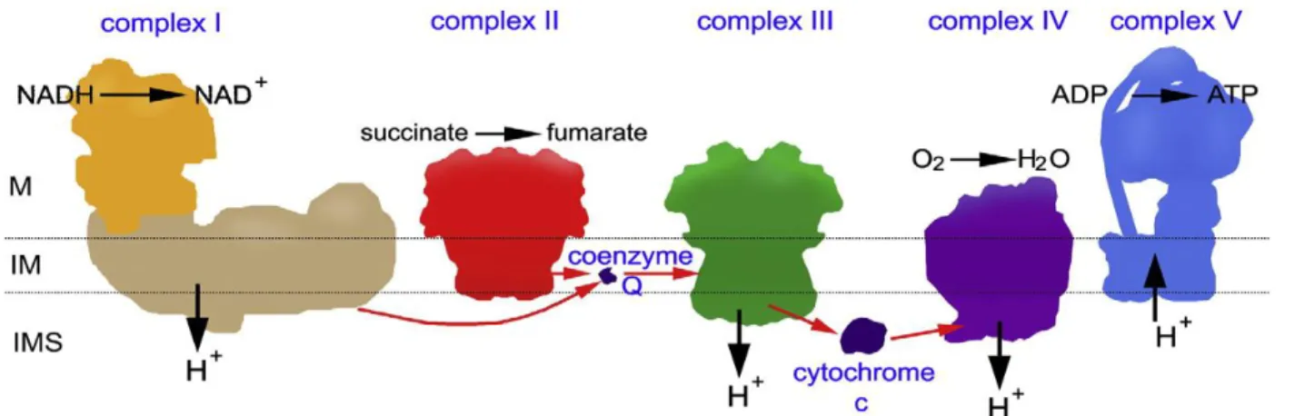
ATP production is usually carried out at the level of the inner mitochondrial membrane through oxidative phosphorylation involving the reduction of oxygen to water by multimeric enzyme complexes. The electron transport chain (ETC) is composed of five enzyme complexes (Figure 8): NADH-ubiquinone oxidoreductase (complex I), succinate dehydrogenase-CoQ oxidoreductase (complex II), cytochrome reductase (complex III), cytochrome oxidase (complex IV), and ATP synthase, which is sometimes referred to as complex V. During oxidative phosphorylation, ETC complexes are usually involved in reduction and oxidation reactions which generate a proton gradient across the inner mitochondrial membrane space that is used by the ATP synthase complex to synthesize ATP from ADP and inorganic phosphate Pi. The transfer of protons from the matrix to the inner mitochondrial membrane leads to the generation of a mitochondrial membrane potential ΔΨm of 150-180 mV.
Figure 8 Representation of the oxidative system. The position of the matrix (M), the
intermembrane space (IMS) and cristae or inner membrane (IM) has been indicated (Dudkina et al., 2010).
Mitochondria and apoptosis
The ETC is an important intracellular source of ROS. During the generation of ATP via oxidative phosphorylation by the ETC components, electrons can randomly interact with oxygen (O2) resulting in the partial reduction of O2 to form superoxide anion O2 -, a majority of which is