HAL Id: tel-02388086
https://tel.archives-ouvertes.fr/tel-02388086
Submitted on 1 Dec 2019HAL is a multi-disciplinary open access archive for the deposit and dissemination of sci-entific research documents, whether they are pub-lished or not. The documents may come from teaching and research institutions in France or abroad, or from public or private research centers.
L’archive ouverte pluridisciplinaire HAL, est destinée au dépôt et à la diffusion de documents scientifiques de niveau recherche, publiés ou non, émanant des établissements d’enseignement et de recherche français ou étrangers, des laboratoires publics ou privés.
Characterization of a MAPK module involved in
Arabidopsis response to wounding
Cecile Sözen
To cite this version:
Cecile Sözen. Characterization of a MAPK module involved in Arabidopsis response to wounding. Vegetal Biology. Université Paris Saclay (COmUE), 2017. English. �NNT : 2017SACLS472�. �tel-02388086�
NNT : 2017SACLS472
T
HESE DE DOCTORAT
DE
L’U
NIVERSITE
P
ARIS
-S
ACLAY
PREPAREE A
L’U
NIVERSITE
P
ARIS
-S
UD
ECOLE DOCTORALE N°567
Sciences du végétal : du Gène à l’Ecosystème
Spécialité de doctorat : Biologie
Par
Mlle Cécile SÖZEN
Characterization of a MAPK module involved
in Arabidopsis response to wounding
Thèse présentée et soutenue à Orsay (IPS2) le 29 novembre 2017 Composition du Jury :
Dr. Christian MEYER Directeur de Recherche - IJPB, Versailles Président du jury Dr. Claudia JONAK Directrice de Recherche - AIT, Vienne Rapportrice Dr. Debora GASPERINI Directrice de Recherche - LIPB, Halle Rapportrice Dr. Heribert HIRT Directeur de Recherche - KAUST,Thuwal Examinateur Dr. Jean COLCOMBET Chargé de Recherche - IPS2, Orsay Directeur de thèse Dr. Axel de ZELICOURT Maître de Conférences - IPS2, Orsay Invité
Characterization of a MAPK module involved in Arabidopsis response to wounding 2
LIST OF ABBREVIATIONS
ABA: Abscisic Acid
APS: Ammonium PerSulfate ATP: Adenosine Tri-Phosphate Cas9: CRISPR-Associated protein 9
CDPK: Calcium-Dependent Protein Kinase CHX: CycloHeXimide
CRISPR: Clustered Regularly Interspaced Short Palindromic Repeats C-terminal: Carboxyl-terminal DMSO: DiMethylSulfOxide DTT: DiThioThreitol ET: Ethylene EtOH: Ethanol GLR: Glutamate Receptor-like HA: HemAgglutinin
hpi: hours post-inoculation H2O2: Hydrogen peroxide JA: Jasmonic Acid
kDa: KiloDalton
MAPK: Mitogen-Activated Protein Kinase
MAP2K: Mitogen-Activated Protein Kinase Kinase
MAP3K: Mitogen-Activated Protein Kinase Kinase Kinase MBP: Myelin Basic Protein
meJA: methyl-Jasmonate NaF: Sodium Fluoride OG: OligoGalacturonide
PRR: Pathogen Recognition Receptor ROS: Reactive Oxygen Species
RT-qPCR: Reverse Transcription - quantitative Polymerase Chain Reaction SA: Salicylic Acid
SDS-PAGE: Sodium Dodecyl Sulfate - PolyAcrylamide Gel Electrophoresis TBS: Tris-Buffered Saline
TEMED: TEtraMEthyleneDiamine T-DNA: Transfer DNA
YFP: Yellow Fluorescent Protein Y2H: Yeast two Hybrid
Characterization of a MAPK module involved in Arabidopsis response to wounding 3
LIST OF FIGURES & TABLES
CHAPTER I: INTRODUCTION
Figure 1.1: Simplified model depicting cellular events triggered by wounding and herbivory – p.11 Figure 1.2: Biosynthetic pathway of Jasmonic Acid (JA) – p.19
Figure 1.3: Perception and signaling of Jasmonic Acid – p.22 Figure 1.4: Relationship of JA with other phytohormones – p.24 Figure 1.5: JA and systemic signaling – p.25
Figure 1.6: MAPK cascades are key elements of the stress signal transduction in plants – p.35 Figure 1.7: 20 MAPKs in Arabidopsis divided into 4 subgroups – p.36
Figure 1.8: MKK3, the sole member of group B – p.36
Figure 1.9: 20 MEKK-like MAP3Ks divided into 3 clades – p.37
Figure 1.10: PAMP-activated MAPK cascades illustrate the complexity of MAPK signaling – p.37 Figure 1.11: Biotic and abiotic stresses activating MKK3-dependent pathways – p.39
Figure 1.12: Identification of a complete MKK3-dependent module activated by ABA – p.40
CHAPTER II: RESULTS & DISCUSSIONS
Figure 2.1: MKK3 and sub-clade III MAP3Ks interact in yeast – p.46
Figure 2.2: Sub-clade III MAP3Ks, MKK3 and MPK2 constitute a functional module in protoplasts –p.47 Figure 2.3: Transcriptional regulation of MEKK-like MAP3Ks in Arabidopsis thaliana – p.48
Table 2.1: Main conditions under which sub-clade III MAP3Ks are regulated – p.48
Figure 2.4: General model based on the conserved transcriptional regulation of sub-clade III MAP3Ks – p.49 Figure 2.5: MPK2 is activated by wounding in an MKK3-dependent way – p.50
Figure 2.6: MPK1, MPK2 and MPK7 are not transcriptionally regulated by wounding – p.51
Figure 2.7: MPK1, MPK2 and MPK7 are activated by wounding in an MKK3-dependent way – p.51 Figure 2.8: MPK2 is not activated by gentle touch – p.52
Figure 2.9: MPK3 and MPK6 activation by wounding is not dependent on MKK3 – p.52 Figure 2.10: MPK3 and MPK6 have no effect on the activation of MPK2 by wounding – p.53
Figure 2.11: MPK1, MPK2 and MPK7 activation by wounding requires a de novo protein synthesis– p.53 Figure 2.12: CHX induces a constitutive activation of MPK3 and MPK6 – p.54
Figure 2.13: Expression of sub-clade III MAP3Ks upon wounding – p.55
Figure 2.14: Sub-clade III MAP3Ks protein accumulation upon wounding – p.55 Figure 2.15: Subcellular localization of MAP3K18-YFP upon ABA and wounding – p.56
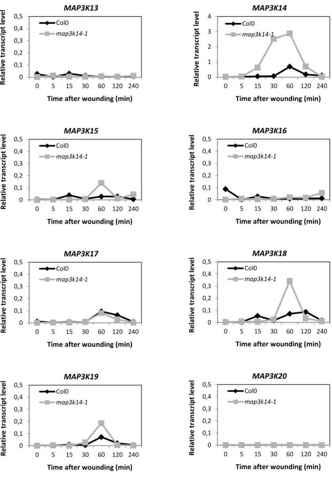
Table 2.2: Testing the wounding-induced activation of MPK2 in map3k mutants led to variable results – p.56 Figure 2.16: MPK2 activity upon wounding is increased in map3k14-1 plants compared to Col-0 – p.56 Figure 2.17: MPK3 and MPK6 activity upon wounding is unchanged in map3k14-1 plants – p.56 Figure 2.18: Some MAP3Ks expression is increased in map3k14-1 plants– p.56
Characterization of a MAPK module involved in Arabidopsis response to wounding 4
Figure 2.19: Truncated MAP3K14 is functional in vivo and leads to a more stable protein – p.57
Figure 2.20: MPK2 activity upon wounding is unchanged in CRISPR map3k14 plants compared to Col-0 p.57 Figure 2.21: Theoretical model depicting the two MAPK groups activated upon wounding – p.58
Figure 2.22: Theoretical model depicting the involvement of specific sub-clade III MAP3Ks depending on the
wounding-induced component – p.59
Figure 2.23: MPK2 is not activated by an exogenous treatment with H2O2, OGs, ATP or flg22 – p.60
Figure 2.24: MPK1, 2 and 7 are activated by JA in an MKK3-dependent manner – p.61 Figure 2.25: MPK3 and MPK6 are not activated by an exogenous treatment with JA – p.62 Figure 2.26: MPK2 activation by JA requires its perception by COI1 – p.62
Figure 2.27: MAP3Ks expression upon exogenous treatment with JA – p.63
Figure 2.28: JA-induced activation of MPK2 is reduced in some map3k mutants – p.63
Figure 2.29: MPK2 activation by wounding is not highly dependent on ATP, OGs or H2O2 signaling – p.64
Figure 2.30: JA, but not ABA, has a major role upstream of MKK3-MPK2 in response to wounding – p.64 Figure 2.31: JA does not have a major role upstream of MPK3/MPK6 upon wounding – p.64
Figure 2.32: Expression of candidate MAP3Ks upon wounding is reduced in coi1-16 plants – p.64 Figure 2.33: Expression of candidate MAP3Ks upon wounding is reduced in coi1-34 plants – p.65 Figure 2.34: MKK3-MPK2 is not activated in systemic leaves – p.66
Figure 2.35: Model depicting the JA-dependent MAPK module activated by wounding – p.67 Figure 2.36: MKK3-MPK2 is activated by B. cinerea – p.68
Figure 2.37: MAP3K19 is highly induced 48 hours after inoculation with B. cinerea – p.69 Figure 2.38: MKK3-MPK2 is activated by Spodoptera littoralis – p.70
Figure 2.39: MKK3-MPK2 is activated by S. littoralis and B. cinerea, two JA-producing pathogens – p.70 Table 2.3: Summary of results obtained with B. cinerea inoculations – p.71
Figure 2.40: Lesion outgrowth after inoculation with B. cinerea spores – p.71
Figure 2.41: Some Botrytis-induced genes expression is misregulated in mkk3-1 plants – p.72 Figure 2.42: MKK3 is not involved in resistance towards Spodoptera littoralis – p.73 Figure 2.43: MKK3 negatively regulates SA and JA accumulation after insect feeding – p.73 Figure 2.44: MKK3 is not involved in hormonal regulation upon wounding – p.74
Figure 2.45: Role of MKK3 in the regulation of genes involved in SA and JA biosynthesis pathways – p.75 Figure 2.46: Role of MKK3 in the regulation of SA- and JA-responsive genes – p.76
Figure 2.47: Wounding-induced trichome formation – p.77
Figure 2.48: Transcriptomic analysis of mkk3-1 plants submitted to wounding did not highlight any
MKK3-regulated genes – p.78
Table 2.4: Few MKK3-regulated genes were found – p.78
Figure 2.49: Theoretical model on the role of MKK3 in phytohormone regulation upon S. littoralis feeding -p.79
Characterization of a MAPK module involved in Arabidopsis response to wounding 5
CHAPTER III: CONCLUSIONS & PERSPECTIVES
Figure 3.1: Hypothetical model on the interdependent activation of two MAPK modules upon wounding – p.82
Figure 3.2: MPK7 is activated by nitrate in an MKK3- and NLP7- dependent manner – p.84 Figure 3.3: mkk3-1 plants show increased dormancy – p.90
CHAPTER IV: MATERIALS & METHODS
Table 4.1: Mutant lines mentioned in this manuscript – p.92
Table 4.2: Exogenous treatments applied on Arabidopsis plantlets – p.97 Table 4.3:Preparation of the enzymatic solution – p.98
Table 4.4:Composition of W5 and WI solutions – p.98
Table 4.5:Composition of the MMg solution – p.99
Table 4.6: Composition of the PEG solution – p.99
Table 4.7: Primers used to amplify loci for the creation of lines expressing tagged versions of MAP3Ks,
MKK3 and MAPKs – p.102
Table 4.8: Composition of the RT mix – p.103
Table 4.9: Genes used for qPCR experiments and corresponding primer couples – p.104 Table 4.10:Description of samples sent for micro-array analyses – p.104
Table 4.11:Dilutions of antibodies used for western blots – p.106
Characterization of a MAPK module involved in Arabidopsis response to wounding 6
TABLE OF CONTENT
CHAPTER I: INTRODUCTION ... 9
I. EARLY CELLULAR EVENTS TRIGGERED BY WOUNDING AND HERBIVORY ... 12
1. Sensing cell wall integrity and cell death ... 12
2. Ion fluxes and other second messengers ... 14
3. Production of Reactive Oxygen Species (ROS) ... 15
4. Damage-Associated Molecular Patterns (DAMPs) are intercellular signaling molecules produced upon wounding ... 16
5. JA is a central actor of plant responses to wounding and insects ... 18
6. Long-distance signaling ... 25
7. How to differentiate herbivory from mechanical wounding? ... 27
II. WOUNDING AND HERBIVORY FEEDING INDUCE A LARGE SET OF RESPONSES ... 29
1. A great transcriptional reprogramming largely dependent on JA ... 29
2. Various defense responses ... 31
III. MITOGEN-ACTIVATED PROTEIN KINASE (MAPK) MODULES ARE KEY SIGNALING ACTORS OF ENVIRONMENTAL PERCEPTION ... 35
1. MAPKs are important stress responsive modules in plants ... 35
2. Spare data suggest a complex MAPK role in wounding and herbivory interaction ... 41
IV. OBJECTIVES OF THE PHD WORK: UNVEILING THE FUNCTION OF MKK3 MODULES IN WOUNDING SIGNALING ... 43
1. MAPKs and signal transduction before this work, a state of knowledge ... 43
2. Objectives: characterizing the activation of a MAPK module upon wounding stress ... 44
CHAPTER II: RESULTS & DISCUSSIONS ... 46
I. TOWARD A GENERAL MODEL FOR THE ACTIVATION OF MKK3-RELATED MODULES ... 46
1. Identification of MAP3Ks able to interact with MKK3 using the yeast 2-hybrid system ... 46
2. Functional validation of MAP3Ks using a transient expression system ... 47
3. Sub-clade III MAP3Ks are strongly transcriptionally regulated by stresses ... 48
4. Discussion ... 49
II. IDENTIFICATION AND CHARACTERIZATION OF NEW MAPK MODULES ACTIVATED BY WOUNDING ... 50
1. Wounding activates an MKK3 module in Arabidopsis thaliana ... 50
2. The wounding-induced activation of the iconic MPK3 and 6 is not dependent on MKK3 ... 52
3. The hunt of upstream MAP3K(s) ... 53
Characterization of a MAPK module involved in Arabidopsis response to wounding 7
III. IDENTIFICATION OF SIGNALING ELEMENTS ACTING UPSTREAM MKK3 UPON WOUNDING
... 60
1. Exogenous application of some candidate second messengers can activate an MKK3-dependent module... 60
2. Wounding-induced activation of the MKK3-dependent module is disrupted in mutants of the JA signaling pathway ... 63
3. MKK3-MPK2 is not activated in systemic leaves ... 65
4. Discussion ... 66
IV. ACTIVATION OF AN MKK3-DEPENDENT MODULE BY CELL WALL-DAMAGING PATHOGENS ... 67
1. Botrytis cinerea activates an MKK3-dependent module in Arabidopsis thaliana 68 2. Spodoptera littoralis activates an MKK3-dependent module in Arabidopsis thaliana ... 69
3. Discussion ... 70
V. STUDY OF THE ROLE OF AN MKK3-DEPENDENT MODULE IN WOUNDING-RELATED RESPONSES ... 71
1. Plants impaired in MKK3 signaling do not show clear detectable phenotype upon Botrytis infection ... 71
2. Plants impaired in MKK3 signaling do not properly respond to herbivorous insects ... 73
3. Wounding-induced trichome formation ... 77
4. Transcriptomic analysis of mkk3-1 plants submitted to wounding did not highlight any MKK3-regulated genes ... 78
5. Discussion ... 79
CHAPTER III:CONCLUSIONS & PERSPECTIVES ... 81
I. AMAPK SIGNALING NETWORK ACTIVATED IN RESPONSE TO WOUNDING ... 81
1. Toward a sequential and interdependent activation of two MAPK modules ... 81
2. Spatial organization of MAPK activation upon wounding ... 82
II. A NOVEL MKK3-DEPENDENT MODULE ACTIVATED BY ENVIRONMENTAL CONSTRAINTS THROUGH TRANSCRIPTIONAL REGULATION OF MAP3K GENES ... 83
1. An emerging general working model… ... 83
2. …coexisting with other models? ... 85
3. Open questions about the functioning of sub-clade III MAP3Ks-MKK3-C-group MAPKs modules ... 85
III. A ROLE FOR MKK3 IN STRESS RESPONSES? ... 88
1. A well-conserved MAPK through evolution ... 88
2. A role in herbivory signaling? ... 88
3. Characterized phenotypes in other stress and developmental contexts ... 89
Characterization of a MAPK module involved in Arabidopsis response to wounding 8
CHAPTER IV: MATERIALS & METHODS ... 92
I. MATERIALS ... 92
1. Plant material ... 92
2. Culture media and conditions ... 93
3. Antibodies ... 93
4. Vector backbones ... 94
5. Buffers and solutions ... 95
II. METHODS ... 96
1. Plant methods ... 96
2. Molecular biology methods ... 100
3. Biochemistry methods ... 105
CHAPTER V: REFERENCES ... 107 ANNEX: Review - Convergence of Multiple MAP3Ks on MKK3 Identifies a Set of Novel Stress MAPK Modules
Characterization of a MAPK module involved in Arabidopsis response to wounding 9
CHAPTER I: INTRODUCTION
Agricultural context
Humans started to cultivate plants more than 10.000 years ago for their alimentation. Nowadays, 62% of crop production is intended for human food, 35% for animal feeding and 3% for bioenergy, seed and other industrial products (Foley et al., 2011). Thus it is clear that agriculture and food production are of great concern for people all over the world. International organizations such as the Food and Agriculture Organization of the United Nations (FAO) or the World Health Organization have developed strategies to prevent worldwide food crises and highlighted the urgent need for research and innovation in agricultural sciences. According to the FAO, the world population will reach 9.1 billion by 2050. In order to feed that number of people, food productivity will have to increase by 60-70% (www.fao.org, 2015). The increase of food productivity is also of great importance to fight poverty in many third-world countries. To reach this objective, different levels are explored. The first one consists in increasing overall plant productivity. Breeding has been performed since crop domestication but new tools coupled to a better knowledge of plant biology are available to accelerate the process. Another possibility will consist in reducing crop losses due to non-optimal growth conditions referred as “stresses”. Such stresses are generally classified in two categories. Abiotic stresses are caused by non-living entities such as drought, salt, heat, cold, whereas biotic stresses are caused by living organisms like bacteria, fungi, viruses or insects. Abiotic and biotic stresses are responsible for about 50% crop losses in the world (Ashraf et al., 2012). It is important to stress the fact that the impact of abiotic stresses is very likely to increase in the future due to global warming issues (increasing temperatures and CO2 emissions) (Intergovernmental Panel on Climate Change,
ipcc.ch). The effects of stresses on plants are diverse: reduction of biomass, decrease of fertility or simply death. It is thus important to find a way to increase crop tolerance to environmental stresses with the aim of improving their quantity and quality. Until now, strategies used to improve crop production have relied on a massive use of chemical products and great deforestations to create surfaces devoted to cultures. Yet these practices have huge impact on human health and environment (destruction of wildlife and plants). Today, the new goal is to develop a sustainable agriculture without impacting the environment. For this reason it became critical to develop new tools based on basic science. Studying and
Characterization of a MAPK module involved in Arabidopsis response to wounding 10
understanding how plant respond to their environment is an important step before being able to engineer plants more resistant to environmental stresses.
Plants are confronted to mechanical stimuli of diverse nature
Like many other organisms, plants are exposed to mechanical stimuli imposed by their environment that can range from a gentle breeze or the touch of neighboring objects during the growth of organs to a strong wounding caused by herbivore feeding or the accidental abscission of aged organs. The sensing of “non-wounding” mechanical stimuli have an important role in plant morphogenesis as it triggers developmental responses of the plant, the best illustrating examples being the shaping of a tree by the wind or the development of roots finding their way through obstacles in the soil (Monshausen & Gilroy, 2009). On the other hand, the wounding of plant tissues can have more detrimental effects as it can cause cell death and create entrances for pathogens thereby facilitating their development inside plant tissues. In the course of my PhD, I have been particularly interested in the mechanical stress caused by herbivorous insects.
Plants and herbivorous insects
Plants and insects have co-evolved for nearly 350 million years (Gatehouse, 2002). Some plant-insect interactions can be beneficial for the plant as in the case of pollination. In some cases, plants can even develop at the expense of insects. An amazing example is the important source of nitrogen that carnivorous plants living on very poor soils find in insects they can catch. Yet many insects have deleterious effects on the plant as in the case of attacks by herbivorous insects that can feed themselves on the targeted plant or deposit their eggs (oviposition). Herbivorous insect species are very diverse and possess different feeding strategies. The majority is composed of beetles and caterpillars that can damage leaves with their mouthparts by chewing, tearing or snipping (Fürstenberg et al., 2013). Sucking insects, like aphids, have specialized organs called stylets that can be inserted into the phloem to suck the liquid content. In this case, the wounding part of the interaction is reduced but aphids can form very large colonies which can have strong effects.
Plants have developed advanced response systems to fight and defend themselves against herbivorous insects. A first layer of defense is constitutive. Plants are naturally equipped with physical barriers like a cuticle layer, trichomes, and thorns. They also
DAMPs HAMPs
PEPR1
PEPR2 DORN1 WAK1/2
apoplast cytosol OGs ATP Pep1 Wounding Oral secretions nucleus Stress response JA and ET biosynthesis PEPR1/PEPR2 … ROS burst NADPHox H2O2 O2 O2- Ca2+
+
CAM CBL CDPK JA ABA ET SA MAPKs activation ?Figure 1.1: Simplified model depicting cellular events triggered by wounding and herbivory Wounding and herbivory can be perceived by the cell through the recognition of Damage-Associated Molecular Patterns (DAMPs) such as OGs, Pep1 or ATP that are self-derived molecules. Plants can also respond to Herbivore-Associated Molecular Patterns (HAMPs) found in insect oral secretions that are recognized through an unknown mechanism. Both DAMPs and HAMPs are able to trigger plant defense responses. Wounding leads to a rapid depolarization of the plasma membrane followed by an important influx of calcium and a burst of Reactive Oxygen Species (ROS). Those events lead to the de novo synthesis of phytohormones and in term to the regulation of stress-responsive genes. Plants will be able to defend themselves by activating specific defense responses in order to prevent further attacks or to repel herbivores.
Membrane depolarization Defense responses -physical barriers (trichomes…) -resistance to further attacks -callose deposition -production of volatile compounds
Characterization of a MAPK module involved in Arabidopsis response to wounding 11
accumulate secondary metabolites that can function against herbivores. But they also sense the presence of insects by detecting compounds of their oral secretions/oviposition fluids and by the mechanical damage caused by the insect feeding. In order to counteract insect effects, plants activate specific immune responses that are controlled by complex signaling pathways. One specific response is the emission of volatile compounds to repulse herbivores, attract their predators and alert neighboring organs and plants.
Mechanical wounding of plant tissues is an unavoidable consequence of herbivory, although the intensity and the extent of damage depend on the mode of feeding of the insect (chewing, sucking…). Insects feed on leaves by continuously wounding them and swallowing small pieces of tissue, simultaneously introducing saliva and other secretions in contact of plant cells. In order to better understand plant defense responses triggered by herbivores, most studies chose to apply a single mechanical wound by crushing or puncturing leaves. Although the major part of responses is similar to the ones observed upon insect attack, a single wound is often not enough to induce the emission of volatiles (Alborn et al., 1997).
Cellular events occurring upon wounding and herbivory include membrane depolarization, increase of cytosolic calcium levels, production of Reactive Oxygen Species (ROS), activation of Mitogen-Activated Protein Kinases (MAPKs) along with phosphorylation events and phytohormone accumulation. These cascades of intracellular events finally lead to a strong transcriptional reprogramming allowing the plant to raise defenses against the perceived stress (Savatin et al., 2014; Zebelo & Maffei, 2015). Those wound-triggered cellular events are described in the first part of the introduction and additional information about insect-induced responses is given when available (figure 1.1).
Arabidopsis, a model plant to study plant defense responses
Although studies on this subject have been conducted on different plants species including tobacco (Nicotiana attenuata), tomato (Lycopersicon esculentum), Lima bean (Phaseolus lunatus) and thale cress (Arabidopsis thaliana) closer attention will be paid to literature data provided by works performed on Arabidopsis. Indeed, the latter is commonly used in the laboratory and provides a wide range of advantages. Arabidopsis thaliana was first proposed to be used as a genetic model organism about 75 years ago and started to be widely used as such during the 1980s (Meyerowitz, 2001). The study of this plant has developed our understanding of plants in general but had also an impact on some aspects of
Characterization of a MAPK module involved in Arabidopsis response to wounding 12
research on animals and humans. Indeed, some important processes of human biology can be more easily studied in this model plant and many discoveries made using Arabidopsis were directly useful for human health issues such as cancer or Alzheimer’s disease (Jones et al., 2008). The use of Arabidopsis as a model offers advantages such as a small genome size (≈ 135 million base pairs), the convenience of crossing linked to a strict Mendelian inheritance, the large progeny of each plant (>10.000 seeds) and the short generation time (≈ 8 weeks from germination to mature seed production). Arabidopsis thaliana genome has been entirely sequenced and very well annotated (The Arabidopsis Genome Initiative, 2000). Mutations of desired target sequences have been largely facilitated by the development of various tools such as the collection of T-DNA insertion lines, the TILLING (Targeting Induced Local Lesions in Genomes) technology, the use of approaches based on RNA-silencing and more recently the CRISPR-Cas9 system based on the action of endonucleases aiming desired sites in the genome (Zhang et al., 2016). Large mutant collections are easily accessible to query using online tools.
I.
Early cellular events triggered by wounding and herbivory
1. Sensing cell wall integrity and cell death
Wounding caused by insect feeding or other mechanical stimuli is a very complex stress. It locally induces cell death on a variable area as well as a mechanical stimulation of the neighboring living cells. The first events and their relative importance in the initiation of wounding signaling processes are poorly known. In one hand, wounding-induced cell death triggers a strong loss of compartmentalization of cellular molecules such as enzymes or ions which may be involved in the initiation of wounding signaling (ATP, Ca2+…). On the other hand, living cells neighboring the wounding site are abruptly disturbed and probably react by the activation of intra and intercellular signaling pathways.
Wounding and herbivore feeding affect the integrity of cuticle and cell wall. Resulting breaches alert the plant of the presence of invading visitors. Among the warning signs are changes in mechanical properties and release of oligosaccharide fragments from the cell wall (Nühse et al., 2012). A loss of cell wall integrity (CWI) can be sensed by Receptor-Like Kinases (RLKs) such as THESEUS 1 but other sensors have been suggested based on studies conducted in yeast where a CWI-dependent pathway has been well characterized
Characterization of a MAPK module involved in Arabidopsis response to wounding 13
(Levin, 2011; Hamann, 2015). Similar sensing systems might be involved in the detection of wounding in plants. Additionally, in the case of a pathogen attack, cell wall-degrading enzymes such as polygalacturonase, pectate lyase and pectin methylesterase, which are released by the pathogen or produced by the plant and activated upon attack, come into play (Walton et al., 1994). Treatment of the plant cell wall with these enzymes can elicit defense responses (Walton, 1994). In most cases, responses are triggered by the recognition of the enzymatic products among which oligogalacturonides have been the most studied (Ferrari et
al., 2013).
Physical signals could also be generated by stretching of the plasma membrane that could be detected by stretch-activated enzymatic activities. There is much evidence from electrophysiological studies that the plasma membrane of plant cells contain a wide diversity of mechanosensitive (MS) ion channels. A simple touch of the leaf surface can be sensed through MS channels that can trigger further signal transduction events. Thus a soft mechanical stress at the leaf surface was shown to be enough to activate the expression of touch (TCH) genes as well as other cellular responses similar to the ones induced by a stronger wounding (ROS production, Ca2+ influx, resistance to pathogens) (Benikhlef et al., 2013). A number of MS channels activities have been functionally characterized in plant membranes and are good candidates to act as first sensors of mechanical stimuli (Peyronnet et
al., 2014; Haswell et al., 2008). Based on their homology to the bacterial MscS channel, the
10 MscS-Like (MSL) proteins of Arabidopsis thaliana have been hypothesized to form mechanosensitive channels in plant cell and organelle membranes. MS channels have the property to convert mechanical stimuli into electrophysiological (potential variation) or biochemical signals. Once they are activated and opened, they often lead to membrane depolarization and to the influx of second messengers such as Ca2+ ions. The link between the activation of MS channels and Ca2+ variation is still not clearly known (Hamilton et al., 2015). Some authors suggest the involvement of a MS calcium channel that could itself generate Ca2+ influx upon a mechanical stimulus. It seems that MSL permeability is more anionic than calcic (Haswell et al., 2008), but as a very low Ca2+ permeability might be enough to induce slight increases of cytosolic Ca2+, a direct role for MS channels in calcium-induced activities cannot be excluded. Alternatively, it is possible that voltage-dependent Ca2+ channels found on plasma membranes (Miedema et al., 2008) could be activated by the depolarization triggered by the opening of MS channels.
Characterization of a MAPK module involved in Arabidopsis response to wounding 14
2. Ion fluxes and other second messengers
As indicated earlier, the depolarization of the plasma membrane causes ion fluxes, the most important being Ca2+ ions as they have been shown to function as second messengers in many signaling pathways (Kudla et al., 2010). Plants can discriminate between various stimuli based on calcium signatures that are defined by their amplitude, frequency, subcellular localization and duration. Peaks of cytosolic Ca2+ are detected within 6 seconds in cells close to the injury site (Beneloujaephajri et al., 2013) and several studies showed that cytosolic calcium levels also increase upon herbivore attack (Arimura & Maffei, 2010). Very recently, the use of a fluorescent calcium biosensor has allowed observing elevations of Ca2+ levels during a biotic interaction with an aphid (Myzus persicae) (Vincent et al., 2017). Using appropriate mutant lines, authors were able to show that these Ca2+ elevations are dependent on BAK1 (Brassinosteroid insensitive-Associated Kinase 1), a well-known co-receptor of Pathogen Recognition Receptors (PRRs), as well as on GLR3.3 and 3.6 channels (GLutamate Receptor-like). In the same study, the vacuolar cation channel TPC1 (Two Pore Channel 1) was shown to importantly contribute to the observed cytosolic elevation. In another study, TPC1-dependent calcium elevations were observed upon feeding of the caterpillar Spodoptera
littoralis, both locally and systemically (Kiep et al., 2015). Inositol trisphosphate (InsP3) was
shown to mediate Ca2+ release from internal stores (Alexandre & Lassalles, 1990). It was recently shown, along with phosphoinositide, to act as a second messenger of wounding signal transduction and regulate the expression of wound-inducible genes (Mosblech et al., 2008). Interestingly, their production was shown to require the biosynthesis of JA as no increase of InsP3 was observed in plants deficient in JA synthesis.
In Arabidopsis, the calcium signal is decoded by three major multigenic families of Ca2+ sensor proteins: calmodulins or calmodulin-like proteins (CAMs), calcineurin B-like proteins (CBLs) and Calcium-Dependent Protein Kinases (CDPKs). Some of those sensors have been studied and were shown to have roles in plant immunity in response to bacterial pathogens but also herbivorous insects (Boudsocq & Sheen, 2013; Arimura & Maffei, 2010). In Arabidopsis, some CDPKs are activated by the bacterial elicitor flg22 and were shown to be important for the transduction of defense responses (Boudsocq et al., 2010). Two CDPKs, CPK3 and CPK13, were directly shown to act as positive regulators of the defense gene
PDF1.2 upon exposure to Spodoptera littoralis caterpillars (Kanchiswamy et al., 2010). A
Characterization of a MAPK module involved in Arabidopsis response to wounding 15
from S. littoralis and to connect Ca2+ to JA signaling (Vadassery et al., 2012). cml42 plants showed enhanced resistance to the caterpillar as well as higher expression levels of the JA-responsive gene VSP2. Another calmodulin-like protein, CML37, was shown to be involved in the regulation of the Jasmonic Acid (JA) pathway and in resistance to herbivory (Scholz et
al., 2014). Three TCH (TouCH) genes encoding calmodulin and calmodulin-like proteins
were shown to be induced by mechanical stimuli such as touch or wind, supporting the important role of calcium signaling in the transduction of such environmental cues (Braam & Davis, 1990). The role of Ca2+ sensor proteins in a context of wounding and/or herbivory was also demonstrated in other plants species. Thus, in tomato, LeCDPK2 is involved in the wound-induced ethylene (ET) production (Kamiyoshihara et al., 2010) and in tobacco (Nicotiana attenuata), NaCDPK4 and NaCDPK5 negatively regulate wounding responses and resistance to the herbivore Manduca sexta (Yang et al., 2012). Finally, in maize, ZmCPK11 is activated by wounding in a JA-dependent pathway and is involved in systemic wound responses (Szczegielniak et al., 2012).
In addition to calcium signaling, phospholipase D (PLD) has been suggested to play an important role in the mediation of wound-induced responses (Wang et al., 2000). PLD hydrolyzes phospholipids and leads to the production of phosphatidic acid (PA). PLD-induced hydrolysis was observed after wounding in bean leaves and appeared to be mediated by an increase in cytoplasmic Ca2+ concentrations (Ryu & Wang, 1996). Interestingly, PLD was shown to be required for the production of JA and the induction of a JA-regulated gene (Wang et al., 2000; Bargmann et al., 2009).
3. Production of
Reactive Oxygen Species (ROS)
Reactive Oxygen Species (ROS) are normally produced in peroxisomes, chloroplasts and mitochondria due to the presence of an oxidizing activity or electron transfer chains in these organelles (Tripathy & Oelmuller, 2012). In addition, ROS production can be induced upon stress conditions and create a ROS burst that is thought to have antimicrobial effects. Stress-induced ROS production was first described in 1983 in potato tubers infected with the oomycete Phytophthora infestans (Doke, 1983). Since this study, many works contributed to show that ROS can also be involved in other stresses including wounding and insect feeding (Suzuki & Mittler, 2012; Maffei et al., 2006).
Characterization of a MAPK module involved in Arabidopsis response to wounding 16
NADPH oxidases are well known producers of ROS (Marino et al., 2012). They transfer electrons from cytosolic NADPH (or NADH) to oxygen in the apoplast, thus leading to the production of superoxide ions (O2-). The latter is then converted to hydrogen peroxide
(H2O2) in the apoplast by the action of superoxide dismutase. NADPH oxidases belong to the
Respiratory Burst Oxidase Homolog (RBOH) family that has 10 members (RBOHA to RBOHJ) in Arabidopsis (Torres & Dangl, 2005). In response to wounding, the observed oxidative burst is believed to be due to NADPH oxidases. Indeed, in Arabidopsis seedlings, the rapid local and systemic ROS burst is impaired in rbohd mutants (Miller et al., 2009). In tomato (Solanum lycopersicum), the inhibition of NADPH oxidase by specific inhibitors blocks the wound-induced production of ROS and the induction of some defense genes. NADPH oxidases are also involved in wound-induced ROS burst in other plant species such as Medicago truncatula or potato (Soares et al., 2011; Bernards & Razem, 2001). In addition to their oxidase domain, NADPH oxidases also possess Ca2+ binding domains and it was demonstrated that wound-induced ROS burst is dependent on Ca2+ peaks (Monshausen et al., 2009; Beneloujaephajri et al., 2013). Besides NADPH oxidases, ROS can also be produced through the action of apoplastic peroxidases but no evidence was found for their action in wound-induced ROS production in Arabidopsis (Minibayeva et al., 2014). Concerning herbivory, it has been shown that H2O2 is released upon Spodoptera littoralis feeding on Lima
bean and, to a lesser extent, upon mechanical damage (Maffei et al., 2006).
4. Damage-Associated Molecular Patterns (DAMPs) are intercellular
signaling molecules produced upon wounding
Plants are able to sense their damaged self and trigger responses very similar to those induced by pathogen infection. Endogenous molecules deriving from wounded tissue can act like Damage-Associated Molecular Patterns (DAMPs) and elicit local and systemic plant defenses. In tomato, systemin, an 18 amino-acid peptide, is released in the apoplast after wounding or insect attack (Jacinto et al., 1997). Sensing of systemin leads to the biosynthesis of JA, an important wound phytohormone, that activate defense responses locally and in the neighboring cells (Orozco-Cardenas et al., 1993). Hydroxyproline-rich systemins have also been identified in other Solanaceae (potato, petunia, black nightshade) and are able to trigger plant immunity in response to herbivore attack. In Arabidopsis thaliana, three self-derived families of molecules have been identified so far. They are all recognized by
Pattern-Characterization of a MAPK module involved in Arabidopsis response to wounding 17
Recognition Receptors (PRRs) localized at the plasma membrane level. Oligogalacturonides (OGs) are released from the plant cell wall after degradation of homogalacturonan, the main component of pectin, through the action of wounding-induced hydrolytic enzymes such as polygalacturonase. The size of OGs is important and can determine their efficiency as eliciting molecules. OGs with a degree of polymerization between 10 and 15 are the most active ones (Ferrari et al., 2013). Wall-Associated Kinases (WAKs) are RLKs associated to the cell wall that are required for cell expansion during development but can also mediate stress responses induced by OGs released upon wounding. WAKs are encoded by 5 genes among which WAK1 and WAK2 are transcriptionally induced by wounding. In rice, OsWAK1 is also upregulated by wounding as well as salicylic acid (SA) and JA. By the use of a chimeric receptor approach, authors have shown the ability of WAK1 to bind OGs (Brutus et
al., 2010). WAK2 also seems to have a predominant role as wak2-1 plants are not able to
activate defense responses induced by OGs (Kohorn et al., 2009). Adenosine Tri Phosphate (ATP) can also act as an extracellular signaling molecule (Tanaka et al., 2014). It is released from plant cells in response to abiotic stresses (Dark et al., 2011), fungal elicitors and mechanical stimuli (Weerasinghe et al., 2009). Concentrations of ATP up to 40 µM were measured after wounding, probably released from neighboring cells whose plasma membrane had lost its integrity (Jeter et al., 2004). Recently the plant receptor of ATP has been identified in Arabidopsis as DORN1 (DOes not Respond to Nucleotides 1), a lectin receptor kinase (Choi et al., 2014). The characterization of dorn1 plants showed the specificity of this receptor for ATP as plants were not impaired in responses to other exogenous stimuli. Based on this observation, transcriptional responses to ATP were compared to those observed upon wounding. 60% of the genes induced by ATP were also induced by wounding and the expression of selected genes was found highly reduced in dorn1 plants after wounding, strongly suggesting that ATP is a major component of wounding response.
Pep1 is a 23 amino-acid peptide released from the C-terminal part of its precursor PROPEP1 (Yamaguchi & Huffaker, 2011). The latter constitutes a family with other 7 members that are all produced in response to wounding or pathogen perception, which means that unlike OGs and systemin, whose production does not require any gene regulation, genetically encoded peptides such as Pep1 are produced in a second wave of events to reinforce responses and/or mediate intercellular signaling. Pep1 is perceived by two Receptor-Like Kinases (RLKs), PEPR1 and PEPR2 (for Pep-Receptor1 and 2), which are functionally
Characterization of a MAPK module involved in Arabidopsis response to wounding 18
and structurally very similar to the RLKs FLS2 and EFR (receptors of bacterial elicitors flg22 and elf18 respectively) (Krol et al., 2010). PEPR1 and PEPR2 are transcriptionally induced by wounding and JA as well as upon herbivore attack and application of oral secretions (OS) (Klauser et al., 2015). Besides, the pepr1 pepr2 mutant shows increased sensitivity to
Spodoptera littoralis feeding as well as a reduction of JA accumulation upon application of
insect OS. Homologues of AtPep1 have been identified in maize; one of them, ZmPep3, triggers the biosynthesis of JA and ET and induces the production of volatile compounds (Huffaker et al., 2013).
DAMP signaling is very similar to the one induced by the recognition of PAMPs (Pathogen-Associated Molecular Patterns) that are pathogen-derived molecules, in opposition to the self-derived nature of DAMPs. First, they are recognized at the cell surface by PRRs (mostly receptor-like kinases) that are structurally very similar: a variable ectodomain potentially involved in ligand binding, a single transmembrane domain and an intracellular kinase domain, which is involved in the initiation of the intracellular signaling pathway (Couto & Zipfel, 2016). Secondly, the recognition of both DAMPs and PAMPs by the appropriate receptor leads to a cellular immune signaling involving the steps described earlier: rapid ion-flux changes at the plasma membrane level, increase of Ca2+ levels, production of ROS. In a coordinated manner, activation of phosphorylation cascades conveys immune signaling to the nucleus, resulting in strong transcriptional reprogramming to establish an immune response.
5. JA is a central actor of plant responses to wounding and insects
Phytohormones are small molecules used in various aspects of plant life ranging from development to stress responses. Jasmonic acid, the major component of jasmine scent, and its major derivative JA-Isoleucine (JA-Ile) help to regulate various aspects of plant biology, from stress responses to development. In non-stressed plants, JA is involved in senescence and reproductive development. Stress responses in which JA is involved include some abiotic stresses (drought, UV, ozone…) and mainly biotic stresses caused by insects or other microbial pathogens (Wasternack & Hause, 2013; Santino et al., 2013). Notably, JA has long been described as the major defense hormone against herbivores (Howe & Jander, 2008). Herbivores can activate the synthesis of JA in damaged host tissue and, in turn, JA can activate specific wound-induced defense responses (Campos et al., 2014). Most
herbivore-Figure 1.2: Biosynthetic pathway of Jasmonic Acid (JA) (from Acosta & Farmer, 2010) JA is synthesized from the fatty acid α-linolenic acid (18:3) (α -LeA) released from galactolipids of chloroplast membranes. After the action of several enzymes (LOXs, AOS, AOC), OPDA is obtained and is relocated to the peroxisome. The reduction of OPDA by OPR3 followed by several cycles of β-oxidations leads to the synthesis of JA which is then translocated to the cytosol where it can be modified to JA-Ile and other derivatives.
JA-Ile is then perceived by the SCF/COI1 complex and allows the expression of defense genes as well as genes encoding enzymes of its own synthesis.
Characterization of a MAPK module involved in Arabidopsis response to wounding 19
induced responses are also induced by treatments with JA. Moreover, wounding is sufficient to trigger increases of JA and JA-Ile, thus demonstrating that insect-derived molecules are not required to activate JA production (Koo et al., 2009). Mutants defective in JA biosynthesis or perception are highly susceptible to insect attack or other pathogens, highlighting the essential role of JA in defense (Thomma et al., 1998; Stotz et al., 2002). Hereafter is an overview of JA functions in the context of wounding and herbivory, including its biosynthesis, signaling, as well as some indications of crosstalks with other phytohormones.
a. J A hom eost asi s i n response to woundi ng and herbivor y
Jasmonates form a family of oxylipins deriving from enzymatic oxygenation of 18 and 16-carbon triunsaturated fatty acids (Wasternack & Kombrink, 2010). JA is synthesized from the fatty acid α-linolenic acid (18:3) (α-LeA) released from galactolipids of chloroplast membranes (figure 1.2). The enzyme responsible for the release of α-LeA was identified as phospholipase 1 (PLA1). α-LeA is then oxidized by a lipoxygenase (LOX) that is responsible for the insertion of an oxygen at the C-13 position of the acid, thus forming 13-HPOT. The genome of Arabidopsis includes 4 LOXs involved in JA synthesis upon wounding (LOX2, 3, 4 and 6). LOX2 was shown to be required for JA production proximal to the wound (Glauser
et al., 2009). However, JA and JA-Ile are still synthesized in the lox2-1 mutant suggesting the
involvement of other LOXs. More recent studies using combinations of multiple LOXs mutants revealed that all 4 LOXs contribute to JA synthesis upon wounding (Chauvin et al., 2013). LOX6 was found to have a particular role in early JA synthesis in distal leaves (Farmer
et al., 2014). The next step of the JA biosynthesis pathway leads to the formation of its
precursor cis-12-oxo-phytodienoic acid (OPDA). The enzymes responsible for this step are ALLENE OXIDE CYCLASE (AOC) and ALLENE OXIDE SYNTHASE (AOS). OPDA is then transported to the peroxisome where it is reduced to OPC-8:0 by 12-oxophytodienoate reductase (OPR3). Next, (+)-7-iso-JA is produced after several rounds of β-oxidations. Upon its subsequent transport to the cytoplasm, JA is modified to methyl-JA (meJA), JA-Ile or other derivatives. JA-Ile is formed by the action of the JAR1 enzyme (Jasmonic Acid Resistant 1) and is the most active form of JA as it seems to be the one that actually plays the major role in Arabidopsis leaves (Staswick & Tiryaki, 2004; Fonseca et al., 2009b).
At the histological level, jasmonate biosynthesis in Arabidopsis mainly takes place in the vascular bundles of the vegetative parts of the plant. However, some data suggest a
Characterization of a MAPK module involved in Arabidopsis response to wounding 20
production of JA in cells outside the vessels. For example, LOX2 is found strongly expressed in mesophyll cells (Montillet et al., 2013). The use of promoter::GUS lines allowed the observation of some genes encoding enzymes of the AOC family in Arabidopsis roots (Stenzel et al., 2012).
The Arabidopsis microarray datasets from various stress conditions and developmental stages revealed transcriptional regulation of all JA biosynthesis genes (Pauwels et al., 2009; van Verk et al., 2011). However, the activity of some enzymes seems to be post-translationally regulated. Thus, the activity of OPR3 seems to result from a monomer/dimer equilibrium (Schaller & Stintzi, 2009). Unstressed leaves of Arabidopsis contain very low amounts of bioactive JA, typically about 20-50 pmol/g fresh mass but this level increases within 5 minutes upon wounding (Glauser et al., 2008b; Glauser et al., 2009). In the wounded leaf JA levels increase up to 500-fold, reaching about 10 nmol/g fresh mass whereas JA-Ile levels reach less than 10% of the level of JA (Suza & Staswick, 2008). In more details, the first significant increase of JA in the wounded leaf is observed less than 30 seconds after wounding and its level doubles every 20 seconds during the first minute. Concerning JA-Ile, its first significant increase is observed 5 min after wounding (Koo et al., 2009). This remarkable speed of production suggests that all biosynthetic enzymes involved in production of JA/JA-Ile are already present in resting cells.
The molecular events linking wounding to the de novo synthesis of JA are largely unknown. Genetic and pharmacological disruption of cell wall integrity has been shown to constitutively activate the JA pathway (Hamann et al., 2009). There is also evidence suggesting that JA/JA-Ile are produced upon perception of DAMPs or bacterial effectors (Campos et al., 2014). In Arabidopsis, the perception of AtPep1 by its receptors PEPR1 and PEPR2 was shown to have a role in JA production as pepr1 pepr2 plants accumulated less JA upon herbivore oral secretion (Klauser et al., 2015). In maize, ZmPEPR3 activates JA synthesis by increasing the level of transcripts encoding the biosynthetic enzymes AOC and AOS (Huffaker et al., 2006). The application of ATP induces the expression of genes encoding enzymes for JA biosynthesis in Arabidopsis (Song et al., 2006; Choi et al., 2014). The responses induced by OGs were however shown to be independent from JA signaling (Ferrari et al., 2007). The exact mechanism by which DAMP signaling leads to JA biosynthesis still remains elusive. Among the intracellular events that could link the perception of self-derived elicitors to JA production are Ca2+ ions, ROS, MAPK cascades and
Characterization of a MAPK module involved in Arabidopsis response to wounding 21
CDPKs (Arimura & Maffei, 2010; Zebelo & Maffei, 2015). For instance, Ca2+ fluxes and associated CDPKs exert a control during the activation of JA biosynthesis genes (Bonaventure et al., 2007). However, it is not clear whether any of the enzymes involved in the biosynthesis of JA are regulated by CDPK/MAPK-dependent phosphorylation or cellular redox changes, although JA-induced phosphorylation of JA signaling components has been observed (Zhai et al., 2013).
In addition to JA-Ile, diverse conjugated and oxidized derivatives of JA are produced. These forms of jasmonates include hydroxyhasmonates (HOJAs) and HOJA-Ile as well as dicarboxyjasmonate (HOOCJA-Ile). HOJAs and HOOCJA-Ile accumulate in midveins of wounded leaves (Glauser et al., 2008) and were shown to be products of JA catabolism (Heitz et al., 2012; Koo et al., 2012). These derivatives being less active than JA-Ile in promoting the binding of COI1 to JAZ proteins, they participate to the attenuation of JA responses. Enzymes involved in JA-Ile catabolism are also rapidly induced upon wounding, herbivory and exogenous JA treatments (Bhosale et al., 2013). This co-expression of catabolism genes with other JA-response genes reflects the tight control of JA signaling.
b. J A si gnaling t hrough CO I1 -JAZ-MYC si gnali ng m odule
In 1994, the first JA resistant mutant coi1 (coronatine insensitive 1) was isolated and shown to be impaired in the receptor of JA-Ile (Feys et al., 1994). In Arabidopsis, plants impaired in COI1 do not respond to external JA application. In addition, biochemical approaches showed direct binding of JA-Ile (or its structural mimic coronatine) to COI1 (Yan
et al., 2009). COI1 is an F-box protein containing 18 leucine-rich repeat (LRR) that forms a
complex with SCF (Skp1/Cullin/F-box), an E3 ubiquitin ligase which mediates protein ubiquitination for targeted degradation by the 26S proteasome (Xie et al., 1998). SCF/COI1 defines a complex specifically involved in jasmonate signaling (Xu et al., 2002). Mutants lacking a functional COI1 are compromised in all known JA-regulated processes and do not respond to exogenous JA, as shown in tobacco, tomato and Arabidopsis (Paschold et al., 2007; Li et al., 2004; Xie et al., 1998). Interestingly, the accumulation of JA-Ile was shown to be dependent on COI1 as wound-induced JA levels were far lower in coi1 plants than in wild-type ones (Chung et al., 2008). Jasmonate ZIM-Domain (JAZ) proteins that form a family of 13 members in Arabidopsis are negative regulators of the transcription of JA-responsive genes (Thines et al., 2007). The absence of DNA-binding domains in JAZ proteins suggests
Figure 1.3: Perception and signaling of Jasmonic Acid (from Acosta & Farmer , 2010)
a) In the absence of JA-Ile, the repressor JAZ is bound to transcription factors of the MYC family, thus restraining their action.
b) When JA-Ile is produced in necessary amounts, it binds to the SCF/COI1 complex, allowing the latter to recruit JAZ repressors and promote their ubiquitination for further degradation by the proteasome. MYC factors are then set free and can activate the expression of defense genes.
Characterization of a MAPK module involved in Arabidopsis response to wounding 22
that the negative regulation is probably not performed by direct binding to gene promoter sequences but rather by interfering with transcription factors. Indeed, JAZ proteins can bind to many transcription factors and repress their activity. Among them the basic helix-loop-helix (bHLH) transcription factors MYC2, MYC3 and MYC4 have been extensively studied (Fernandez-Calvo et al., 2011). In the absence of JA-Ile, JAZ proteins are stable and repress the action of MYCs, and thus the expression of the downstream MYC-dependent genes, by recruiting two co-repressors TPL (TOPLESS) and NINJA (NOVEL INTERACTOR OF JAZ) and by competing with MED25 (MEDIATOR 25) for interaction with MYCs (figure 1.3). The myc2 myc3 myc4 triple mutant was shown to be highly susceptible to Spodoptera
littoralis feeding (Schweizer et al., 2013). JA-Ile binding to COI1 promotes ubiquitination of
JAZ proteins and their subsequent degradation by proteolysis (Chini et al., 2007). Protein-protein interaction assays showed that the COI1-JAZ interaction is stimulated by JA-Ile but not by meJA or OPDA (Thines et al., 2007). Poly-ubiquitination of JAZ proteins has been indirectly observed using proteomic approaches (Nagels et al., 2016) and their proteasome-dependent degradation was demonstrated by the use of MG132 treatments that stabilized JAZ proteins (Thines et al., 2007).
Interestingly, the expression of JAZ repressors is induced upon wounding and herbivory (Chung et al., 2008). More particularly, the JAZ10 gene is a well-described early marker for JA signaling in wounded leaves (Yan et al., 2007; Acosta & Farmer, 2010). The authors showed that this induction was correlated with the accumulation of JA and JA-Ile and moreover that it was dependent on COI1. Although most of JAZ proteins were shown to be rapidly degraded upon JA-Ile perception, some studies also showed a relative stability of some JAZ proteins following fluctuating JA-Ile levels (Chung et al., 2010). This negative feedback loop probably acts to restrain the duration and amplitude of JA-triggered responses. However, JA is also involved in a so-called positive feedback loop where it can stimulate its own synthesis by inducing MYC2-dependent expression of genes like LOX2, OPR3, AOS and
AOC (Browse, 2009). The wound-induced expression of AOS and OPR3 was shown to be
COI1-dependent (Devoto et al., 2005).
c. J A crosst al k with other st ress related ph yt ohormones
Plant pathogens are usually divided into biotrophs and necrotrophs based on their lifestyles. Biotrophic pathogens feed on living tissue whereas necrotrophs kill the host tissue
Characterization of a MAPK module involved in Arabidopsis response to wounding 23
and feed on dead residues. While it is generally admitted that JA has a role in defense against necrotrophic pathogens and herbivorous insects, SA rather activates resistance against biotrophic pathogens (Thomma et al., 1998). At first glance, SA and JA have antagonistic roles in plants as mutants impaired in one of them display enhanced expression of marker genes of the other (Kloek et al., 2001; Glazebrook, 2005). However, the reality is more complicated. For instance, the hemi-biotrophic bacteria Pseudomonas syringae induces both JA and SA signaling pathways in tomato (Stout et al., 1999).
NPR1 was described as the receptor of SA. When SA levels increase, NPR1, which is present as a homodimer, is “monomerized” and goes to the nucleus where it can regulate the expression of specific genes. The JA/SA antagonism principally occurs at the transcriptional level where SA can suppress the induction of JA-dependent genes (Spoel et
al., 2003; Caarls et al., 2015). The well-known JA marker gene PDF1.2 is under the control
of several transcription factors among which ORA59 from the ERF family (Ethylene Responsive Factor) that is activated by ethylene (ET) and JA. ORA59 was shown to be degraded by SA thus blocking the expression of PDF1.2. This suppression of PDF1.2 expression still occurs in coi1-1 mutants showing that the SA/JA crosstalk is independent on COI1 and is likely to occur downstream. Moreover, redox signaling was shown to be important for SA-induced suppression of JA responses. High SA levels lead to a reduced redox state. Inhibition of glutathione synthesis, a reducing agent, blocked the SA-mediated antagonism of PDF1.2 expression. However, the results of JA and SA crosstalk can vary with hormone concentrations and the type of stimuli (Mur et al., 2006; Spoel et al., 2007).
There are evidence of a JA/SA crosstalk in response to wounding. First, external application of aspirin and SA can block JA biosynthesis in wounded tomato leaves and thus inhibit the expression of some wound-induced genes (Peña-Cortés et al., 1993). Moreover, SA was shown to inhibit the JA-induced production of proteinase inhibitor (PI) in wounded tomato leaves (Doares et al., 1995). In addition, the SA-inducing pathogen Hyaloperonospora
arabidopsidis was shown to strongly suppress JA-mediated defenses that are activated by
caterpillars of Pieris rapae (Koornneef et al., 2008). Less is known about the effect of JA on SA. However, SA-mediated defense responses seem to be enhanced in JA mutants, as demonstrated in wounded tobacco leaves (Niki et al., 1998). Very recently, JA was shown to induce the expression of EDS5, a gene involved in SA biosynthesis, upon elicitation with flg22 (Mine et al., 2017). The same study showed that JA also represses the expression of
Figure 1.4: Relationship of JA with other phytohormones
JA acts in synergy with ABA for the activation of the MYC branch and with ET for the activation of the ERF branch. The antagonism of SA and JA occurs at the transcriptional level where SA inhibits the action of the ORA59 transcription factor from the ERF family thus inhibiting the expression of PDF1.2.
PDF1.2
ORA59 SA
ORA59 = ERF family MYC2
VSP2, LOX2
EIN3 ERF EIL1 JA ABA ET PDF1.2 MYC branch ERF branch
Characterization of a MAPK module involved in Arabidopsis response to wounding 24 PAD4, a positive regulator of EDS5 expression. Overall, the study points out the complexity
of immune signaling networks and hormonal crosstalks.
In the context of herbivores, SA and JA were shown to mediate defense responses towards Spodoptera littoralis, the Egyptian cotton worm (Stotz et al., 2002). By analyzing
coi1 and npr1 mutants in Arabidopsis, authors showed that SA and JA have opposite effects
on plant. Moreover, exogenous application of SA was able to reduce JA amounts induced by the insect. Another study showed this inhibition of JA-induced resistance by SA in plants challenged by the herbivore Spodoptera exigua (Cipollini et al., 2004). Using a phloem-feeding insect (Bemicia tabaci) another study showed that Arabidopsis plants accumulate SA-responsive defense genes both locally and systemically (Zarate et al., 2006). On the contrary, JA-responsive genes were repressed or not affected by the insect challenge. Moreover, the insect development was boosted in plants constitutively activating SA defenses (cim10, for
Constitutive IMmunity 10) and plants impaired in JA defenses (coi1). Finally a more recent
study showed that different patterns of phytohormones production can be observable depending on the herbivore species (Diezel et al., 2009).
JA-induced responses are usually described as being dependent on two different branches (figure 1.4). MYC2 is the master regulator of the MYC branch that is co-regulated by JA and abscisic acid (ABA) and leads to the activation of the downstream marker genes
VSP2 and LOX2 (Lorenzo et al., 2004; Vos et al., 2013). The ERF branch is co-regulated by
JA and ET and involves transcription factors of EIN3, EIL1, and ERF families such as ERF1 and ORA59. This branch activates the downstream marker gene PDF1.2 (Zhu et al., 2011). Like JA, ET and ABA generally antagonize SA responses.
ET is produced in response to various stress conditions including herbivore attacks or mechanical injury and can modulate the expression of specific ET-regulated genes but also plant physiology (von Dahl & Baldwin, 2007). In Arabidopsis thaliana, the production of ET was observed during interaction with the lepidopteran Pieris rapea and the homopteran Myzus
persicae (De Vos et al., 2005). Since then, several studies have elucidated the signaling
pathway of ET from the sensing by its receptors to the downstream signaling components and the ethylene-responsive genes (Ju & Chang, 2015). ET can act both positively and negatively on the immune response. In the case of an attack by necrotrophic pathogens, ET acts in synergy with JA for the control of the signaling branch dependent on ERF transcription factors whereas it antagonizes the MYC-dependent branch. It can also affect SA responses.
Figure 1.5: JA and systemic signaling (Modified from Nature Protocols - Farmer lab)
Wounding of a leaf leads to the production of JA and JA-Ile in distal leaves within a restricted area of the plant. Consequently, JAZ repressors are also degraded in distal leaves and transcriptional regulations can occur. For instance, JAZ10, a marker of JA signaling is found induced in distal leaves. JAZ10 JA JA JA JA JA JAZ10 JAZ10 JAZ10
Characterization of a MAPK module involved in Arabidopsis response to wounding 25
For example, when plants are treated with both ET and SA, the SA-induced expression of
PR1 (Pathogenesis-Related 1) is strongly amplified (De Vos et al., 2006). On the other hand,
ET-responsive transcription factors EIN3 and EIL1 were shown to repress the expression of SA biosynthesis gene SID2 leading to a reduced SA accumulation (Chen et al., 2009). Finally, ET also plays an important role in the JA/SA interaction as it was shown to be able to stop the SA-mediated suppression of JA responses (Leon-Reyes et al., 2010a). Indeed, when ET and JA are produced prior to SA, the antagonistic effect of SA is abolished. This event is no more observable when an inhibitor of ET is used, confirming its important role.
ABA is the major drought and salt hormone. However, a production of ABA was observed in potato and tomato plants after wounding (Peña-Cortés et al., 1995). In
Arabidopsis thaliana, ABA was shown to affect the expression of herbivore-induced genes
and JA biosynthesis (Bodenhausen et al., 2007). ABA acts in synergy with JA to induce MYC2-dependent gene expression during wound responses (Anderson et al., 2004). It was also shown to affect JA-inducible defense responses in maize (Erb et al., 2009) and resistance to herbivores in tomato (Thaler et al., 2004). These data suggest that the regulation of ABA levels in response to herbivory or wounding can modulate JA-induced defense responses. Conversely, JA signaling can have a positive effect on ABA signaling by inducing genes, such as PYL4 and PYL5 that encode proteins of the ABA receptor family as it was observed in Arabidopsis and tobacco (Lackman et al., 2011). Plants mutated in those genes are more sensitive to JA-induced growth inhibition but less sensitive to JA-induced anthocyanin accumulation. Thus, JA-inducible responses depend on ABA and vice versa. In Arabidopsis, ABA and SA response mechanisms also affect each other at multiple levels from biosynthesis to signal transduction pathways (Yasuda et al., 2008).
6. Long-distance signaling
The events described above start locally at the wounding site but can spread systemically throughout the plant, the aim being to alert the rest of the plant of the imminent danger. The existence of a systemic signaling molecule was first reported in tomato in 1972 (Green & Ryan, 1972). JA and other members of the oxylipins family related to JA were shown to be crucial components of the systemic wound signal. Indeed, wounding of Arabidopsis leaves results in a rapid accumulation of JA-Ile in distal leaves (Koo et al., 2009) (figure 1.5). The events leading to jasmonate accumulation distal to wounds can be reduced