Université de Montréal
Transforaminal versus intra-articular facet steroid
injections for the treatment of cervical radiculopathy: a
randomized, double-blinded, controlled study.
par
Nathalie J Bureau MD
Département de radiologie, radio-oncologie et médecine nucléaire Faculté de Médecine
Mémoire présenté à la Faculté des études supérieures et postdoctorales en vue de l’obtention du grade de
Maîtrise en Sciences biomédicales, option musculo-squelettique
Avril, 2014
Université de Montréal
Faculté des études supérieures et post-doctorales
Ce mémoire intitulé:
Transforaminal versus intra-articular facet steroid injections for the treatment of cervical radiculopathy: a randomized, double-blinded, controlled study.
présenté par : Nathalie J Bureau MD
a été évalué par un jury composé des personnes suivantes :
Monsieur Jeffrey Chankowski MD, président-rapporteur Monsieur Jacques de Guise Ing., PhD, directeur de recherche
Madame Nicola Hagemeister Ing. PhD, co-directrice Madame Josée Dubois MD MSc, membre du jury
i
Résumé
Les infiltrations foraminales cervicales sont associées à un risque de complications neurologiques majeures. Cette étude compare l’efficacité des infiltrations facettaires, plus sécuritaires, à celle des infiltrations foraminales dans le traitement de la cervico-brachialgie secondaire à une spondylose et/ou à une hernie discale, à 4 semaines post traitement.
Cinquante-six sujets ont été randomisés pour recevoir une infiltration foraminale (15 hommes, 13 femmes ; âge moyen 52 ans) ou facettaire (8 hommes, 20 femmes ; âge moyen 44 ans). L’issue principale était l’intensité de la douleur mesurée sur une échelle visuelle analogique (0 – 100). Les issues secondaires étaient le Neck Disability Index et le Medication
Quantitative Scale.
Suivant les analyses en intention-de-traiter et en intention-du-protocole, pour un score de douleur initial moyen, une réduction significative de l’intensité de la douleur a été observée avec les infiltrations facettaires [45.3% (95%CI: 21.4; 69.2) et 37.0% (95%CI: 9.2; 64.7)] contrairement aux infiltrations foraminales [9.8% (95%CI: +11.5; 31.2) et 17.8% (95%CI: +6.6; 42.2)]. Les infiltrations facettaires ont procuré une amélioration cliniquement (mais non statistiquement) significative du Neck Disability Index [24.3% (95%CI: +2.9; 51.5) et 20.7% (95%CI: +6.2; 47.6),], contrairement aux infiltrations foraminales [9.6% (95%CI: +15.2; 34.4) et 12.8% (95%CI: +11.2; 36.7)]. Les infiltrations facettaires étaient au moins aussi efficaces que les infiltrations foraminales pour un score initial de douleur ≤ 60, alors que l’analyse de non infériorité n’était pas concluante pour un score initial ≥ 80, de même que pour le Neck
Disability Index. Les infiltrations n’ont pas été associées à une réduction du score de Medication Quantitative Scale.
Les infiltrations facettaires sont efficaces dans le traitement de la névralgie cervico-brachiale et représentent une alternative valable et plus sécuritaire aux infiltrations foraminales.
Mots-clés : Cervico-brachialgie, rachis cervical, hernie discale, spondylose, intervention
Abstract
Transforaminal corticosteroid injections can be performed in the management of cervical radiculopathy but carry the risk of catastrophic complications. This study compares the efficacy of transforaminal and facet corticosteroid injections at 4 weeks post treatment.
We randomly assigned 56 subjects to receive CT-guided transforaminal (15 men, 13 women; mean age 52 years; range 28 – 72 years) or facet (8 men, 20 women; mean 44 years; range 26 – 60 years) injections. The primary outcome was pain severity rated on a visual analog scale (0-100). Secondary outcome measures were the Neck Disability Index and the Medication Quantitative Scale.
In the intention-to-treat and as-treated analyses, for a mean baseline score, facet injections demonstrated a significant pain score reduction of 45.3% (95%CI: 21.4; 69.2) and 37.0% (95%CI: 9.2; 64.7), while transforaminal injections showed nonsignificant pain score reduction of 9.8% (95%CI: +11.5; 31.2) and 17.8% (95%CI: +6.6; 42.2). While facet injections demonstrated an improvement in Neck Disability Index score of [24.3% (95%CI: +2.9; 51.5); 20.7% (95%CI: +6.2; 47.6),] as opposed to transforaminal injections [9.6% (95%CI: +15.2; 34.4); 12.8% (95%CI: +11.2; 36.7)], the results did not reach statistical significance. Noninferiority of facet to transforaminal injections was demonstrated for baseline pain score ≤ 60, while noninferiority analysis was inconclusive for baseline pain score ≥ 80 and for the Neck Disability Index score. Neither intervention showed a significant medication intake score reduction over time.
Facet injections are effective for the treatment of cervical radiculopathy and represent a valid and safer alternative to transforaminal injections.
Keywords: Cervical spine, cervical radiculopathy, cervical disc herniation, cervical
spondylosis, cervical foraminal stenosis, spine injections, transforaminal steroid injections, intra-articular facet steroid injections, spine intervention, pain management.
iii
Table of Contents
Résumé ... i!
Abstract ... ii!
Table of Contents ... iii!
List of Tables ... vi!
List of Figures ... vii!
List of Appendices ... ix!
List of Abbreviations and Symbols ... x!
Dedication ... xi!
Acknowledgements ... xii!
Introduction ... 1!
Chapter 1. Cervical radiculopathy ... 4!
1.1. Epidemiology ... 4!
1.2. Anatomy ... 5!
1.3. Physiopathology ... 11!
1.4. Clinical symptoms and signs ... 13!
1.5. Differential diagnosis ... 14!
1.6. Imaging ... 15!
1.7. Treatment ... 16!
Chapter 2. Transforaminal steroid injections ... 19!
2.1. Efficacy ... 19!
2.2. Technique ... 21!
2.3. Potential complications ... 24!
2.3.1. Vertebral artery injury ... 25!
2.3.2. Intravascular injections of corticosteroids ... 25!
2.3.2.1. The role of the technique ... 26!
2.3.2.2. The role of corticosteroids ... 27!
2.4. Alternative techniques to transforaminal corticosteroids injections ... 28!
2.4.1 Interlaminar epidural corticosteroid injections ... 28!
Chapter 3. Hypothesis and Objectives of the Study ... 31!
3.1. Hypothesis... 31!
3.2. Primary objective ... 31!
3.3. Secondary objective ... 31!
Chapter 4. Transforaminal versus intra-articular facet steroid injections for the treatment of cervical radiculopathy: a randomized, double-blinded, controlled study. .. 32!
4.1. Authors ... 32!
4.2. Manuscript accepted pending revisions in the American Journal of Neuroradiology AJNR ... 32!
4.2.1. Abstract ... 34!
4.2.2. Abbreviation key ... 35!
4.2.3. Introduction ... 36!
4.2.4. Materials and Methods ... 37!
4.2.4.1. Subjects ... 37!
4.2.4.1.1.Recruitment ... 37!
4.2.4.1.2. Enrollment in the study ... 38!
4.2.4.1.3. Randomization ... 38!
4.2.4.2. Interventions ... 39!
4.2.4.3. Outcome measures ... 41!
4.2.4.4. Follow-up time points ... 41!
4.2.4.5. Statistical analysis ... 42!
4.2.5. Results ... 43!
4.2.5.1. Clinical efficacy of IFSI and TFSI ... 47!
4.2.5.2. Analysis of group differences in efficacy ... 48!
4.2.6. Discussion ... 51!
4.2.7. Conclusions ... 55!
4.2.8. Acknowledgments ... 56!
4.2.9. References ... 57!
4.3. Published manuscript ... 60!
Chapter 5. Correlation of contrast distribution patterns with pain severity outcome ... 60!
v
5.2. Material and Methods ... 61!
5.2.1. Subjects ... 61!
5.2.2. CT scan image analysis ... 61!
5.2.3. Statistical analysis ... 62!
5.3. Results ... 63!
5.3.1. Frequency of contrast distribution patterns following intra-articular facet and transforaminal steroid injections ... 63!
5.3.2. Clinical efficacy of the contrast distribution patterns following intra-articular facet and transforaminal steroid injections ... 68!
5.3.3. Analysis of contrast distribution patterns differences in clinical efficacy ... 69!
5.4 Discussion ... 71!
Conclusion ... 72!
Bibliography ... 73!
List of Tables
Table I: Physical findings associated with cervical radiculopathy ... 14!
Table II: Efficacy of transforaminal corticosteroid injections: non-controlled studies ... 20!
Table III: Size of corticosteroid particles, comparison with red blood cells (7.5 – 7.8 µm) ... 28!
Table IV. Baseline characteristics of subjects (per randomization) treated either with
transforaminal or intra-articular facet corticosteroid injections. ... 46!
Table V. Pain severity scores for intra-articular facet and transforaminal corticosteroid
injections according to the baseline score. ... 48!
Table VI. Frequency of contrast distribution patterns following intra-articular facet and
transforaminal steroid injections ... 63!
Table VII. Clinical efficacy of contrast distribution patterns for a mean baseline VAS pain
score ... 69!
Table VIII. Difference in clinical efficacy between the contrast distribution patterns following
vii
List of Figures
Figure 1. Spinal cord and nerves. ... 5!
Figure 2. Cervical spinal nerves. ... 6!
Figure 3. Intervertebral foramen. ... 7!
Figure 4. Vascular supply to the cervical spinal cord. ... 8!
Figure 5. Anatomy of the facet joint. ... 9!
Figure 6. Retrodural space of Okada. ... 10!
Figure 7. Degenerative cervical spondylosis. ... 11!
Figure 8. Magnetic resonance imaging of a cervical disc herniation. ... 12!
Figure 9. Combination of disc herniation and spondylosis. ... 16!
Figure 10. Fluoroscopically guided transforaminal corticosteroid injection. ... 21!
Figure 11. CT guided transforaminal corticosteroid injection. ... 23!
Figure 12. A) CT-guided transforaminal corticosteroids injection. The needle is positioned in the posterolateral aspect of the foramen with contrast media flowing in the foramen. B) CT-guided intra-articular facet corticosteroids injection. The needle is positioned in the facet joint. Contrast media injection confirms an intra-capsular distribution. ... 40!
Figure 13. Flow diagram of the progress of subjects through the phases of the study ... 44!
Figure 14. Relative visual analog scale (VAS) pain score differences between intra-articular facet (IFSI) and transforaminal (TFSI) corticosteroid injections, according to VAS baseline value with adjustment for age, gender, and employment status. The intention-to-treat (A) and as-intention-to-treated (B) analyses are presented. Error bars indicate two-sided 95% CI. Squares indicate mean difference. Dotted vertical line marks the 15% margin of noninferiority. LCL = lower confidence limit; UCL = upper confidence limit. ... 49!
Figure 15. Medication quantitative scale (MQS) scores over time adjusted for age, gender, and employment status, for the intra-articular facet (IFSI) and transforaminal (TFSI) corticosteroid injections groups. The intention-to-treat (A) and as-treated (B) analyses are presented. Error bars indicate two-sided 95% CI. ... 51!
Figure 17. Intra-articular facet injection with contrast spreading in the retro-ligamentous
space, also called retrodural space. ... 65!
Figure 18. Juxta-articular facet injection. ... 66!
Figure 19. Transforaminal injection with extra-foraminal extension of contrast. ... 66!
Figure 20. Transforaminal injection with contrast spreading in the foraminal, extra-foraminal
and epidural spaces. ... 67!
Figure 21. Transforaminal injection with contrast spreading in the foraminal and epidural
ix
List of Appendices
Appendix I. Research Ethics Committee approval ... i!
Appendix II. Consent form ... ii!
Appendix III. Eligibility criteria ... xiv!
Appendix IV. Eligibility / Exclusion criteria ... xv!
Appendix V. Neck Disability Index ... xvi!
Appendix VI. Visual Analog Scale ... xix!
Appendix VII. Notification of manuscript acceptance pending revisions from AJNR. ... xx!
Appendix VIII. Permission to use copyrighted material in a thesis granted by the American Society of Neuroradiology. ... xxi!
Appendix IX. Published manuscript : Transforaminal versus intra-articular facet steroid injections for the treatment of cervical radiculopathy: a randomized, double-blind, controlled study. Reproduced with permission from licensed content publisher American Society of Neuroradiology and all co-authors. ... xxii!
List of Abbreviations and Symbols
ANCOVA = Analysis of covariance
ANOVA = Analysis of variance
CI = Confidence interval
CT = Computed tomography
G = Gauge
IFSI = Intra-articular facet steroid injections ILSI = Interlaminar steroid injections
LIO = Laboratoire de recherche en Imagerie et Orthopédie
MDCT = Multidetector computed tomography
mg = Milligram
mL = Milliliter
MN = Minnesota
MQS = Medication Quantitative Scale
MRI = Magnetic resonance imaging
NDI = Neck Disability Index
NSAIDs = Nonsteroidal anti-inflammatory drugs TFSI = Transforaminal steroid injections
µm = microns
VA = Vertebral artery
xi
Acknowledgements
My most humble and sincere thanks to:
Gilles Soulez MD, MSc who, through several years of sustained efforts and resilience, has shaped the University of Montreal, Department of Radiology Research Program. The program gave me the opportunity to embark on this improbable journey; Gilles gave me his unconditional support.
Jacques de Guise ing., PhD for his enthusiasm for collaborative research. Jacques welcomed me into the Laboratoire de recherche en Imagerie et Orthopédie (LIO) and helped to foster the idea of this Master of Science degree.
Nicola Hagemeister ing., PhD for her management skills and her philosophical approach to research and to life in general. Nicola gave me the opportunity to collaborate at the forefront on her research projects during the past five years, and has taught me much.
The entire team at the LIO for its warm welcome, its creativity, and collaborative efforts on behalf of my research projects, with very special thanks to Alexandre Fuentes, Caroline Lau, Gerald Parent, Laurence Marck and Youssef Ouami.
My resourceful research assistant Assia Belblidia, for her dedication and for expertly navigating through the recruitment of the participants, the planning of the interventions, the communication between the investigators and the collection of data. Without her, this randomized controlled study would not have materialized.
My co-investigators and co-authors Thomas Moser MD MSc, Bernard E Leduc MD, Jehane Dagher MD, Paul Brassard MD MSc, Maria Li MD CM MSc, and Daniel Shedid MD MSc, for their long-term commitment and brilliant expertise.
My statistician, Anne-Sophie Julien, for her expertise and for constantly providing perspective along the way.
Manon Choinière PhD for her generous support and for sharing her critical and knowledgeable observations with me on various dimensions of research.
Introduction
Cervical radiculopathy is a debilitating condition that affects approximately one person per 1 000 of population per year. Most patients will respond favorably to medical treatment, which may include rest, the use of analgesics and/or nonsteroidal anti-inflammatory drugs (NSAIDs), physical therapy and corticosteroid injections. Surgery is indicated when red flags such as, signs of myelopathy and deleterious sensory or motor impairment have been identified, or when medical treatment has failed. The prognosis following medical or surgical treatment is generally good to excellent. Although debilitating, cervical radiculopathy is not a life-threatening condition. Therefore, the expected benefits of our therapeutic interventions should outweigh significantly the risk of complications.
In recent years, catastrophic neurological complications following transforaminal steroid injections (TFSI) have been reported. TFSI are used to treat cervical radiculopathy on the basis of several non-controlled observational studies, which have reported encouraging results, but their efficacy has yet to be demonstrated in randomized controlled trials. As musculoskeletal radiologists who perform spinal interventions, we were faced with a dilemma:
Should we continue performing TFSI in view of the potential risk of serious neurological complications?
Preliminary work by Kim et al. in 2005, had suggested that intra-articular facet steroid injections could be effective in patients with cervical disc herniations1. Anatomically, the facet joint ventral recess is in close proximity to the spinal nerve root. Therefore, using a facet joint injection approach to deliver corticosteroids in the vicinity of the injured spinal nerve root seem to be a viable alternative to the riskier transforaminal approach.
These considerations prompted me to design a study to test the hypothesis that intra-articular facet steroid injections (IFSI) would be at least as effective as TFSI at treating cervical radiculopathy. My objective was to design a randomized controlled study that would yield good-quality data that could impact on the clinical management of patients with cervical radiculopathy.
With the help of my co-investigators, I submitted a grant application to the Fonds de recherche du Québec – Santé and I was awarded, as principal investigator, a $50,000 grant (FRQ-S, grant # 21230-2; 2010-2013) to conduct this study. Later on, I was also awarded an $8,000 grant from the Fonds Académique du Département de Radiologie, Radio-oncologie et Médecine nucléaire de l’Université de Montréal (2012 – 2013) to complete the research project. From the conception and design of the study, to the drafting of the manuscript, and the submission of the final manuscript to the American Journal of Neuroradiology (AJNR), I was responsible for the coordination of the entire research project and I take responsibility for the integrity of the work as a whole.
The Research Ethics Committee of the Centre hospitalier de l’Université de Montréal approved our study (Appendix I) and all participants received written and verbal information and gave their written consent (Appendix II). The documents pertaining to the Eligibility criteria, Exclusion criteria, Neck Disability Index questionnaire and Visual Analog Scale that were used in the study are presented in the Appendices III to VI respectively.
My co-investigators played essential roles in the realization of this study:
My colleague, Thomas Moser MD MSc is a musculoskeletal radiologist at the Centre hospitalier de l’Université de Montréal and a specialist of spinal interventions. He participated in the analysis and interpretation of the available literature, and in the conception and design of the study. Dr Moser and I performed all the CT-guided interventions and we retrospectively reviewed, in consensus the imaging data.
Bernard E Leduc MD is a physiatrist at the Centre hospitalier de l’Université de Montréal and a long-time collaborator. He participated in the conception and design of the study and in the recruitment and clinical evaluation of the subjects.
Paul Brassard MD MSc is a clinical epidemiologist at the McGill University Health Center. He is also a long-time collaborator. He participated in the conception of the study, and provided advice on the statistical design. He was responsible for generating the randomization sequence of the interventions.
3
Jehane Dagher MD is a physiatrist at the Institut de réadaptation Gingras-Lindsay-de-Montréal. Daniel Shedid MD MSc is a neurosurgeon at the Centre hospitalier de l’Université de Montréal. Maria Li MD CM MSc is a neurosurgeon at the Hôpital Maisonneuve-Rosemont of Montreal. These specialists participated in the recruitment and clinical evaluation of the subjects.
As co-authors of the manuscript, all these specialists also participated in the interpretation of the data, in critical revision of the manuscript and they approved the final manuscript.
Chapter 1. Cervical radiculopathy
1.1. Epidemiology
Cervical radiculopathy refers to a group of symptoms and signs caused by the dysfunction of a cervical nerve root. It typically presents with neck pain radiating to the upper limb with a combination of sensory disorder, altered reflexes or motor weakness in the anatomical distribution of the affected spinal nerve 2. In the population-based retrospective cohort study from Rochester, Minnesota (MN), from 1976 to 1990, the annual age-adjusted incidence of cervical radiculopathy was approximately 1 in 1000 people, with a male to female ratio of 1.7 3. The peak incidence was at 50 to 54 years of age with an average incidence of 203 per 100,000 people. In 1996, the authors of another epidemiological study on cervical radiculopathy conducted in a Sicilian municipality reported a prevalence of 3.5 cases per 1000 people 4. In both of these studies, the incidence of cervical radiculopathy decreased significantly after the age of 60 years 3, 4.
In Radhakrishnan’s study, irritation of the nerve roots was caused by cervical spondylosis, a disc herniation or both in 68% of the cases, and by a disc herniation in 22% of the cases. Only 15 % of the patients reported a history of trauma or physical exertion preceding the onset of symptoms. The most frequently involved nerve root was C7 in 46% of the cases, followed by the C6 nerve root in 18% of the cases and by the combined involvement of the C5 and C6 nerve roots in 10% 3.
Data on the natural history of cervical radiculopathy are still relatively scarce 5-8. According to the data from Rochester, MN, during the median duration of follow-up of 4.9 years, recurrence of cervical radiculopathy occurred in 31.7% of the cases and 26% of the patients underwent surgery 3. Long-term prognosis in patients treated either conservatively (analgesics, anti-inflammatory drugs, physical therapy) or with surgery, is equally good with improvement in pain, neurological symptoms and functional status, although pain relief occurs more rapidly in patients treated with surgery 6.
5
1.2. Anatomy
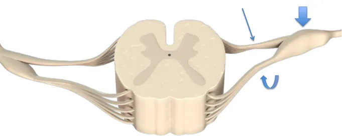
The cervical spinal nerves are constituted by the reunion of the ventral and dorsal nerve roots that emerge from the spinal cord (Figure 1). As it progresses through the intervertebral foramen, the dorsal nerve root enlarges to form the spinal ganglion proximal to joining the ventral nerve root to form the spinal nerve. The spinal nerve exits the spinal canal through the intervertebral foramen above the vertebra, which bares the same number. Hence, the C7 spinal nerve exits through the C6-C7 foramen and the C8 nerve exits through the C7-T1 foramen (Figure 2).
Figure 1. Spinal cord and nerves.
3D illustration of the spinal cord demonstrates the dorsal nerve root (curved arrow) that enlarges to form the spinal ganglion (thick arrow) before joining the ventral nerve root (long arrow) to form the spinal nerve. www.Anatomy.TV by Primal Pictures.
Figure 2. Cervical spinal nerves.
Coronal 3D illustration of the cervical spine shows the dorsal ganglions (yellow segments) and spinal nerves (pink segments) passing obliquely infero-laterally to their exit through the intervertebral foramina. www.Anatomy.TV by Primal Pictures.
The intervertebral foramen has an anterolateral orientation relative to the coronal plane. It is bounded superiorly and inferiorly by the pedicles of the adjacent vertebrae. The posterolateral aspect of the uncovertebral joint forms the anterior wall of the foramen, while the facet joint forms its posterior wall. The lateral margin of the foramen is demarcated by a line joining the anterolateral vertebral body and the lateral aspect of the facet joint. The vertebral artery (VA), which originates from the subclavian or innominate artery, runs in a caudad-to-cephalad direction immediately in front of the external opening of the intervertebral foramen and enters, in most cases, the foramina transversaria at C6 9.
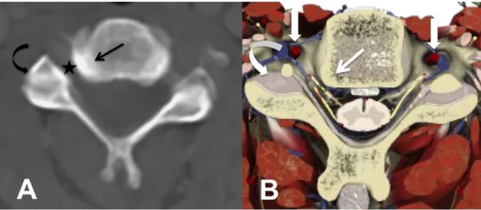
7
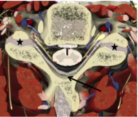
Figure 3. Intervertebral foramen.
Axial CT image (A) and corresponding illustration (B) of a cervical vertebra demonstrate the intervertebral foramen (star), bordered anteriorly by the uncinate process (long arrows) and posteriorly by the facet joint (curved arrows). The illustration (B) also shows the ventral and dorsal nerve roots emerging from the spinal cord and coursing through the intervertebral foramen. The vertebral arteries (thick arrows) are seen within the foramina transversaria, in front of the external opening of the intervertebral foramina. www.Anatomy.TV by Primal Pictures.
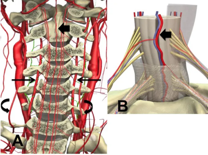
The nerve root is enclosed within its dural sleeve and runs within the lower half of the foramen while the epiradicular veins occupy the superior half. The spinal arterial branches originate from the vertebral, the ascending cervical, the deep cervical or the superior intercostal arteries 10. These spinal arterial branches traverse the intervertebral foramen alongside the spinal nerves and continue as radicular arteries, which in turn will join the anterior and posterior spinal arteries to supply the spinal cord 11. The majority of these vessels traversing the intervertebral foramen occur in the lower levels of the cervical spine. Although the radicular arteries are most commonly located inferiorly and anteriorly to the spinal nerve roots, a wide anatomic variation in the origin and location of these vessels exists. Hence, it has
been demonstrated that branches from the ascending and deep cervical arteries course within the posterior aspect of the intervertebral foramen and occasionally participate to the anterior and posterior vascular supply to the spinal cord 12.
Figure 4. Vascular supply to the cervical spinal cord.
Coronal illustration (A) of the cervical spine depicts the vertebral arteries (long arrows), deep cervical arteries (curved arrows) and ascending arteries (shown in green color) coursing in a caudad-to-cephalad direction. These arteries have several branches including the spinal arteries, which run through the intervertebral foramina alongside the spinal nerve roots, as shown on the 3D illustration of the spinal cord (B), to join the anterior and posterior spinal arteries that supply the spinal cord. www.Anatomy.TV by Primal Pictures.
9
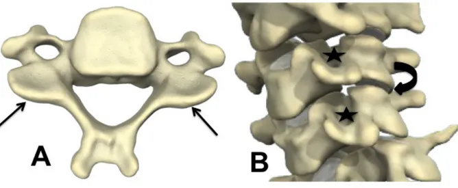
The cervical facet joint (also called zygapophyseal joint) forms the posterior limit of the intervertebral foramen. It is a diarthrodial joint formed by the articulation of the inferior articular process (facet) of the superior vertebra and the superior articular process (facet) of the inferior vertebra at each spinal level. The articular processes arise from the lateral mass of the vertebra at the confluence between the lamina and the pedicle. A fibrous capsule, lined by a synovial membrane, surrounds the joint and the articular surfaces are covered by hyaline cartilage. From C3-C4 through C8-T1, the facet joints are innervated by the medial branches of the dorsal rami of the spinal nerves above and below the joint 13.
Figure 5. Anatomy of the facet joint.
3D illustration of a cervical vertebra (A) demonstrates the articular facets (long arrows). 3D illustration of the cervical spine (oblique view) (B) shows the facet joint space (curved arrow). Note that the facet joints form the posterior limit of the intervertebral foramina (stars) where the spinal nerves exit. www.Anatomy.TV by Primal Pictures.
The facet joint can communicate with the interlaminar region, interspinous region and contralateral facet joint via an extradural space located dorsal to the ligamentum flavum. As cited by Murthy et al. 14, this potential space is called the retrodural space of Okada after the physician who described it in 1981 15. This potential space can be a pathway for the dissemination of contrast agent, corticosteroids, infection and tumor.
Figure 6. Retrodural space of Okada.
3D illustration of a cervical vertebra demonstrates the facet joint capsule (stars) in continuity with a potential anatomical space (long arrow) located between the ligamenta flava (grey linear band) anteriorly and the spinous process posteriorly. www.Anatomy.TV by Primal Pictures.
11
1.3. Physiopathology
In the vast majority of cases, cervical radiculopathy is the result of degenerative cervical spondylosis, a disc herniation or a combination of both. Other causes such as infection, tumor or trauma are infrequent. Cervical spondylosis occurs from degenerative disc disease, which starts with the dessication of the nucleus pulposus. With age, the nucleus pulposus loses water, proteins and mucopolysaccharides and consequently, becomes smaller and stiffer. As the annulus fibrosus assumes more of the weight bearing charge, radial and concentric tears develop leading to subsequent disc bulging and loss of disc height 16 (Figure 7).
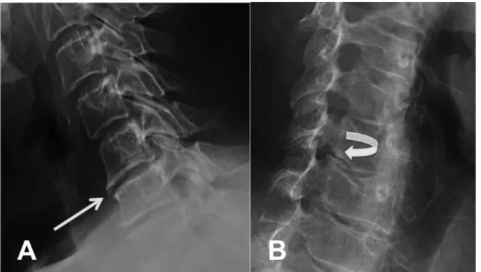
Figure 7. Degenerative cervical spondylosis.
(A) Lateral view radiograph of the cervical spine shows degenerative disc disease at the C5-C6 level characterized by loss of intervertebral disc height and anterior marginal osteophytes (long arrow). (B) Oblique view radiograph depicts marginal osteophytes arising from the uncinate processes (curved arrow) and protruding into the right C5-C6 neural foramen.
Loss of disc height occurs preferentially on the ventral aspect of the disc, at least initially. Segmental instability ensures which in turn leads to the development of osteophytes at the anterior margin of the vertebral body. As the intervertebral space decreases, the height of the neural foramina also diminishes causing stenosis. With altered biomechanics, degenerative changes also occur at the uncovertebral joints and the facet joints, with joint space narrowing and osteophyte formations, which protrude into the neural foramina compromising the diameter of the foramen and encroaching upon the nerve root ganglion.
Disc herniations, which result from nucleus pulposus material protruding through a radial tear and beyond the margin of the disc may occur alone or be part of the degenerative changes of cervical spondylosis (Figure 8).
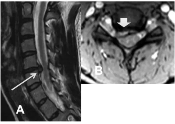
Figure 8. Magnetic resonance imaging of a cervical disc herniation.
(A) Sagittal view of the cervical spine demonstrates herniated disc material protruding into the spinal canal (long arrow) at the C5-C6 level. (B) Corresponding axial view shows the disc herniation (short arrow) indenting the spinal cord.
13
Both degenerative discs and herniated disc material can produce mediators of inflammation such as matrix metalloproteinases, nitric acid, interleukin-6 and prostaglandin E2 17. These biochemical agents play an integral role in tissue degradation and inflammation.
Ultimately, two major mechanisms can potentially cause irritation to the nerve root: (1) nucleus pulposus material secondary to a disc herniation leaking onto the nerve root and (2) compression of the nerve by degenerative changes occurring at the intervertebral disc level, at the uncovertebral joints and/or at the facet joints. Furthermore, either of these pathophysiological mechanisms will induce 2 processes in the nerves: (1) an inflammatory reaction and, related to this (2) changes in ion-channel functioning thought to cause hyperexcitability and dysfunction of the nerve root ganglion responsible for radicular pain 18.
1.4. Clinical symptoms and signs
Cervical radicular pain should be distinguished from cervical radiculopathy, a disorder, which entails objective sensory disturbances, diminished or absent reflexes and motor dysfunction corresponding to the nerve root(s) involved. Patients may experience sharp pain, tingling or a burning sensation in the involved area. Two examination maneuvers that may help confirm involvement of a nerve root at the level of the foramen have been described 19.
Shoulder abduction relief sign: the patient holds the arm over his or her head, typically
resting the wrist or forearm onto the top of the head. A positive sign is relief of the radicular symptoms. Spurling’s sign: while holding his or her neck in extension, the patient turns his head toward the affected side. A positive test exacerbates the radicular symptoms.
In a large retrospective study of patients who had surgery for cervical radiculopathy, Henderson et al. reported the clinical presentation in 736 patients: 99.4% had arm pain, 85.2% had sensory deficits, 79.7% had neck pain, 71.2% had reflex deficits, 68% had motor deficits, 52.5% had scapular pain, 17.8% had anterior chest pain, 9.7% had headaches, 5.9% had anterior chest and arm pain and 1.3% had left-sided chest and arm pain 20. Neurologic symptoms and signs corresponded with the affected disc level and nerve root, in approximately 80% of the patients.
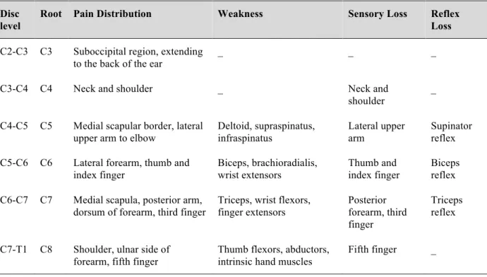
Findings on physical examination vary depending on the intervertebral disc level and nerve root(s) that are involved. These findings, adapted from Carette et al. 2 and Rao et al. 21 are summarized in Table I.
Table I: Physical findings associated with cervical radiculopathy Disc
level
Root Pain Distribution Weakness Sensory Loss Reflex
Loss
C2-C3 C3 Suboccipital region, extending to the back of the ear
_ _ _
C3-C4 C4 Neck and shoulder _ Neck and shoulder
_ C4-C5 C5 Medial scapular border, lateral
upper arm to elbow
Deltoid, supraspinatus, infraspinatus Lateral upper arm Supinator reflex C5-C6 C6 Lateral forearm, thumb and
index finger Biceps, brachioradialis, wrist extensors Thumb and index finger Biceps reflex C6-C7 C7 Medial scapula, posterior arm,
dorsum of forearm, third finger
Triceps, wrist flexors, finger extensors Posterior forearm, third finger Triceps reflex C7-T1 C8 Shoulder, ulnar side of
forearm, fifth finger
Thumb flexors, abductors, intrinsic hand muscles
Fifth finger _
1.5. Differential diagnosis
Other entities that may cause neck and arm pain including, intraspinal or extraspinal tumor, angina, reflex sympathetic dystrophy, infection, peripheral entrapment syndromes, thoracic outlet syndrome, brachial neuritis, rotator cuff and shoulder pathology, must be differentiated from cervical radiculopathy 16. Another common mimicker of cervical radiculopathy is cervical facet joint syndrome that may result from traumatic, inflammatory or degenerative processes 22. Studies have shown that each facet joint produces a distinct pattern of pain with a characteristic distribution 23-25. Hence, cervical facet syndrome may cause axial neck pain radiating to the occipital region, the posterolateral cervical region and the scapulo-humeral region.
15
1.6. Imaging
After clinical history and physical examination, radiographs of the cervical spine should be obtained in patients presenting with cervical radiculopathy. The study should include at least, anteroposterior and lateral views. In addition, lateral views in flexion and extension may be useful to assess the biomechanics of the cervical columns, while oblique views allow for assessment of the intervertebral foramina. Radiographs can show evidence of degenerative disc disease, uncovertebral and facet joints degenerative disease as well as foraminal stenosis resulting from bony degenerative changes (Figure 7).
Magnetic resonance imaging (MRI) is the imaging modality of choice to investigate patients with cervical radiculopathy. Indications include the presence of symptoms and signs suggestive of associated myelopathy, infection, tumor or in view of progressive neurologic deficits. For most other patients, MRI should be limited to those who remain symptomatic after four to six weeks of conservative therapy which may include rest, NSAIDs and physical therapy 2. MRI should also be obtained before performing a corticosteroid injection to confirm the diagnosis, to correlate the lesion with the patient’s symptoms and to assess for any particular anatomic variant that could influence the performance of the procedure. It is also important to recognize that structural abnormalities, including disc herniations, disc bulging, central canal and foraminal stenosis may be asymptomatic and that these findings are commonly seen in older individuals 26.
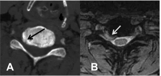
Although the contrast resolution of CT is limited compared to MRI, its spatial resolution is by far superior and it is very useful to characterize the extent of bony spurs, foraminal encroachment or the presence of calcified disc herniation or ossification of the posterior longitudinal ligament which may cause central canal stenosis (Figure 9). Furthermore, with the advent of the multidetector CT scans, the global resolution of the CT images has increased improving the diagnosis of disc herniation and disco-osteophytic complexes.
Figure 9. Combination of disc herniation and spondylosis.
(A) CT scan axial image of a cervical vertebra demonstrates degenerative changes with bony proliferation at the uncinate process of the vertebral body (black arrow) but the disc herniation is more difficult to identify. (B) Conversely, the MR axial image in the same subject depicts the herniated disc material (white arrow) in a more comprehensive manner.
1.7. Treatment
Cervical radiculopathy resulting from cervical spondylosis and/or disc herniation is typically, initially managed by non-surgical measures unless the patient has detrimental extremity weakness, intractable pain, associated signs of myelopathy or when non-surgical measures have failed to result in symptoms reduction 27. Non-surgical management, often referred to as conservative or medical treatment, may consist of rest, use of a rigid collar, analgesics, NSAIDs, physical therapy and corticosteroid injections.
In a study by Saal et al. 28, 26 patients with cervical radiculopathy secondary to a disc herniation were treated with a systematic conservative management approach. 20 (77%)
17
patients achieved an excellent or good recovery and 2 (1%) patients required subsequent surgery.
In a randomized study of 81 patients with chronic cervical radiculopathy, Persson et al. 6 demonstrated that surgery by an anterior approach, a customized physical therapy treatment or the use of a cervical collar were equally effective in the long-term (follow-up of 12 months). At short-term (1 month of follow-up) the patients treated with surgery reported less pain and as the physical therapy group, better function.
In a retrospective cohort study, Heckmann et al. 5 analyzed the functional outcomes of 60 patients with cervical disc herniation with radiculopathy but without signs of cervical myelopathy. The 60 patients were prospectively assessed with an average follow-up time of 5.5 years. 39 (65%) patients had been treated with conservative measures and 21 (35%) patients had undergone surgery (anterior approach). Patients in the surgical group initially presented with more severe and long-lasting neurological disturbances. In the group of patients treated conservatively, brachialgia improved in 100% and neurological disturbances, namely sensory disorders, reflex abnormalities and motor weakness improved in 97%, 59% and 94% respectively. In the surgical group, brachialgia improved in 97% of the patients and the neurological disturbances improved in 75%, 53% and 50% respectively. Residual intermittent neck pain was common in the conservatively treated group.
The Cervical Spine Research Society Study was undertaken to assess the outcome of patients with recognized subacute or chronic cervical disorders referred to spine surgeons for evaluation and treatment 8. Forty-one spine surgeons participated in this multicentric study and patients were assessed at 1-year follow-up, by way of questionnaires that were reviewed by an independent blinded observer. Two-hundred-and-forty-six patients diagnosed with cervical radiculopathy were enrolled in the study. In 160 (65%) patients, medical treatment was recommended on the basis of the surgeon’s evaluation and in 86 (35%) surgery was recommended. Of the 246 patients, 155 (63%) returned for follow-up. Of these patients, 104 (67%) received medical treatment and 51 (33%) underwent surgery. Both medically treated patients and surgically treated patients demonstrated significant improvement in overall pain and overall functional status.
These studies suggest that unless red flags have been identified, patients presenting with cervical radiculopathy should initially be treated with an optimized medical treatment management. In patients with subacute or chronic symptoms refractory to medical treatment, approximately 30% will be treated with surgery. In the appropriately selected patients, both medical management and surgery can treat cervical radiculopathy successfully.
Chapter 2. Transforaminal steroid injections
2.1. Efficacy
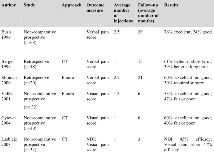
The presumed therapeutic mechanism of corticosteroid injections is the suppression of the inflammatory cascade that is believed to be responsible for the symptoms and signs of cervical radiculopathy that result from a disc herniation and/or degenerative spondylosis. TFSI allow for delivering high concentration of corticosteroids directly and precisely at the site of the involved spinal nerve root. Many non-controlled, observational studies have evaluated the efficacy of TFSI for the treatment of cervical radiculopathy (Table II).
Bush et al. treated 68 subjects for cervical radiculopathy with an average of 2.5 injections and reported a complete resolution of symptoms at long term follow-up in 76% of the cases 29.
In a retrospective study of 18 patients treated with one CT-guided TFSI, Berger et al. reported good or excellent results in 61% of their patients 30.
Slipman et al. obtained good or excellent results in 60% of cases, at 12 to 45 months follow-up and an average of 2.2 injections, in a retrospective study of 20 patients suffering from a degenerative foraminal stenosis 31.
Vallée et al. treated 32 patients with an average of 1.3 injections performed with fluoroscopy guidance, and obtained good or excellent results at 6 months in 53% of their patients 32.
Similarly, Cyteval et al. obtained good or excellent results at one month, in 60% of 30 patients, treated with one CT-guided TFSI 33.
Finally, Lasbleiz et al. reported 45% efficacy using the Neck Disability Index (NDI) and 47% of efficacy using a visual pain scale at 3 months, in 34 patients treated with one CT-guided injection 34.
Overall these results suggest that image-guided TFSI provide complete resolution of the radiculopathy in one third of patients, partial resolution of symptoms in another one third and are inefficacious in one third.
More recently, Anderberg et al. randomized 40 patients presenting with chronic cervical radiculopathy (mean duration of symptoms of 31 months) from degenerative spondylosis, to receive one fluoroscopically-guided TFSI 35. The treatment group received an injection of mepivicaïne + methylprednisolone while the control group received an injection of mepivicaïne + saline. There was a positive response in only 30% of the patients in the treatment group at 3 weeks follow-up. There were no significant differences in treatment results between the two groups.
Table II: Efficacy of transforaminal corticosteroid injections: non-controlled studies
Author Study Approach Outcome
measure Average number of injections Follow-up (average number of months) Results Bush 1996 Non-comparative prospective (n=68) Verbal pain score 2.5 39 76% excellent; 24% good Berger 1999 Retrospective (n=18) CT Verbal pain score
1 15 61% better at short term; 39% better at long term Slipman
2000
Retrospective (n=20)
Fluoro Verbal pain score 2.2 21 60% excellent or good; 30% required surgery Vallée 2001 Non-comparative prospective (n= 32)
Fluoro Visual pain score 1.3 6 53% excellent or good; 47% fair or poor Cyteval 2004 Non-comparative prospective (n=30) CT Visual pain score 1 6 60% excellent or good; 40% fair or poor Lasbleiz 2008 Non-comparative prospective (n=34) CT NDI; Visual pain score 1 3 NDI 45% efficacy; Visual pain score 47% efficacy
21
2.2. Technique
Traditionnally, TFSI, for the treatment of cervical radiculopathy, have been performed with fluoroscopic 36-39, computed tomography (CT) 32, 33, 38 or a combined technique of CT-fluoroscopy 40-42 guidance. More recently, some authors have advocated the use of ultrasound for real-time monitoring of these procedures 43.
For a fluoroscopically guided injection, the patient is placed in the supine position with the head turned slightly opposite the side to be injected. The C-arm is angled obliquely approximately 45 degrees towards the side to be injected, as to obtain a well-defined view of the targeted foramen with the superior facet overlying the lamina (Figure 10).
Figure 10. Fluoroscopically guided transforaminal corticosteroid injection.
(A) Oblique view radiograph shows the targeted point (thin arrow) at the posterior aspect of the intervertebral foramen. (B) Oblique view radiograph demonstrates the needle in place with contrast flowing in the posterior aspect of the foramen (curved arrow). (C) Anteroposterior view radiograph shows the spinal nerve root and exiting spinal nerve outlined by contrast (large arrow).
A needle is advanced in the plane of the xray beam, with intermittent fluoroscopic monitoring, until it contacts the superior facet at the level of the equator of the foramen. The needle is then redirected slightly anteriorly and advanced a few millimeters to penetrate into the posterior aspect of the foramen. Needle position is verified with a frontal view where the needle should project over the sagittal midline of the articular pillars. Injection of approximately 1 ml of nonionic contrast medium is then performed to confirm the adequate placement of the needle and the absence of intra-arterial injection. With optimal placement of the needle, the contrast should outline the exiting spinal nerve and the epidural space.
To perform the procedure with CT guidance, the patient is placed in the supine position with the head turned approximately 45 degrees to the contralateral side from the injection. Axial images limited to the target cervical foramen are obtained from a topogram of the cervical spine and an appropriate entry site is chosen to avoid the carotid and vertebral arteries and the jugular vein, and to gain access to the posterior aspect of the foramen, in a plane parallel to the anterior surface of the superior facet of the facet joint. The needle is advance to the outer aspect of the foramen using intermittent CT fluoroscopy. Once the needle is in the desired position, approximately 0.5 to 1.0 ml of nonionic contrast agent is injected and controlled with CT fluoroscopy to exclude intra-arterial injection and confirm adequate distribution of the contrast (Figure 11).
23
Figure 11. CT guided transforaminal corticosteroid injection.
(A) Axial CT image shows the path of the needle, which is parallel to the articular facet (long arrow). The needle tip is located within the posterior aspect of the neural foramen and posterior to the spinal nerve root. (B) Axial CT image demonstrates the distribution of contrast within the neural foramen (black arrow) and extending to the posterior epidural space (curved arrow) within the spinal canal. V = vertebral artery; C = Carotid artery; J = Jugular vein.
2.3. Potential complications
Steroid injections for the treatment of cervical radiculopathy occasionally cause minor adverse immediate reactions such as: vasovagal reaction, increased pain at the site of injection, increased radicular pain, numbness or weakness of the arm, headache, dizziness, nausea, hypersensitivity reaction and transient global amnesia 44. In a large series of 1036 fluoroscopically guided extraforaminal injections, Ma et al. 45 reported a prevalence of 1.6% (14 cases) of minor adverse effects. In the past 15 years, an alarming number of severe neurological adverse events has been reported in the literature, paralleling the rapid growth of interventional pain management procedures performed over the same period of time 46. Although a confirmed case of direct puncture of the spinal cord has been reported 47, the mechanism causing other numerous serious complications is still debated. The true risks of severe complications associated with these procedures remain unknown since only relatively few cases are being published in the literature. Nevertheless, in view of these dramatic complications and the lack of randomized controlled trials establishing their real efficacy, some authors have raised doubt about the justification for performing TFSI 36, 48.
In an effort to determine the prevalence of severe complications, Scanlon et al. 49 performed an anonymous survey among the members of the American Spine Society. At the time of the study, the membership of the society was composed of anesthesiologists (76%), physiatrists (17%), orthopedic surgeons (3%) and radiologists (< 1%). Among 1340 members, the response rate was 21.4% (287). In all, 78 complications were reported including:
• 16 vertebrobasilar brain infarcts (cerebellum, brainstem or posterior cerebral artery territory)
• 12 cervical spinal cord infarcts
• 2 combined brain / spinal cord infarcts
• 13 deaths (5 with brain infarcts; 1 with combined brain / spinal infarct; 1 following high spinal anesthesia; 1 associated with seizure; 5 with unspecified etiology)
25
2.3.1. Vertebral artery injury
Suresh et al. reported the case of a 60-year-old man who suffered a cerebellar and brainstem infarction following a CT-guided C4-C5 TFSI performed with a 25G needle 50. A magnetic resonance angiography was performed and did not reveal evidence of a VA dissection. The patient’s condition improved and he had a progressive recovery over the next month. The authors concluded that the most likely cause of this dramatic incident was a VA vasospasm secondary to unintended direct puncture of the VA.
Wallace et al. reported the imaging findings of two cases of VA dissection following TFSI 51. Rozin et al. reported the autopsy findings in a 44-year-old woman who died following a C6-C7 TFSI 52. The cause of death was a brainstem and cerebellar ischemic infarct with massive cerebral edema, secondary to a dissection and subsequent thrombosis of the left vertebral artery (VA), caused by inadvertent perforation of the artery with a 25G spinal needle during the procedure. Reflecting on this case, and on another similar case reported by Beckman et al. 53, de-Leon-Casasola questioned the likelihood that a puncture with a 25G needle would cause a dissection of a normal VA 54. He suggested that these patients might already have had a small VA dissection that was aggravated by the 25G needle puncture. Considering the relative prevalence of VA dissection in the low cervical levels of patients with a history of neck manipulations or accelerating / decelerating injuries, and the wide anatomic variation of the VA at low cervical levels, recognizing this potential additional risk factor is important.
2.3.2. Intravascular injections of corticosteroids
Arterial injection of particulate steroids causing an embolic phenomenon is the most frequently cited presumptive cause of brain and spinal cord infarcts 55-59. While performing a TFSI, despite using careful and precise technique, it is possible to cause inadvertent injection of material into radicular arteries that feed the spinal column 60. Cadaveric studies have demonstrated the variability and complexity of the vasculature in the vicinity of the intervertebral foramen and the inconsistent presence of critical arteries in the posterior aspect of the foramen susceptible to needle cannulation in the course of a TFSI 12.
In a prospective study of 504 consecutive fluoroscopically guided TFSI, Furman et al. reported a 19.4% rate of fluoroscopically confirmed venous or arterial intravascular contrast injections using a 25G spinal needle 61. Remarkably, using the spontaneous return of blood in the needle hub or after attempted aspiration with a syringe, predicted an intravascular injection with 97% specificity but only 45.9% sensitivity.
2.3.2.1. The role of the technique
Although there is no consensus in the literature as to what constitutes the most effective and safe method of performing epidural steroid injections 39, 62, the necessity to use image guidance and contrast medium injection to achieve accurate needle placement has long been recognized 63.
To minimize the risk of inadvertent intra-vascular injection, some authors have further recommended using live fluoroscopy or real time fluoroscopy with digital substraction angiography 60 to monitor contrast media injection more accurately and confirm the extravascular location of the needle tip before injecting the steroids 36. Other authors have advocated using CT fluoroscopy to perform TFSI because it offers better anatomic details reducing the risk of puncturing the VA and allows for more precise needle positioning within the intervertebral foramen 40-42.
Other proposed measures to avoid serious complications include the use of a test dose of local anesthetic before injection of steroids, to confirm the absence of signs and symptoms of central nervous system hyperirritability 64, 65.
Most interventionalists agree that TFSI should be performed without or with little sedation to keep the patient alert to report any unusual sensation during the procedure.
The choice of needles has also been examined. Although blunt needles in general are less likely than sharp needles to puncture vital structures and produce hemorrhage, this distinction is less apparent with the smaller 22G or 25G needles 66. Some authors favor the use of 22G instead of 25G needles to decrease the risk of penetrating the smaller vessels 40.
27
2.3.2.2. The role of corticosteroids
The risk of severe neurologic complications following TFSI is linked to the type of corticosteroids used and a growing body of evidence supports an embolic mechanism. In the study by Scanlon et al. 49 methylprednisolone, which consists of the type of corticosteroid with the largest particles, was most frequently involved. There is also incriminating evidence against triamcinolone, another particulate corticosteroid 49, 55, 59, 65, 67, 68. These types of corticosteroids have a tendency to coalesce into larger aggregates increasing their potential for embolic occlusion of the small radicular arteries during accidental intra-arterial injection 65. Betamethasone acetate-betamethasone sodium phosphate is another type of particulate corticosteroid which appears to be safer, although complications have been reported following its use 49. Derby et al. 69 tested different types of particulate and nonparticulate corticosteroid preparations in various solutions in vitro, under light microscopy (Table III). In this study, dexamethasone sodium phosphate demonstrated negligible particle size (approximately 10 times smaller than red blood cells) did not appear to form aggregates and had the lowest density. These findings suggest that because of its high solubility and small particle size, dexamethasone would be less likely to cause microvascular occlusion if inadvertent intra-arterial injection occurred in the course of a TFSI. To date, no severe adverse effects have been reported with the use of dexamethasone sodium phosphate in TFSI. In terms of metabolic activity, dexamethasone is recognized to have a rapid onset, but short duration of action compared to particulate corticosteroids. In a non-randomized retrospective study comparing the efficacy of TFSI with triamcinolone and dexamethasone at short-term follow-up, Lee et al. found no statistically significant difference in effectiveness between the two compounds 70. Similarly, in a randomized trial of 30 patients comparing the effectiveness, at 4 weeks, of a single TFSI with triamcinolone or dexamethasone, Dreyfuss et al. found that the yield of dexamethasone was slightly less than that of triamcinolone but the difference was neither statistically nor clinically significant 71. Hence, dexamethasone appears to be a valid alternative to particulate corticosteroids.
Table III: Size of corticosteroid particles, comparison with red blood cells (7.5 – 7.8 µm) Corticosteroids Dexamethasone sodium phosphate 10mg/ml Triamcinolone acetonide 40mg/m l Methylprednisolone 40mg/ml Betamethasone acetate-betamethasone sodium phosphate 6mg/ml
Particle size < 7.6 µm 0.5 – 100 µm < 7.6 µm Varied
Aggregation None Extensive Few > 100
Adapted from Derby et al. 69
2.4. Alternative techniques to transforaminal corticosteroids
injections
In view of the growing body of reports of catastrophic neurologic injuries following TFSI, some authors have questioned its use in the treatment of cervical radiculopathy 48, 72. As previously discussed, emphasis has been put on technical strategies to improve the safety of the technique. Alternative approaches, which potentially carry fewer risks, have also been advocated by some authors, even though their safety and efficacy, as is the case for TFSI, has yet to be validated by controlled randomized studies 48.
2.4.1 Interlaminar epidural corticosteroid injections
In the cervical spine, two approaches for direct delivery of corticosteroids into the epidural space may be used: transforaminal and interlaminar. The choice of which technique to use depends on the cervical pathology, the ability of the interventionalist to perform each technique and the efficacy versus the risks associated with each technique. The interlaminar route allows for more diffuse spread of the medication and is the preferred method to treat multilevel pathology. More localized spread of the medication and theoretically, higher concentration of medication is achieved with the transforaminal route and many believe that it
29
is the most effective technique to relieve the inflammation caused by mechanical or chemical nerve root irritation in the foramen.
In recent years, there appears to be a renewed interest for the interlaminar epidural corticosteroid injections (ILSI). In 2004, Derby et al. performed a retrospective survey among the course instructors of the International Spine Intervention Society (ISIS) 73. Questions were asked regarding the type of cervical epidural injections performed for neck and / or arm pain and any complications during the preceding 12 months period. Seventeen out of 29 (59%) instructors replied. In all, they performed an estimated total of 5968 cervical epidural injections with 74% ILSI and 24% TFSI. Twenty-three (0.52%) cases of minor complications in 4389 ILSI and 5 (0.32%) cases in 1579 TFSI were reported. When performing an ILSI, most respondents chose either the C7-T1 or T1-T2 levels, which provide the most distance between the ligamentum flavum and the dural sac and is believe to be the most secure approach. This survey could not assess the prevalence of major complications although one physician reported 2 cases of radicular artery injection confirmed by digital substraction angiography during 354 TFSI. Although serious complications after ILSI at the cervical level are recognized to be exceedingly rare, some authors have reported severe complications following ILSI such as subdural hematoma 74, direct cord injury 75 and quadriparesis 76 emphasizing the omnipresent risks associated with all interventional procedures.
2.4.2 Facet joint corticosteroid injections
IFSI are indicated for the treatment of cervical facet joint syndrome 77, 78, although evidence for their short-term and long-term efficacy in the cervical spine is limited 79, 80.
The study by Kim et al. suggested that IFSI could be effective in patients with cervical disc herniation 1. More recently, Hwang and colleagues suggested the use of a technique of IFSI with intentional capsular rupture, as an alternative to epidural steroid injections for lumbar radiculopathy caused by spinal stenosis, in patients who are at increased risk of bleeding 81. The exact mechanism of this apparent beneficial effect of IFSI for the treatment of cervical and lumbar radiculopathy remains uncertain and unclear, but could potentially be explained by the proximity of the facet joint ventral capsular recess to the intervertebral
foramen and/or leakage of the medication from the facet joint into the epidural and/or foraminal spaces.
In 1983, Dory described his technique of arthrography of the cervical facet joint 82. In his study, Dory showed that the ventral recess of the facet joint capsule is part of the posterior wall of the intervertebral foramen and may even bulge into the foramen FIGURE. Furthermore, in 4/21 intra-articular facet injections, Dory reported leakage of contrast in the epidural space or intervertebral foramen after over distention of the joint capsule.
In our experience and that of others 81, leakage of contrast into the epidural space following intra-articular facet injection is not uncommon, and may occur either spontaneously or intentionally. In 2001, we described a percutaneous technique to treat lumbar facet joint synovial cysts causing lower limb radiculopathy, consisting of an IFSI combined with forceful injection of bupivacaine 0.5% and normal saline under image guidance, to rupture the synovial cyst 83. Following rupture of the cyst, dispersion of contrast into the epidural space was well documented either at fluoroscopy or CT.
IFSI are procedures that are not technically challenging and can be easily performed with image guidance. As opposed to TFSI, IFSI are more widely available to patients because they are performed by a greater number of physicians. IFSI are considered to be safe, with only scarce reports of complications in the literature, namely a case of septic arthritis 84 and a case of ‘blind’ IFSI that resulted in transient tetraplegia 85.
Chapter 3. Hypothesis and Objectives of the Study
3.1. Hypothesis
Intra-articular facet steroid injections are at least as effective as transforaminal steroid injections or are worse by less than 15%, for the treatment of cervical radiculopathy due to degenerative spondylosis and/or a disc herniation.
3.2. Primary objective
The primary objective of this study was to compare the efficacy of CT-guided intra-articular facet steroid injections to CT-guided transforaminal steroid injections in subjects with cervical radiculopathy of at least one-month duration, due to degenerative spondylosis and/or a disc herniation, at 4 weeks follow-up.
3.3. Secondary objective
The secondary objective of this study was to examine the contrast distribution patterns following the CT-guided intra-articular facet and transforaminal steroid injections, and to correlate to the pain severity outcome rated on a visual analog scale (0 – 100).
Chapter 4. Transforaminal versus intra-articular facet
steroid injections for the treatment of cervical
radiculopathy: a randomized, double-blinded, controlled
study.
4.1. Authors
The following authors have contributed to the research project and to the elaboration of the manuscript. The authors’ affiliations are given herein. The role played by each author was detailed in the Introduction section of this mémoire.
Nathalie J Bureau MD1,2, Thomas Moser MD MSc1,2, Jehane H Dagher MD3, Daniel Shedid MD MSc4, Maria Li MD, CM, MSc5, Paul Brassard MD MSc6, Bernard E Leduc MD7. 1. Department of Radiology, Centre hospitalier de l’Université de Montréal, Montreal
Quebec, Canada.
2. Research center, Centre hospitalier de l’Université de Montréal, Montreal, Quebec, Canada.
3. Institut de réadaptation Gingras-Lindsay-de-Montréal, Université de Montréal, Montreal, Quebec, Canada
4. Department of Surgery, Division of Neurosurgery, Centre hospitalier de l’Université de Montréal, Montreal, Quebec, Canada.
5. Department of Surgery, Division of Neurosurgery, Hôpital Maisonneuve-Rosemont, Université de Montréal, Montreal, Quebec, Canada.
6. Division of Clinical Epidemiology, McGill University Health Center, Montreal, Quebec, Canada.
7. Department of Medicine, Centre hospitalier de l’Université de Montréal, Montreal, Quebec, Canada.
4.2. Manuscript accepted pending revisions in the American
Journal of Neuroradiology AJNR
33
Transforaminal versus intra-articular facet corticosteroid injections
for the treatment of cervical radiculopathy: a randomized, double-blinded,
controlled study
Nathalie J Bureau MD1,2, Thomas Moser MD MSc1,2, Jehane H Dagher MD3, Daniel Shedid MD MSc4, Maria Li MD, CM, MSc5, Paul Brassard MD MSc6, Bernard E Leduc MD7.
8. Department of Radiology, Centre hospitalier de l’Université de Montréal, Montreal Quebec, Canada.
9. Research center, Centre hospitalier de l’Université de Montréal, Montreal, Quebec, Canada.
10. Institut de réadaptation Gingras-Lindsay-de-Montréal, Université de Montréal, Montreal, Quebec, Canada
11. Department of Surgery, Division of Neurosurgery, Centre hospitalier de l’Université de Montréal, Montreal, Quebec, Canada.
12. Department of Surgery, Division of Neurosurgery, Hôpital Maisonneuve-Rosemont, Université de Montréal, Montreal, Quebec, Canada.
13. Division of Clinical Epidemiology, McGill University Health Center, Montreal, Quebec, Canada.
14. Department of Medicine, Centre hospitalier de l’Université de Montréal, Montreal, Quebec, Canada.
Corresponding author: Nathalie J Bureau MD
Department of Radiology, Centre hospitalier de
l’Université de Montréal, 1058 St-Denis, Montreal, Quebec, Canada, H2X 3J4. Phone: 514-942-1516.
Fax: 514-412-7359.
This research project was funded by the Fonds de recherche du Québec - Santé (Quebec Government Funding Agency) (FRQ-S, grant # 21230 – 2).
This paper will be presented at the American Society of Neuroradiology 52nd Annual Meeting & the Foundation of the ASNR Symposium, Montreal, Quebec, Canada, May 17 – 22, 2014.