HAL Id: hal-00317440
https://hal.archives-ouvertes.fr/hal-00317440
Submitted on 1 Jun 2020HAL is a multi-disciplinary open access archive for the deposit and dissemination of sci-entific research documents, whether they are pub-lished or not. The documents may come from teaching and research institutions in France or abroad, or from public or private research centers.
L’archive ouverte pluridisciplinaire HAL, est destinée au dépôt et à la diffusion de documents scientifiques de niveau recherche, publiés ou non, émanant des établissements d’enseignement et de recherche français ou étrangers, des laboratoires publics ou privés.
Phosphorylation of CDC25B by Aurora-A at the
centrosome contributes to the G2-M transition.
Stéphanie Dutertre, Martine Cazales, Muriel Quaranta, Christine Dozier,
Gladys Mirey, Jean-Pierre Bouché, Nathalie Theis-Febvre, Estelle Schmitt,
Claude Prigent, Bernard Ducommun
To cite this version:
Stéphanie Dutertre, Martine Cazales, Muriel Quaranta, Christine Dozier, Gladys Mirey, et al.. Phos-phorylation of CDC25B by Aurora-A at the centrosome contributes to the G2-M transition.. Journal of Cell Science, Company of Biologists, 2004, 117 (Pt 12), pp.2523-31. �10.1242/jcs.01108�. �hal-00317440�
Introduction
The cdc25 cell cycle regulator was identified almost twenty years ago in the fission yeast Schizosaccharomyces pombe as a dose-dependent inducer of mitosis (Russell and Nurse, 1986). It was later shown that the CDC25 protein is a member of the dual specificity phosphatase family that dephosphorylates critical residues and activates the CDK1 cell cycle ‘master regulator’ (Galaktionov and Beach, 1991; Kumagai and Dunphy, 1991; Millar and Russell, 1992). CDC25 activation is therefore an essential step in the triggering of the biochemical events that initiate the structural changes accompanying entry into mitosis. Despite the identification of numerous other regulators, partners and substrates, a clear picture of the activation process of CDK1 at the G2–M transition remains to be established.
The CDC25B phosphatase is one of the three members of the CDC25 family in mammals. It is a short half-life protein (Baldin et al., 1997a; Nishijima et al., 1997) that is expressed and active in late G2 (Baldin et al., 1997b; Gabrielli et al., 1996). CDC25B localisation was shown to depend on nuclear export signal (NES) and nuclear localisation signal (NLS) sequences (Davezac et al., 2000) and to be regulated through its interaction with 14-3-3 proteins (Forrest and Gabrielli, 2001; Giles et al., 2003; Mils et al., 2000). CDC25B
accumulation in the cytoplasm has been correlated with prophase spindle formation (Gabrielli et al., 1996) and it was suggested that this phosphatase pool located in close vicinity of the centrosome was responsible for the activation of the cytoplasmic pool of CDK1-cyclin B and the subsequent dependent increase in microtubule dynamics (Gabrielli et al., 1996). It has been reported that centrosomal and cytoplasmic CDK1-cyclin B activation precedes nuclear mitotic events (De Souza et al., 2000), and recently it has been shown that active CDK1-cyclin B first appears at centrosomes in prophase (Jackman et al., 2003). Thus, if as suggested, CDC25B acts as an initiator of the early mitotic events (Nilsson and Hoffmann, 2000), it could well play a role in the activation of a centrosomal sub-population of CDK1-cyclin B1 that is next translocated to the nucleus where activation of CDC25C will initiate an amplification loop driving the cell into mitosis (Hoffmann et al., 1993; Kumagai and Dunphy, 1992).
The Aurora-A protein kinase, encoded by the STK15 oncogene is of major interest since its overexpression has been correlated with high grade tumours (reviewed by Dutertre et al., 2002; Katayama et al., 2003). This protein is located at the centrosome from S-phase to mitosis (reviewed by Bischoff and Plowman, 1999; Dutertre et al., 2002; Giet and Prigent, 1999). It is thought to be involved in centrosome separation (Giet et
Aurora-A protein kinase, which is the product of an oncogene, is required for the assembly of a functional mitotic apparatus and the regulation of cell ploidy. Overexpression of Aurora-A in tumour cells has been correlated with cancer susceptibility and poor prognosis. Aurora-A activity is required for the recruitment of CDK1-cyclin B1 to the centrosome prior to its activation and the commitment of the cell to mitosis. In this report, we demonstrate that the CDC25B phosphatase, an activator of cyclin dependent kinases at mitosis, is phosphorylated both in vitro and in vivo by Aurora-A on serine 353 and that this phosphorylated form of CDC25B is located at the centrosome during mitosis. Knockdown experiments by RNAi confirm that the centrosome phosphorylation of CDC25B on S353 depends on Aurora-A kinase.
Microinjection of antibodies against phosphorylated S353 results in a mitotic delay whilst overexpression of a S353 phosphomimetic mutant enhances the mitotic inducing effect of CDC25B. Our results demonstrate that Aurora-A phosphorylates CDC25B in vivo at the centrosome during mitosis. This phosphorylation might locally participate in the control of the onset of mitosis. These findings re-emphasise the role of the centrosome as a functional integrator of the pathways contributing to the triggering of mitosis.
Supplemental data available online
Key words: Aurora-A, CDC25B phosphatase, Centrosome, Mitosis Summary
Phosphorylation of CDC25B by Aurora-A at the
centrosome contributes to the G2–M transition
Stéphanie Dutertre1, Martine Cazales2, Muriel Quaranta2, Carine Froment3, Valerie Trabut2, Christine Dozier2,
Gladys Mirey2, Jean-Pierre Bouché2, Nathalie Theis-Febvre2, Estelle Schmitt2, Bernard Monsarrat3,
Claude Prigent1and Bernard Ducommun2,*
1Groupe Cycle Cellulaire – CNRS UMR6061 – IFR97, Génomique Fonctionnelle et Santé, Université de Rennes I, 2 avenue du Pr Léon Bernard,
35043 Rennes, France
2LBCMCP-CNRS UMR5088-IFR109, Institut d’Exploration Fonctionnelle des Génomes, Université Paul Sabatier, 118 route de Narbonne,
31062 Toulouse, France
3IPBS – CNRS UMR5089, 205 route de Narbonne, 31077 Toulouse, France *Author for correspondence (e-mail: [email protected])
Accepted 19 January 2004
Journal of Cell Science 117, 2523-2531 Published by The Company of Biologists 2004 doi:10.1242/jcs.01108
2524
al., 2002; Glover et al., 1995) and maturation as well as in bipolar spindle assembly and stability (Giet and Prigent, 2000). Inhibition of its function using siRNA leads to the formation of monopolar spindles. Its overexpression results in centrosome amplification and polyploidy of the cell (Bischoff et al., 1998; Zhou et al., 1998) probably through cytokinesis failure (Meraldi et al., 2002). Furthermore, it has very recently been shown that Aurora-A activity is required for the recruitment of CDK1-cyclin B1 to the centrosome, which correlates with its activation and for the commitment of the cells to mitosis (Hirota et al., 2003). Since a major post-translational modification required for the activation of the CDK1 kinase is tyrosine 15 dephosphorylation, we investigated whether CDC25B might participate in that process.
We report that CDC25B is phosphorylated in vitro and in vivo at serine 353 by the Aurora-A kinase. This phosphorylated form of CDC25B is detected at the centrosome from prophase to anaphase. This modification appears to participate in the regulation of entry into mitosis suggesting a local function of CDC25B. These results emphasise the centrosome as an integrator of cellular signals participating in mitosis.
Materials and Methods
Cell culture conditions
HeLa cells, HeLa C1 stably expressing a GFP-centrin fusion protein (a generous gift from M. Bornens) (Piel et al., 2001) and U2OS expressing epitope-tagged CDC25B were grown as previously described (Davezac et al., 2000).
Immunofluorescence microscopy
HeLa cells seeded on glass coverslips were then fixed and permeabilised after 18 or 24 hours. DNA was visualised using DAPI. Polyclonal antibody against phosphorylated serine 353 (SE96) was raised against the QNKRRRS(p)VTPPEEQ peptide then affinity-purified on a phosphorylated peptide column then on an unphosphorylated peptide column to eliminate antibodies reacting against the unphosphorylated peptide. SE96 was used at the dilution of 1/1000 and the incubation was performed overnight at 4°C. A polyclonal antibody against total CDC25B was raised against the ITNSQAPDGRRKSEA internal peptide then affinity-purified on a peptide column. This antibody was used at a dilution of 1/1500 and incubated overnight at 4°C. Monoclonal anti-Aurora-A human (35C1) was used as previously described (Cremet et al., 2003). Rabbit anti-phosphorylated Aurora-A (Thr288) antibody (Cell Signalling Technology) was used at the dilution of 1/250. Mouse anti-human lamin (Santa-Cruz, SC-7292) and anti-gamma-tubulin (Sigma T6557) were used at the dilution of 1/2000 and 1/50 respectively. Secondary antibodies labelled with Alexa 488 or Alexa 594 (Molecular probes) were used at 1/800 for 1 hour at room temperature.
Mitotic inducing activity of CDC25B was monitored 24 hours after transfection of YFP-tagged expressing vectors using EXGEN and following the manufacturer’s instructions. HeLa cells were grown on polylysine-coated coverslips to more efficiently retain the rounded mitotic cells that are otherwise frequently lost. Cells were stained with DAPI and the percentage of transfected cells (YFP positives) displaying a premature condensed chromatin (PCC) phenotype (Gabrielli et al., 1996; Karlsson et al., 1999) was determined.
Images were acquired on a DMRXA2 or a DMIRE2 microscope (Leica) using CCD cameras and subsequently processed by the Metamorph or the Qfluoro softwares.
Production of recombinant proteins, in vitro kinase assays, mass spectrometry analyses, western blot
MBP-CDC25B3, A and C were produced in JM109 bacteria and affinity purified on amylose beads following the manufacturer’s instructions. Recombinant proteins (1 µg) were incubated in 20 µl of kinase buffer (50 mM Tris-HCl, pH 7.5, 10 mM MgCl2, 25 mM NaCl,
1 mM DTT, 0.01% Triton X-100 and 10 µM ATP) with 1 µg of purified recombinant Aurora-A (Cremet et al., 2003) at 37°C for 30 minutes. The reaction was stopped by addition of 20 µl of 2×Laemmli sample buffer and separated by SDS-PAGE and analysed by western blotting. In-gel digestion and on-line Nano-LC-MS/MS analyses were performed using capillary HPLC (LC Packings) coupled to an LCQ ion trap mass spectrometer (ThermoFinnigan) as described previously (Theis-Febvre et al., 2003). Mouse anti-maltose binding protein (Sigma MBP-17) was used in western blots at the dilution of 1/4000. To analyse in vivo phosphorylation of CDC25B, CDC25B was affinity purified from a conditionally expressing U2OS human cell line that was engineered to express a polyHis-tagged version of CDC25B under the control of the tetracycline inducible promoter (J.-P.B., unpublished data).
Microinjection experiments
Affinity-purified antibodies (SE96) or control purified rabbit antibodies were injected into the nucleus of HeLa cells either asynchronous or synchronised by thymidine block, using an Eppendorf micromanipulator and microinjector devices. At the indicated time after microinjection, cells were fixed then processed for immunofluorescence microscopy with anti-rabbit immunoglobulin antibodies as described above. The cells were scored according to the protocol proposed by Lane and Nigg (Lane and Nigg, 1996), which consists of injecting scattered cells, thus allowing the determination of whether a microinjected cell has undergone mitosis or not. Divided cells were detected as two fluorescent cells next to each other.
Interference RNA
Human Aurora-A siRNA oligonucleotide with the following sequence 5′-AUGCCCUGUCUUACUGUCA-3′(Kufer et al., 2002) and as a control a scrambled siRNA were used. Human CDC25B siRNA were purchased from DHARMACON. Oligonucleotides were transfected using oligofectamine (Invitrogen, Life Technologies) following the manufacturer’s instructions.
Results
Aurora-A phosphorylates CDC25B in vitro on serine 353
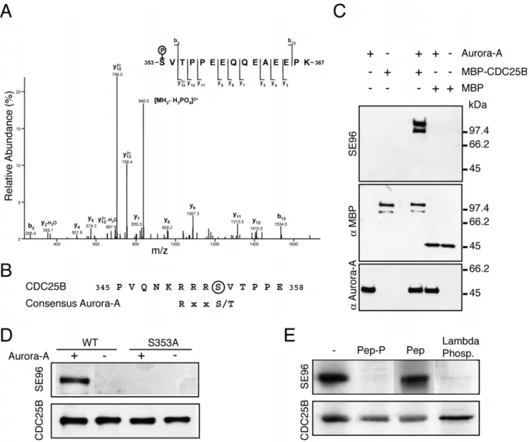
We first examined whether CDC25B was a substrate of the Aurora-A kinase in vitro. We found that recombinant CDC25B was heavily phosphorylated in vitro by purified Aurora-A kinase. Mass spectrometry analysis of Aurora-A-phosphorylated CDC25B was performed and indicated a mono-phosphorylation on the peptide sequence 353SVTPPEEQQEAEEPK367 (Fig. 1A). A single phosphate group was detected on this peptide and was found to be associated with serine 353. This serine residue was also found by mass spectrometry analysis to be phosphorylated in CDC25B overexpressed in baculovirus-infected Sf9 cells (data not shown). S353 is located in a sequence that matches the consensus (RxxS) phosphorylation site for the Aurora-A kinase (Fig. 1B).
An affinity-purified polyclonal antibody (SE96) was raised against phospho-S353. SE96 recognised Aurora-A-phosphorylated recombinant MBP-CDC25B but not MBP nor Journal of Cell Science 117 (12)
Aurora-A itself (Fig. 1C) and the immunolabelling was competitively inhibited by the phosphorylated immunogenic peptide (QNKRRRS(p)VTPPEEQ) (not shown).
Recombinant MBP-CDC25A and MBP-CDC25C were not phosphorylated by Aurora-A kinase and neither were they recognised by the SE96 antibody (Fig. S1, http://jcs.biologists.org/supplemental/). In vitro phosphorylation of wild-type and S353A mutant CDC25B protein performed in the presence of [γ-32P]ATP showed a 71%
lower incorporation in the mutant, indicating that serine 353 is a major Aurora-A phosphorylation site on CDC25B. S353A mutant protein incubated with Aurora-A was not detected by the SE96 antibody (Fig. 1D). The effect of CDC25B phosphorylation by Aurora-A on the phosphatase activity was tested using phosphorylated CDK/cyclin as a substrate (Cans et al., 1999). No change in the catalytic activity of CDC25B upon phosphorylation by Aurora-A was detectable (data not shown).
Fig. 1. Aurora-A phosphorylates CDC25B on serine 353 in vitro and in vivo. (A) MS/MS spectrum of the monophosphorylated peptide, 353-SVTPPEEQQEAEEPK-367 (doubly charged precursor ion, MH22+, at m/z 889.38) displays series of b- and y-ions [according to Biemann’s
nomenclature (Biemann, 1990)], intense doubly charged y13 (at m/z 756,4) together with weak mono-charged b2 (at m/z 266.9) and indicating that the serine 353 residue is phosphorylated and not threonine 355. (B) CDC25B alignment with Aurora-A consensus phosphorylation site. (C) Recombinant proteins were incubated with purified recombinant Aurora-A at 37°C for 30 minutes. The samples were analysed by western blotting with the 35C1 monoclonal anti-Aurora-A or affinity-purified polyclonal anti-serine 353 phosphorylated epitope – SE96 or monoclonal anti-maltose binding protein (MBP). MBP-CDC25B migrated as a doublet because of the presence of a degradation product. (D) Recombinant MBP-CDC25B and MBP-CDC25B S353A mutant were phosphorylated or not by Aurora-A as in C. Western blot analysis was performed with SE96 and anti-CDC25B antibodies. (E) Western blot analysis of CDC25B affinity-purified (15 ng/lane) from human U2OS cells expressing polyHis-tagged CDC25B, with SE96 antibody and anti-CDC25B polyclonal antibody was performed in the presence of 10003 molar excess of the phosphorylated peptide (lane 2), the unphosphorylated peptide (lane 3), or after prior incubation of the sample for 60 minutes at 30°C in the presence of λphosphatase.
2526
Aurora-A phosphorylates CDC25B in vivo on serine 353
CDC25B was affinity purified from a U2OS human osteosarcoma cell line conditionally expressing an epitope-tagged version of CDC25B under the control of the tetracycline-inducible promoter. As shown by western blot analysis with SE96, serine 353 was found to be phosphorylated on purified CDC25B protein (Fig. 1E). The immunolabelling was competitively inhibited by the phosphorylated immunogenic peptide but not by the unphosphorylated one. This labelling was also abolished by prior dephosphorylation of purified CDC25B with lambda phosphatase.
To further confirm the phosphorylation of CDC25B in vivo on S353 and the recognition specificity of SE96, CDC25B expression was suppressed by RNA-mediated interference in HeLa cells. Western blot analysis of cell extracts using an antibody against CDC25B or the SE96 antibody detected a major single band at the expected molecular mass, that was abolished when RNA-mediated interference was used (Fig. 2A). Detection of actin was performed to ensure for equivalent loading.
Together, these results indicate that CDC25B is phosphorylated in vivo on serine 353 and can specifically be detected using the SE96 anti-phosphoserine 353 antibody.
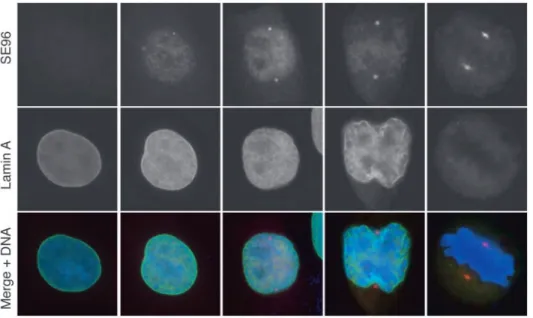
CDC25B phosphorylated on serine 353 is located at the centrosomes in mitotic cells
We then investigated the subcellular localisation of the S353 phosphorylated form of CDC25B in HeLa cells. As shown in Fig. 2B, the phosphorylated epitope recognised with the SE96 antibody was exclusively detected in mitotic cells. Staining was in the form of two dots located on both sides of dividing cells from prophase to anaphase (see also Figs 3, 4; see also Fig. S3, http://jcs.biologists.org/supplemental/). Interphase cells displayed only a faint and diffuse staining (Fig. 2B,C). Labelling was totally competed away by the immunogenic peptide but not by the non-phosphorylated peptide, and not by an irrelevant phosphorylated peptide (Fig. S2, http://jcs.biologists.org/supplemental/). Labelling was also abolished using recombinant MBP-CDC25B that was phosphorylated in vitro by Aurora-A (Fig. S2, http://jcs.biologists.org/supplemental/).
The centrosome localisation of S353-phosphorylated CDC25B in mitotic cells was confirmed using an anti-γ-tubulin monoclonal antibody (Fig. 2C) and an anti α-tubulin antibody (Fig. S3, http://jcs.biologists.org/supplemental/). No
centrosomal staining with the SE96 antibody was detected in interphase cells. The centrosome localisation of CDC25B in mitotic cells was also examined in HeLa C1 cells that were engineered to express a GFP-centrin fusion protein (Piel et al., 2001). SE96 staining was only detected after centrosome separation, not at the centrioles per se but rather with the pericentriolar material as shown in Fig. 2D.
Nuclear lamina staining with monoclonal anti-lamin A antibody was performed together with SE96 staining to determine more accurately the timing of serine 353 Journal of Cell Science 117 (12)
Fig. 2. Serine 353-phosphorylated CDC25B is located at the centrosome. (A) CDC25B RNA interference. Western blot with anti-CDC25B polyclonal antibody, SE96 and anti-actin of total extracts from HeLa cells transfected or not with CDC25B RNAi or control (cont.) scrambled RNAi. (B,C) HeLa cells were fixed and
immunofluorescence staining was performed as described previously (Davezac et al., 2000). Cells were also stained with 4′-6 diamino-2-phenylindole (DAPI). (B) Staining with SE96 antibodies of cells in prometaphase and interphase (top panels), and anaphase and interphase (lower panels). (C) Double immunofluorescent staining with SE96 and a γ-tubulin monoclonal antibody of cells in interphase (top panels) and prometaphase (lower panels); SE96 (red), γ-tubulin (green) and DAPI (blue). (D) HeLa C1 expressing a GFP-centrin fusion protein (Piel et al., 2001) were stained with SE96 antibodies; SE96 (red), GFP-centrin (green) and DAPI (blue).
phosphorylation. As shown in Fig. 3, the SE96 epitope was detected in the separated centrosomes of early prophase cells when the nuclear envelope was still intact, although very faint traces of lamina reorganisation were already visible (Beaudouin et al., 2002).
Phosphorylated CDC25B colocalises with active Aurora-A
Immunofluorescence double staining using SE96 and 35C1, a monoclonal antibody raised against Aurora-A (Cremet et al., 2003), showed that CDC25B phosphorylated on serine 353 also colocalised with Aurora-A at the centrosome during mitosis (Fig. 4A). However, Aurora-A was detected at the centrosome and the poles of the mitotic spindle whereas phosphorylated serine 353 was only detectable at the centrosome from early prophase to anaphase (Fig. 4A). We then asked whether S353-phosphorylated CDC25B localises at the centrosome together with the activated Aurora-A. Phosphorylation of Aurora-A on threonine 288 (activation loop) results in a significant increase in catalytic activity (Walter et al., 2000). Activated Aurora-A was detected by indirect immunofluorescence using a polyclonal antibody raised against phosphorylated T288 (Fig. 4B). We were therefore able to examine whether the phosphorylation of CDC25B on serine 353 was coincident with Aurora-A kinase activity in HeLa cells. We found that the activated form of Aurora-A (phosphorylated on the activated threonine of the activation loop) appeared at the level of the centrosome concomitantly with the appearance of phosphorylated S353.
Taken together, these results strongly suggested that activated Aurora-A and serine-353-phosphorylated CDC25B appear simultaneously at the centrosome in early prophase and that Aurora-A might be responsible for this phosphorylation.
In vivo phosphorylation of CDC25B on serine 353 is dependent on Aurora-A activity
To further confirm this hypothesis, we used RNA-mediated
interference to suppress Aurora-A expression and ask whether the kinase activity of Aurora-A was required for the phosphorylation of serine 353 in vivo. The suppression efficiency of Aurora expression varied from cell to cell but led to major mitotic spindle abnormalities. We therefore used conditions in which the Aurora-A level was partially, but significantly, reduced but in which the spindle remained bipolar (see western blot in Fig. 5D). We used HeLa C1 cells expressing GFP-centrin in order to localise the centrosome. As shown in Fig. 5A, Aurora-A expression was knocked down to a low level around the centrosome (Fig. 5A). In these conditions, CDC25B phosphorylated on S353 also dramatically decreased (Fig. 5B,E). CDC25B staining with an anti-peptide antibody that recognised the phosphatase independently of its phosphorylation on serine 353, showed that inhibition of Aurora-A expression did not affect the level of CDC25B localised to the spindle poles (Fig. 5C). These results demonstrate that Aurora-A phosphorylates CDC25B on S353 in vivo.
Phosphorylation of serine 353 and entry into mitosis
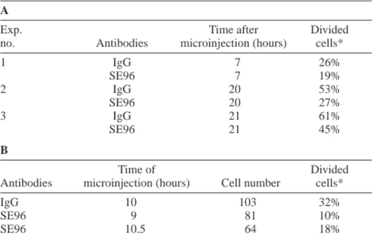
Because both Aurora-A and CDC25B are proposed to be involved in the activation of CDK1/cyclin B to trigger the G2–M transition, we asked whether phosphorylation of S353 could contribute to mitosis entry. To assess the contribution of serine 353 phosphorylation in the progression into mitosis, affinity-purified SE96 immunoglobulins were microinjected in scattered (Lane and Nigg, 1996) asynchronous HeLa cells or in G2 cells that had been synchronised by double-thymidine block and release (Table 1A and B, respectively). Cells were fixed and stained with anti-rabbit Ig antibodies to detect the microinjected cells and to determine the percentage that had divided. Compared to control antibodies, SE96 strongly delayed entry into mitosis (Table 1), as does an antibody against all forms of CDC25B (Lammer et al., 1998), indicating that specific targeting of this phosphorylation event is sufficient to impair the role of CDC25B in the control of the progression into mitosis.
Fig. 3. Timing of serine 353 phosphorylation of CDC25B at the centrosome. Double
immunofluorescence staining of HeLa cells selected at different stages of mitosis with SE96 and lamin A monoclonal antibodies (mouse anti-human lamin); SE96 (red), lamin A (green) and DAPI (blue).
2528
To further investigate this question, HeLa cells were transiently transfected with YFP-tagged wild-type CDC25B or S353A or S353E mutants and the ability of these proteins, expressed at a similar level (data not shown), to cause premature and unscheduled initiation of mitosis (PCC, Premature Chromosome Condensation) in the transfected cells was examined (Karlsson et al., 1999). As already described (Gabrielli et al., 1996; Karlsson et al., 1999), expression of wild-type CDC25B lead to PCC in about 20% of cells. Overexpression of the S353E mutant, which is supposed to mimic phosphorylation, enhanced this effect with 40% of the transfected cells undergoing premature and aberrant mitosis, similarly to the overexpression of Aurora-A (41% of PCC in transfected cells). In contrast, expression of a S353A mutant had a mitotic inducing effect weaker than that of the wild type (14% of PCC). These data demonstrate that phosphorylation of CDC25B on S353 plays an important function during progression through G2–M.
Discussion
Since the first demonstration that CDC2 was associated with the centrosome (Bailly et al., 1989), several component of the cell cycle control machinery have been found to be located there or to the spindle pole body in yeast (Alfa et al., 1990). These include, cyclin B1 (Alfa et al., 1990; Jackman et al., 2003), PLK1 (Tsvetkov et al., 2003), Aurora-A (Bischoff and Plowman, 1999; Dutertre et al., 2002) and CHK2 (Tsvetkov et al., 2003). We have determined that a subpopulation of the CDC25B phosphatase phosphorylated by Aurora-A on S353 localises to the centrosome during mitosis where its probable function is to regulate the function of CDK1-cyclin B complexes that also localise to the centrosome at this time Journal of Cell Science 117 (12)
Table 1. Microinjection of antibodies against phosphorylated serine 353 delays entry into mitosis A
Exp. Time after Divided
no. Antibodies microinjection (hours) cells*
1 IgG 7 26% SE96 7 19% 2 IgG 20 53% SE96 20 27% 3 IgG 21 61% SE96 21 45% B Time of Divided
Antibodies microinjection (hours) Cell number cells*
IgG 10 103 32%
SE96 9 81 10%
SE96 10.5 64 18%
Exp. no., experiment number.
*The percentage of divided cells among the microinjected cells population (detected by staining with anti-rabbit IgG antibody as two fluorescent cells next to each other).
Affinity-purified anti-CDC25B antibodies (SE96) or normal rabbit IgG were injected into the nucleus of scattered HeLa cells (as described by Lane and Nigg, 1996) either asynchronous or synchronised by thymidine block. (A) Asynchronous HeLa cells fixed at the indicated time after microinjection. (B) HeLa cells synchronised by thymidine block, injected in G2 at the indicated time after release, and fixed 16 hours after block release.
Fig. 4. Active Aurora-A and serine 353-phosphorylated CDC25B localise at prophase and metaphase centrosomes. (A) HeLa cells were fixed and subjected to double immunofluorescence staining with SE96 polyclonal and Aurora-A monoclonal antibodies; SE96 (green), Aurora-A (red) and DAPI (blue). Photomicrographs of cells
representative of each stage of mitosis are shown. Interphase (A), prophase (B), prometaphase (C), metaphase (D), anaphase A (E) and anaphase B (F). (B) HeLa cells were fixed and stained with monoclonal Aurora-A antibodies and polyclonal antibodies against threonine 288-phosphorylated Aurora-A. Selected cell cycle phases are shown.
(Bailly et al., 1989; Bailly et al., 1992; Jackman et al., 2003). This finding reinforces the view of the centrosome as a functional integrator of the control pathways that participate in the triggering of mitosis (Jackman et al., 2003).
We have reported that CDC25B is phosphorylated both in vitro and in vivo by Aurora-A. Mass-spectrometry analysis and the use of phosphospecific antibodies indicate that phosphorylation occurs on serine 353. Suppression of Aurora-A expression using RNAurora-A interference confirms that CDC25B S353 phosphorylation is dependent on Aurora-A. This phosphorylated form of the CDC25B phosphatase is localised at the centrosome at an early stage of mitotic entry, concomitant with very early events in nuclear envelope disassembly (Beaudouin et al., 2002) and with T288-phosphorylation and activation of the Aurora-A kinase (Walter et al., 2000).
We have found that mimicking S353 phosphorylation enhances the mitotic inducing activity of CDC25B leading to premature chromosome condensation (PCC), as does the overexpression of Aurora-A. Furthermore, the targeting of the S353 phosphorylated form of CDC25B, by microinjection of affinity-purified antibodies to the phosphopeptide, delays entry into mitosis as does an antibody against all forms of CDC25B
(Lammer et al., 1998). Because this observation is very similar to the mitotic delay observed by Marumoto et al. (Marumoto et al., 2002) using antibodies against Aurora-A, these data strongly argue that a major function of Aurora-A at prophase might be the phosphorylation of S353 of CDC25B.
The in vitro catalytic activity of CDC25B does not change upon phosphorylation by Aurora-A. In vivo it might trigger additional modifications, interaction with associated proteins, or conformational changes required for the docking of a substrate or a partner.
CDC25B has been proposed to act as one of the starters of mitosis (Karlsson et al., 1999) and the CDK cyclins that are putative CDC25B substrates, are responsible for change in microtubule dynamics at the centrosome level in early prophase (Buendia et al., 1992). Localisation of CDC25B to the centrosome and its phosphorylation by Aurora-A strongly argues in favour of this hypothesis (Gabrielli et al., 1996). Several reports have shown that centrosomal ablation does not block mitotic entry (Bobinnec et al., 1998; Hinchcliffe et al., 2001; Keryer et al., 2003; Khodjakov et al., 2000). However, such cells do not complete cytokinesis and do not resume S-phase, indicating a critical role for the centrosome in the overall control of cell cycle progression. Thus, our results Fig. 5. Inhibition of Aurora-A expression by RNA interference shut down serine 353 phosphorylation. HeLa C1 cells expressing GFP-centrin (Piel et al., 2001) were transfected with A RNAi or control scrambled RNAi. Immunofluorescence staining was performed with Aurora-A monoclonal antibodies (Aurora-A), with SE96 anti-serine 353-phosphorylated CDC25B polyclonal antibody (B), or with a CDC25B polyclonal antibody. Cells were also stained with DAPI (blue). (D) Lysates from HeLa cells treated as above were subjected to western blot analysis with anti-Aurora-A and anti β-tubulin as a loading control. (E) Quantification of immunofluorescence signal was performed using Metamorph software on images taken with a CoolsnapHQ camera.
2530
suggest that Aurora-A-dependent phosphorylation of CDC25B at the centrosome may participate in the temporal and spatial regulation of critical biochemical events that are required for a synchronised mitosis entry.
We gratefully acknowledge Jeremy Hyams for invaluable comments and suggestions, Michel Bornens for the gift of the HeLa GFP-centrin cell line. Thanks to the IFR97 microscopy platform in Rennes. S.D. is a recipient of a post-doctoral fellowship from ARC (Association pour la Recherche sur le Cancer). G.M. is a recipient of a post-doctoral fellowship from la Ligue Contre le Cancer. E.S. is a recipient of a post-doctoral fellowship from the Fonds de la Recherche en Santé du Québec. B.M. is supported by the CNRS, l’Université Paul Sabatier, la Région Midi-Pyrénées, le pôle ARECA ‘Protéomique et Cancer’, and B.D. and C.P. by la Ligue Nationale Contre le Cancer (Equipe labellisée 2001) and to (Equipe labellisée 2003).
References
Alfa, C., Ducommun, B., Beach, D. and Hyams, J. S. (1990). Distinct
nuclear and spindle pole body populations of cyclin-cdc2 in fission yeast.
Nature 347, 680-682.
Bailly, E., Dorée, M., Nurse, P. and Bornens, M. (1989). P34cdc2 is located
in both nucleus and cytoplasm; part is centrosomally associated at G2/M and enters vesicles at anaphase. EMBO J. 8, 3985-3995.
Bailly, E., Pines, J., Hunter, T. and Bornens, M. (1992). Cytoplasmic
accumulation of cyclin B1 in human cells: association with a detergent-resistant compartment and with the centrosome. J. Cell Sci. 101, 529-545.
Baldin, V., Cans, C., Knibiehler, M. and Ducommun, B. (1997a).
Phosphorylation of human CDC25B phosphatase by CDK1/cyclin A triggers its proteasome-dependent degradation. J. Biol. Chem. 272, 32731-32735.
Baldin, V., Cans, C., Superti-Furga, G. and Ducommun, B. (1997b).
Alternative splicing of the human CDC25B tyrosine phoshatase. Possible implications for growth control? Oncogene 14, 2485-2495.
Beaudouin, J., Gerlich, D., Daigle, N., Eils, R. and Ellenberg, J. (2002).
Nuclear envelope breakdown proceeds by microtubule-induced tearing of the lamina. Cell 108, 83-96.
Biemann, K. (1990). Appendix 5. Nomenclature for peptide fragment ions
(positive ions). Methods Enzymol. 193, 886-887.
Bischoff, J. R., Anderson, L., Zhu, Y., Mossie, K., Ng, L., Souza, B., Schryver, B., Flanagan, P., Clairvoyant, F., Ginther, C. et al. (1998). A
homologue of Drosophila aurora kinase is oncogenic and amplified in human colorectal cancers. EMBO J. 17, 3052-3065.
Bischoff, J. R. and Plowman, G. D. (1999). The Aurora/Ipl1p kinase family:
regulators of chromosome segregation and cytokinesis. Trends Cell Biol. 9, 454-459.
Bobinnec, Y., Khodjakov, A., Mir, L. M., Rieder, C. L., Edde, B. and Bornens, M. (1998). Centriole disassembly in vivo and its effect on
centrosome structure and function in vertebrate cells. J. Cell Biol. 143, 1575-1589.
Buendia, B., Draetta, G. and Karsenti, E. (1992). Regulation of the
microtubule nucleating activity of centrosomes in Xenopus egg extracts: role of cyclin A-associated protein kinase. J. Cell Biol. 116, 1431-1442.
Cans, C., Sert, V., Derycke, J., Baldin, V. and Ducommun, B. (1999). Use
of cdc2 from etoposide-treated cells as substrate to assay CDC25 phosphatase activity. AntiCancer Res. 19, 1241-1244.
Cremet, J. Y., Descamps, S., Verite, F., Martin, A. and Prigent, C. (2003).
Preparation and characterization of a human aurora-A kinase monoclonal antibody. Mol. Cell Biochem. 243, 123-131.
Davezac, N., Baldin, V., Gabrielli, B., Forrest, A., Theis-Febvre, N., Yashida, M. and Ducommun, B. (2000). Regulation of CDC25B
phosphatases subcellular localization. Oncogene 19, 2179-2185.
De Souza, C. P., Ellem, K. A. and Gabrielli, B. G. (2000). Centrosomal and
cytoplasmic Cdc2/cyclin B1 activation precedes nuclear mitotic events. Exp.
Cell Res. 257, 11-21.
Dutertre, S., Descamps, S. and Prigent, C. (2002). On the role of aurora-A
in centrosome function. Oncogene 21, 6175-6183.
Forrest, A. and Gabrielli, B. (2001). Cdc25B activity is regulated by
14-3-3. Oncogene 20, 4393-4401.
Gabrielli, B. G., De Souza, C. P. C., Tonks, I. D., Clarck, J. M., Hatward, N. K. and Ellem, K. A. O. (1996). Cytoplasmic accumulation of CDC25B
phosphatase in mitosis triggers centrosomal microtubule nucleation in HeLa cells. J. Cell Sci. 109, 1081-1093.
Galaktionov, K. and Beach, D. (1991). Specific activation of cdc25 tyrosine
phosphatases by B-type cyclins: Evidence for multiple roles of mitotic cyclins. Cell 67, 1181-1194.
Giet, R., McLean, D., Descamps, S., Lee, M. J., Raff, J. W., Prigent, C. and Glover, D. M. (2002). Drosophila Aurora A kinase is required to
localize D-TACC to centrosomes and to regulate astral microtubules. J. Cell
Biol. 156, 437-451.
Giet, R. and Prigent, C. (1999). Aurora/Ipl1p-related kinases, a new
oncogenic family of mitotic serine-threonine kinases. J. Cell Sci. 112, 3591-601.
Giet, R. and Prigent, C. (2000). The Xenopus laevis aurora/Ip11p-related
kinase pEg2 participates in the stability of the bipolar mitotic spindle. Exp.
Cell Res. 258, 145-151.
Giles, N., Forrest, A. and Gabrielli, B. (2003). 14-3-3 acts as an
intra-molecular bridge to regulate cdc25B localization and activity. J. Biol. Chem.
278, 28580-28587.
Glover, D. M., Leibowitz, M. H., McLean, D. A. and Parry, H. (1995).
Mutations in aurora prevent centrosome separation leading to the formation of monopolar spindles. Cell 81, 95-105.
Hinchcliffe, E. H., Miller, F. J., Cham, M., Khodjakov, A. and Sluder, G.
(2001). Requirement of a centrosomal activity for cell cycle progression through G1 into S phase. Science 291, 1547-1550.
Hirota, T., Kunitoku, N., Sasayama, T., Marumoto, T., Zhang, D., Nitta, M., Hatakeyama, K. and Saya, H. (2003). Aurora-A and an interacting
activator, the LIM protein Ajuba, are required for mitotic commitment in human cells. Cell 114, 585-598.
Hoffmann, I., Clarke, P., Marcote, M. J., Karsenti, E. and Draetta, G.
(1993). Phosphorylation and activation of human cdc25-C by cdc2-cyclin B and its involvement in the self amplification of MPF at mitosis. EMBO J.
12, 53-63.
Jackman, M., Lindon, C., Nigg, E. A. and Pines, J. (2003). Active cyclin
B1-Cdk1 first appears on centrosomes in prophase. Nat. Cell Biol. 5, 143-148.
Karlsson, C., Katich, S., Hagting, A., Hoffmann, I. and Pines, J. (1999).
Cdc25B and Cdc25C differ markedly in their properties as initiators of mitosis. J. Cell Biol. 146, 573-584.
Katayama, H., Brinkley, W. R. and Sen, S. (2003). The Aurora kinases: role
in cell transformation and tumorigenesis. Cancer Metastasis Rev. 22, 451-464.
Keryer, G., Witczak, O., Delouvee, A., Kemmner, W. A., Rouillard, D., Tasken, K. and Bornens, M. (2003). Dissociating the centrosomal matrix
protein AKAP450 from centrioles impairs centriole duplication and cell cycle progression. Mol. Biol. Cell 14, 2436-2446.
Khodjakov, A., Cole, R. W., Oakley, B. R. and Rieder, C. L. (2000).
Centrosome-independent mitotic spindle formation in vertebrates. Curr.
Biol. 10, 59-67.
Kufer, T. A., Sillje, H. H., Korner, R., Gruss, O. J., Meraldi, P. and Nigg, E. A. (2002). Human TPX2 is required for targeting Aurora-A kinase to the
spindle. J. Cell Biol. 158, 617-623.
Kumagai, A. and Dunphy, W. (1991). The cdc25 protein controls tyrosine
dephosphorylation of the cdc2 protein in a cell free system. Cell 64, 903-914.
Kumagai, A. and Dunphy, W. (1992). Regulation of the cdc25 protein during
the cell cycle in Xenopus extracts. Cell 70, 139-151.
Lammer, C., Wagerer, S., Saffrich, R., Mertens, D., Ansorge, W. and Hoffman, I. (1998). The cdc25B phosphatase is essential for the G2/M
phase transition in human cells. J. Cell Sci. 111, 2445-2453.
Lane, H. A. and Nigg, E. A. (1996). Antibody microinjection reveals an
essential role for human polo-like kinase 1 (Plk1) in the functional maturation of mitotic centrosomes. J. Cell Biol. 135, 1701-1713.
Marumoto, T., Hirota, T., Morisaki, T., Kunitoku, N., Zhang, D., Ichikawa, Y., Sasayama, T., Kuninaka, S., Mimori, T., Tamaki, N. et al. (2002).
Roles of aurora-A kinase in mitotic entry and G2 checkpoint in mammalian cells. Genes Cells 7, 1173-1182.
Meraldi, P., Honda, R. and Nigg, E. A. (2002). Aurora-A overexpression
reveals tetraploidization as a major route to centrosome amplification in p53–/– cells. EMBO J. 21, 483-492.
Millar, J. and Russell, P. (1992). The cdc25 M-phase inducer: An
unconventional protein phosphatase. Cell 68, 407-410.
Mils, V., Baldin, V., Pinta, I., Goubin, F., Papin, C., Waye, M., Eychene,
A. and Ducommun, B. (2000). Specific interaction between 14.3.3 isoforms
and the human CDC25B phosphatase. Oncogene 19, 1257-1265.
Nilsson, I. and Hoffmann, I. (2000). Cell cycle regulation by the Cdc25
phosphatase family. Prog. Cell Cycle Res. 4, 107-114.
Nishijima, H., Nishitani, H., Seki, T. and Nishimoto, T. (1997). A
dual-specificity phosphatase Cdc25B is an unstable protein and triggers p34(cdc2)/cyclin B activation in hamster BHK21 cells arrested with hydroxyurea. J. Cell Biol. 138, 1105-1116.
Piel, M., Nordberg, J., Euteneuer, U. and Bornens, M. (2001).
Centrosome-dependent exit of cytokinesis in animal cells. Science 291, 1550-1553.
Russell, P. and Nurse, P. (1986). cdc25+ functions as an inducer in the mitotic
control of fission yeast. Cell 45, 145-153.
Theis-Febvre, N., Filhol, O., Froment, C., Cazales, M., Cochet, C., Monsarrat, B., Ducommun, B. and Baldin, V. (2003). Protein kinase CK2
regulates CDC25B phosphatase activity. Oncogene 22, 220-232.
Tsvetkov, L., Xu, X., Li, J. and Stern, D. F. (2003). Polo-like kinase 1 and
Chk2 interact and co-localize to centrosomes and the midbody. J. Biol.
Chem. 278, 8468-8475.
Walter, A. O., Seghezzi, W., Korver, W., Sheung, J. and Lees, E. (2000).
The mitotic serine/threonine kinase Aurora2/AIK is regulated by phosphorylation and degradation. Oncogene 19, 4906-4916.
Zhou, H., Kuang, J., Zhong, L., Kuo, W. L., Gray, J. W., Sahin, A., Brinkley, B. R. and Sen, S. (1998). Tumour amplified kinase STK15/BTAK
induces centrosome amplification, aneuploidy and transformation. Nat.