UNIVERSITÉ DE MONTRÉAL
MULTIMODAL DIFFUSE OPTICAL IMAGING EVIDENCE OF AGE-RELATED CHANGES IN NEURAL SUBSTRATES OF SEMANTIC WORDS PROCESSING
MAHNOUSH AMIRI
INSTITUT DE GÉNIE BIOMÉDICAL ÉCOLE POLYTECHNIQUE DE MONTRÉAL
THÈSE PRÉSENTÉE EN VUE DE L’OBTENTION DU DIPLÔME DE PHILOSOPHIAE DOCTOR
(GÉNIE BIOMÉDICAL) AVRIL 2016
UNIVERSITÉ DE MONTRÉAL
ÉCOLE POLYTECHNIQUE DE MONTRÉAL
Cette thèse intitulée:
MULTIMODAL DIFFUSE OPTICAL IMAGING EVIDENCE OF AGE-RELATED CHANGES IN NEURAL SUBSTRATES OF SEMANTIC WORDS PROCESSING
présentée par : AMIRI Mahnoush
en vue de l’obtention du diplôme de : Philosophiæ Doctor a été dûment acceptée par le jury d’examen constitué de :
M. DEHAES Mathieu, Ph. D., président
M. LESAGE Frédéric, Ph. D., membre et directeur de recherche M. JOANETTE Yves, Ph. D., membre et codirecteur de recherche M. JOLICOEUR Pierre, Ph. D., membre
DEDICATION
To my family Able to stay so close while so far, making me believe I could achieve such ambitious works under such circumstances.
REMERCIEMENTS
Je tiens en tout premier lieu à remercier mes deux directeurs de thèse.
Yves Joanette qui, au-delà de son excellence académique, m’a apporté un soutien décisif par son humanité, son expérience et son approche de la vie de recherche. Il a beaucoup influencé mon parcours doctoral et mes choix personnels depuis que nous nous sommes rencontrés et continuera à jamais à marquer mes travaux de chercheuse par la suite. Yves, merci d’avoir toujours eu confiance en moi, de m’avoir portée vers le haut et parfois au-delà de ce que semblaient m’autoriser mes capacités. Avec cette confiance et ces orientations, avec cet accompagnement, j’ai été conviée à trouver des pistes pour avancer en restant motivée par notre terrain et les réalisations de notre laboratoire. Je garderai toujours en mémoire la façon dont Yves a pu en tant que Directeur de laboratoire, à la fois communiquer son expertise et valoriser les connaissances de tous les étudiants pour permettre une saine. Comment oublier cette présentation du poster à distance en Californie alors que des difficultés de visa vous avaient obligés à me laisser à Montréal ?
J’aimerais également remercier Frédéric Lesage. Frédéric a accepté de partager cette aventure qu’est la thèse en cotutelle en apportant toute son expertise en ingénierie par un outillage de recherche innovant. Frédéric m’a apporté la confiance véhiculée par son expérience et sa remarquable maîtrise des technologies les plus avancées au service de nos expériences. Merci de m’avoir accueillie au sein du laboratoire d’imagerie optique moléculaire LIOM et de m’avoir aidée à concevoir et à réaliser le protocole nous conduisant aux résultats tant attendus. Ces différentes rétroactions n’ont jamais cessé de fertiliser mon travail de recherche. Frédéric a toujours su rendre la recherche vivante.
Je tiens aussi à préciser que je n’étais pas citoyenne canadienne à l’origine de ce travail mais le suis devenu avec beaucoup de fierté. Ce pays m’a accueillie en attendant de moi que je lui apporte le meilleur de moi-même et j’espère que cette première pierre à l’édifice sera à la hauteur des attentes qui ont été placées en moi. Notons au passage l’appui financier du laboratoire pour cette recherche sans lequel je n’aurais pu concilier ambitions académiques, contraintes de bien-être d’une résidente de plein-droit. En tant que boursière, j’ai pu me dédier à temps plein à mes études doctorales, à la vie du laboratoire et à mon projet de recherche. J’en remercie l’organisation IRSC.
Je me dois d’exprimer ma gratitude envers Clément Bonnéry, généreux par ses encouragements, qui m’a continuellement appuyée. Toujours présent. Clément, qui m’a beaucoup soutenue lorsque j’étais sous tension lors de la validation du protocole de recherche, m’a aidée pour l’acquisition et la mise en place du projet pilote. Je remercie Philippe Pouliot pour sa disponibilité, ses connaissances et son accompagnement. Mes amis collègues de laboratoire à l’École Polytechnique pour l’ambiance agréable du labo, et ceux des Universités de Montréal et McGill (Pariya Salami, Maximiliano Wilson, Jean-Sébastien Provost, Perrine Ferré, Ikram Metqal, et Samuel Laventure) que j’ai eu la chance de rencontrer durant mon parcours.
Je réserverai le mot de la fin pour remercier du fond du cœur mon cher ami, Jean-Marc Ayme, pour son appui et sa foi en moi. Il m’a dirigée vers ce programme dès les premiers jours où je me suis sentie habitée par une vive passion pour ce domaine, en m’encourageant à m’y investir. Il m’a accompagnée dans ma première phase d’approche de doctorat, malgré les difficultés que j’éprouvais. Au collège de France par exemple, où il m’a soutenu pour que je puisse assister aux conférences annuelles du Professeur Stanislas Dehaene, dont le nom m’a ouvert tant de portes lors des phases de candidature. Grâce à cela, j’ai pu changer d’orientation depuis l’imagerie vers la neuroscience. Merci Jean-Marc d’avoir été toujours à l’écoute et prêt à dispenser ton appui si fort et si chaleureux.
AKNOWLEDGEMENTS
First of all, I would like to express my gratitude to both of my Ph.D. thesis supervisors.
Yves Joanette, who, beyond his renowned academic excellence, has provided me a critical support with profound humanity and his experience. He has influenced so much this journey from the issue submissions up to the thesis defence. Even my personal choices have been drastically influenced by Yves since we first met and we can be sure that this will define the everlasting pattern of my further works as a researcher. Yves, thank you so much for your reliable trust in me and for your ability to drive me up sometimes to higher points than I would have expected to reach. With this trust and this support, I have been invited to find ways to move forward while remaining motivated by our field and achievements in our laboratory. I will always keep in mind how Yves was able as Lab Director to communicate his expertise and enhance the knowledge of all students to allow a nourishing competition. How can we forget this remote poster presentation in California as visa difficulties forced you all to leave me in Montreal? I would also like to thank wholeheartedly Frédéric Lesage. Frédéric agreed to share this thesis adventure jointly supervised by bringing his expertise in engineering to make possible the observation of participants' results through innovative research equipment and an original protocol designed specifically during these years. Frédéric gave me the required confidence together with his scientific intuitions to conduct our experiments under the supervision of his remarkable command of advanced technologies. Thank you for welcoming me to the Lab-community of LIOM and for helping me to design the methodology and produce the results leading us to our participants’ comprehensive observations. You offered the technical facilities and to get in touch with some contacts to support this thesis approach. Frédéric has always been able to stimulate a living research
Thanks to both of you, I am encouraged to further contribute to our field to make our research community progress by crossing skills, opening myself to broader contributions, preserving the intuition of future research tracks. You will never be thanked enough for your support, your scientific orientations and especially for the trust you have placed in me from the start-off.
I also want to clarify that I was not a Canadian citizen at the origin of this work but have become very proudly Canadian by the end of last year. This country welcomed me expecting that I would bring the best of myself and I hope that this first building-block will stand up to the expectations
that have been placed in me. By the way, the financial support from the laboratory for this research has decisively made possible to reconcile academic ambitions, daily life’s stress and plain-rights resident welfare. As a studentship recipient, I could fully dedicate myself to my doctoral studies, to the laboratory life and to my research project. I thank the organization for its generosity.
I must also express my gratitude to Clément Bonnéry for acquisition and implementation of the pilot project that was very supportive when I was under pressure during the validation of the research protocol. Always present. My dear laboratory mates, thank you for your support and thank you for having created a unique working atmosphere during these last years. I would like to thank my fellow students and faculty of the University of Montreal (Pariya Salami, Maximiliano Wilson, Jean-Sébastien Provost, Perrine Ferré, Ikram Metqal, and Samuel Laventure) I've met during my journey.
I will reserve the last word for my dear friend Jean-Marc Ayme to have such confidence on me and have always provided a warm and strong support. He supported me in the maintenance of passion and energy I placed in my Ph.D. approach phase, while living a difficult era in my life. At the Collège de France for instance, he helped me to assist my first sessions in neurosciences through Professor Stanislas Dehaene’s seminars, whose name has opened so many doors during my application candidacy. This later facilitated the path to operate a shift from my engineering background towards cognitive neuroscience. Being away from my beloved ones is always an added difficulty to overcome. This materialized delivery, finally tangible, is a way to represent this intense presence that definitely helped me along the way.
RÉSUMÉ
Le vieillissement rapide de la population au Canada changera les aspects démographiques à l’avenir par le fait que les personnes âgées de 65 ans et plus vont dépasser en volume les jeunes de moins de 20 ans. En sachant les coûts associés au traitement et au soutien des personnes âgées atteintes par une ou plusieurs démences cognitives, on admet l’importance des études gériatriques pour mieux comprendre les mécanismes neurophysiologiques du vieillissement. L’intérêt principal est de trouver un lien entre les effets neurologiques du vieillissement et ceux du déclin cognitif afin d’établir des stratégies qui encourageront un vieillissement en santé. Notre compréhension du cerveau a beaucoup évolué au cours des dernières décennies grâce à de nouvelles techniques en imagerie cérébrale. Pourtant, l’interprétation de ces données reste un défi. Dans le cas de l’imagerie fonctionnelle par résonance magnétique (IRM) ou par optique diffuse (IOD), la réponse neuronale est indirectement dérivée de l’hémodynamique. Cette dernière est sujette à de complexes interactions entre l’oxygénation du cerveau, le volume et le débit sanguin, ainsi que la structure hétérogène du cortex. Ces interactions rendent difficile une interprétation quantitative des données. Dans le cas des études en vieillissement cognitif, ces paramètres sont de plus modifiés par l’âge, ce qui mène à une importante variabilité interindividuelle dans l’interprétation des données. La caractérisation des effets neurophysiologiques du vieillissement sur les signaux d’imagerie cérébrale est donc essentielle pour permettre des études rigoureuses du déclin cognitif avec l’âge. Vu les limites en rapport avec les signaux intrinsèques de chacune des modalités d’imagerie non-invasive, l’intérêt pour les études multimodales s’accroît car elles permettent de calibrer avec plus de précision les données fonctionnelles. L’intégration des données complémentaires acquises via différentes modalités de neuroimagerie, dans cette étude, nous a permis de quantifier les activations neuronales et de surveiller leurs modifications reliées au vieillissement. Un montage de spectroscopie en temps résolu, fait au laboratoire, nous a fourni des données au repos sur la concentration en oxy- et déoxyhémoglobine, ainsi que sur le volume sanguin. En imagerie par résonance magnétique, une séquence anatomique nous a servi à 1) évaluer une potentielle corrélation entre l’épaisseur corticale (matières grise et blanche) et le niveau de la réponse hémodynamique et 2) Recaller la carte d’activité cérébrale de chaque participant sur son image anatomique. On rajoute ces dernières mesures comme régresseur à un modèle linéaire généralisé de la réponse hémodynamique. En faisant l’hypothèse que ces changements de bases sont liés à la physiologie,
leur régression permet de prendre en compte le changement de la physiologie de base. Celui-ci renforce les inférences statistiques basées sur les changements de la concentration d’hémoglobine qui sont dus à l’activité neuronale et pas aux effets du vieillissement.
Cette recherche ouvre des perspectives cliniques intéressantes en termes de diagnostic et d’amélioration de la qualité de vie, en proposant notamment des axes de réflexions pour traiter les déficits cognitifs. De plus, ce projet, en haussant la rigueur dans les analyses statistiques de groupes, a le potentiel d’améliorer la puissance d’interprétation de futures études d’imagerie.
ABSTRACT
The demographic features of the population of Canada will experience an unprecedented historical change in the near future by the number of individuals above 65 years surpassing the number of youngsters under 20 years. Considering the costly consequences of age-related cognitive decline, both for individuals and the society, studying the neurophysiological mechanisms of these unfavorable changes has become an utmost priority in health research. The main goal of this field is to link the effects of cerebral aging to those of cognitive aging in order to stablish strategies promoting healthy aging. Normal cerebral aging is accompanied by some neurophysiological and neuroanatomical alterations depending on epigenetics of individuals. Amongst neurophysiological deteriorations causing cognitive decline, one should account for the neural loss, cortical density reduction, neurovascular, metabolic, and neurotransmission dysfunctions. Taken together these alterations with age, we were interested to determine whether older adults are affected in their cognitive abilities by more than one simple factor. In another word, we aimed at exploring the potential relationship between the abovementioned age-related alterations with cognitive performance. However, the main challenge of such study appears when interpreting functional data regarding baseline measures of each individual. Thus, the increased inter and intra-individual variability in cognitive studies is mainly due to their large variations in structural and neurophysiological characteristics in the course of their lifespan.
In this project, we defined a multi-modal neuroimaging protocol with the aim of calibrating the functional measures of task-related activity by measured individual baseline neurophysiological characteristics. To assess individuals` cerebral blood flow at rest, one of the constituent of hemodynamic response, we used an arterial-spin labeling sequence of magnetic resonance imaging. This technique based on tagging water in blood, gives the blood quantity emerging to brain. Carotids, the main arterial vessels that supply blood to brain, neck and face, are well known to be affected by age inter-individually and play as a non-functional moderator in hemodynamic response formation. As an estimate of total blood volume and baseline concentration of oxy- and deoxyhaemoglobin, we used a home-made 4-chanel time resolution optical device to acquire data from each participant’s prefrontal lobe. To refine the spatial resolution of non-invasive optical imaging, we also acquired anatomical MR images of each participant to 1) calculate cortical thickness with the objective of evaluating the correlation
between grey matter and white matter volumes and the task-evoked hemodynamic response and 2) co-register functional map of each participant on her/his anatomical image. The hemodynamic response was measured by an optical imaging system using near-infrared light. This emerging technique is based on the absorption properties of the biological tissue illuminated with near-infrared range of light.
This research opens up interesting perspectives in terms of clinical diagnosis and quality of life improvements. It includes new reflection axes to deal with cognitive deficits. In addition, this project has the potential to improve the power of interpretation of future imaging studies showing more rigors in groups’ statistical analysis.
TABLE OF CONTENTS
DEDICATION ... III REMERCIEMENTS ... IV AKNOWLEDGEMENTS ... VI RÉSUMÉ ... VIII ABSTRACT ... X TABLE OF CONTENTS ...XII LIST OF FIGURES ... XVI LIST OF TABLES ... XVIII LIST OF SYMBOLS AND ABBREVIATIONS... XIX LIST OF APPENDICES ... XXICHAPTER 1 INTRODUCTION ... 1
1.1 Summary of problems in studies on Aging ... 6
1.2 Goals and hypothesis ... 7
CHAPTER 2 GENERAL CONTEXT ... 9
2.1 Cognitive neuroscience of aging ... 9
2.1.1 Neural correlates of age-related cognitive decline ... 10
2.1.2 Aging brain and language ... 13
2.1.3 Shedding light on words processing ... 14
2.2 Functional neuroimaging ... 15
2.2.1 From firing neurons to the hemodynamic response ... 16
2.3 The necessity of calibrated functional imaging in aging studies ... 17
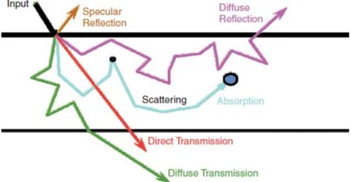
CHAPTER 3 DIFFUSE OPTICAL IMAGING ... 19
3.2 Diffuse optical tomography ... 20
3.3 Near-infrared spectroscopy ... 21
3.4 Issues with NIRS ... 24
CHAPTER 4 GENERAL METHODOLOGY ... 26
4.1 Language task ... 26
4.2 Image acquisition protocol ... 27
4.2.1 Magnetic resonance imaging (MRI) ... 27
4.2.2 Diffuse optical imaging ... 30
4.3 Efficient experimental paradigm ... 32
4.3.1 Design matrix ... 34
4.3.2 Statistical inference ... 35
CHAPTER 5 ARTICLE 1: FNIRS EXPLORATION OF THE HEMODYNAMIC CHANGES ALLOWING FOR THE SEMANTIC PROCESSING OF WORDS IN NORMAL AGING……… ... 36
5.1 Abstract ... 36
5.2 Introduction ... 37
5.3 Materials and methods ... 41
5.3.1 Participants and protocol ... 41
5.3.2 Diffuse optical measurements ... 43
5.3.3 MRI acquisition ... 45
5.3.4 Coregistration ... 45
5.4 Data analysis ... 45
5.4.1 Behavioral and task performance ... 45
5.4.2 NIRS data signal processing and statistical analysis ... 46
5.4.4 Anatomical MRI ... 48 5.4.5 ASL data ... 48 5.5 Results ... 49 5.5.1 Neuropsychological performance ... 49 5.5.2 Task performance ... 50 5.5.3 TRS ... 51 5.5.4 Resting-state CBF ... 52
5.5.5 Functional optical recordings ... 52
5.5.6 Correlation analyses ... 55
5.6 Discussion ... 56
5.7 Acknowledgement ... 59
5.8 References ... 60
CHAPTER 6 ISSUES & CHALLENGES ... 66
CHAPTER 7 ARTICLE 2: THE EFFECT OF CORTICAL MORPHOLOGY OF NORMAL AGING ON THE HEMODYNAMIC RESPONSE MEASURED BY FNIRS; A LANGUAGE STUDY……….. ... 72 7.1 Abstract ... 72 7.2 Introduction ... 73 7.3 Methods ... 76 7.4 Results ... 79 7.5 Discussion ... 85 7.6 Conclusion ... 87 7.7 Reference ... 89
CHAPTER 8 GENERAL DISCUSSION ... 116
8.2 Neurophysiology of Aging ... 118
8.3 Neuroanatomy of aging ... 119
CONCLUSION AND RECOMMENDATION ... 122
BIBLIOGRAPHY ... 124
APPENDIX 1 ... 155
LIST OF FIGURES
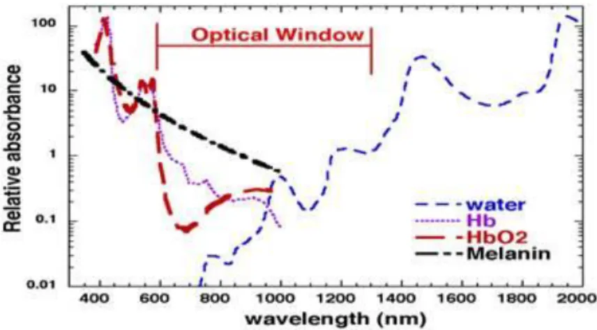
Figure 3-1 The probability of the photon migration in the biological tissue. Image taken from Boas et al, 2001 ... 21 Figure 3-2 Optical windows in biological tissue. The graph is taken from SPIE Newsroom. DOI:
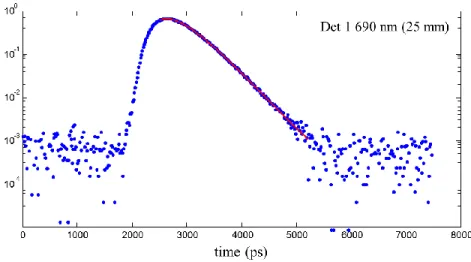
10.1117/2.1200906.1669 ... 22 Figure 4-1 : TRS measurements (x-axis: number of photons) for detector 1 at the distance of 25
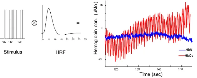
mm from the light source at 690 nm. The red curve shows here a perfect fit to the raw data (blue dots). ... 31 Figure 4-2 Predicted data to the convolution of HRF by a random SOA stimuli is shown by
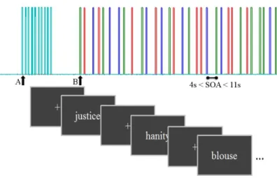
measured HbR and HbO2 as hemodynamic response to stimuli. ... 33 Figure 5-1 A schema of the task diagram with inter stimulus interval = 1.36 s and stimulus onset
asynchrony from 4 s to 11 s. Triggers from the computer presenting the task were sent to the NIRS computer after synchronization: A = start control task, and B = start main task. Each color bar represents a different condition: concrete, abstract and pseudo words. ... 42 Figure 6-1 Here there are 2 examples of the incongruent co-registration of the optodes on the
cortex, resulting from different positioning of the head in the MRI scanner and head size. Left) native cortical space. Right) template MNI space. ... 67 Figure 6-2 Age-related difference of beta values for concrete words processing, calculated by
GLM for both chromophores HbO2 and HbR. Green line: Young adults and blue line: Older adults. Note that the fSD channels stand for the short source-detector separation. ... 69 Figure 6-3 Means and standard deviations of beta-values for some random channels are presented
for young and older adults. ... 70 Figure 7-1. Description of cortical measurements: Mean values in voxels for cortical, subcortical
and total gray volumes, as well as total white matter. There are significant differences in cortical and gray matter volumes. ... 80 Figure 7-2 Difference of the cortical thickness between young and old adults. Clusters are FDR
C=right hemisphere medial view; D=left hemisphere medial view. Hemispheres are inflated. ... 81 Figure 7-3 Difference of cortical thickness between young and old with age as co-variate.
Clusters are uncorrected at p=0.001; A=right hemisphere lateral view; B=left hemisphere lateral view; Plot presented for the left middle temporal cluster. Red squares=young; blue circles=old. Hemispheres are inflated. ... 82 Figure 7-4 Difference of cortical thickness between young and old with cerebral blood flow
measures in gray matter (cbfgm) as co-variates. Clusters are FDR corrected at p=0.05. A=right hemisphere lateral view; B=left hemisphere lateral view; C=left hemisphere medial view. Hemispheres are inflated. Plot presented for the right orbitofrontal cluster. Red squares=young; blue circles=old. ... 83 Figure 7-5 cortical maps of the activation for HbR in response to semantic words processing A)
Shows neural activity measures of HbR for concrete (A1) and abstract (A2) words processing. In this panel measures of cortical thickness are used as a regressor to the group analysis. ... 84 Figure 7-6 cortical maps of the activation for HbO2 in response to semantic words processing A)
Shows neural activity measures for concrete (A1) and abstract (A2) words processing. In this panel measures of cortical thickness are used as a regressor to the group analysis. Similarly B1 and B2 show activations for concrete and abstract words respectively, but without cortical measures of thickness as a regressor. ... 85 Figure Annexe-1 10-20 system of EEG electrode placement used in optical helmet placement. A1
represent below auricular left. Fp1 and Fp2 were the limits of the beginning of the optical helmet. ... 155 Figure Annexe-2 ROI formation according to the language-related anatomical areas. ... 155
LIST OF TABLES
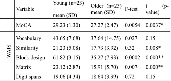
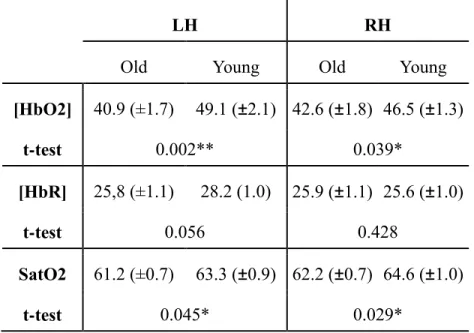
Table 5.1: Demographic variables and cognitive characteristics. Results from neuropsychological batteries by age group. MoCA: Montreal Cognitive Assesment; WAIS: Wechsler Adult Intelligent Scale; Hayling: test for inhibitory control. * p < .05, ** p < .0001 ... 49 Table 5.2: Results from TRS measurements. Oxy- and deoxyhaemoglobin concentrations and
oxygen saturation calculated in both hemispheres from TRS measures in micromoles, * p < .05, ** p < .001 ... 51
LIST OF SYMBOLS AND ABBREVIATIONS
aMRI Anatomical Magnetic Resonance ImagingASL Arterial-Spin Labeling
BOLD Blood Oxygen Level Dependent CBF Cerebral blood flow
CBV Cerebral blood volume
CMRO2 Cerebral metabolic rate of oxygen consumption CSF Cerebrospinal fluid
CW Continuous wave
DPF Differential pathlength factor DOI Diffuse optical imaging GLM General linear model
fMRI functional magnetic resonance imaging HbO2 Oxygenated haemoglobin
HbR Deoxygenated haemoglobin HbT Total haemoglobin
HRF Hemodynamic response function MTLs Middle temporal lobes
MTG Middle temporal gyri NIRS Near-infrared spectroscopy OAs Old adults
PFC Prefrontal cortex
SatO2 Blood oxygen saturation SNR Signal-to-noise ratio
TD-NIRS Time domain-NIRS
LIST OF APPENDICES
CHAPTER 1
INTRODUCTION
By 2025, the demographic features of the population in Canada will experience an unprecedented historical change: the number of individuals above 65 years will surpass the number of youngsters under 20 years. The undeniable decline in age-related sensory processing, motor performance, and cognitive processes affects the quality of older adults’ life in their intellectual, physical and social activities. In an ideal framework, in which our aging population is merely undergoing a natural process of decline, understanding the mechanisms underlying healthy aging is very important to both individuals and society, to encourage a healthy life style. Normal aging is characterized by changes in brain’s anatomy and physiology, a phenomenon that varies depending on brain’s region and component (Raz et al., 2005). For instance, the global volume of the cerebral cortex decreases by 0.35% per year in adult individuals of over 52 years. Yet it is important to note that this reduction differs across brain regions and individuals. Raz and colleagues, in their meta-analysis of 14 studies, have found that the frontal lobe has the steepest rate of atrophy among others. However, neural loss and myelin breakdown causing white matter integrity reduction are not the only indicators of aging (Bucur et al., 2008). For instance, neurovascular architecture, brain metabolism as well as neurotransmitter dysfunctions should also be taken into account as indicators of aging. Hence, exploring the correlation between theses physical substrates of cognitive aging and performance requires a comprehensive research protocol in the aging studies. With this objective in mind, we believed that contemplating a multifaceted investigation would clarify the increased inter and intra-individual variability in cognitive performances. In other words, the reduced performance caused by aging is dependent on the type of cognitive domains (Grady, 2008; Grady, Springer, Hongwanishkul, Mcintosh, & Winocur, 2006) and on the individual characteristics (Cabeza, 2002), developed in the course of lifespan.
To explore the above-mentioned structural and neurophysiological features underlying age-related cognitive decline in different task environments, neuroscientists have been using neuroimaging techniques of different modalities. Soon after the advent of X-Ray (Wilhelm Röentgen 1895), imaging has found its way in the field of medical studies (Spiegel, 1995). Now after more than a century, with the emergence of new techniques, both structural and functional brain characteristics can be studied using several methods such as: X-ray computed tomography
(CT) scan (Allan Cormack & Godfrey Hounsfield, early 1970s), nuclear positron emission tomography (PET), electroencephalography (EEG), event-related potentials (ERP), magnetic resonance imaging (MRI), ultrasound and optical imaging.
One of the most extensively used functional neuroimaging technique, functional magnetic resonance imaging (fMRI), is based on quantifying neural activities evoked by hemodynamic changes in cerebral tissues. Neuronal firing in the brain demands energy that gives rise to blood tissue perfusion (Raichle, 1998) and concomitantly venous blood oxygenation (Ogawa et al., 1992). In other words, the hemodynamic consequences of neural firing are the summation result of vasodilation/constriction signaling, glucose uptake, and tissue oxygenation changes. So far, two hypotheses explain the local regulation of the cerebral blood flow (CBF) increase and cerebral metabolism; the metabolic hypothesis and the neurogenic hypothesis. The metabolic hypothesis states that CBF increases by neuronal activity-induced metabolic energy consumption. This later sends signal to the feeding vasculature causing a local increase in CBF. In the neurogenic hypothesis, neural activities provoke parallel mechanisms of blood flow and energy consumption, and provide feed-forward signaling to release neural activity related neurotransmitter. Numerous functional neuroimaging studies have proven that the augmentation of CBF measured via hemodynamic response is correlated with post-synaptic activity (Arthurs & Boniface, 2002; Logothetis, Pauls, Augath, Trinath, & Oeltermann, 2001; Ritter & Villringer, 2006). Yet, a thorough comprehension of the metabolic state change and CBF increase, which is known as neurovascular coupling, is not well achieved (Logothetis, 2008).
Functional MRI (fMRI) signal is based on the susceptibility effects of deoxygenated haemoglobin in venous blood in a strong magnetic field (more than 1.5T) reflecting blood oxygen level dependent (BOLD) (Bandettini, Wong, Hinks, Tikofsky, & Hyde, 1992; Ogawa, Lee, Nayak, & Glynn, 1990). Since the advent of fMRI, a growing number of studies were published on the use of endogenous BOLD contrast as a marker of neural activity. With time, as critiques became important on the ambiguous representation of neural activity by BOLD signal, significant progress has been made in exploring BOLD signals by models which examined the degree of relation between the BOLD contrast in one hand and cerebral blood volume (CBV) and CBF on the other hand (Buxton and Frank, 1996, Ogawa et al, 1998).
Another technique in brain imaging has been developed in the end of last century, using optical properties of trans-illuminated cortical tissues. The principle of diffuse optical imaging (DOI) is based on the light intensity changes as a result of its interaction with cortical tissue. The optical properties of biological tissues change depending on the light intensity and frequency with the occurrence of a neuronal activity (Jobsis, 1977). Since this discovery, an increasing number of researchers in various fields of neuroscience have used DOI to assess changes in the brain. The use of non-invasive DOI technique in research studies was first established for mapping cortical activity (Grinvald, Lieke, Frostig, Gilbert, & Wiesel, 1986). The principle of optical measures lies on optical property changes that are intrinsic to the tissue itself (Aitken, Fayuk, Somjen, & Turner, 1999). This technique uses haemoglobin, the main absorbing chromophore in the range of visible to near-infrared light spectrum, as an endogenous contrast mechanism in the brain. The changes in the haemoglobin concentration following neural activities affect optical density (effective photon absorption) and lead to a contrast in optical signal, proportional to the extent of activation. Since both oxy- and deoxyhaemoglobin (HbO2 and HbR respectively) are the components of hemodynamic response, acquiring measurements at two wavelengths is then necessary to resolve each species. The advantage of near-infrared spectroscopy (NIRS) sensitivity to both HbO2 and HbR (instead of HbR in BOLD signal) has put forward the utility of NIRS in non-invasive brain mapping in human studies. Measuring the alterations in HbO2 and HbR concentration ([HbO2] and [HbR] respectively) gives an estimate of blood volume (Total hemoglobin concentration [HbT] = [HbO2] + [HbR]) as well as blood oxygenation ([HbO2] / [HbT]) which is more advantageous in studies seeking to map the cortical surface, due to the limited depth of penetration with DOI. Moreover, the low cost along with the natural setting of the signal acquisition have allowed researchers in neuropsychology to assess behavioral tests in a much more convenient environment.
Presently, the compromise in choosing NIRS over the well-established fMRI is the lack of spatial resolution and sensitivity in the former. There are different DOI instrumentation techniques offering different types of measurement, which will be discussed in another chapter in details. Continuous-wave (CW) NIRS measures HbO2 and HbR concentration changes with sampling frequency as high as 100Hz, while other methods (frequency-domain and time-resolution spectroscopy) provide an absolute change of tissue haemoglobin concentrations with slower acquisition rates (Wolf, Ferrari, & Quaresima, 2007). The CW NIRS device measurements are
dependent on the baseline physiological and vasculature state, so the measured hemodynamic response could change uncorrelated from the extent of neural activities. To overcome this flaw, we proposed a multimodal imaging protocol to assess baseline cerebral blood flow, haemoglobin concentrations, and oxygenation, which will be described further later in this manuscript.
The growing corpus of brain imaging technologies has advanced research in fields of neuropsychology and cognitive aging. Indeed in the last few decades, new mechanistic insights have emerged on the psychological constructs of aging in each domain. Behavioral assessments of aging, both in longitudinal and cross-sectional studies, have found a consistent pattern of cognitive declines; including working memory, inhibition, and processing speed (Hedden & Gabrieli, 2004). Regarding the converging findings of the importance of age-related prefrontal cortex atrophy (Naftali Raz, 2000) it is believed that those cognitive tasks that demand mental efforts and involve executive functions are more severely affected by age. On the other hand, some studies have shown no significant decline in autobiographical memory, semantic knowledge and short-term memory. It is suggested that a compensatory mechanism expresses some degree of neural recruitment to maintain performance while anatomo-physiological changes appear as a natural consequence of aging.
Evidences from various studies suggest that older adults confront cognitive aging by either adapting compensatory processing procedure or changing cognitive strategies (Backman et al., 2002). In that sense, numerous neuroimaging studies clearly indicate the presence of an inter- and intra-hemispheric functional reorganization associated with aging (Cabeza, 2002; Grady et al., 2006). Decreased or increased brain activities accompanied by reduced or preserved performance have all been observed in different experimental conditions. Decreased neuronal activity has often been interpreted as a deficit; however, an increase could be seen as either a reflection of inefficient neural underpinning, compensatory mechanism or a reduction in the selectivity of the supporting neural circuitry (Grady, Springer, Hongwanishkul, McIntosh, & Winocur, 2011; Reuter-Lorenz & Park, 2010; Salthouse, 2011). From all these observations, we can infer that aging has distinctive effects on cognitive faculties and thus needs to be investigated more comprehensively by overpassing intrinsic challenges the age-related studies present. To do so, one should bear in mind the unique adult lifespan which gives rise to variable vulnerability and compensatory functional reorganization, both at structural and physiological levels, in face of aging. This issue involves cross-sectional studies in which we compare older adults (OAs) to
their younger counterparts, and also, those neglecting salient neurophysiological declines that compromise cognition.
The concept of “Active Aging1” does not only consist of longevity of the population but also the maximum span of effective functioning. Amongst cognitive abilities, language and communication are undoubtedly of profound importance in social aspect of a successful aging and life quality. Investigating the neuronal substrates of this complex cognitive function would delineate a framework in which we could explore the brain mechanism supporting a preservation of cognitive function and/or adapting the clinical management of elderly stroke survivors. For instance, studying semantic word processing could lead neuroscientists in research on aging, to explore semantic markers in the diagnosis of degenerative dementia. Also, despite age-equivalent performance accuracy to semantic processing tasks, imaging studies have revealed activation differences in brain regions. In reaction time (RT) measures of lexical decision task, the observed age-related slowing is justified by white matter integrity decline (Bucur et al., 2008). This later could be one of the potential moderators of between individual differences. My goal during my PhD was to assess the morphological and physiological changes underpinning preserved semantic memory, specifically in the lexical-semantic brain networks, during normal healthy aging. The research protocol consisted of a well-established lexical-semantic decision task and two imaging modalities; MRI and DOI. Anatomical and blood perfusion (Arterial Spin Labeling; ASL) MRI as well as time-resolution spectroscopy (TRS) optical imaging were used to measure age-related baseline changes in the brain for each individual. A functional CW NIRS device was utilized in the protocol to acquire indirectly stimulus-evoked neuronal activities by means of hemodynamic response. We intended to explore the importance of complementary measures of hemodynamic responses components, i.e. brain circulation, tissue and venous-blood oxygenation, and cerebral metabolism, for the understanding of healthy aging brain. We believed that hemodynamic changes provoked by brain activity are biased by baseline state of neurovascular and neurophysiological factors. Thus, to quantify and localize the neural activation, studying the ill effects of aging seemed primordial to us while designing the research protocol. There is a corpus of inconsistency in specific aspects of aging process affecting neuroimaging data. Here we
covered the most important ones in consequential physiological events resulting to the formation of hemodynamic response. In addition a priori morphological information of each individual, which would contribute to functional data measures, was provided by means of anatomical MRI (aMRI). Cortical thickness as well as white matter integrity acquired by aMRI would give insights on the background support to neural activation in response to cognitive stimuli. In order to understand if whether cognitive-specific declines have neural basis or brain mechanistic correlations, in comparison of the degree and the extent of neural activity in elder population with their younger counterparts, it was then essential to consider an analytical methodology. We set the main objective of this study in normalizing acquired data to cancel out the effects of baseline age-group differences since in case of hemodynamic-based NIRS technique, provoked neural activity is observed via oxy- and deoxyhaemoglobin concentration changes, resulting from a complex physiological and neurovascular interaction. Moreover, this approach would certainly augment the power of statistical analysis inference in group studies by taking into account the inter-subject differences.
1.1 Summary of problems in studies on Aging
Age-related changes in brain vascular architecture and physiological systems affect the hemodynamic response profile, thus hamper the field of cognitive neuropsychology of aging aiming to understand the physical substrates of cognitive declines. In addition to the vague relationship between hemodynamic response components, there are large numbers of evidences depicting age-related changes of these factors. Aging processes influence baseline values of (CBF), cerebral blood volume (CBV), and cerebral metabolic rate of oxygen consumption (CMRO2) as well as their functional changes. Yet no firm consensus does exist on either global decrease or increase of one or another factor with age. As a result, the associated geriatric studies that aim to study the causal relation of small cognitive changes and neural activity become even more complicated (Mark D’Esposito, Deouell, & Gazzaley, 2003; Farkas & Luiten, 2001; Nagasawa et al., 1979). In this regard, to account for individual’s neurophysiological characteristics, a multimodal brain imaging approach seems essential. In the case of hemodynamic-based imaging techniques, the incongruent pattern of physiological aging has led scientists in fMRI studies to calibrate the functional measures of neural activity (Richard D. Hoge, 2012). The term calibrated fMRI was first introduced by Davis and colleagues (Davis,
Kwong, Weisskoff, & Rosen, 1998) as a method to quantify BOLD signals. He proposed a hypercapnia approach (carbon dioxide breathing) to measure oxidative metabolism changes independent of CBF.
The promising applicability of NIRS in clinical studies has put forward this technique of brain mapping over fMRI. Still one would need to normalise fNIRS data to surpass the above-mentioned confounds in results interpretation. Since we have felt the necessity of understanding the neural substrates of semantic processing with a perspective on clinical rehabilitation (i. e., aphasic patients), the necessity of a comprehensive research protocol was undeniable. For a more quantitative analysis of optical measurements, the idea in this work was to perform a multimodal experimental protocol combining optical imaging and MRI to acquire complementary data which would allow us identifying factors of aging that impact hemodynamic based neuroimaging data (i. e., brain tissue shrinking, decreased blood volume and flow as well as diminished cerebral metabolism).
1.2 Goals and hypothesis
As mentioned in section 1.1, we intended to compare the hemodynamic response of young adults versus elderly while undergoing a lexical-semantic decision task to study the neuronal response and cognitive performance related to age. We believed that validating the applicability of a low cost, convenient, and thoroughly non-invasive imaging technique could play a trivial role tracing the early onsets of physiological decline and the presence of compensatory functional reorganization. By improving insights into a high-functioning aging brain, clinical researchers and neuropsychologists could ameliorate the precision of diagnosis and the effectiveness in treatment once more personalized therapies would be applied.
Objective 1:
I. To evaluate the neural response to a semantic task by including the measures of baseline blood flow with ASL and haemoglobin concentration with NIRS, and also evaluate the impact of the structural changes between subjects as a function of age.
II. To compare the hemodynamic response of young adult versus elderly ones while undergoing a lexical decision task to study the neuronal response and cognitive performance correlation with age.
Hypothesis 1: We evaluate the impact of age-related neurophysiological changes on the cognitive performance, measured by fNIRS, with the perspective of cancelling out the baseline level difference. This approach would reinforce the existence of compensatory mechanisms by exploring any changes in the activated neural substrates. This hypothesis is motivated by the importance of precision in interpreting functional data, due to the complex nature of neurovascular coupling and inter-subject variability in brain aging pattern.
Objective 2:
I. To develop and to validate a method to estimate the structural parameters underlying neural activation by the use of combining anatomical MRI and NIRS, in order to characterize different population as a function of their ages.
II. To measure the cortical thickness and brain volume in order to study any potential correlation between neural architecture and hemodynamic response.
Hypothesis 2: By correlating the gray and white matters thickness with performance, we could evaluate the individual differences by their anatomical differences along with the cognitive reserve hypothesis that posits the mode in which tasks are processed (more or less efficient manner). On the other hand, exploring any correlation between the cortical brain volume along with the GM thickness and hemodynamic response would shed lights on the interpretation of age-group functional differences with the same behavioural performances.
CHAPTER 2
GENERAL CONTEXT
Increased life expectancy in developing countries, due to the improvement in health conditions, has brought the attention of health organizations to the issue of population aging. Policy makers in health organizations face the challenge of how to bring supports to the fragile older population with age-related cognitive problems. The concerns of health care and insurance systems as well as family members about the life quality of older people have put forward the interest in health promotion and prevention policies. The answer to this strategic plan is in the hands of multidisciplinary researchers in any kind of aging-related studies. In the following sections, I will discuss the context of this problematic, works done in related studies, drawbacks and advantages of multidisciplinary research, and finally introduce my research proposal for the designed protocol realized in this PhD project.
2.1
Cognitive neuroscience of aging
We all desire to age well, but why would some people maintain their cognitive abilities into old age (i.e. memory, attention, reaction speed, multitasking, etc.) while others do not? Similar to other organs in the body, we do need to have strategies to maintain brain’s health as we age. Studies show that by doing exercise, eating well and avoiding stressful environment we may improve our health conditions (Bherer, Erickson, & Liu-Ambrose, 2013; Marioni, Valenzuela, van den Hout, Brayne, & Matthews, 2012; Rohwedder & Willis, 2010), however neuroscientists actively search for efficient strategies (e.g., adopting healthy lifestyle) to encourage a healthy brain aging and to prevent degenerative diseases.
Before describing the problem, it is important to distinguish between neurogenic and psychogenic effects of aging (Cabeza, Nyberg, & Park, 2004) Neurogenic effects consist of any structural or functional changes in the brain causing a modification in the process of cognitive abilities. Post-mortem analyses were the origin of neurogenic investigation on behavioral and perceptual declines. Recently the progressing technologies in neuroimaging allow researchers to assess neural measures in-vivo. Psychogenic effects are changes in the neural substrates of a particular task influenced by changes in the cognitive patterns. For instance, a recent trend of studies on the effect of age of retirement in different countries acclaims that after the retirement, the brain suffers in an accelerated pace (Rohwedder & Willis, 2010). This axe of aging studies
suggests that an intellectually active life leads to buffering the aging population, against brain declines.
To establish a background for this work, first I review existing models of cognitive aging on one hand and the neural mechanisms of aging on the other hand. At last, I depict the necessity of studying the relationship between these two domains.
2.1.1 Neural correlates of age-related cognitive decline
Since the advent of neuroimaging techniques, cognitive abilities were portrayed by their neural bases at the specific brain regions. Aging decline in fluid cognitive functions could be mostly explained by structural and physiological changes in the aging brain. We can acquire information on brain’s structures, baseline physiology, and neuronal activities by means of different imaging modalities. In this regard, cognitive neuroscientists, in the field of aging, trust to find a solid pattern between neural structure, neural function, and behavioural age-related changes. In accordance with earlier post-mortem studies, evidences from neuroimaging studies depict gray matter (GM) atrophy and white matter (WM) integrity degradation with age, but in a heterogeneous fashion (for review Kemper, 1994). In addition, aging causes changes in neurovascular architecture, in cerebral metabolism, as well as in oxygen uptake. A complex combination of these causes leads older adults to undergo cognitive challenges in everyday life. Here I go through an ensemble of evidences for each of these alterations with the aim of analyzing the major problem in the field of cognitive aging.
Neurophysiology of aging
Aging is influenced by a combination of both neurovascular and nonvascular dysfunctions which are difficult to classify as normal or pathological age-related indices. Nonvascular changes consist of physiological characteristics’ alterations such as synaptic loss, neural death, and metabolic slowing (Bertsch et al., 2009; Kennedy & Raz, 2009). Those of neurovascular changes include blood oxygen saturation and blood flow decreases (Ances et al., 2009); Meyer et al., 1994). However there is great variability among aging individuals. The main challenge in aging studies is then to dissect these mechanisms of aging and evaluate their influences on cognitive performance. Even though any changes in cognitive faculties imply changes in structural components, and vice versa, it is important to explore the direction of neural characteristics’
changes underpinning the cognitive decline. Roberto Cabeza and colleagues (Cabeza et al., 2004) have introduced a simple model to account for the principal elements of the problem; “Aging is a gradual process during which molecular and cellular processes deteriorate progressively, often leading to such pathological conditions as vascular and metabolic disorders and cognitive decline.” Yet, it is not obvious that the direction of this deterioration is as described by Cabeza. Moreover, it has been shown that vascular dysfunctions were implicated as a potential mechanism for age-related global neural tissue deterioration (Chen, Rosas, & Salat, 2013).
Blood supply. Aging is accompanied by a decrease of resting cerebral blood flow (CBF) in
studies with healthy older adults (Kawamura et al., 1993; Lu et al., 2010). This observation is explained by age-related changes in the vascular architecture (Gauthier et al., 2015; Gazzaley & D’Esposito, 2004) and alterations in glial and neuronal control of brain blood flow (Attwell et al., 2010; Takano et al., 2006). Since an increase in cerebral blood flow following neuronal activation is one of the initiators of hemodynamic response formation, the age-related changes in any component of CBF would affect the amplitude of the hemodynamic response.
Metabolism. Blood oxygen saturation, cerebral metabolic rate of oxygen (CMRO2), and oxygen extraction fraction are variables that are subject to aging (Lu et al., 2011; Peng et al., 2014). Supposed that CBF decreases with age, in order to provide the metabolic demand, oxygen extraction should augment. Peng and colleagues have shown that the resting brain metabolic rate increases with aging, while earlier studies showed a decrease in older adults (Aanerud et al., 2012). Either these changes are due to brain atrophy, cerebrovascular reactivity (CVR) or physiological modulators; this phenomenon causes an age-related difference in functional measures of hemodynamic response.
Neuroanatomy of aging
There is a consensus in aging studies that brain volume decreases as we age, manifested by cortical thinning and ventricular expansion due to the reduction in neuronal density (Reuter-Lorenz & Park, 2010). Imaging techniques such as high resolution anatomical MRI is the most reliable non-invasive method for such structural assessment. However in MRI studies of cortical thickness, there are discrepancies in results which hinder the understanding of neuroanatomical changes of aging. We could enumerate two possible reasons for this observation; on the one hand, cumulative evidence suggests that brain regions are affected heterogeneously by age (Raz
et al., 1997; Raz et al., 2005; Salat et al., 2004). On the other hand, different brain imaging sequences and image processing procedures could give rise to the inconsistency of results (Salthouse, 2011) in order to assess whether these discrepancies are group samples’ or technique’s dependent. Fjell and colleagues (Fjell et al., 2009) conducted a meta-analysis over 6 studies with different groups of participants for a total of 883 individuals. Overall, they observed widespread age-related cortical thickness and volume differences. The frontal cortices (superior, middle, and inferior frontal cortices) though were mostly affected by age across all 6 samples, and lateral inferior temporal lobes were the best preserved with age. In another study over 465 individuals, Good and colleagues found that gray matter decreases linearly with age (Good et al., 2002), but white matter volume doesn’t change significantly. Evidences from longitudinal studies have shown a relation between gray matter thickness in frontal areas and executive function (Marquis et al, 2001).
Synaptic density. Aging can affect the neural networks by synaptic dysfunction and contact loss.
Any alteration in white matter microstructure can reflects breakdown of myelin, certain constituents of cytoskeleton, and axon density.
Vascular structure. Aging affects the morphology of arteries by reducing the diameter of
arterioles and capillary loss due to the dysfunctional blood pressure regulation and blood vessel stiffness. Known as arteriosclerosis, the manifestation of aging on the neuro-vasculature is the result of either hypertension or the mere aging process. Anyhow, these alterations cause a reduction in both flow and volume of the blood supply to a focal cluster of firing neurones.
White matter integrity. With aging, the myelinated axon's cytoskeleton is disrupted as the
manifestation of white matter hyperintensity in MR images (Gunning-Dixon & Raz, 2000) or hypodensity in computed tomography scans (Freedman et al., 1984). This later is the result of disruption in the integrity of myelin sheath and/or the linear orientation of neurofilaments (Alan Peters, 2002), causing connection loss. Normal aging has been linked to the reduction in the number and length of myelinated fibers (Marner, Nyengaard, Tang, & Pakkenberg, 2003). White matter integrity evaluated with diffuse-tensor imaging (DTI) is positively related to cognitive functions (Brickman et al., 2006; Naftali Raz, 2000).
2.1.2 Aging brain and language
A consensus amount of recent functional brain imaging studies have shown that the human conceptual knowledge is represented in distinct brain regions with specific roles working in parallel to associate knowledge (Jeffrey R. Binder, Desai, Graves, & Conant, 2009; Bookheimer, 2002; Démonet, Thierry, & Cardebat, 2005; Price, 2012). Language processing is comprised of various independent tasks from acoustic analysis to categorical concept identification. Depending on the nature of the task, one should first identify phonemes and access lexical skills to yield the recognition of words, and later perform a semantic analysis in the context of sentence. At the most basic level in linguistics, semantics refers to the meaning of words. Thus, to understand language, the brain must carry out connections between the perceptual representations of words and the associated conceptual knowledge. Considering this complex procedure of language processing, it is interesting to remind that older adults do relatively preserve their language abilities comparing to other cognitive faculties, such as working memory and speed of processing. For instance, reading skills remain relatively stable in aging populations, but neurophysiological measures such as the reduction in left-lateralization could suggest lower efficiency of lexical information processing (Kemmotsu et al., 2012). In response to this observation, another study shows that the performance in the older adults is preserved despite gray matter atrophy by increased activity in right hemisphere frontotemporal regions. Another study also has showed that right hemispheric homologous of Broca’s and Wernicke’s areas are engaged in healthy older adults (Van Ettinger-Veenstra, Ragnehed, McAllister, Lundberg, & Engström, 2012) with good performance at reading, language ability, fluency, and non-word discrimination tasks. Madden and collaborators investigated semantic decision processing among older adults and found that both age groups activated left inferior prefrontal cortex (IPFC) and occipitotemporal regions responsible for word form recognition (Laurent Cohen et al., 2000). The quest for the understanding of a healthy aging brain by means of studying a preserved cognitive skill is getting more attention since last decade. Relying on functional brain imaging evidence, researchers believe in the existence of compensatory neural networks, which take part in language processing.
Shedding light on words processing
Isolated visual word recognition has been central to researches aiming at comprehending different levels of language processing in human. Lexical units are ideal to analyze functional roles of letters, graphemes, phonemes, and morphemes apart from their semantics (Petersen, Fox, Snyder, & Raichle, 1990); a journey from features to meanings. There are distributed brain’s region representations in object categories’ recognition (i. e., object form, object motion, use-associated motor movement, and unique object), which are in parts distinct from lexical entities. Several studies have shown that there is a fine-grained selective brain specification in the semantic system about identification of nonanimate objects versus living things and food, and even a within category specific organization (Hart, Berndt, & Caramazza, 1985; Warrington & Shallice, 1984). Studies aiming at semantic memory identification, define semantic processing by non-living and non-action semantic entities versus pseudo-word stimuli (Petersen et al., 1990) (M. E. Raichle et al., 1994). Such a task could be presented using visual stimuli or audio single word assessments. Several studies (M. Beeman et al., 1994; M. J. Beeman & Chiarello, 1998; Bouaffre & Faita-Ainseba, 2007; Hagoort, Brown, & Swaab, 1996; Nocentini, Goulet, Roberts, & Joanette, 2001) have provided support to the idea of different role of two brain hemispheres for lexical-semantic processing. More specifically, the left hemisphere is understood to be responsible for close semantic relationships (fine coding) whereas the right hemisphere is hypothesized to deal with more distantly related items (coarse coding). Specifically, the left inferior prefrontal cortex (LIPFC) is involved in semantic memory manipulation to access semantic information restored elsewhere.
Modern neuroimaging techniques have allowed neuroscientists to investigate the neural networks underlying semantic word processing. Starting from the very basic element in pattern recognition, Peterson et al., (1990) have shown some blood flow changes in the striate cortex of the occipital lobe for feature-like detection. This activation occurred when analyzing visual word stimuli in contrast to letter-like forms of pseudo-words. A decade later, Cohen et al., endorsed this finding by providing evidence from a hemispheric split-field word presentation to the middle portion of left fusiform gyrus and left inferior temporal region engagements (Laurent Cohen et al., 2000; McCandliss, Cohen, & Dehaene, 2003). This later known as visual word form (VWF) system devoted to the processing of letter strings. Notably, and to the best of our knowledge, most of the studies on the word imageability and concreteness issues of semantic properties (Bedny &
Thompson-Schill, 2006; Jessen et al., 2000; Pexman, Hargreaves, Edwards, Henry, & Goodyear, 2007; Sabsevitz, Medler, Seidenberg, & Binder, 2005) have been done by means of fMRI. Here, for the first time, we aim to investigate the age-related changes in brain semantic networks and the impact of possible cognitive enrichment effects by an event-related experimental paradigm with fNIRS.
2.2 Functional neuroimaging
When the great experimental physiologist Ivan Pavlov said “if we could look through the skull into the brain of a consciously thinking person… , then we should see playing over the cerebral surface, a bright spot with fantastic, waving borders constantly fluctuating in size and form…”, his vision was seen for many years a mere fantasy. It has taken almost forty years since then for the emerging technology of brain imaging permitting scientists to fill the gap between physiology and psychology. Meanwhile, the only opportunity to study brain-behavior relationship was through animal studies and experiments on patients with brain deficits. In this perspective, the very first scientist who showed interest in brain blood flow and mental activity was the Italian physiologist Angelo Mosso. In 1881 he recorded the pulsation of the human cortex during a mental activity and acclaimed a regional increase of the brain circulation depending on the neuronal activity (particularly mental arithmetic). Since then, a new chapter in studies of the brain circulation and metabolism has been opened. The very first recordings of quantitative changes in regional blood flow (Landau, Freygang, Roland, Sokoloff, & Kety, 1955; Lewis, Sokoloff, Wechsler, Wentz, & Kety, 1960; Sokoloff & Kety, 1960; Sokoloff, Mangold, Wechsler, Kenney, & Kety, 1955) in the brain following neuronal function gave the insight to the developing brain imaging techniques in the 1970s. These emerging techniques were mainly positron emission tomography (PET) and magnetic resonance imaging (MRI). With the later introduction of glucose metabolism measurements (Sokoloff et al., 1977), more recognition was received by the pioneers of these quantitative methods to enhance our understanding of brain function through bridging human behavior and neural events (Marcus E. Raichle, 2011). In the last few decades, functional NIRS (fNIRS) also has found its way among functional neuroimaging techniques. I will go through more details of fNIRS in next chapter because of its importance in this thesis study.
Vascular-based functional imaging approaches, as opposed to the cellular electrophysiological assessments techniques (EEG, ERP), measure neurovascular changes, which are induced by local arteriolar vasodilation. These methods measure the magnitude and the localisation of the brain activation by following the vascular response (Buxton, Uludag, Dubowitz, & Liu, 2004; Logothetis et al., 2001). This change in local blood supply, known as neurovascular coupling, consists of the consequent increase in cerebral blood flow (CBF) and blood volume (CBV). Since brain activities lead to glucose and oxygen consumption, an increase in the oxygenated blood, provided through neurovascular coupling, changes local blood oxygenation (Villringer & Dirnagl, 1994). This coupling is the basis of most of the functional neuroimaging techniques. The increase in oxygen delivery exceeds the demand associated with a metabolic rise for oxygen consumption (Fox & Raichle, 1986; Richard D. Hoge et al., 1999), therefore cerebral oxygenated-haemoglobin increases locally and deoxygenated-haemoglobin decreases (washout). There are two hypotheses explaining neurovascular coupling, which is discussed in the next section.
2.2.1 From firing neurons to the hemodynamic response
Neurons are able to transmit information by generating electrical pulse signals known as action potentials. A neuronal response to a stimulus, such as light or sound, is represented by depolarization of the membrane potential to fire a postsynaptic neuron. This action potential can be measured by measuring relative changes of spiking activity from an active to resting state (Arthurs & Boniface, 2002). This direct measurement of neuronal activity was used invasively in animal studies for more than a century and in patients when clinical intervention obliged. The method of implanting electrodes to measure single-unit cell electrical activity had given its place gradually to non-invasive techniques that were indexing neural activity by measuring electrical or electromagnetic fields on the surface of the scalp. Yet the limitation of these non-invasive techniques was their spatial resolution. Another way to assess neuronal activity was the indirect measurements of its metabolic correlates. In 1930s, two American chemists found the magnetic properties of haemoglobin. They showed that deoxyhaemoglobin is paramagnetic, which means that it is able to distort magnetic fields (Pauling & Coryell, 1936). It took several decades for this discovery to be used in a neuronal activity assessment device; functional MRI (Kwong et al., 1992; Ogawa et al., 1992). As oxygen supply augments following oxygen consumption induced
by neuronal activity, local deoxygenated haemoglobin diminishes. Knowing deoxygenated haemoglobin as a compound of brain activity, Thulborn and colleagues showed that T2* contrast of MRI changed when its concentration locally changes (Thulborn, Waterton, Matthews, & Radda, 1982). Although, it is fair to mention that PET imaging was used prior to MRI for functional brain imaging, it was considered invasive because of the use of radioactive contrast agents. I don’t discuss the principles of PET here for the matter of consistency
Changes in brain metabolism following neuronal activity provoke an increase in CBF by an important percent change, but less in CBV (Fox, Raichle, Mintun, & Dence, 1988). This uncoupling in the vascular system supply causes an initial increase in deoxygenated haemoglobin for a few seconds and an important decrease thereafter. This result is explained by the fact that excessive CBF flushes deoxyhaemoglobin from capillaries and downstream venules (Malonek & Grinvald, 1996; Mandeville et al., 1999). Measuring these post-stimulus changes in oxy- and deoxyhaemoglobin accompanying neuronal activity is the indirect way used in functional hemodynamic-based brain imaging methods. While the coarse correspondence between neuronal activity and hemodynamic response is well established through thousands of MRI studies, the detail of its mechanism giving rise to it is not clearly understood.
2.3 The necessity of calibrated functional imaging in aging studies
Up to here, I offered an assessment of hemodynamic response itself and in the next chapter will entail NIRS methodology, leaving aside intrinsic problematic of neurobiology of aging. Recalling the origin of hemodynamic response, which is constituted of concomitant local changes in CBF, CBV, and oxidative metabolism, we assume that any changes with age in these compounds could significantly affect the signal. For instance, increased rigidity in brain vessels could potentially lead to a diminished blood supply in response to a given fixed metabolic demand. If we assume that hemodynamic-based modalities are limited to the mass action measurements (Logothetis, 2008), we note that any potential alteration in factors underlying the hemodynamic response would create an ambiguity in interpreting signals. Moreover, across populations, there are various sources of confounds in the hemodynamic response measured via neuroimaging methods such as fMRI and fNIRS. For example, one important issue is that the functional data acquired through neuroimaging techniques may be dependent on both global brain structures and those underpinning the activity region of interest. As a result, baseline age-related difference in