HAL Id: dumas-01147146
https://dumas.ccsd.cnrs.fr/dumas-01147146
Submitted on 29 Apr 2015HAL is a multi-disciplinary open access
archive for the deposit and dissemination of sci-entific research documents, whether they are pub-lished or not. The documents may come from teaching and research institutions in France or abroad, or from public or private research centers.
L’archive ouverte pluridisciplinaire HAL, est destinée au dépôt et à la diffusion de documents scientifiques de niveau recherche, publiés ou non, émanant des établissements d’enseignement et de recherche français ou étrangers, des laboratoires publics ou privés.
Results of early versus delayed carotid surgery after
acute ischemic stroke
Sébastien Perou
To cite this version:
Sébastien Perou. Results of early versus delayed carotid surgery after acute ischemic stroke. Human health and pathology. 2015. �dumas-01147146�
AVERTISSEMENT
Ce document est le fruit d'un long travail approuvé par le
jury de soutenance et mis à disposition de l'ensemble de la
communauté universitaire élargie.
Il n’a pas été réévalué depuis la date de soutenance.
Il est soumis à la propriété intellectuelle de l'auteur. Ceci
implique une obligation de citation et de référencement
lors de l’utilisation de ce document.
D’autre part, toute contrefaçon, plagiat, reproduction illicite
encourt une poursuite pénale.
Contact au SICD1 de Grenoble :
thesebum@ujf-grenoble.fr
LIENS
LIENS
Code de la Propriété Intellectuelle. articles L 122. 4
Code de la Propriété Intellectuelle. articles L 335.2- L 335.10
http://www.cfcopies.com/V2/leg/leg_droi.php
Année : 2015 FACULTE DE MEDECINE DE GRENOBLE
THESE
PRESENTEE POUR L’OBTENTION DU DOCTORAT EN MEDECINE DIPLÔME D’ETAT
PEROU SEBASTIEN
Né le 17 Octobre 1986 à REIMS
THESE SOUTENUE PUBLIQUEMENT A LA FACULTE DE MEDECINE DE GRENOBLE* Le 24 Avril 2015
Devant le jury composé de :
Monsieur le Professeur MAGNE Jean-‐Luc : Président du jury et Directeur de thèse Monsieur le Professeur BRICHON Pierre-‐Yves
Monsieur le Professeur CHAFFANJON Philippe Monsieur le Docteur DETANTE Olivier
Monsieur le Docteur COCHET Emmanuel
*La Faculté de Médecine de Grenoble n’entend donner aucune approbation ni improbation aux opinions émises dans les thèses ; ces opinions sont considérées comme propres à leurs auteurs.
Results of early versus delayed carotid
surgery after acute ischemic stroke
Année : 2015 FACULTE DE MEDECINE DE GRENOBLE
THESE
PRESENTEE POUR L’OBTENTION DU DOCTORAT EN MEDECINE DIPLÔME D’ETAT
PEROU SEBASTIEN
Né le 17 Octobre 1986 à REIMS
THESE SOUTENUE PUBLIQUEMENT A LA FACULTE DE MEDECINE DE GRENOBLE* Le 24 Avril 2015
Devant le jury composé de :
Monsieur le Professeur MAGNE Jean-‐Luc : Président du jury et Directeur de thèse Monsieur le Professeur BRICHON Pierre-‐Yves
Monsieur le Professeur CHAFFANJON Philippe Monsieur le Docteur DETANTE Olivier
Monsieur le Docteur COCHET Emmanuel
*La Faculté de Médecine de Grenoble n’entend donner aucune approbation ni improbation aux opinions
Results of early versus delayed carotid
surgery after acute ischemic stroke
... ... ...
Affaire suivie par Marie-Lise GALINDO sp-medecine-pharmacie@ujf-grenoble.fr
Doyen de la Faculté : M. le Pr. Jean Paul ROMANET
Année 2014-2015
ENSEIGNANTS A L’UFR DE MEDECINE
CORPS NOM-PRENOM Discipline universitaire
PU-PH ALBALADEJO Pierre Anesthésiologie réanimation
PU-PH APTEL Florent Ophtalmologie
PU-PH ARVIEUX-BARTHELEMY Catherine chirurgie générale
PU-PH BALOSSO Jacques Radiothérapie
PU-PH BARRET Luc Médecine légale et droit de la santé
PU-PH BENHAMOU Pierre Yves Endocrinologie, diabète et maladies métaboliques
PU-PH BERGER François Biologie cellulaire
PU-PH BETTEGA Georges Chirurgie maxillo-faciale, stomatologie
MCU-PH BIDART-COUTTON Marie Biologie cellulaire
MCU-PH BOISSET Sandrine Agents infectieux
PU-PH BONAZ Bruno Gastro-entérologie, hépatologie, addictologie
MCU-PH BONNETERRE Vincent Médecine et santé au travail
PU-PH BOSSON Jean-Luc Biostatiques, informatique médicale et technologies de communication
MCU-PH BOTTARI Serge Biologie cellulaire
PU-PH BOUGEROL Thierry Psychiatrie d'adultes
PU-PH BOUILLET Laurence Médecine interne
MCU-PH BOUZAT Pierre Réanimation
PU-PH BRAMBILLA Christian Pneumologie
PU-PH BRAMBILLA Elisabeth Anatomie et cytologie pathologiques
MCU-PH BRENIER-PINCHART Marie Pierre Parasitologie et mycologie
PU-PH BRICAULT Ivan Radiologie et imagerie médicale
PU-PH BRICHON Pierre-Yves Chirurgie thoracique et cardio- vasculaire
MCU-PH BRIOT Raphaël Thérapeutique, médecine d'urgence
PU-PH CAHN Jean-Yves Hématologie
MCU-PH CALLANAN-WILSON Mary Hématologie, transfusion
PU-PH CARPENTIER Françoise Thérapeutique, médecine d'urgence
PU-PH CARPENTIER Patrick Chirurgie vasculaire, médecine vasculaire
PU-PH CESBRON Jean-Yves Immunologie
PU-PH CHABARDES Stephan Neurochirurgie
UFR de Médecine de Grenoble
DOMAINE DE LA MERCI
38706 LA TRONCHE CEDEX – France TEL : +33 (0)4 76 63 71 44
FAX : +33 (0)4 76 63 71 70
PU-PH CHABRE Olivier Endocrinologie, diabète et maladies métaboliques
PU-PH CHAFFANJON Philippe Anatomie
PU-PH CHAVANON Olivier Chirurgie thoracique et cardio- vasculaire
PU-PH CHIQUET Christophe Ophtalmologie
PU-PH CINQUIN Philippe Biostatiques, informatique médicale et technologies de communication
PU-PH COHEN Olivier Biostatiques, informatique médicale et technologies de communication
PU-PH COUTURIER Pascal Gériatrie et biologie du vieillissement
PU-PH CRACOWSKI Jean-Luc Pharmacologie fondamentale, pharmacologie clinique
PU-PH DE GAUDEMARIS Régis Médecine et santé au travail
PU-PH DEBILLON Thierry Pédiatrie
MCU-PH DECAENS Thomas Gastro-entérologie, Hépatologie
PU-PH DEMATTEIS Maurice Addictologie
PU-PH DEMONGEOT Jacques Biostatiques, informatique médicale et technologies de communication
MCU-PH DERANSART Colin Physiologie
PU-PH DESCOTES Jean-Luc Urologie
MCU-PH DETANTE Olivier Neurologie
MCU-PH DIETERICH Klaus Génétique et procréation
MCU-PH DOUTRELEAU Stéphane Physiologie
MCU-PH DUMESTRE-PERARD Chantal Immunologie
PU-PH EPAULARD Olivier Maladies Infectieuses et Tropicales
PU-PH ESTEVE François Biophysique et médecine nucléaire
MCU-PH EYSSERIC Hélène Médecine légale et droit de la santé
PU-PH FAGRET Daniel Biophysique et médecine nucléaire
PU-PH FAUCHERON Jean-Luc chirurgie générale
MCU-PH FAURE Julien Biochimie et biologie moléculaire
PU-PH FERRETTI Gilbert Radiologie et imagerie médicale
PU-PH FEUERSTEIN Claude Physiologie
PU-PH FONTAINE Éric Nutrition
PU-PH FRANCOIS Patrice Epidémiologie, économie de la santé et prévention
PU-PH GARBAN Frédéric Hématologie, transfusion
PU-PH GAUDIN Philippe Rhumatologie
PU-PH GAVAZZI Gaétan Gériatrie et biologie du vieillissement
PU-PH GAY Emmanuel Neurochirurgie
MCU-PH GILLOIS Pierre Biostatiques, informatique médicale et technologies de communication
PU-PH GODFRAIND Catherine Anatomie et cytologie pathologiques (type clinique)
MCU-PH GRAND Sylvie Radiologie et imagerie médicale
PU-PH GRIFFET Jacques Chirurgie infantile
MCU-PH GUZUN Rita Endocrinologie, diabétologie, nutrition, éducation thérapeutique
PU-PH HOFFMANN Pascale Gynécologie obstétrique
PU-PH HOMMEL Marc Neurologie
PU-PH JOUK Pierre-Simon Génétique
PU-PH JUVIN Robert Rhumatologie
PU-PH KAHANE Philippe Physiologie
PU-PH KRACK Paul Neurologie
PU-PH KRAINIK Alexandre Radiologie et imagerie médicale
PU-PH LABARERE José Epidémiologie ; Eco. de la Santé
PU-PH LANTUEJOUL Sylvie Anatomie et cytologie pathologiques
MCU-PH LAPORTE François Biochimie et biologie moléculaire
MCU-PH LARDY Bernard Biochimie et biologie moléculaire
MCU-PH LARRAT Sylvie Bactériologie, virologie
MCU-PH LAUNOIS-ROLLINAT Sandrine Physiologie
PU-PH LECCIA Marie-Thérèse Dermato-vénéréologie
PU-PH LEROUX Dominique Génétique
PU-PH LEROY Vincent Gastro-entérologie, hépatologie, addictologie
PU-PH LETOUBLON Christian chirurgie générale
PU-PH LEVY Patrick Physiologie
MCU-PH LONG Jean-Alexandre Urologie
PU-PH MACHECOURT Jacques Cardiologie
PU-PH MAGNE Jean-Luc Chirurgie vasculaire
MCU-PH MAIGNAN Maxime Thérapeutique, médecine d'urgence
PU-PH MAITRE Anne Médecine et santé au travail
MCU-PH MALLARET Marie-Reine Epidémiologie, économie de la santé et prévention
MCU-PH MARLU Raphaël Hématologie, transfusion
MCU-PH MAUBON Danièle Parasitologie et mycologie
PU-PH MAURIN Max Bactériologie - virologie
MCU-PH MCLEER Anne Cytologie et histologie
PU-PH MERLOZ Philippe Chirurgie orthopédique et traumatologie
PU-PH MORAND Patrice Bactériologie - virologie
PU-PH MOREAU-GAUDRY Alexandre Biostatiques, informatique médicale et technologies de communication
PU-PH MORO Elena Neurologie
PU-PH MORO-SIBILOT Denis Pneumologie
MCU-PH MOUCHET Patrick Physiologie
PU-PH MOUSSEAU Mireille Cancérologie
PU-PH MOUTET François Chirurgie plastique, reconstructrice et esthétique, brûlogie
MCU-PH PACLET Marie-Hélène Biochimie et biologie moléculaire
PU-PH PALOMBI Olivier Anatomie
PU-PH PARK Sophie Hémato - transfusion
PU-PH PASSAGGIA Jean-Guy Anatomie
PU-PH PAYEN DE LA GARANDERIE Jean-François Anesthésiologie réanimation
MCU-PH PELLETIER Laurent Biologie cellulaire
PU-PH PELLOUX Hervé Parasitologie et mycologie
PU-PH PEPIN Jean-Louis Physiologie
PU-PH PERENNOU Dominique Médecine physique et de réadaptation
PU-PH PERNOD Gilles Médecine vasculaire
PU-PH PIOLAT Christian Chirurgie infantile
PU-PH PISON Christophe Pneumologie
PU-PH PLANTAZ Dominique Pédiatrie
PU-PH POLACK Benoît Hématologie
PU-PH POLOSAN Mircea Psychiatrie d'adultes
PU-PH PONS Jean-Claude Gynécologie obstétrique
PU-PH RAMBEAUD Jacques Urologie
MCU-PH RAY Pierre Génétique
PU-PH REYT Émile Oto-rhino-laryngologie
MCU-PH RIALLE Vincent Biostatiques, informatique médicale et technologies de communication
PU-PH RIGHINI Christian Oto-rhino-laryngologie
PU-PH ROMANET J. Paul Ophtalmologie
MCU-PH ROUSTIT Matthieu Pharmacologie fondamentale, pharmaco clinique, addictologie
MCU-PH ROUX-BUISSON Nathalie Biochimie, toxicologie et pharmacologie
PU-PH SARAGAGLIA Dominique Chirurgie orthopédique et traumatologie
MCU-PH SATRE Véronique Génétique
PU-PH SAUDOU Frédéric Biologie Cellulaire
PU-PH SCHMERBER Sébastien Oto-rhino-laryngologie
PU-PH SCHWEBEL-CANALI Carole Réanimation médicale
PU-PH SCOLAN Virginie Médecine légale et droit de la santé
MCU-PH SEIGNEURIN Arnaud Epidémiologie, économie de la santé et prévention
PU-PH STAHL Jean-Paul Maladies infectieuses, maladies tropicales
PU-PH STANKE Françoise Pharmacologie fondamentale
MCU-PH STASIA Marie-José Biochimie et biologie moléculaire
PU-PH TAMISIER Renaud Physiologie
PU-PH TONETTI Jérôme Chirurgie orthopédique et traumatologie
PU-PH TOUSSAINT Bertrand Biochimie et biologie moléculaire
PU-PH VANZETTO Gérald Cardiologie
PU-PH VUILLEZ Jean-Philippe Biophysique et médecine nucléaire
PU-PH WEIL Georges Epidémiologie, économie de la santé et prévention
PU-PH ZAOUI Philippe Néphrologie
Remerciements
A notre Maître, Directeur de thèse et Président du Jury, A Monsieur le Professeur MAGNE,
Nous vous remercions de nous avoir fait l’honneur d’accepter la présidence de cette thèse. Vous nous avez confié ce travail. Durant sa réalisation ainsi qu’au long des trois années passées, vous nous avez accordé votre bienveillance et votre soutien. Votre expérience et votre sagesse resteront un exemple dans notre pratique de la spécialité. Nous vous adressons ce témoignage de notre reconnaissance et de notre profond respect.
A nos Maîtres et juges,
A Monsieur le Professeur BRICHON,
Vous nous avez fait l’honneur d’accepter de prendre part à ce Jury. Nous vous remercions pour l’enseignement chirurgical que vous nous avez prodigué. Vous nous avez appris la rigueur nécessaire à la chirurgie. Vos paroles sont rares mais toujours justes. Soyez assuré de notre profond respect. Acceptez nos remerciements.
A Monsieur le Professeur CHAFFANJON,
Nous sommes très honorés de votre présence parmi nos juges. Votre savoir de l’anatomie et votre compétence chirurgicale sont pour nous source de respect et d’admiration. Nous avons eu le privilège d’apprécier la richesse de votre enseignement. Veuillez trouver ici le témoignage de notre sincère considération.
A Monsieur le Docteur DETANTE,
Vous nous avez fait l’honneur de juger cette thèse. Pour votre disponibilité et votre gentillesse, nous vous prions d’agréer nos remerciements les plus sincères et toute notre gratitude.
A Monsieur le Docteur COCHET,
Tu m’as fait l’honneur de prendre part à ce Jury. Le dévouement, la rigueur et la patience qui te définissent quotidiennement ont été un réel privilège. Pour les précieux conseils que tu m’as enseignés et ta maitrise chirurgicale, ta présence m’était évidente. Trouve ici le témoignage de ma plus grande estime.
A Barbara,
Merci pour ta tendresse, ton soutien et ta patience au quotidien. L’amour que tu me portes fait de chaque jour passé à tes côtés un éternel bonheur. Merci d’être toi, merci d’être là. A tous nos projets et à notre futur. Je t’aime.
A ma Famille,
Maman, Papa,
Vous qui m’avez toujours tout donné ; merci pour votre amour, votre confiance et votre soutien de chaque jour. Je suis fier des valeurs que vous m’avez inculquées qui font de moi qui je suis. Je vous exprime toute ma reconnaissance et mon amour.
Audrey, ma grande sœur plus petite que moi, merci pour cette complicité et ton affection indéfectible. Tu as fait entrer dans notre vie Sébastien puis Léa et Camille pour notre plus grand bonheur.
A mes trois Grands Parents, merci pour tout votre amour.
A Sandrine, Gilles et Bastien, merci pour votre soutien et d’accueillir Titi toujours si chaleureusement.
A ma Belle-Famille, merci pour votre générosité et votre accueil parmi vous. Je vous suis très reconnaissant.
A mes Amis,
De la première heure de médecine à Dijon,
Les éternels Insupportables® ; parce que les années passent, la vie nous éloigne mais c’est toujours le même plaisir et bonheur de se retrouver. Votre présence me touche.
A Camille, la seule venue dans les montagnes. J’espère que ta vie en station te plait, c’est toujours un plaisir de te voir.
A François, pour ses boulettes légendaires. Laisse Pierre Richard dans sa boite ce soir.
A Geoffrey, pour ses traversées nocturnes de la France. A notre future spécialité commune.
A Jean-Sébastien, pour nos origines troyennes et son éternel optimisme. A Louis-Thomas, le plus sage d’entre nous, pour ses mythiques rouflaquettes.
A Maxence pour nos révisions FIFA/Granola et la simplicité de notre amitié. Et désormais à Laëtitia et ma filleule Athénaïs.
A Raphaël, pour sa générosité sans fin, et pour être mon copain de calvitie. A Romain, pour son insouciance permanente et ses plans toujours simples. A Thibaut, pour son sens du rythme sur le dancefloor. Vivement ton mariage, encore tous mes vœux de bonheur.
A Victor, pour son gros cœur sous sa carrure de déménageur. Je suis sûr que tu vas t’épanouir dans ta vie de famille prochaine.
De la seconde heure de médecine à Grenoble,
Camille, Jeremy et Oscar. Merci pour cette belle rencontre. On a hâte de partager tout cela avec vous.
A mes co-internes et néanmoins amis,
Les vasculaires. Amandine, Nicolas et Thibaut. Pour les bons moments
passés au 10ème et parce que bientôt les PAC et les BAT, ça ne sera plus que
pour vous.
Les digestifs. Edouard ma doublette du 12ème, un futur Professeur mais nous
on sait très bien qui ça cache. Nicolas et son calme légendaire, en mémoire de notre ortho harassante d’Annecy. Bertrand, l’homme à l’épi, organisé mais que dans sa tête. Pierre-Yves, l’homme encore plus râleur que moi. Le pédiatrique. Pierre-Yves et son humour décapant ; Ah, on t’a pas dit ?.. Les orthopédistes. Medhi, le seul ortho qui essaye de comprendre ce qu’il fait, merci pour ce grain de folie qui est le tien. Mickaël, qui est heureux de travailler sous microscope pendant 8h pour un bout de doigt condamné.
A l’équipe du mardi soir,
Pour ces bons moments de détente sportive. Cela fait toujours plaisir de retrouver une équipe plaisante comme celle-ci dans laquelle vous m’avez vite intégré.
Nathalie, Edouard et Manon, c’est toujours un plaisir de passer une soirée avec vous.
A mes Maîtres en Chirurgie,
Le Dr Abba, pour toute l’estime que je te porte, tant pour tes qualités humaines que chirurgicales (et pour ton accent).
Le Dr Blaise, pour avoir été ma première assistante et pour son sens aigu de la répartie.
Le Dr De Lambert, pour m’avoir encadré à mon arrivée puis enseigné par la suite. Que ta vie sur Chambéry se passe comme tu l’espères.
Le Dr Ducos, pour son calme permanent et sa facilité chirurgicale.
Le Dr Guigard, pour sa capacité à commencer 5 choses en même temps, vivement ton retour en Novembre.
Le Dr Guillaud, pour ses BBQ, parce qu’on n’a jamais vraiment travaillé ensemble mais ca aurait été un plaisir.
Le Dr Hireche, pour nous faire opérer, même à notre rythme.
Le Dr Jager, pour sa gentillesse et pour m’avoir fait apprécier l’orthopédie. Le Dr Pirvu, pour sa disponibilité de tout instant et sa vision simple des choses.
Le Dr Skowron, pour m’avoir enseigné les bases de la chirurgie et m’y avoir donné goût.
A mes collègues du 10ème,
Qui m’ont vu débarquer tout jeune et qui sont partis : Karine, Anne-Laure, Audrey, Marion, Anaïs, Mylène, Elodie et Dimitri.
A celles qui sont restées ou arrivées et qui m’ont vu grandir : Aude et son humour, Audrey B. l’efficace, Audrey C. l’indéboulonnable, Aurore la discrète, Cécile et son sourire, Céline et son accent, Chloé l’insouciante, Elisabeth notre kiné à nous, Elodie la rigolote, Gwenaëla la tornade, Isabelle et son rire inimitable, Laure toujours joviale et efficace, et Maud la chanceuse.
Les amatrices du septique ; Anne-Marie, Elisabeth, Rachelle et toutes les nouvelles.
Les fidèles de la consultation : Jeanine, Martine, Nicole.
Toute l’équipe des secrétariats pour leur bonne humeur et leur disponibilité ainsi que tous nos AS.
A mes collègues du bloc,
Les anesthésistes ; Bruno tantôt râleur tantôt blagueur, Luis mon péruvien préféré, Martine et ses hypnoses, Anne-Caroline, Thierry, tous nos AR de Réa 9 et les IADE.
Les filles du bloc : Cathy, Martine et Doudou mes mamans d’en bas, le duo gagnant du thoracique Bernadette et Monique, Nathalie ou le plaisir de travailler avec toi, les Véronique, Kristel, Carine, Hayat et Sylvie.
Nos brancardiers et leur passion pour le foot.
Aux différentes équipes que j’ai croisé à Annecy, au 9ème et au 12ème.
Merci pour leur présence et leur participation à : Mme AMIER N. (Laboratoire TAKEDA) Mme De CHARRY S. (Laboratoire BARD)
Mme MAZON J. (Laboratoire MAQUET) Mr DESPROGES C. (Laboratoire MEDTRONIC)
Table of contents
Titre et résumé (français)………..12
Article………..13 Abstract………..13 Introduction……….…14 Methods………..15 Study design……….15 Definitions……….15
Emergency diagnostic workup……….16
Operative procedure and outcomes………..16
Statistical analysis………17 Results……….18 Patients characteristics……….18 Peri-‐operative management………..19 Revascularization……….22 Outcomes………...22 Discussion……….25 Conclusion………31 Bibliography………32
List of abbreviations………..35
Résumé
Titre : Résultats du traitement chirurgical précoce et tardif des sténoses carotidiennes
symptomatiques responsables d’infarctus cérébral.
Objectif : Evaluer les résultats des patients opérés d’une sténose carotidienne
symptomatique responsable d’un infarctus cérébral, après une revascularisation précoce (< 14 jours) ou tardive (de 14 jours à 3 mois).
Méthodes : Etude rétrospective monocentrique incluant tous les patients ayant
bénéficié d’une chirurgie carotidienne dans les 3 mois après un infarctus cérébral de Juillet 2010 à Juin 2014. Le Groupe A concernait les patients qui ont bénéficié d’une revascularisation dans les 14 premiers jours après le début des symptômes et le Groupe B ceux qui ont été opérés entre 15 jours et 3 mois après le début des symptômes. La mortalité, le taux d’infarctus cérébral et les complications cardiaques post-‐opératoires ont été analysés.
Résultats : Soixante et onze patients ont été opérés d’une sténose carotidienne
symptomatique avec un infarctus cérébral (73.2% d’hommes, âge médian de 71 ans). Dix-‐neuf patients ont été opérés précocement avec un délai moyen de prise en charge chirurgicale de 9.5 jours [3-‐14] et 52 ont été opérés tardivement avec un délai moyen de prise en charge chirurgicale de 42 jours [15-‐92]. Le suivi à un mois de 98.6% des patients, n’a montré dans le groupe A aucune complication post-‐opératoire ; et 2 hématomes cérébraux (3.8%) responsables d’AVC dans le groupe B.
Conclusion : Cette étude confirme le résultat satisfaisant des revascularisations
carotidiennes précoces après infarctus cérébral. Une sélection clinique et radiologique rigoureuse des patients permet d’éviter les complications neurologiques précoces.
Mots-‐clefs : sténose carotidienne, symptomatique, revascularisation précoce, résultats.
Abstract
Title: Results of early versus delayed carotid surgery after acute ischemic stroke.
Objective: Evaluate the 30-‐day outcomes of patients with an internal carotid artery
(ICA) stenosis responsible for an acute brain infarction who underwent early (≤ 14 days) or delayed (15 days to 3 months) carotid endarterectomy (CEA).
Methods: From July 2010 to June 2014, we studied all patients with a recent cerebral
infarction who underwent CEA within 3 months after the onset of symptoms. This was a continuous, monocentric, and retrospective series. Two groups were identified: Group A with patients who underwent CEA in the first fourteen days after symptom onset; and Group B with patients who underwent CEA from the fifteenth day to the third month after symptom onset. Death, stroke and major cardiac events were analyzed.
Results: Seventy-‐one patients with ICA stenosis responsible for an acute brain
infarction underwent CEA (73.2% men, with a median age of 71). Nineteen patients underwent early CEA and 52 underwent delayed CEA. The mean interval from initial examination to surgery was 9.5 days (range, 3-‐14 days) in Group A and was 42 days (range, 15-‐92 days) in Group B. After one month, and with a 98.6% follow-‐up, no postoperative complication occurred in Group A and 2 patients in Group B had a post-‐ operative stroke (3.8%).
Conclusion: This retrospective study confirms the satisfactory outcomes of early CEA
after acute brain infarct. A strict clinical and radiological selection of patients prevents early neurological complications.
Keywords: carotid stenosis, early surgery, results, brain infarct.
Introduction
Stroke management is a public health issue. The establishment of stroke units and increased accessibility to high-‐performance brain imaging have improved the immediate care of patients presenting acute neurologic focal deficit. When significant carotid stenosis is responsible for neurologic symptoms, surgical care is recommended.1-‐3 Nevertheless, the timing from symptom onset to surgery is a key
question. Indeed, early carotid revascularization decreases the risk of stroke recurrence, but can be complicated by a new cerebral infarction or of the hemorrhagic transformation of the preexisting ischemic lesion. Many randomized trials have shown the benefit of early carotid revascularization for symptomatic carotid stenosis in stable patients4. According to the recent literature, carotid endarterectomy (CEA) is
recommended within the first two weeks of a transient ischemic attack (TIA) or minor stroke,5-‐8 or even in the hyper-‐acute period (within the first 48h).9-‐10 In contrast, for
major stroke or strokes in evolution, the main studies recommend that surgery should be delayed until 4 to 6 weeks after the first neurological symptoms, and to perform surgery when the clinical and radiological lesions have stabilised.11-‐12 However, some
recent series have questioned the purpose of delaying revascularization, showing excellent outcomes after early revascularization during the acute phase of major stroke.13-‐15 The main objective of our study was to evaluate the 30-‐day outcomes of
patients who underwent early or delayed CEA in our department after an acute brain infarction due to internal carotid artery (ICA) stenosis.
Methods
Study design. This was a descriptive retrospective single-‐center study of the outcomes
of CEA after acute ischemic stroke in Grenoble University Hospital. We consulted the vascular surgery database from July 2010 to June 2014 and extracted data on all patients who had undergone carotid revascularization. Patients with asymptomatic carotid stenosis or with CEA combined with cardiac bypass surgery were excluded. For patients with a symptomatic carotid stenosis, we cross-‐referenced the vascular surgery database with the stroke unit database. For all patients, brain images were studied and patients who had suffered TIA were excluded. Thus, we obtained a continuous series of patients who underwent CEA after a recent stroke and we retained only those who had been operated in our department during the first 3 months after symptom onset. Data collected included demographics, medications, neurologic presentation (symptom onset, neurologic deficit and NIHSS score at admission, evolution of symptoms), radiologic findings (carotid duplex ultrasound imaging and brain imaging), preoperative workup, operative details, complications, and clinical and ultrasound imaging follow-‐ups at 1 and 3 months. Primary outcomes were 30-‐day postoperative mortality, stroke, and the rate of cardiac complications.
Definitions. An internal carotid artery (ICA) stenosis was symptomatic when it caused
a focal neurologic deficit corresponding to the territory of the lesion considered. We defined a single transient ischemic attack (TIA) as a focal neurologic deficit lasting < 1 hour without any recent ipsilateral lesion on brain imaging. We defined a stroke as a focal neurologic deficit associated with a radiologic lesion in the brain territory concerned. We prefer to use the term brain infarct rather than stoke to describe
radiologic lesions in order to differentiate ischemic lesions from hemorrhagic lesions. The cohort was divided in two groups. Group A included all patients who underwent CEA in the first 14 days after symptom onset. Group B gathered patients who underwent CEA from day fifteen to three months after the initial examination.
Emergency diagnostic workup. Patients received standardized preoperative care. At
admission, a neurologic evaluation was immediately performed by a neurologist who described symptoms with their severity and duration. The NIHSS score was always calculated or recalculated using the initial complete neurologic description for patients secondarily transferred to the university hospital. Brain imaging (CT scan or MRI) was systematic to confirm the diagnosis and to rule out a hemorrhagic infarction. In case of hemorrhagic lesion, second control brain imaging was performed. Patients were hospitalized in the stroke unit and received optimal medical therapy with antiplatelet agents and statins. Eligible patients received thrombolytic therapy. Duplex ultrasound imaging combined with CT or MR angiography was used for the diagnosis of ICA stenosis (> 60% according to the North American Symptomatic Carotid Endarterectomy Trial11 criteria). After diagnosis, if the patient showed clinical improvement with no sign
of hemorrhage or massive stroke on brain imaging he was referred to the vascular surgery team to confirm the indication and timing of CEA.
Operative procedure and outcomes. The operative procedure was always performed
under general anesthesia with a standardized surgical technique. Blood pressure was monitored by invasive blood pressure measurement. Heparin (50 IU/kg intravenously) was administered before clamping. Shunting was used if the residual pressure was < 50mmHg or if low reflux in the internal carotid artery was observed. Carotid duplex ultrasound imaging was systematically realized by the operator at the end of the
revascularization. In the recovery room, blood pressure was checked every 15 minutes and hypertension > 16mmHg was treated. Postoperative hospitalization was in the vascular surgery department and neurologic evaluation was routinely repeated. A new persistent neurologic deficit with concordant brain imaging was classified as a new stroke. A major cardiac event was defined as thoracic pain associated with an ischemic change in the electrocardiogram and an elevated troponin concentration. Follow-‐up neurologic examinations were performed by the vascular surgeon at 1 and 3 months and by the neurologist at 6 months. After discharge, all patients were scheduled for carotid duplex ultrasound imaging at 3 months.
Statistical analysis. Results are expressed as means and standard deviations or as
median and interquartile range (IQR) as appropriate. Statistical analysis was performed using the Wilcoxon-‐Mann-‐Whitney test, the Chi2 test or, when appropriate, the Fisher
exact test. Significance was assumed at p < 0.05.
Results
From July 2010 to June 2014, 447 patients (154 symptomatic patients (34.4%) and 293 asymptomatic patients) were treated for carotid disease in the vascular surgery department of Grenoble University Hospital. Patients with combined CEA and cardiac bypass surgery were excluded. Among symptomatic patients, clinical presentation was TIA in 61 patients (39.6%) and stroke in 93 patients (61.4%). Among these 93 patients, we excluded 22 for whom the onset of symptoms was more than three months prior to CEA. In total, 71 consecutive patients who had undergone CEA after an acute ischemic stroke were eligible for our study. Nineteen patients underwent surgery within the first fourteen days (Group A) and 52 patients were treated between day fifteen and 3 months (Group B).
Patient characteristics. The study population was composed of 52 men (73.2%) and
19 women (26.8%), and their median age at presentation was 71 years (IQR, 65-‐78 years). The two groups did not significantly differ in age and sex (Table I). The prevalence of cardiovascular risk factors such as smoking, diabetes, obesity or hypercholesterolemia was similar across groups. Group B showed a significantly higher proportion of patients with systemic hypertension (80.8% vs 57.9%; p = 0.05). Demographic characteristics are presented in Table I.
Table I. Baseline characteristics and medical history.
Variables a Total n = 71 Group A
n = 19 Group B n = 52 p Age, years 71 [65 ; 78] 69 [65 ; 79] 71.5 [65.5 ; 78] 0.90 Male sex 52 (73.2) 16 (84.2) 36 (69.2) 0.21 Smokers (current/former) 47 (66.2) 15 (78.9) 32 (61.5) 0.17 Hypertension 53 (74.6) 11 (57.9) 42 (80.8) 0.05 Hypercholesterolemia 39 (54.9) 10 (52.6) 29 (55.8) 0.81 Diabetes 22 (31) 4 (21.1) 18 (34.6) 0.27 Obesity (BMI > 30) 9 (12.7) 2 (10.5) 7 (13.5) 0.74 Chronic lung disease 14 (19.7) 3 (15.8) 11 (21.2) 0.61 Renal impairment 6 (8.5) 1 (5.3) 5 (9.6) 0.56 Atrial fibillation / valvular heart disease 6 (8.5) 2 (10.5) 4 (7.7) 0.70 Coronary artery disease 18 (25.4) 7 (36.8) 11 (21.2) 0.18 Previous stroke or TIA 14 (19.7) 5 (26.3) 9 (17.3) 0.40 American Society of anesthesiologists 0.59 Grade I 1 (1.4) 0 1 (1.9) -‐ Grade II 19 (26.8) 7 (36.8) 12 (23.1) -‐ Grade III 50 (70.4) 12 (63.2) 38 (73.1) -‐ Grade IV 1 (1.4) 0 1 (1.9) -‐ Optimal medical therapy
(antiplatelet + statin)
26 (36.6) 8 (42.1) 18 (34.6)
0.56
BMI, Body Mass Index ; TIA, transient ischemic attack.
aCategoric data are shown as number (%) and continuous data as median [Q1 ; Q3].
Peri-‐operative management. At admission, the mean NIHSS score in Group B was
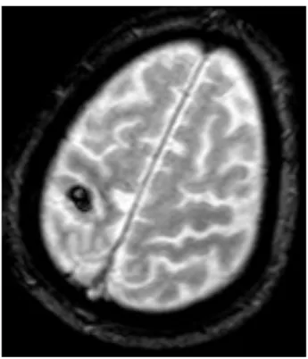
significantly higher than in Group A (5 [range, 0-‐27] vs 3 [range, 0-‐7] respectively; p = 0.04) and in 3 patients the NIHSS score was >15 with a major neurologic deficit. All patients underwent brain imaging: 56 had MRI and 15 had CT scans. All patients had sustained one or more recent ischemic infarcts < 3cm in the appropriate hemisphere. MRI was more often performed in Group A (89.5% vs 75%; p = 0.32) but the difference was not significant. Figure 1 illustrates imaging evaluation in a 87-‐year-‐old man with a left hemiplegia (NIHSS score 6 at admission) who underwent surgery 15 days after onset of symptoms, after a control brain MRI because of a hemorrhagic lesion in this first image.
Figure 1. Preoperative MRI (T2*-‐weighted sequence) showing a recent hemorrhagic
conversion (8 mm) of a superficial right territory brain infarction.
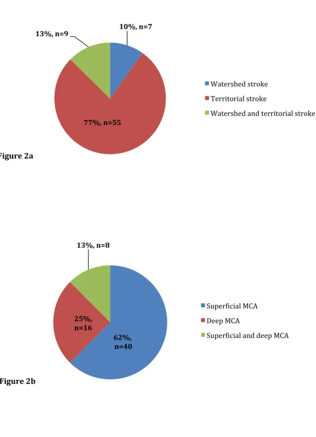
For each patient, the topographic pattern of the cerebral ischemic lesion was analyzed. Sixteen watershed strokes were noted with a similar distribution between the both groups. Among these 16 patients, we found combined border-‐zone and territorial (in the middle cerebral artery (MCA) territory) stroke caused by multiple emboli in 9 patients. Among territorial strokes, we noticed a high proportion of superficial strokes (Figure 2). Systematic carotid duplex ultrasound imaging associated with CT angiography (or MRA in case of renal impairment) was performed to evaluate the internal carotid stenosis. Results are listed in Table II. All patients received medical therapy (antiplatelet and statin) before surgery. Ten eligible patients (14.1%) received thrombolytic therapy, all belonging to Group B (0% vs 19.2%; p = 0.04).
Figure 2. Topographic pattern of stroke in the whole cohort (Figure 2a) and in the
specific middle cerebral artery (MCA) territory (Figure 2b).
10%, n=7
77%, n=55 13%, n=9
Watershed stroke Territorial stroke
Watershed and territorial stroke
Figure 2a 62%, n=40 25%, n=16 13%, n=8 Superwicial MCA Deep MCA
Superwicial and deep MCA
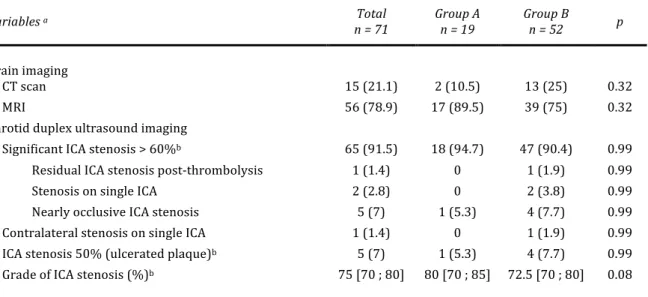
Table II. Brain imaging and results of carotid duplex ultrasound imaging.
Variables a Total
n = 71 Group A n = 19 Group B n = 52 p
Brain imaging CT scan 15 (21.1) 2 (10.5) 13 (25) 0.32 MRI 56 (78.9) 17 (89.5) 39 (75) 0.32 Carotid duplex ultrasound imaging Significant ICA stenosis > 60%b 65 (91.5) 18 (94.7) 47 (90.4) 0.99
Residual ICA stenosis post-‐thrombolysis 1 (1.4) 0 1 (1.9) 0.99 Stenosis on single ICA 2 (2.8) 0 2 (3.8) 0.99 Nearly occlusive ICA stenosis 5 (7) 1 (5.3) 4 (7.7) 0.99 Contralateral stenosis on single ICA 1 (1.4) 0 1 (1.9) 0.99 ICA stenosis 50% (ulcerated plaque)b 5 (7) 1 (5.3) 4 (7.7) 0.99
Grade of ICA stenosis (%)b
75 [70 ; 80] 80 [70 ; 85] 72.5 [70 ; 80] 0.08
ICA, Internal Carotid Artery.
aCategoric data are shown as number (%) and continuous data as median [Q1 ; Q3]. bAccording to the NASCET
criteria.
Revascularization. The mean interval from initial examination to surgery was 9.5 days
(range, 3-‐14 days) in Group A and 42 days (range, 15-‐92 days) in Group B. All carotid revascularizations were done under general anesthesia. On the basis of residual pressure measurement and an evaluation of reflux in the ICA, 5 patients in Group A and 7 patients in Group B required shunting, without significant difference (26.4% vs 13.5%; p = 0.20). The type of surgery used was similar in both groups, and the median duration of surgery was 112 minutes (IQR, 100-‐135 minutes). Carotid duplex imaging was systematically performed by the operator at the end of the revascularization. The operative results are summarized in Table III.
Outcomes. At one month, no death, no recurrent stroke or TIA and no cardiac complication had occurred in Group A with a 100% complete follow-‐up. In Group B, with a 98.1% follow-‐up (1 lost to follow-‐up), 2 patients had a postoperative stroke (Table III). The combined morbidity/mortality rate for both groups was 2.8%. The
combined morbidity/mortality rate for the Group B was 3.8%. At 3 months, with a 96.4% follow-‐up, all patients had favorable functional outcome. Follow-‐up carotid duplex imaging at 3 months showed 2 recurrent stenoses in patients who underwent bypass venous grafting.
Table III. Operative details and outcomes.
Variables a Group A
n = 19 Group B n = 52 p
Surgery Timing. Onset -‐ surgery, days 9.5 [3-‐14] 42 [15-‐92] -‐ General anesthesia 19 (100) 52 (100) -‐ Type of surgery
Endarterectomy and direct closure 1 (5.3) 2 (3.8) 0.79 Endarterectomy and reversal 6 (31.6) 14 (26.9) 0.70 Endarterectomy + patch angioplastyb 6 (31.6) 9 (17.3) 0.19
Bypass venous grafting 5 (26.3) 25 (48.1) 0.10 Bypass prothetic graftingb 1 (5.3) 2 (3.8) 0.79
Shunting 5 (26.3) 7 (13.5) 0.20 Duration of surgery, minutes 110 [75-‐180] 120 [64-‐168] 0.02 Postoperative hypertension, >16mmHg 15 (78) 43 (82.7) 0.40 30-‐day clinical outcomes Stroke 0 2 (3.8) 0.39 Mortality 0 0 -‐ Cardiac event 0 0 -‐ 90-‐day duplex ultrasound imaging outcomes Normal exam 17 (100) 48 (96) 0.40 Carotid stenosis 0 2 (4) 0.40 Lost to follow-‐up
2 (10.5) 2 (3.8) -‐
aCategoric data are shown as number (%) and continuous data as mean [range]. bPolytetrafluroethylene was used.
The two neurologic complications were acute postoperative strokes in the first 48 hours after surgery. The first was a 80-‐year-‐old man (ASA III) who had presented with left hemiplegia with an NIHSS score of 10. Initial brain imaging had shown a right superficial territorial stroke and he had undergone thrombolysis. Surgery had been
angioplasty without shunting. The patient had presented postoperative hypertension (> 20mmHg) and suffered a new stroke in the recovery room. Control brain imaging showed a hematoma in the deep right MCA territory. The second patient was a 65-‐year-‐ old man (ASA III) who had consulted for left hemiplegia with an NIHSS score of 7. Initial brain imaging had shown right deep territorial stroke. The 70% ICA stenosis had been operated 28 days after symptom onset with bypass venous grafting. Postoperative follow-‐up had been complicated by hypertension and a new stroke one day after surgery. A right occipital hematoma was found on control brain images.
Among the 12 patients who underwent shunting, 33.3% had a former history of stroke or TIA (vs 15.3% in the rest of the cohort). Their median grade of ICA stenosis was more severe (80% [IQR, 72.5-‐90%] vs 75% [IQR, 70-‐80%] ; p = 0.10) and more near occlusive (>95%) ICA stenoses were revascularized (25% vs 3.4%; p = 0.03). More watershed strokes were found in these patients (58.3% vs 15.3% ; p = 0.004). None of these patients received thrombolysis and no postoperative complication occurred.
Among the ten patients who received thrombolysis, all had systemic hypertension. At admission, their neurologic status was more severe; the mean NIHSS score was 12 (range, 2-‐27) (p <0.0001) and all patients were classified as ASA III (100% vs 65.6% ; p = 0.03). No patient underwent early surgery and the mean interval from initial examination to surgery was 46 days (range, 16-‐92 days).
Discussion
This study is the result of a retrospective analysis of data from prospectively identified patients who underwent CEA for symptomatic carotid stenosis responsible of brain infarcts in our hospital from 2010 to 2014. Performing CEA in an urgent setting after a neurologic deficit requires strong collaboration between the vascular surgeons and neurologists that is nowadays possible thanks to stroke units. During the preoperative consultation, the timing of surgery is one of the most important issues.
For patients who require CEA after brain infarct, the key question is the extent to which the absolute benefits and risks of surgery are increased by early operation. In fact, the aim of early surgery is to restore blood flow to the ischemic penumbra, which improves neurologic symptoms, and to reduce the risk of carotid occlusion and recurrent stroke. However, the major risk of the ischemic lesion developping into a hemorrhagic stroke. Nevertheless, a recent literature review by Naylor et al16 of 30-‐day outcomes and
conversion rates from ischemic stroke to hemorrhagic stroke after early CEA, concluded that the rate of symptomatic intracranial hemorrhage was reassuring, from 0% to 2%. The timing of surgery for patients who have suffered a TIA due to internal carotid stenosis is codified and is conventionally performed precociously.5-‐8 For this reason, we
decided to exclude from our study all patients who consulted for TIA, in order to focus on stroke outcomes. Results of urgent CEA depend on the operating team and the severity of the initial stroke, and this surgery is considered to be high risk. Nevertheless, several small monocentric studies have reported good results, including in patients with major strokes in the acute phase.15,17,18 Lesèche et al described no death and no
of these studies tend to consider a prompt surgical approach. The aim of our study was to analyze our 30-‐day outcomes after early CEA compared to delayed CEA, and to evaluate the quality of our selection criteria.
In our study population, the results are satisfying with an overall combined morbidity/mortality rate of 2.8%. The lack of postoperative complication in Group A confirms the good selection of patients who underwent early CEA. These results can be explained by :
-‐ Close cooperation between vascular surgeons, neurologists and neuroradiologists.
-‐ Rapid diagnosis and easy access to brain imaging (80.3% of patients had a brain MRI that allows a more accurate analysis compared to brain CT scan). -‐ Optimal preoperative medical management.
-‐ Standardized surgery performed by trained surgeons. In order to minimize perioperative risks, we implement: low mobilization of the ICA and the carotid bifurcation before clamping to prevent embolization, long arteriotomie with fixation of the stop of atheromatous plaque, and closure with patch angioplasty.
-‐ Using systematic residual pressure measurement and/or brain monitoring, and duplex ultrasound imaging by the operator at the end of the surgery.
Selection criteria for early CEA follow local guidelines which are based on the experience of the surgical and neurologic teams. In fact, several conditions are necessary in order for us to recommend early CEA. The first condition is clinical improvement. Early CEA is proposed only if patients show signs of neurologic
improvement during the workup. When at admission the patient has a NIHSS score > 8, we are usually more cautious and don’t hesitate to delay surgery. Early surgery is not performed if the patient suffers from a major disabling stroke. For patients with advanced age (> 85 years), the surgical indication depends of medical history, clinical condition and neurological improvement. We don’t propose early surgery to patients who had undergone thrombolysis. On the other hand, decision depends on brain imaging. Hemorrhagic transformation, major ischemic infarct of 3cm or more, or brain edema are contradictions for early CEA. In those cases, we repeat brain imaging and regularly revalue the timing of surgery.
Our study has several strengths. First, the data collection was detailed and included more than 60 clinical, radiologic, anesthesia and surgical management variables. Second, all patients had a standardized preoperative and operative management, with prescheduled follow-‐up consultations at 30-‐days and 90-‐days and duplex ultrasound imaging to 90-‐days. Only one patient was lost to follow-‐up in the 30-‐day perioperative period. Lastly, the exclusion of patients with TIA reduced the interpretation biases towards good results of early CEA, which could have been increased by the usually excellent outcomes in these patients. However, this selection decreased the number of patients in both groups and the power of the study.
We recognize several limitations to our study. First, it was a retrospective study. This allowed a more thorough data collection and when the NIHSS score was missing, a recalculation was possible. In fact, the determination of the NIHSS score is partly subjective. Nonetheless, the neurologic examinations of patients secondarily addressed to our department were highly detailed enabling us to avoid major bias when recalculation was needed, as in 17 patients. Second, the Group A was rather small due to