Research Article
The 2.4-Å crystal structure of the penicillin-resistant
penicil-lin-binding protein PBP5fm from Enterococcus faecium in
complex with benzylpenicillin
E. Sauvagea,*, F. Kerffa, E. Fonzéa, R. Hermana, B. Schootb, d, J.-P. Marquetteb, Y. Taburetb, D. Prevostb,
J. Dumasb, G. Leonardc, P. Stefanica, J. Coyetteaand P. Charliera
aCentre d’Ingénierie des Protéines, Université de Liege, Institut de Physique B5 and Institut de Chimie B6,
Sart Tilman, 4000 Liège (Belgium), Fax + 32 4 366 3667, e-mail: [email protected]
bAventis Pharma, 102, route de Noisy, 93235 Romainville Cedex (France)
cMacromolecular Crystallography, European Synchrotron Radiation Facility, BP 220, 38043 Grenoble Cedex (France) dPresent address: Analyse et Biologie Structurale, Galderma Research and Development, 635, route des Lucioles,
06902 Sophia Antipolis (France)
Reveived 30 April 2002; received after revision 6 June 2002; accepted 6 June 2002
Abstract. Penicillin-binding proteins (PBPs) are
mem-brane proteins involved in the final stages of peptidogly-can synthesis and represent the targets of b-lactam
tibiotics. Enterococci are naturally resistant to these an-tibiotics because they produce a PBP, named PBP5fm in
Enterococcus faecium, with low-level affinity for
b-lac-tams. We report here the crystal structure of the acyl-en-zyme complex of PBP5fm with benzylpenicillin at a res-olution of 2.4 Å. A characteristic of the active site, which distinguishes PBP5fm from other PBPs of known struc-ture, is the topology of the loop 451 – 465 defining the left
© Birkhäuser Verlag, Basel, 2002
CMLS
Cellular and Molecular Life Sciencesedge of the cavity. The residue Arg464, involved in a salt bridge with the residue Asp481, confers a greater rigidity to the PBP5fm active site. In addition, the presence of the Val465 residue, which points into the active site, reducing its accessibility, could account for the low affinity of PBP5fm for b-lactam. This loop is common to PBPs of
low affinity, such as PBP2a from Staphylococcus aureus and PBP3 from Bacillus subtilis. Moreover, the insertion of a serine after residue 466 in the most resistant strains underlines even more the determining role of this loop in the recognition of the substrates.
Key words. Penicillin-binding protein; PBP5fm; Enterococcus faecium; resistance; benzylpenicillin; peptidoglycan
synthesis.
Penicillin-binding proteins (PBPs) are essential mem-brane enzymes functioning at the late stages of peptido-glycan assembly. They are members of the penicilloyl ser-ine transferase family, a family of enzymes that catalyze the transfer of the penicilloyl moiety of penicillin to their active site serine [1]. PBPs represent the lethal targets of penicillins and more generally of b-lactam antibiotics in
susceptible bacteria. b-Lactams exhibit structural
anal-*Corresponding author.
ogy with the PBP transpeptidation substrate, the D-alanyl-D-alanine carboxy terminus of peptidoglycan pep-tides. They acylate the penicillin-binding domain of PBPs, forming a rather stable acyl-enzyme devoid of transpeptidase activity.
PBPs fall into two major groups: the low-molecular-mass (LMM) PBPs are monofunctional enzymes acting mainly as DD-peptidases [2], while the high-molecular-mass (HMM) PBPs are multimodular enzymes also displaying a DD-peptidase activity sometimes associated with a
transglycosylase activity. HMM-PBPs are further divided into classes A and B according to their function and the sequence similarities found in their non-penicillin-bind-ing module [3, 4]. The penicillin-bindnon-penicillin-bind-ing module of the multimodular PBPs is associated with at least one other domain which is assumed to have a transglycosylase ac-tivity in class A HMM-PBPs. The role of the non-peni-cillin-binding domain of class B HMM-PBPs is not yet understood. Transglycosylase activity has not been de-tected in this class of PBPs and the non-penicillin-bind-ing module has been proposed to interact with other pro-teins to form multiprotein complexes [5 – 7]. Enterococci, which are involved in many nosocomial infections, are naturally resistant to b-lactam antibiotics. Furthermore,
they have become increasingly resistant to many antibi-otics and are thus responsible for an increasing number of therapeutic failures [8 – 10]. Enterococcus faecium is the most penicillin resistant enterococcal species, with MICs ranging from 0.5 to more than 64 mg/ml for clinical
iso-lates. MICs as high as 256 and 512 mg/ml have been
found in very resistant strains [11, 12]. Some highly
b-lactam resistant E. faecium strains are also vancomycin resistant [12 – 14], although vancomycin and b-lactam
an-tibiotics can act in a synergistic way against vancomycin-resistant enterococci [15]. Since ampicillin resistance and vancomycin-vanB-type resistance have recently been shown to be cotransferable within a large mobile element [14 – 16], this double resistance has become a serious public health problem.
The natural resistance of enterococci to b-lactam
antibi-otics is due to the presence of one PBP (named PBP5fm in E. faecium) with low-level affinity for penicillin. Re-sistance to b-lactam in E. faecium may be enhanced by
higher levels of PBP5fm production. However, overpro-duction is not the only path to resistance within resistant strains of E. faecium. Point mutations in the PBP5fm fur-ther reduce the affinity of the protein for b-lactams,
lead-ing to very high levels of resistance [13, 17]. PBP5fm is thought to be able to take over the role of the other HMM-PBPs in peptidoglycan synthesis when these proteins are inhibited by an antibiotic [10, 11, 18, 19]. In E. faecalis, the peptidoglycan synthesized by PBP5 in the presence of benzylpenicillin is slightly different from that produced by the usual PBPs in the absence of benzylpenicillin. The main differences are the lack of oligomers higher than tetramers and a reduction in the number of tetramers in the muropeptide composition of the peptidoglycan, sug-gesting that PBP5 is unable to use trimeric peptides as donors or acceptors in transpeptidase reactions to form oligomeric cross-linked muropeptides. Nevertheless, un-der these conditions, PBP5 still cross-links the unmodi-fied peptidoglycan pentapeptide precursors [20]. In con-trast, strains showing vancomycin resistance are now as-sumed to synthesize a pentapeptide with an abnormal carboxyterminal unable to bind vancomycin. The
syner-gic effect between vancomycin and b-lactam can be
ex-plained by the inability of the PBP5fm to cross-link the modified pentapeptide [15, 21].
The PBP5fm crystal structure is the first structure of a low-level-affinity PBP. PBP5fm serves as a paradigm of the B1 subgroup of class B HMM-PBPs (B1-PBPs) that reassembles PBPs from enterococci (E. hirae PBP5, E.
faecalis PBP5 or PBP4), bacilli (Bacillus subtilis PBP3, B. halodurans PBP3, B. cereus PBP3), staphylococci
(Staphylococcus aureus PBP2a, S. sciuri PBP2, S.
epi-dermis PBP2) and Clostridium acetobutylicum PBP2.
PBP5fm also shares sequence similarities with a protein from the Listeria innocua genome (comparison computa-tion with PBP5fm sequence was performed at the SIB us-ing the BLAST network service [22]). The B1 subgroup enzymes are assumed to be responsible for the b-lactam
resistance of their respective bacterial species. Structural information on class B HMM-PBPs has been made avail-able through the crystallographic structure of PBP2x from Streptococcus pneumoniae [23]. PBP2x belongs to the B4 subgroup of class B HMM-PBPs and is sensitive to benzylpenicillin and other b-lactam antibiotics.
Typi-cal MICs of benzylpenicillin for non-resistant S.
pneu-moniae strains are 0.02 mg/ml (strain R6) whereas values
of 4 mg/ml (strain BM4107) are characteristic of E. fae-cium strains. While high resistance observed in some S. pneumoniae clinical isolates rests on a PBP2x harboring
many mutations [24], resistance to b-lactams is a
consti-tutive element of E. faecium and a few point mutations in PBP5fm can lead to very high levels of resistance.
Materials and methods
Construction and expression of recombinant PBP5fm molecules
DNA encoding PBP5fm was amplified by PCR using pDML517 [12] as template and primers designed to in-troduce NcoI and NdeI cloning sites, to replace the hy-drophobic anchoring peptide and putative polar region by a Met-Gly peptide in positions 32 and 33 and to introduce a Leu-Glu dipeptide at the C terminal end of the protein. The PCR product was first cloned into a pUC18 deriva-tive generating pDML536 and subcloned in the pET28a expression vector generating pDML537. The recombi-nant protein was overproduced at 25 °C in the cytoplasm of Escherichia coli BL21 (DE3) cells that were grown in medium containing kanamycin (50 mg/l) by a fed-batch process [25]. To obtain a selenium-labeled PBP5fm de-rivative, the pDML537 insert was subcloned into NcoI and EcoRI restriction sites of the pTRC99a expression vector. The labeled recombinant protein was overpro-duced at 33 °C in the cytoplasm of methionine-aux-otrophic E. colib180 cells that were grown in a medium
Purification and crystallization
IPTG-induced cells were sonicated (15 min–1) and
cen-trifuged to discard cell debris. Both PBP5fm forms were purified by anion exchange (sepharose HP and Q-sepharose Hitrap), hydroxyapatite (ceramic-hydroxyap-atite type I) and gel filtration (Superdex 200 pf) chro-matography to reach an approximate 95 % purity. Several crystals were obtained from the purified native protein and its selenium-labeled derivative, at various protein concentrations ranging from 5 to 90 mg/ml. All crystals were grown by the sitting-drop vapor diffusion method. For the crystal leading to the structure determination, the purified seleno-methionine-labeled PBP5fm protein was concentrated and cocrystallized with benzylpenicillin by mixing at the ratio 1:5. The crystal was grown at 20 °C by mixing equal volumes of PBP5fm (15 mg/ml), 10 % PEG8000 (w/v), 0.4 M Li2SO4and 100 mM Na acetate at
pH 4.5. The crystal appeared within 1 week and reached its maximum size (0.300 ¥ 0.15 ¥ 0.10 mm) in about 2
weeks.
Data collection, phase determination and structure refinement
The crystal was cryoprotected in the same buffer con-taining 20 % (v/v) glycerol and flash-cooled at 100 K for data collection at the ID14-eh4 beamline (European Synchrotron Radiation Facility-ESRF, Grenoble) using an ADSC Quantum 4RCCD detector. MAD data were collected at three different wavelengths (Se-peak, inflec-tion, remote). At each wavelength, 540 frames were col-lected with an exposure time of 0.5 s and an oscillation of one-third of a degree. The three data sets were com-plete up to 2.4 Å (statistics are given in table 1). The
crystal belongs to the space group C2 (a = 79.3 Å, b = 128.8 Å, c = 236.1 Å and b = 93.92°) with three
mole-cules in the asymmetric unit. Integration was performed with MOSFLM [27] and scaling with the program SCALA from the CCP4 program suite [28]. PBP5fm is a 648-amino-acid protein with 13 methionines. Thirty out of the 39 possible selenium sites were localized using SHAKE-AND-BAKE [29] and coordinates further refined by use of the MLPHARE program from the CCP4 suite. The multiwavelength anomalous diffraction method (MAD) map calculated after density modifica-tion with DM [30] was suitable for initiating the protein construction. The major part of the C-terminal and the N-terminal domain (without residues 193 – 256) were built in the MAD map using TURBO-FRODO [31]. Simulating annealing with CNS [32] and manual re-building allowed completion of the model except residues 32 – 38, 193 – 256 and 623 – 632. The three monomers were refined individually. The Ramachandran plot analysis revealed that 84.4 % of the non-proline, non-glycine residues are in the most favored region, and 14.7 % are in the additionally allowed region.
Results
Structure of PBP5fm
The crystal structure of PBP5fm was solved at 2.4 Å by MAD using data collected from one crystal of the Se-Met-substituted protein cocrystallized in the presence of benzylpenicillin. The space group is C2 with three mole-cules in the asymmetric unit. The structure was refined against the data set collected at the remote wavelength with an R-factor of 22.7 % and an Rfreeof 28.6 %. The
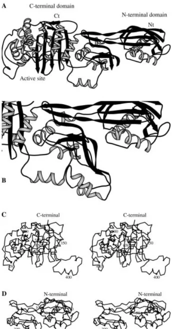
fi-nal model contains 570 residues per monomer and one benzylpenicillin molecule bound to the active-site serine of each monomer. PBP5fm presents two clear distinct do-mains (fig. 1), the N- and the C-terminal domain, the lat-ter being the transpeptidase domain that shows weak penicillin-binding activity.
The N-terminal domain exhibits a new type of folding. The N-terminal helix begins at residue 39, residues 33 – 38 being invisible in the electron density map. The region up to residue 162 is globular and composed of two helices (aN1and aN2) and a three-stranded antiparallel b
sheet (bN1to bN3). A long b sheet connects this globular
domain to the C-terminal domain. It is comprised of the strands bN5, bN10and bN11that form a three-stranded
an-tiparallel b sheet on the N-terminal side and becomes a
two-stranded parallel b sheet (segments 170 – 177 and
337 – 344) on the C-terminal side. In the central region (residues 190 – 260), the electron density maps only sug-gest some secondary-structure elements that were not in-cluded in the final model. Its conformation must differ from the equivalent PBP2x region since there would be a
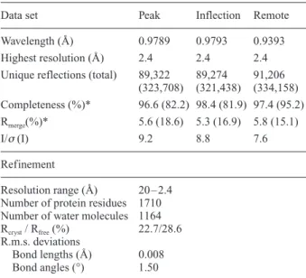
Table 1. Data collection and refinement statistics.
Data set Peak Inflection Remote
Wavelength (Å) 0.9789 0.9793 0.9393 Highest resolution (Å) 2.4 2.4 2.4 Unique reflections (total) 89,322 89,274 91,206
(323,708) (321,438) (334,158) Completeness (%)* 96.6 (82.2) 98.4 (81.9) 97.4 (95.2) Rmerge(%)* 5.6 (18.6) 5.3 (16.9) 5.8 (15.1) I/s(I) 9.2 8.8 7.6 Refinement Resolution range (Å) 20 – 2.4 Number of protein residues 1710 Number of water molecules 1164 Rcryst/ Rfree(%) 22.7/28.6
R.m.s. deviations
Bond lengths (Å) 0.008 Bond angles (°) 1.50
* Statistics for the highest-resolution shell (2.55 – 2.4 Å) are given in parentheses.
steric clash between this region and a symmetrical protein in the crystal packing. An attempt to determine the back-bone trace in monomer A is shown in figure 1 B. The N-terminal domain has been shown to be important for the folding of the PBP5 enzyme from E. hirae, the se-quence of which is nearly identical to that of PBP5fm [33]. The deletion of some segments in the N-terminal domain resulted in proteins unable to bind penicillin, a
situation that also prevails in E. coli PBP3 [5] and S.
au-reus PBP2a [34]. Transglycosylase activity has not been
demonstrated in PBP5fm and no clear biological function could be attributed to the N-terminal domain, which could, according to the most widely accepted assump-tions, interact with other proteins to form a multiprotein complex responsible for the synthesis of the cell wall pep-tidoglycan.
The C-terminal domain shows structural similarities with the transpeptidase domain of the penicillin-sensitive PBP2x and more generally with the penicillin-recogniz-ing enzymes (b-lactamases of classes A, C or D [35], E. coli PBP5 [36], and the Streptomyces R61 [37] and K15
[38] DD-peptidases). The active-site serine 422 is located between two subdomains, one a five-stranded b sheet
covered by helices, the other being all a helices. The
Ser422 – Lys425 segment at the beginning of helix aC2
(the secondary-structural elements defined for class A
b-lactamases will be used with subscript C to describe the transpeptidase domain) and the Ser480-Asp-Asn and Lys618-Thr-Gly-Thr motifs define the active-site cleft as in most active serine PBPs. The loop at the bottom of the cavity contains residues Gly537-Tyr-Gly-Gln-Gly and adopts the same conformation as in PBP2x. Downstream, the bC3strand that bears the Lys618-Thr-Gly motif, the
bC3-bC4 connecting loop (residues 623 – 632), is
com-pletely invisible in the electron density map of the three monomers. In PBP2x, this loop has three additional residues and defines one side of a long groove that stretches on both parts of the active site. The other side of the groove is delimited by residues 461 – 465 which cover the Ser480-Asp-Asn motif. In PBP5fm, the groove is not as deep as in PBP2x and the residues bordering the ac-tive-site entry are different in both structures, maybe re-flecting the specificity of the natural substrate of each en-zyme.
The main structural difference between PBP5fm and the
b-lactamases or the LMM-PBPs is the insertion of three
loops extending in the direction of the N-terminal do-main. One of these loops (388 – 410) is close to the poorly defined region 190 – 260 and a double hydrogen bond is formed between the Asn388 side chain and the Tyr265 backbone.
Structure of class B HMM-PBPs
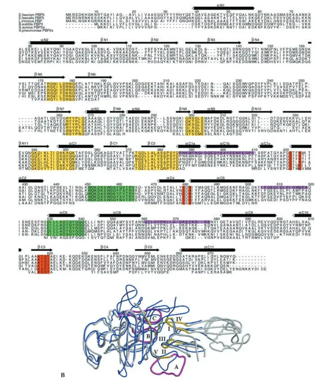
Both PBP5fm and PBP2x belong to the class B HMM-PBPs. All the members of this class share five common sequence motifs not directly involved in the enzymat-ic activity (motifs I – V: Arg173-Gly-Xaa-Xaa-Xaa-Asp-Arg-Asn-Gly, Arg263-Xaa-Tyr-Pro-Xaa-Gly, Gly299-Xaa-Xaa-Gly-Xaa-Glu, Gly340-Xaa-Asp-Xaa-Xaa-Xaa-Thr-Xaa-Asp-Xaa-Xaa-Xaa-Glu and Thr373-Gly-Asp (Glu)-Xaa-Leu-Ala-Xaa-Xaa-Xaa-Xaa-Pro-Ser-Xaa-Asp) [3]. These motifs are located at the interface between the N- and C-terminal domains (fig. 2). Motifs I and IV
con-Figure 1. Overall structure of PBP5fm. (A) Ribbon representation of the tertiary structure of PBP5fm. The C-terminal domain is the transpeptidase domain, while the function of the N-terminal do-main is unknown. (B) Ribbon representation of the central region of PBP5fm. The backbone trace of residues 192 – 257 has been tenta-tively determined in the monomer A. (C) Stereoview of the Catrace of the C-terminal domain (346 – 680). (D) Stereoview of the Ca trace of the N-terminal domain (39 – 346).
Figure 2. Structure of class B high-molecular mass PBPs. (A) Sequence alignment of five representatives of subgroup B1 of class B HMM-PBPs and partial alignment with Streptococcus pneumoniae PBP2x that belongs to subgroup B4. The following sequences are available from GenBank (accession numbers in parentheses): Enterococcus faecium PBP5 (CAA59287), Enterococcus faecalis PBP5 (CAB89865),
Listeria innocua PBP (CAC95693), Bacillus subtilis PBP3 (CAB12221), Staphylococcus aureus PBP2a (CAA68684), Streptococcus pneumoniae PBP2x (CAA34412). PBP2x alignment is based on a structural comparison with PBP5fm. Only PBP2x residues with
struc-tural analogs in PBP5fm are shown. Active-site catalytic residues of PBPs are colored red, conserved strucstruc-tural motifs of class B-PBPs are colored yellow and B1-PBPs conserved boxes near the active site are colored green. The loops in magenta define a groove surrounding the conserved structural motifs of class B-PBPs. The secondary structures of the transpeptidase domain are labeled following their usual name in class A b-lactamases. Color scheme is conserved for figures 2 and 4. (B) Conserved structural motifs in the class B HMM-PBPs. PBP5fm is shown in gray and PBP2x is shown in blue. The conserved structural motifs of class B HMM-PBPs are colored yellow and the loops defining the groove around the conserved motifs are colored magenta.
A
stitute the two parallel b strands of the ‘degenerated’ b
sheet described above, while motif V is the b2C strand
defining the C-terminal domain interface. Motifs II and III are small loops connecting strand bN7to helix aN3and
strand bN9 to strand bN10, respectively. The N-terminal
globular region (39 – 162) is connected to motif I through strand bN5. The region between motifs I and II (190 – 260)
is not seen in the density map. The region between motifs II and III (313 – 332) defines the central b sheet and a
lit-tle loop constituted by residues 279 – 297 is connected to motifs III and IV. These four regions are very different in sequence and in length in PBP2x, whereas all these ele-ments are of similar length and exhibit partially con-served amino acid sequences in all B1-PBPs.
Motifs I – IV may be schematically represented as the four fingers of a right hand pointing into the palm of a left hand. The thumb of this left hand is the fifth motif and the palm is defined by the three loops (loop A: 388 – 410, loop B: 505 – 519 and loop C: 561 – 581) be-longing to the C-terminal transpeptidase domain. The three loops do not exist in class A b-lactamases or in
LMM-PBPs. They are characteristic of class B HMM-PBPs even if they differ in length and amino acid se-quences. The 561 – 581 loop C is more elongated in PBP2x than in PBP5fm, while the 388 – 410 loop A is shorter. Some strong interactions maintain these fingers together (Arg179-Asp375, Arg263-Glu304) and anchor them into the transpeptidase module (Arg173-Glu304). Whatever the structures of the N-non-penicillin-binding and C-transpeptidase-penicillin-binding terminal do-mains of class B HMM-PBPs, they are held together by a tight interface structure of about 150 Å2, defined by the
conserved motifs I – V.
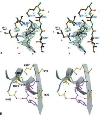
Interaction with penicillin
The electron density of the PBP5fm active site unam-biguously reveals a covalent acyl-enzyme with ben-zylpenicillin (fig. 3 A). The thiazolidine ring and the car-boxylate are well defined and the electron density is con-tinuous between the active site serine hydroxyl group and the b-lactam carbonyl group. The side chain is easily
traced in the electron density of monomer A but is less well defined in the other monomers. The benzylpenicillin conformation (fig. 3 B) is similar to the one observed in the acyl-enzyme formed by benzylpenicillin and the Glu166-Asn inactive mutant of TEM1 class A
b-lacta-mase [39]. The carbonyl oxygen lies in the oxyanion hole defined by the Ser422 and Thr618 main chain amine groups and the carboxylate is at hydrogen bond distance from Thr618 Og and Thr620 Og. The amide group of the
benzylpenicillin side chain is wedged between Asn482 and the bC3strand. In class A and class D b-lactamases,
the carboxylate interacts with the guanidinium group of an arginine that can occupy different positions in the pri-mary structure of these enzymes. The presence of such a
guanidinium group in b-lactamases is favorable to
acyla-tion by b-lactams [40].
Acylation of PBP5fm is very slow (second-order rate constant k2/K = 20 M–1s–1) [12]. It can proceed according
to a two-step model where Lys425 abstracts a proton from the active-site serine hydroxyl group to promote the nu-cleophilic attack of the b-lactam carbonyl by the activated
serine Og [39, 41]. The proton is subsequently back
do-nated by Lys425 to the b-lactam nitrogen via Ser480. An
alternative interpretation of the acylation mechanism re-lies upon a concerted one-step process where covalent-bond formation between penicillin and the active-site ser-ine Ser422 is concomitant with the transfer of the Ser422 proton directly to Ser480 and to the transfer of the Ser480 hydroxyl hydrogen to the b-lactam nitrogen [42]. Both
charged Lys425 and Lys617 support the correct orienta-tion of the Ser480 hydroxyl group. This explanaorienta-tion could prevail for all penicilloyl serine transferases in-cluding enzymes possessing a tyrosine residue instead of a serine in the second conserved active-site motif, such as class C b-lactamases. Both interpretations are in
accor-dance with the distances between the active-site residues observed in PBP5fm.
As for most PBPs, the deacylation step is extremely slow compared to the efficiency of the deacylation step in class A b-lactamases. In the latter, a glutamic acid residue in
Figure 3. Benzylpenicillin binding in PBP5fm. (A) Stereo view of an Fobs– Fcalcelectron density omit map calculated without the
ben-zylpenicillin molecule, contoured at 2.5 s. (B) Stereoview of the interactions between the benzylpenicillin and the PBP5fm active-site residues. Benzylpenicillin is colored magenta. Hydrogen-bonded interactions are shown as thin black lines.
A
position 166 (bottom of the active site) is responsible for the high deacylation rate, by activating a conserved water molecule that attacks the acyl-enzyme carbonyl [43]. Though a water molecule is present in the PBP5fm active site at a position equivalent to the hydrolytic water mole-cule of class A b-lactamases, there is no counterpart to
Glu166 that could be relevant to activate this molecule.
Low-level affinity
A low-level affinity for b-lactams is the main
character-istic of the B1-PBPs. The second-order rate constant k2/K
for the acylation of PBP2a from S. aureus by penicillin V is about 16 M–1 s–1 [44]. The corresponding values for
PBP5fm from E. faecium strains vary from 15 to 24 M–1
s–1and they are at least ten times lower for PBP5fm from
highly resistant strains [12]. In comparison, a k2/K value
of 58,000 M–1s–1has been reported for the acylation of
PBP2x from a sensitive S. pneumoniae strain by ben-zylpenicillin [45].
The common motifs required to allow efficient acylation by penicillin are present in PBP5fm, with the exception of the guanidinium group found in b-lactamases that
in-creases the efficacy of the acylation process, but this group is not essential for the acylation step [46]. The ac-tive-site cleft is delimited by the bC3strand on one side,
by residues 461 – 465 on the opposite side, and the bottom of the cavity is covered with residues 537 – 541. Among the residues surrounding the active-site groove, only Glu622 on the bC3strand and Val465 on the opposite side
can hinder the incoming antibiotic. In PBP2x, the muta-tion of Gln552 (equivalent to Glu622 in PBP5fm) into a glutamic acid results in a decreased affinity for ben-zylpenicillin by a factor of three [47]. Thus Glu622 could partially contribute to the low-level affinity of PBP5fm. On the other side of the groove, residues 461 – 465 are part of a loop (451 – 465) very well conserved in B1-PBPs, whereas the primary structure of this loop is dif-ferent in the other classes of PBPs (figs 2 A, 4). Arg464 might play a critical structural role. It is within hydrogen-bonding distance from the backbone carbonyl oxygen of residues 446, 466 and 468, and is involved in a salt bridge with Asp481. Val465 points into the active site and its side chain is close to the benzylpenicillin thiazolidine ring. The distance between the valine side chain Cg2and the sulfur atom of the benzylpenicillin is about 3.5 Å. A backward movement of the valine and of the loop bearing this residue is perhaps necessary to allow access of ben-zylpenicillin into the active site. Comparison between the PBP2x structure and its adduct with cefuroxime (pdb codes 1QME and 1QMF [48]) shows that a tryptophan, lying at a position equivalent to Val465, is slightly pushed away from the active site by cefuroxime and that the he-lix bearing the tryptophan is also somewhat displaced. In PBP5fm, the loop movement is accompanied by a shift of the Ser480-Asp-Asn loop via the Arg464-Asp481 salt bridge. Ser480 would then be too far away from the tive-site serine to allow the proton shuttle between the ac-tive-site serine and the b-lactam nitrogen. According to
this interpretation, the interaction between the 451 – 465 loop and the second active-site motif, and more precisely the constraints induced by the Arg464 interactions, con-fer a great rigidity to this side of the groove that would be responsible for the low-level affinity of PBP5fm. Among the penicilloyl transferase enzymes, only B1-PBPs and class A b-lactamases have an aspartic acid in the second
active-site element (Ser480-Asp-Asn). Nevertheless, the aspartic acid interactions as well as the enzyme structure around this residue are completely different in both types of enzyme. The strong interaction between Arg464 and Asp481 is thus typical of B1-PBPs.
The bottom of the cavity is covered with residues 537 – 541. These residues and the 530 – 536 helix are well conserved in B1-PBPs whereas the whole 530 – 541 se-quence is different in other class B PBPs (fig. 4). Never-theless, no side chain position or particular interaction occurring in this region can easily be related to the low-level affinity of PBP5fm.
Highly resistant mutants
The protein described above is responsible for the high resistance to b-lactam antibiotics of the E. faecium D63r
strain (MIC of penicillin = 70 mg/ml). The mechanism
in-volved in the resistance is the overproduction of the pro-tein [12]. The past two decades have seen the emergence
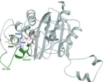
Figure 4. Ribbon representation of the C-terminal domain of PBP5fm. Color scheme as in figure 2. The conserved loops of sub-group B1 HMM-PBPs are colored green. Active-site catalytic residues are colored red, benzylpenicillin is colored black and the residues Val465 and Met485 are colored blue. On the left of the fig-ure, Arg464, Asp481 and the backbone carbonyl of residues 446, 466 and 468 are shown in yellow with oxygen atoms in red and ni-trogen atoms in blue. Arg464 is involved in a salt bridge with Asp481 and is at hydrogen bond distance from the carbonyl oxygen of residues 446, 466 and 468. See text for details.
of some clinical isolates highly resistant to ampicillin, not by means of PBP5fm overproduction but by a reduction of the protein affinity for the antibiotic. Following an analysis by Rybkine et al. [13] who studied a series of clinical isolates with various levels of resistance to ampi-cillin (well correlated with the resistance to benzylpeni-cillin), a first level of resistance is associated with PBP5fm overproduction but higher levels of resistance can be achieved by mutations of Met485 (fig. 4) or the in-sertion of an additional serine in position 466¢.
MICs of ampicillin for strains retaining Met485 are not higher than 16 mg/ml (observed in four strains).
Replace-ment of the methionine by threonine results in an MIC increase to values as high as 64 mg/ml (seven strains)
while its mutation into alanine gives MICs ranging from 128 to 256 mg/ml (five strains). Met485 belongs to helix aC5(three residues after the Ser480-Asp-Asn triad) and
its side chain is located behind the Lys425 side chain. The removal of the bulky methionine side chain would set some free space, giving the lysine more conformational freedom and resulting in a less efficient acylation process. In a similar way, the substitution of Met426 by an isoleucine, as observed in the ampicillin-resistant strain 9439 [49], could be responsible for the resistance of this strain to ampicillin. As Met426 is near Lys425, modification of its side chain could slightly destabilize the active site. Nevertheless, changing a methionine into an isoleucine only leads to minor steric modifications and can hardly account for an important reduction in the acy-lation of PBP5fm by ampicillin.
Insertion of a serine in position 466¢ (residue 466 is also
a serine) in the most resistant strains emphasizes the im-portance of the 451 – 465 loop on the left-hand side of the active site. This insertion is associated with a twofold-in-creased MIC for penicillin (strains AR9, 6885 and H80721) and seems to be independent of Met485 muta-tions [13]. As mentioned above, Val465 is close to the thiazolidine ring of the penicilloyl moiety and the struc-ture rigidity of the left side of the active-site cleft rests upon the Arg464 – Asp481 salt bridge. The insertion of a residue in position 466¢ may slightly displace Val465
in-side the active site, reducing its accessibility for peni-cillin. The serine insertion would therefore affect sub-strate recognition.
Discussion
PBP5fm is a multimodular protein with two main do-mains. The C-terminal one, commonly named the peni-cillin-binding domain, is expected to be responsible for the DD-transpeptidase activity and is inhibited by
b-lac-tam antibiotics. The function of the N-terminal domain is still unclear. However, the two domains seem to be inter-dependent. As the PBP5fm N-terminal domain is far from
the penicillin-binding module active site (about 50 Å), the N-terminal module has been hypothesized to be nec-essary for correct folding of the C-terminal one. This as-sumption remains to be demonstrated but, presumably, a misfolded or unfolded truncated N-terminal domain may somehow modify the transpeptidase domain structure or interfere with its penicillin-binding ability. An interaction between the N-terminal domain and one or several other proteins remains the most attractive proposal. Since PBP5fm is deprived of the sequence motifs correlated with the transglycosylase activity of class A HMM-PBPs and seems, accordingly, to be unable to carry out this ac-tivity, the final step of the peptidoglycan synthesis may require its transpeptidase module to be near the transgly-cosylase domain of another protein. The recent finding that the transglycosylase domain of S. aureus PBP2, to-gether with the PBP2a transpeptidase domain, is needed for the optimal expression of methicillin resistance [50] is supporting this hypothesis.
The N-terminal extensions of the three representative B1-PBPs (PBP5fm, S. aureus PBP2a and B. subtilis PBP3) are of similar length and share common motifs which are missing in other subgroups of class B HMM-PBPs. Nev-ertheless, the homology between the primary structures of the three enzymes is lower in the globular region of the N-terminal module (from the anchor peptide to the first class B conserved motif, residues 39 – 162) than in the rest of the molecule. This lends further support to the idea of an N-terminal module interacting with other proteins, since protein-protein interactions occurring in the supramolecular assembly involved in the late steps of peptidoglycan synthesis can be specific for each species. Yet, biological function of the N-terminal domain re-mains unclear and the validity of a model associating a normal PBP tranglycosylase activity and a rescue PBP transpeptidase activity to express b-lactam resistance
re-mains to be demonstrated in enterococci.
The reason why PBP5fm is naturally resistant to peni-cillin is not evident at first sight. Sequence alignments give supportive evidence that all B1-PBPs are structurally alike and probably play a similar functional role in pepti-doglycan synthesis. The active-site region is surrounded by sequence elements (451– 465, 530 – 540) that are strongly conserved in B1-PBPs, while the sequence ho-mology is lower in other regions. These sequence ele-ments are not conserved in other classes of PBPs, in their primary structure and probably also in their tertiary struc-ture, as shown by the comparison with the PBP2x three dimensional structure. Accordingly, the presence of Val465 near the binding site and the salt bridge Arg464-Asp481, characteristic of B1-PBPs, can be invoked as structural elements responsible for the low-level affinity for penicillin of these enzymes. PBP5fm mutations asso-ciated with highly resistant clinical isolates are the re-placement of Met485 by threonine or alanine, potentially
leading to a decrease of k2, and the insertion of a serine in
position 466¢, which might in turn affect b-lactam
recog-nition. The latter modification is found in the sequence of the most highly ampicillin resistant strains and seems to be an essential determinant in PBP5fm-mediated
b-lac-tam resistance.
Acknowledgements. This work was supported in part by the Belgian
Program on Interuniversity Poles of Attraction initiated by the Bel-gian State, Prime Minister’s Office, Science Policy Programming (PAI P4/03).
1 Ghuysen J. M. (1994) Molecular structure of penicillin-binding proteins and b-lactamases. Trends Microbiol. 2: 372 – 380. 2 Frère J. M., Nguyen-Distèche M., Coyette J. and Joris B.
(1992). Mode of action: interaction with the penicillin-binding proteins. In: The Chemistry of b-Lactams, pp. 148-197, Page M. I. (ed.), Blackie, London
3 Goffin C. and Ghuysen J. M. (1998) Multimodular penicillin-binding proteins: an enigmatic family of orthologs and par-alogs. Microbiol. Mol. Biol. Rev. 62: 1079 – 1093.
4 Massova I. and Mobashery S. (1998) Kinship and diversifica-tion of bacterial penicillin-binding proteins and b-lactamases. Antimicrob. Agents Chemother. 42: 1 – 17
5 Nguyen-Distèche M., Fraipont C., Buddelmeijer N. and Nan-ninga N. (1998) The structure and function of Escherichia coli penicillin-binding protein 3. Cell. Mol. Life Sci. 54: 309 – 316 6 Vicente M. and Errington J. (1996) Structure, function and
con-trols in microbial division. Mol. Microbiol. 20: 1 – 7
7 Ghuysen J. M. (1997) Penicillin-binding proteins: wall pepti-doglycan assembly and resistance to penicillin: facts, doubts and hopes. Int. J. Antimicrob. Agents 8: 45 – 60
8 Williamson R., LeBouguenec C., Gutmann L. and Horaud T. (1985) One or two low affinity penicillin-binding proteins may be responsible for the range of susceptibility of Enterococcus
faecium to benzylpenicillin. J. Gen. Microbiol. 131: 1933 –
1940
9 Hakenbeck R. and Coyette J. (1998) Resistant penicillin-bind-ing proteins. Cell. Mol. Life Sci. 54: 332 – 340
10 Murray B. E. (1990) The life and times of the Enterococcus. Clin. Microbiol. Rev. 3: 46 – 65
11 Fontana R., Aldegheri M., Ligozzi M., Lopez H., Sucari A. and Satta G. (1994) Overproduction of a low-affinity penicillin-binding protein and high-level resistance in Enterococcus
fae-cium. Antimicrob. Agents Chemother. 38: 1980 – 1983
12 Zorzi W., Zhou X. Y., Dardenne O., Lamotte J., Raze D., Pierre J. et al. (1996) Structure of the low-affinity penicillin-binding protein 5 PBP5fm in wild-type and highly penicillin-resistant strains of Enterococcus faecium. J. Bacteriol. 178: 4948 – 4957 13 Rybkine T., Mainardi J. L., Sougakoff W., Collatz E. and Gut-mann L. (1998) Penicillin-binding protein 5 sequence alter-ation in clinical isolates of Enterococcus faecium with different levels of b-lactam resistance. J. Infect. Dis. 178: 159 – 163 14 Hanrahan J., Hoyen C. and Rice L. B. (2000) Geographic
dis-tribution of a large mobile element that transfers ampicillin and vancomycin resistance between Enterococcus faecium strains. Antimicrob. Agents Chemother. 44: 1349 – 1351
15 Al-Obeid S., Billot-Klein D., Heijenoort J. van, Collatz E. and Gutmann L. (1992) Replacement of the essential penicillin-binding protein 5 by high-molecular mass PBPs may explain vancomycin-b-lactam synergy in low-level vancomycin-resis-tant Enterococcus faecium D366. FEMS Microbiol. Lett. 91: 79 – 84
16 Carias L. L., Rudin S. D., Donskey C. J. and Rice L. B. (1998) Genetic linkage and cotransfer of a novel, vanB-containing transposon (Tn5382) and a low-affinity penicillin-binding
pro-tein 5 gene in a clinical vancomycin-resistant Enterococcus
faecium isolate. J. Bacteriol. 180: 4426 – 4434
17 Klare I., Rodloff A. C., Wagner J., Witte W. and Hakenbeck R. (1992) Overproduction of a penicillin-binding protein is not the only mechanism of penicillin resistance in Enterococcus
fae-cium. Antimicrob. Agents Chemother. 36: 783 – 787
18 Rice L. B., Carias L. L., Hutton-Thomas R., Sifaoui F., Gut-mann L. and Rudin S. D. (2001) Penicillin-binding protein 5 and expression of ampicillin resistance in Enterococcus
fae-cium. Antimicrob. Agents Chemother. 45: 1480 – 1486
19 Sifaoui F., Arthur M., Rice L. and Gutmann L. (2001) Role of penicillin-binding protein 5 in expression of ampicillin resis-tance and peptidoglycan structure in Enterococcus faecium. Antimicrob. Agents Chemother. 45: 2594 – 2597
20 Signoretto C., Boaretti M. and Canepari P. (1998) Peptidogly-can synthesis by Enterococcus faecalis penicillin binding pro-tein 5. Arch. Microbiol. 170: 185 – 190
21 Bugg T. D. H., Wright G. D., Dutka-Malen S., Arthur M., Courvalin P. and Walsh C. T. (1991) Molecular basis for van-comycin resistance in Enterococcus faecium BM4147: biosyn-thesis of a depsipeptide peptidoglycan precursor by van-comycin resistance proteins VanH and Van A. Biochemistry
30: 2017 – 2021
22 Altschul S. F., Madden T. L., Schaffer A. A., Zhang J., Zhang Z., Miller W. et al. (1997) Gapped BLAST and PSI-BLAST: a new generation of protein database search programs. Nucleic Acids Res. 25: 3389 – 3402
23 Pares S., Mouz N., Petillot Y., Hakenbeck R. and Dideberg O. (1996) X-ray structure of Streptococcus pneumoniae PBP2x, a primary penicillin target enzyme. Nat. Struct. Biol. 3: 284 – 289
24 Dessen A., Mouz N., Hopkins J. and Dideberg O. (2001) Crys-tal structure of PBP2x from a highly penicillin-resistant
Strep-tococcus pneumoniae clinical isolate: a mosaic framework
con-taining 83 mutations. J. Biol. Chem. 30: 45106 – 45112 25 Korz D. J., Rinas U., Hellmuth K., Sanders E. A. and Deckwer
W. D. (1995) Simple fed-batch technique for high cell density cultivation of Escherichia coli. J. Biotechnol. 39: 59 – 65 26 LeMaster D. M. and Richards F. M. (1985) 1H-15N
heteronu-clear NMR studies of Escherichia coli thioredoxin in samples isotopically labeled by residue type. Biochemistry 24: 7263 – 7268
27 Leslie A. G. R. W. (1996) MOSFLM Version 5.40 Mosflm Users Guide
28 CCP4 (1994) Collaborative computing project. Acta Crystal-logr. D56: 791 – 794
29 Miller R., Gallo S. M., Khalak H. G and Weeks C. M. (1994)
SnB: crystal structure determination via Shake-and-Bake. J.
Appl. Cryst. 27: 613 – 621
30 Cowtan K. and Main P. (1998) Miscellaneous algorithms for density modification. Acta Crystallogr. D54: 487 – 493 31 Roussel A. and Cambillau C. (1989). Silicon Graphics
Geome-try Partner Directory, pp. 77 – 78, Silicon Graphics, Mountain View, Calif.
32 Brünger A. T., Adams P. D., Clore G. M., DeLano W. L., Gros P., Grosse-Kunstleve R. W. et al. (1998) Crystallography & NMR system: a new software suite for macromolecular struc-ture determination. Acta Crystallogr. D54: 905 – 921
33 Mollerach M. E., Partoune P., Coyette J. and Ghuysen J. M. (1996) Importance of the E46 – D160 polypeptide segment of the non-penicillin-binding module for the folding of the low-affinity, multimodular class B penicillin-binding protein 5 of
Enterococcus hirae. J. Bacteriol. 178: 1774 – 1775
34 Wu C. Y. E., Alborn W. E., Flokowitsch J. E., Hoskins J., Ünal S., Blaszczak L. C. et al. (1994) Site-directed mutagenesis of the mecA gene from a methicillin-resistant strain of
Staphylo-coccus aureus. J. Bacteriol. 176: 443 – 449
35 Maveyraud L., Golemi D., Kotra L. P., Tranier S., Valulenko S., Mobashery S. et al. (2000) Insights into class D b-lactamases
are revealed by the crystal structure of the OXA10 enzyme from Pseudomonas aeruginosa. Structure 8: 1289– 1298 36 Davies C., White S. W. and Nicholas R. A. (2001) Crystal
struc-ture of a deacylation-defective mutant of penicillin-binding protein 5 at 2.3 Å resolution. J. Biol. Chem. 276: 616 – 623 37 Kelly J. A., Knox J. R., Moews P. C., Hite G. J., Bartolone J. B.,
Zhao H. et al. (1985) 2.8-Å structure of penicillin-sensitive D-alanyl carboxypeptidase-transpeptidase from Streptomyces R61 and complexes with beta-lactams. J. Biol. Chem. 260: 6449 – 6458
38 Fonzé E., Vermeire M., Nguyen-Distèche M., Brasseur R. and Charlier P. (1999) The crystal structure of a penicilloyl-serine transferase of intermediate penicillin sensitivity: the DD-transpeptidase of Streptomyces K15. J. Biol. Chem. 274: 21853 – 21860
39 Strynadka N. C. J., Adachi H., Jensen S. E., Johns K., Sielecki A., Betzel C. et al. (1992) Molecular structure of the acyl-en-zyme intermediate in b-lactam hydrolysis at 1.7 Å resolution. Nature 359: 700 – 705
40 Zafaralla G., Manavathu E. K., Lerner S. A. and Mobashery S. (1992) Elucidation of the role of arginine-244 in the turnover processes of class A beta-lactamases. Biochemistry 31: 3847 – 3852
41 Wladkowski B. D., Chenoweth S. A., Sanders J. N., Krauss M. and Stevens W. J. (1997) Acylation of b-lactams by class A b -lactamase: an ab initio theoretical study on the effects of the oxy-anion hole. J. Am. Chem. Soc. 119: 6923 – 6431
42 Dive G. and Dehareng D. (1999) Serine peptidase catalytic ma-chinery: cooperative one-step mechanism. Int. J. Quant. Chem.
73: 161 – 174
43 Adachi H., Ohta T. and Matsuzawa H. (1991) Site-directed mu-tants, at position 166, of RTEM-1 b-lactamase that form a
sta-ble acyl-enzyme intermediate with penicillin. J. Biol. Chem.
266: 3186 – 3191
44 Lu W. P., Sun Y., Bauer M. D., Paule S., Koenigs P. M. and Kraft W. G. (1999) Penicillin-binding protein 2a from methicillin re-sistant Staphylococcus aureus: kinetic characterization of its interactions with b-lactams using electrospray mass spectrom-etry. Biochemistry 38: 6537 – 6546
45 Jamin M., Damblon C., Millier S., Hakenbeck R. and Frère J. M. (1993) Penicillin-binding protein 2x of Streptococcus
pneu-moniae: enzymic activities and interactions with beta-lactams.
Biochem. J. 292: 735 – 741
46 Delaire M., Labia R., Samama J. P. and Masson J. M. (1992) Site-directed mutagenesis at the active site of Escherichia coli TEM-1 beta-lactamase: suicide inhibitor-resistant mutants re-veal the role of arginine 244 and methionine 69 in catalysis. J. Biol. Chem. 267: 20600 – 20606
47 Mouz N., Di Guilmi A. M., Gordon E., Hakenbeck R., Dide-berg O. and Vernet T. (1999) Mutations in the active site of penicillin-binding protein PBP2x from Streptococcus
pneumo-niae. J. Biol. Chem. 274: 19175 – 19180
48 Gordon E., Mouz N., Duée E. and Dideberg O. (2000) The crys-tal structure of the penicillin-binding protein 2x from
Strepto-coccus pneumoniae and its acyl-enzyme form: implication in
drug resistance. J. Mol. Biol. 299: 477 – 485
49 Ligozzi M., Pittaluga F. and Fontana R. (1996) Modification of penicillin-binding protein 5 associated with high-level ampi-cillin-resistance in Enterococcus faecium. Antimicrob. Agents Chemother. 40: 354 – 357
50 Pinho M. G, de Lencastre H. and Tomasz A. (2001) An acquired and a native penicillin-binding protein cooperate in building the cell wall of drug-resistant staphylococci. Proc. Natl. Acad. Sci. USA 98: 10886 – 10891