Published in: Annals of Thoracic Surgery (1992), vol. 54, pp. 983-984. Status Postprint (Author’s version)
Aneurysm of the Ascending Aorta After Cardiac Transplantation
Jean-Olivier Defraigne, Olivier Vahdat, Jean-Paul Lavigne, Jean-Claude Demoulin, and Raymond Limet, Departments of Cardiovascular Surgery and Cardiology, C.H.U. Sart-Tilman, Liège, Belgium
ABSTRACT
We report the case of a 57-year-old female cardiac transplant patient in whom an aneurysm of the recipient side of the ascending aorta developed 1 year after transplantation. Although a mycotic origin was the likely cause, histologic examination diagnosed an atherosclerotic aneurysm.
After cardiac transplantation, infection and rejection remain the major causes of morbidity and death [1, 2]. Besides infection, immunosuppression itself accounts for some morbidity [1, 3]. Immunosuppressive agents have deleterious side effects that may alter both quality of life and survival. However, graft atherosclerosis appears progressively as a major impediment to long-term survival [4, 5]. We present here a case of an aneurysm of the ascending aorta that appeared within the first year after transplantation. The immunosuppressive regimen could have been a contributive factor for rapid development of this aneurysm.
A 57-year-old woman underwent transplantation for ischemic cardiomyopathy. The donor aorta was normal. The diameter of the recipient aorta was 35 mm, and there was no evidence of atherosclerotic plaque formation. Immunotherapy included azathioprine, cyclosporin, and prednisone. The postoperative course was complicated by right pleural empyema successfully treated with intensive antibiotic therapy and drainage.
At 5 months, a stage IIB rejection required increased dosages of corticoid. Muscular atrophy, systemic
hypertension, hypercholesterolemia, and insulin-dependent diabetes occurred as side effects of immunotherapy. After 8 months, a decompensation of the diabetes occurred, and several temperature peaks were noted. Although chest roentgenograms and cultures of sputum and urine had negative results, she was treated with
broad-spectrum antibiotics.
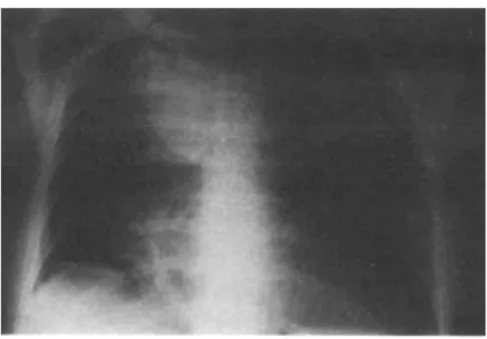
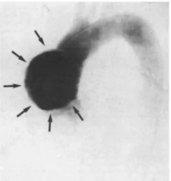
Two months later, a right basal bronchopneumonia and left lower limb edema developed, with purulent cutaneous necrosis in the anterolateral region of the left leg. Serratia marcescens and Proteus vulgaris were isolated from the exudate. At this time, she complained of retrosternal chest pain. On chest roentgenogram, the upper mediastinum was widened with loss of the mediastinal border of the right upper lobe (Fig 1). An aneurysm of the ascending aorta was demonstrated on computed tomographic scan of the chest (Fig 2) and confirmed on aortography (Kg 3).
The left femoral vessels were cannulated for cardiopulmonary bypass. After sternotomy, a saccular aneurysm was discovered, located on the right anterolateral side of the recipient ascending aorta, 2 cm above the suture line. The wall of the donor aorta appeared normal, whereas the wall of the recipient aorta was severely atherosclerotic.
After cross-clamping of the aorta and cold cardioplegia (St. Thomas'), the aneurysm was resected and a patch of collagenated Dacron sutured over the defect. The patient was easily weaned from cardiopulmonary bypass and was extubated after 24 hours. Major sepsis occurred due to bilateral confluent bronchopneumonia 3 days postoperatively. Refractory hypoxemia caused death on day 4.
Histologic examination of the aneurysm showed fibrous thickening of the intima and rarefaction of the media, consistent with atherosclerosis. No signs of mycotic aneurysm or of an inflammatory response could be detected. The cultures of the aortic wall had negative results. Postmortem examination was not obtained because of family refusal.
Published in: Annals of Thoracic Surgery (1992), vol. 54, pp. 983-984. Status Postprint (Author’s version)
Comment
Infection remains a major cause of morbidity and mortality after heart transplantation. Considering the previous empyema, repeated bronchopneumonia, and febrile episodes, and particularly the pyodermitis of the left leg, a mycotic origin could have been considered the most likely cause for aneurysm formation. This pyodermitis could be consecutive to septic embolization from the aneurysm. Nevertheless, histologic examination failed to demonstrate inflammatory infiltration of the arterial wall, and the cultures of the aneurysm wall had negative results, although previous antibiotic therapy could be responsible for this. Surprisingly the anatomopathologic findings were consistent with atherosclerosis of the recipient aorta. Minick and associates [6] induced
atherosclerosis in rabbits after exposure to an allergic injury and lipid-enriched diet. Nevertheless, the relevance of animal models to human disease is not clear. Graft atherosclerosis is not entirely related to the risk factors classically considered for the development of "ordinary" coronary artery disease [6, 7]. It is considered to be a consequence of initial immune injury of the coronary endothelium with exposure of a thrombogenic surface. Subsequent platelet exposition and lipid infiltration lead to fibrous tissue accumulation and to intimal thickening. In a multivariate analysis, mismatches at the HLA-A2 locus and elevated serum triglyceride levels (but not cholesterol levels) were suggested as associated factors [4, 5]. However, the pathogenesis of graft atherosclerosis is not well understood and may eventually be considered to be a manifestation of chronic rejection [7].
In our case, the localization of the aneurysm on the recipient aorta eliminates an immunological origin. In contrast, our patient, who initially underwent transplantation for ischemic cardiomyopathy, was predisposed to the development of such an atherosclerotic aneurysm. At time of the graft, the risk factors included non-insulin-dependent diabetes mellitus and mild hypertension [8]. After transplantation, all these factors were aggravated. With cyclosporin therapy, the hypertension worsened [8]. Hypercholesterolemia and loss of control of diabetes occurred as a consequence of corticotherapy [7]. After the transplantation, improvement of cardiac output increased the shear stresses on the ascending aorta. Combinations of these factors, to varying degrees, could explain the development of accelerated atherosclerosis in the recipient aorta, with consequent rapid growth of the aneurysm until the time of operation [8]. Nevertheless, on a clinical ground, a mycotic origin was the likely origin but was not confirmed by histopathologic examination.
Fig 1. Chest roentgenogram shows widening of the superior medistinum with loss of the right mediastinal
Published in: Annals of Thoracic Surgery (1992), vol. 54, pp. 983-984. Status Postprint (Author’s version)
Fig 2. Computed tomographic scan shows an aneurysm of the ascending aorta (arrow).
Fig 3. Angiogram shows the aneurysm originating from the right side of the ascending aorta.
References
1. Cabrol C, Gandjbakhch J, Pairie A, Cabrol A, Mattei MF, Leger P. Heart transplantation in Paris, at "La Pitié Hospital." Heart Transplant 1985;4:476-80.
2. Kaye MP. The international heart transplant registry. First official report. Heart Transplant 1984;3:278-83.
3. Baumgartner WA. Infections in cardiac transplantation. Heart Transplant 1983;3:75-80.
4. Zusman DR, Stinson EB, Oyer PE, et al. Determinants of accelerated graft atherosclerosis (AGAS) in conventional and cyclosporine treated heart transplant recipients [Abstract]. J Heart Transplant 1985;4:587.
Published in: Annals of Thoracic Surgery (1992), vol. 54, pp. 983-984. Status Postprint (Author’s version)
5. Taylor DO, Thompson JA, Hastillo A, et al. Hyperlipidemia after clinical heart transplantation. J Heart Transplant 1989;8: 209-13.
6. Minick R, Stermeman MB, Insull W. Role of endothelium and hypercholesterolemia in intimal thickening and lipid accumulation. Am J Pathol 1979;85:131-51.
7. Hess ML, Hastillo A, Mohanahumar T, et al. Accelerated atherosclerosis in cardiac transplantation: role of cytotoxic B-cell antibodies and hyperlipidemia. Circulation 1983; 68(Suppl 2):94-101.