Université de Montréal
Faculty of Medicine
Environmental tobacco smoke exposure and risk for Crohn’s disease in children
Ali Lalavi
Biomedical Sciences Program Université de Montréal
Mémoire submitted to the faculty of Medicine of Université de Montréal, in order to obtain the master's degree in Biomedical Sciences, General Option
July 2016 © Ali Lalavi, 2016
i
Résumé
L'importance des déterminants génétiques de la maladie de Crohn (MC) chez l'enfant est bien connue, mais nos connaissances sur la contribution des facteurs de risque environnementaux demeurent limitées. Parmi les facteurs de risque du déclenchement de la MC chez l'adulte, figure le tabac. Le lien entre le tabagisme actif et le déclenchement de la MC a été maintes fois démontré. Cependant, les études menées jusqu'à présent sur l'influence de la fumée secondaire sur le déclenchement de la MC chez l'enfant ne sont pas consistantes, et ont souvent montré des résultats contradictoires. Le principal objectif de notre étude était donc de déterminer l'influence de l'exposition à la fumée secondaire pendant la grossesse et durant l'enfance sur le déclenchement de la MC chez l'enfant.
Méthodes: Nous avons mené une étude cas-témoins auprès d'enfants caucasiens. Les cas avaient reçu un diagnostic de MC avant l'âge de 20 ans à la clinique de gastroentérologie pédiatrique du CHU-Sainte-Justine de Montréal (n=132), et les témoins (n=131) ont été sélectionnés parmi les patients du service de gastroentérologie ou d'orthopédie du même hôpital, sans histoire de maladie chronique intestinale. Nous avons apparié les cas et les témoins selon le moment du diagnostic (± 3 mois) et leur lieu de résidence (à l'aide du code postal). L'information sur l'exposition à la fumée secondaire au cours de la grossesse et durant l'enfance, ainsi que les autres facteurs de risque ont été colligés à l'aide d'un questionnaire. L'analyse des déterminants du déclenchement de la MC a été faite par régression logistique pour estimer le ratio de cote (RC) ainsi que les intervalles de confiance correspondant (IC95%).
Résultats: L'âge moyen (± ET) des cas était légèrement plus élevé que celui des témoins (12,7 ± 4,0 vs. 11,4 ± 4,7; p=0,01). Le sexe était réparti de manière égale entre les groupes. L’histoire familiale s'est avérée significativement associée à la MC (p=0,01). La régression logistique multivariée n'a montré aucun lien statistiquement significatif entre le tabagisme de la mère pendant la grossesse et la MC, en comparant les mères qui ont fumé pendant la grossesse avec celles qui n’ont fumé ni pendant la grossesse
ni après l’accouchement (RC= 1,55 ; IC95% = 0,84-2,86). Le tabagisme chez le père non plus ne semble pas augmenter le risque de la MC chez l'enfant (RC= 0,95 ; IC95% = 0,33-2,75). Bien que durant l’enfance, le tabagisme chez la mère et l'exposition à la fumée secondaire semblent augmenter le risque de la MC, les résultats ne sont pas statistiquement significatifs (RC= 3,54 ; IC95% = 0,71-17,57). Par contre, le tabagisme chez le père durant l'enfance augmente significativement le risque du déclenchement de la MC (RC= 2,52 ; IC95% = 1,11-5,72) et ce particulièrement quand les parents avaient fumé durant la grossesse.
Conclusions: L'exposition à la fumée secondaire durant la grossesse ne semble pas influencer le risque du déclenchement de la MC chez l'enfant. Cependant, durant l'enfance l'exposition à la fumée secondaire, particulièrement quand le père est fumeur, devient déterminante et contribue au risque du déclenchement de la MC. D’autres études sont nécessaires pour mieux élucider ces liens.
iii
Abstract
While the genetic contributors to pediatric-onset Crohn’s disease (CD) have been well identified, there is limited information on the putative environmental risk factors. In adult-onset CD, active smoking has been consistently shown to be positively associated with the disease. In children, there is interest in understanding whether passive exposure to environmental tobacco smoke (ETS) could confer similar risks. However, current studies have provided inconsistent results. The major objective of our study was thus to comprehensively ascertain whether ETS exposure during pregnancy and childhood was associated with the risk of developing CD in children.
Methods: We carried out a case-control study based on Caucasian children diagnosed with CD (n=132) prior to age 20 at a pediatric gastroenterology clinic in Montreal (CHU-Sainte-Justine). Controls (n=131) were children having visited the orthopedic or gastroenterology clinics, who did not have a past/current history of IBD, were diagnosed within ± 3 months of case diagnosis and resided in the same geographic area (based on the first 3 digits of the postal code) as the cases. Information on ETS during and post-pregnancy and other potential risk factors for CD was acquired using a structured questionnaire. Associations between ETS and CD were analyzed using unconditional logistic regression. Odds ratios (OR) and corresponding 95% confidence intervals (95% CI) were estimated.
Results: The mean age (±SD) of the cases 12.7 (±4.0) was slightly higher than the controls (11.4±4.7) (p-value=0.01). Gender was equally distributed between the groups. Family history was positively associated with CD (p-value=0.01). Multivariate logistic regression did not reveal any association with CD when mothers who smoked during pregnancy were compared to those who neither smoked during pregnancy nor post-pregnancy (OR=1.55, 95% CI=0.84-2.86). Paternal smoking during pregnancy was also not associated with risk of CD (OR=0.95, 95% CI=0.33-2.75). Exposure of ETS to the child during childhood via maternal smoking appeared to increase risk (OR=3.54, 95% CI=0.71-17.57) but the risks were not
significant. Paternal smoking during childhood also appeared to enhance risk of CD, in particular when the parents also smoked during pregnancy (OR=2.52, 95% CI=1.11-5.72).
Conclusions: ETS exposure per se during pregnancy does not seem to confer risks of CD in children. However, ETS exposure during childhood either from maternal or paternal smoking appears to contribute to risk of CD in the child. Further studies are required to validate these associations.
v TABLE OF CONTENTS List of Tables ... ix List of Figures ... x List of Abbreviations ... xi Acknowledgements ... xii Introduction ... 1 I) Literature Review ... 3
1. Crohn’s Disease: an overview... 3
Symptoms of CD ... 3
Diagnosis of CD ... 8
Treatment of CD ... 9
2. Paediatric Crohn’s disease ... 12
Challenges ... 13 3. Epidemiology of CD ... 15 Temporal trends ... 15 Geographical trends ... 18 4. CD Risk Factors ... 22 Genetic Predisposition ... 22 Environmental factors ... 24 Smoking and CD ... 26
Active smoking and CD ... 26
Passive smoke/ETS exposure & adult-onset CD ... 27
ETS exposure in childhood and risk childhood-onset CD (Table 3) ………....………33
Mechanisms linking the association between ETS/smoking and CD ... 34
Summary of the literature and rationale for current study ... 35
Objectives... 35 Methods ... 36 Study design ... 36 Study population ... 36 Cases ... 36 Controls ... 37
Study participation & exposure information ... 38
Environmental tobacco smoke ... 38
Potential Confounders ... 39
Ethical considerations ... 39
Statistical Analysis ... 40
Statistical program ... 40
Descriptive Analysis ... 41
Definition of ETS exposure ……….41
Strategy of dissection of ETS exposure effects ………..42
Sample size/Power ... 45
RESULTS ... 46
Introduction ... 47
Methods ... 47
vii
Statistical analysis ... 50
Results... 52
Discussion ... 53
References ... 57
Table 1: Characteristics of the study population ... 60
Table 2: Association between environmental tobacco smoke exposure during pregnancy and post-pregnancy and risk for Crohn’s disease in children. Univariate analysis ... 61
Table 3: association between environmental tobacco smoke exposure during pregnancy & post-pregnancy and risk for Crohn’s disease in children. Multivariate analysis* ... 63
Discussion ... 64
Comparison with previous studies ………...67
Study impact and future challenges ... 66
References ... i
Appendices ... i
Appendix 1 ... ii
Schema of study design Appendix 2………...ii
Environmental Tobacco smoke exposure Questionnaire Appendix 3 ... ix
Model creation and diagnostics ... ix
Assessment of linearity of continuous variables ... ix
Assessing model specification ... x
Extreme and influential Observations ... xi Influential observations ... xii
Appendix 4……….xiv Power estimation
ix LIST OF TABLES
Table 1-Extra-intestinal manifestations of inflammatory bowel disease
Table 2 -Childhood Passive Smoke Exposure and risk for adult-onset CD
LIST OF FIGURES
Figure 1: CD incidence rate by geographical area
Figure 2: Pediatric CD incidence rate by geographical area
Figure 3: Variation of incidence rates for Crohn’s disease within Canada showing an east–west disease gradient
xi LIST OF ABBREVIATIONS
CD: Crohn’s disease
CI: confidence interval
ETS: environmental tobacco smoke
GI: gastrointestinal
IBD: inflammatory bowel disease
IC: indeterminate colitis
OR: odds ratio
SMR: standardized mortality ratio
TNF: tumor necrosis factor
ACKNOWLEDGEMENTS
I would like to thank my thesis supervisor, Dr Devendra Amre, for his guidance. I would also like to extend my gratitude to the CHU Ste-Justine Research Center and the Ste-Justine Foundation, as well as the Department of Biomedical Sciences of the University of Montreal, for their generous support.
INTRODUCTION
Crohn’s disease (CD) and ulcerative colitis (UC), which make up a collection of disorders known as inflammatory bowel diseases (IBD), affect approximately 1 in 150 Canadians. Canada has the highest reported rates of IBD in the world, with Quebec having one of the highest rates in the country (Fedorak et al, 2010). Once thought to be a rare condition among children and adolescents, recent estimates from a population-based, province-wide study in Canada indicate that the incidence of CD among children between 9 and 20 years of age now approaches that in adults (Bernstein et al, 2006).
CD presenting in the paediatric age group presents numerous challenges, which differ from those facing adults. The disease is more severe in children and they are susceptible to more complications and surgical interventions (Griffiths et al, 2004).
Although the precise aetiologies of both disorders are unknown, genetic predisposition, in combination with environmental risk factors, are thought to contribute to their development (Baron et al, 2005; Tysk et al, 1988; Loftus et al, 2004). Family studies and, more recently, genome-wide association studies have shown an important role of genetic predisposition to these diseases (Jostins et al, 2012). However, the absence of these diseases among monozygotic twins, the absence of a family history in the majority of cases, and the evolution of disease incidence in developed nations have all heightened the importance of environmental factors in disease aetiology.
The one factor consistently associated with adult-onset CD is active smoking. CD patients are more likely to be smokers; this association is supported by data indicating that smokers have higher relapse rates and more aggressive disease, suggesting that an element of tobacco smoke exacerbates disease (Bernstein et al, 2006). On the other hand, active smoking has been shown to be protective for UC. The divergent roles of tobacco smoke as a deleterious factor in CD and a protective agent in the development of UC have been a source of debate over the past three decades. The exact mechanisms of these opposing effects have yet to be resolved (Ananthakrishnan, 2015).
Though tobacco exposure is a confirmed risk factor for adult-onset CD, whether tobacco exposure during childhood is a risk factor for childhood-onset CD remains unclear (Aspberg et al, 2006). Approximately twenty-five percent of Quebec women between 20 and 40 years of age are smokers, indicating a high potential for environmental tobacco smoke (ETS) exposure to influence disease development in children (Dubois et al, 2005). However, while some studies have suggested that ETS exposure to the foetus or newborn, either during pregnancy or post-pregnancy may be positively associated with CD in childhood (Lashner et al, 1993; Russell et al, 2005; Roberts et al, 2011), others do not support such an association (Rigas et al, 1993; Baron et al, 2005). Most of the inconsistency in previous studies is likely due to lack of comprehensive data on ETS exposure during different time-periods of development and/or small sample sizes. Considering the high prevalence of childhood exposure to ETS in Quebec, studying its role in CD is of the utmost importance. In the subsequent sections we present a detailed review of the epidemiology of CD, highlight current research on the links between ETS exposure and CD, and present a rationale for the purpose of our study.
I) LITERATURE REVIEW
1. CROHN’S DISEASE: AN OVERVIEW
Inflammatory bowel disease (IBD) is comprised of two major disorders: ulcerative colitis (UC) and Crohn’s disease (CD) (Silverberg et al, 2005). IBD is characterized by symptomatic flare-ups alternating with periods of disease inactivity (Cohen, 2003). UC is a chronic inflammatory condition characterized by relapsing and remitting episodes of inflammation limited to the mucosal layer of the colon. It almost invariably involves the rectum and typically extends in a proximal and continuous fashion to involve other portions of the colon. CD is characterized by transmural inflammation and by skin lesions. The transmural inflammatory nature of CD may lead to fibrosis and strictures, and to obstructive clinical presentations that are not typically seen in UC. More commonly, the transmural inflammation results in sinus tracts, giving rise to micro-perforations and fistulae (Gasche et al, 1998). Classification of IBD facilitates clinical decisions, discussions with the family, eligibility for clinical trials, and epidemiologic research. This classification is usually accomplished with the combination of endoscopy and imaging of the upper gastrointestinal tract. The distinction between the two types of IBD is not always obvious, as some patients may present with characteristics of both UC and CD. If the disease type remains uncertain after complete evaluation, the term "indeterminate" colitis is used (Silverberg et al, 2005). IC (indeterminate colitis) makes up 10-15% of cases (Geboes et al, 2008). Some newer classification schemes suggest using the term "colonic IBD, type unclassified", reserving "indeterminate colitis" for patients in whom the type of IBD remains uncertain after colectomy and pathological evaluation (Silverberg et al, 2005).
SYMPTOMS OF CD
The clinical manifestations of CD are more variable than those of ulcerative colitis. Patients can have symptoms for many years prior to diagnosis (Farmer et al, 1975). Fatigue, prolonged diarrhoea with abdominal pain, weight loss, and fever, with or without gross bleeding, are the hallmarks of CD (Pimentel et al, 2000)(Mekhjian et al, 1979). CD symptoms alternate between periods of activity and periods of
remission. CD and UC share a number of extra-intestinal manifestations generally related to inflammatory disease activity (Burgmann et al, 2006; Ziv et al, 2015).
Table 1- Extra-intestinal manifestations of inflammatory bowel disease
Common extra-intestinal manifestations
Musculoskeletal
Arthritis colitic type, ankylosing spondylitis, isolated joint involvement such as sacroilitis Hypertrophic osteoarthropathic clubbing, periostitis, metastatic Crohn’s disease
Miscellaneous osteoporosis, aseptic necrosis, polymyositis, osteomalacia Skin and mouth
Reactive lesions: erythema nodosum, pyoderma gangrenosum, aphthous ulcers, vesiculopustular eruption, necrotizing vasculitis, Sweet syndrome, metastatic Crohn’s disease
Specific lesions: fissures and fistulas, oral Crohn’s disease, drug rashes
Nutritional deficiency: acrodermatitis enteropathica (zinc), purpura (vitamins C & K), Glossitis (vitamin B), hair loss and brittle nail (protein)
Associated diseases: vitiligo, psoriasis, amyloidosis, epidermolysis bullosa acquisita Hepatobiliary
Specific complications: primary sclerosing cholangitis (PSC) and bile duct carcinoma, small duct PSC, cholelithiasis
Associated inflammation: autoimmune chronic active hepatitis, pericholangitis, portal fibrosis and cirrhosis, granuloma in Crohn’s disease
5 Ocular
Uveitis iritis, episcleritis, scleromalacia, corneal ulcers, retinal vascular disease, gastrobulbar neuritis, Crohn keratopathy
Metabolic
Growth retardation in children and adolescents, delayed sexual maturation
Less common extraintestinal manifestations
Blood and vascular
Anemia due to iron, folate, or B12 deficiency or autoimmune hemolytic anemia, anemia of chronic disease, thrombocytopenic purpura, leukocytosis and thrombocytosis,
thrombophlebitis and thromboembolism, arteritis and arterial occlusion, polyarteris nodosa, Takayasu arteritis, cutaneous vasculitis, anticardiolipin antibody, hyposplenism
Renal and genitourinary tract
Urinary calculi (oxalate stones in ileal disease), local extension of Crohn’s disease involving ureter or bladder, amyloidosis, drugrelated nephrotoxicity
Renal tubular damage with increased urinary excretion of various enzymes (eg, beta-N-acetyl-D-glucosaminidase)
Neurological
Up to 3 percent of patients may have noniatrogenic neurologic involvement, including peripheral neuropathy, myelopathy, vestibular dysfunction, pseudo-tumor cerebri,
myasthenia gravis, and cerebrovascular disorders. Incidence equal in ulcerative colitis and Crohn’s disease. These disorders usually appear 5 to 6 years after the onset of
inflammatory bowel disease and are frequently associated with other extra-intestinal manifestations.
Airway and parenchymal lung disease
Pulmonary fibrosis, vasculitis, bronchitis, acute laryngo-tracheitis, interstitial lung disease, sarcoidosis.
Abnormal pulmonary function tests without clinical symptoms are common (up to 50 percent of cases).
Cardiac
Pericarditis, myocarditis, endocarditis, and heart block (more common in ulcerative colitis than in Crohn’s disease); cardiomyopathy, cardiac failure due to anti-TNF therapy
Pericarditis may also occur from sulfasalazine/5aminosalicylates Pancreas
Acute pancreatitis: more common in Crohn’s disease than in ulcerative colitis. Risk factors include 6-mercaptopurine and 5-aminosalicylate therapy, duodenal Crohn’s disease Autoimmune
Drug induced lupus and autoimmune diseases secondary to anti-TNF-alpha therapy Positive DNA, anti-double stranded DNA, cutaneous and systemic manifestations of lupus
Modified from: Das KM. Relationship of extra-intestinal involvements in inflammatory bowel disease: New insights into autoimmune pathogenesis. Dig Dis Sci 1999; 44:1.
The reasons for the variable disease course in CD are not completely known. There is a dysregulation of pro-inflammatory cytokines towards Th1 cell predominance along with mesenchymal cell and fibroblast proliferation. This is accompanied by an altered expression of adhesion molecules and co-stimulatory molecular species and supplemented by an altered production of protective mucosal mucins, weakening the mucosal barrier and enabling chemically induced mucosal injury (Ziv et al, 2015). The symptoms are most probably due to a chronic inflammation caused by an abnormally permeable gut
7
wall (Baumgart et al, 2012). This allows the antigens to cross the epithelial cell lining of the GI (gastrointestinal) tract and to produce a reaction from the underlying immune system. The permeability of the gut wall is related to faulty tight junctions (proteins that are supposed to render the space between the epithelial cells of the GI tract impenetrable) (Baumgart et al, 2012). Repetitive inflammation of the GI tract results in lesions along its walls, which consecutively, may cause the symptoms associated with CD (Cosnes et al, 2011). Inflammation can also cause more serious damage, for instance, a narrowing of the GI tract (stricture), swelling of the gut wall (abscesses) or abnormal passages between different regions of the GI tract (fistulas) (Cosnes et al, 2011). However, there is no evidence that the severity of symptoms are interrelated to the degree of damage done to the GI tract wall (Cosnes et al, 2011).
Inflammation involves the intestinal wall full-thickness, and extends to mesenteric fat and lymph nodes. In the early stages, lesions typically manifest as cryptic abscesses and aphthous ulcers. The chronicity of inflammation leads to the onset of non-caseating granulomas that, although representing the histological hallmark of CD, are present in fewer than 50% of endoscopic biopsies and in 70% of surgical specimens, since they localize more often in the submucosa than in the mucosa. Other common features are lymphoid aggregates in the submucosa, abundant lympho-monocytic infiltrates in the lamina propria, and mucosal fissures, which eventually become real penetrating fistulas through the gut wall. Aphthous ulcers, which are initially small and superficial, converge and surround areas of unaffected mucosa giving the gut mucosa the typical ‘‘cobblestone’’ appearance. Possible complications are intra-abdominal or intra-parietal-perianal abscesses, fistulas, linking the gut with another intestinal tract or a contiguous organ (e.g., bladder, ureter, and vagina) or the skin, and strictures, which are caused by intestinal fibrosis and lead to the subsequent development of pre-stenotic dilatations (Sabatino et al, 2011).
A consensus hypothesis is that, in genetically predisposed individuals, both exogenous factors (e.g., composition of normal intestinal microbiota) and endogenous host factors (e.g., intestinal epithelial cell barrier function, innate and adaptive immune function) interact to cause a chronic state of
dysregulated mucosal immune function that is further modified by specific environmental factors (e.g., smoking, enteropathogens). Although chronic activation of the mucosal immune system may represent an appropriate response to an unidentified infectious agent, a search for such an agent has thus far been unrewarding in IBD. As such, IBD is currently considered an inappropriate immune response to the endogenous commensal microbiota within the intestines, with or without some component of autoimmunity. Importantly, the normal intestines contain a large number of immune cells in a chronic state of so-called physiologic inflammation, in which the gut is restrained from full immunologic responses to the commensal microbiota and dietary antigens by very powerful regulatory pathways that function within the immune system (e.g., FoxP3+ T regulatory cells). During the course of infections in the normal host, full activation of the gut-associated lymphoid tissues occurs, but is rapidly superseded by dampening of the immune response and tissue repair. In IBD, this process may not be regulated normally (Harrison, 2011).
DIAGNOSIS OF CD
Due to the complex nature of the disease and absence of a gold standard for evaluation, CD cannot be diagnosed by a single test (Ferkolj et al, 2008). Therefore, a combination of multiple tests is used (Ferkolj et al, 2008) (Morrison et al, 2009). A medical history and physical exam will provide descriptive characteristics of symptoms and their frequency and severity (Baumgart et al, 2012). The diagnosis of this group of disorders, particularly small bowel disease, has proven considerably difficult in the past, due to a myriad of clinical presentations, and the paucity of diagnostic tests to effectively evaluate the small bowel. The recent evolution in diagnostic modalities holds great promise in overcoming these limitations of the past. Novel techniques including laboratory tests (serologic and fecal markers), endoscopic modalities (capsule endoscopy and double-balloon enteroscopy), radiologic studies
9
(CT enterography, MR enterography, CT colonography (CTC), and MR colonography (MRC)), and endoscopic mucosal imaging techniques (magnification endoscopy, chromoendoscopy, confocal laser endomicroscopy, and optical coherence tomography (OCT)) represent significant advancements both in the diagnosis and long-term management of IBD (Shabana et al, 2011). Biopsies of the GI tract can help to detect lesions and other pathologies of the intestinal wall (Baumgart et al, 2012). Endoscopy, more specifically ileo-colonoscopy, is a more invasive technique normally used in CD diagnosis. It permits the visualization of lesions, abscesses and fistulas in the GI tract. Endoscopy combined with biopsies is the current gold standard of CD diagnosis (Baumgart et al, 2012) (Morrison et al, 2009). The use of imaging techniques, both as a diagnostic tool and as a tracking instrument, for disease progression is becoming ever more popular, as the field is continuously evolving (Al-Hawary et al, 2012). Imaging techniques, including computed tomography enterography and magnetic resonance enterography, let the specialists evaluate a cross-sectional image of the bowel, instead of being limited to the superficial mucosal layer of the gut, as seen with more traditional endoscopy (Al-Hawary et al, 2012).
When the CD diagnosis has been established, further classification of the disease according to the Montreal, Rome or Vienna classification systems will help in determining the suitable therapy and course of action. These classification systems are based on the structural location, the type, the severity of the intestinal damage, and the demographic characteristics of the patient (Baumgart et al, 2012) (Morrison et al, 2009).
TREATMENT OF CD
Because no curative therapies are available for CD, treatment objectives include slowing the course of disease and treating the symptoms by repairing the damage caused to the GI tract wall (strategy denoted as “mucosal healing”) (Baumgart et al, 2012) (De Cruz et al, 2013). The primary end-points of therapy in CD are induction of remission, maintenance of remission and management of complications.
Therapeutic approaches are different according to disease location (ileocecal or colonic disease), the presence of complications (fistula, abscesses, strictures) and disease severity (Sabatino et al, 2011).
The disease progression varies from one CD patient to another, hence, careful monitoring of the disease phenotype is critical in maximizing the benefits of therapy (Cosnes et al, 2011) (Baumgart et al, 2012) (Vermeire et al, 2012). The disease phenotype, in addition to patient characteristics, for instance age at CD onset, the site and the behaviour of the disease and medical history, may be used to predict prognosis and to adapt the treatment to the patient. The most recent classification system, the Paris system, was established to improve treatment of paediatric IBD by containing additional patient characteristics when classifying the disease phenotype (Vermeire et al, 2012).
To regulate the inflammation, medical therapy is used: steroids or anti-TNF (tumour necrosis factor) agents, either as mono- or combination therapy (Baumgart et al, 2012). During the last decade, anti-TNF agents and the emergence of new therapeutic concepts have dramatically changed IBD management, especially in the early phases of the disease. Salicylates remain the therapeutic basis in UC, while their efficacy in CD has not been confirmed. A rapid step-up approach is considered for managing early-phase IBD by providing early immunomodulators such as immunosuppressant and anti-TNF in case of poor disease course. Some specific situations (severe, extended or complicated forms) require the most efficient first-line therapy consisting of a combination of anti-TNF and immunosuppressant (Poullenot et al, 2014). Fast acting drugs, like steroids, are usually combined with slower acting drugs, like immunotherapy drugs (Baumgart et al, 2012) (Morrison et al, 2009). Different types of medical therapies are chosen based on the nature and the severity of the symptoms, associated illnesses and personal factors (Baumgart et al, 2012).
Surgery is reserved for specific indications described below (Schwart, 2015). The majority of CD patients (70-80%) require surgery within 20 years of diagnosis, and the majority of patients undergoing
11
surgery will experience recurrence of the disease (Cosnes et al, 2011) (De Cruz et al, 2013). The percentage of patients whose endoscopy results stay normal 10 years post-surgery is less than 5% (Cosnes et al, 2011). The risk of recurrence is related to tobacco use, the extent of the damage to the GI tract wall and surgical history (Baumgart et al, 2012). In addition, nutritional support in the form of aggressive enteral regimens or, if necessary, parenteral nutrition is used to manage the malnutrition that is common in patients with CD (Schwart, 2015).
Biologic therapies are new advances in CD management. They involve the use of drugs, which target specific components of the inflammation process, including drugs that bind TNF-alpha (Morrison et al, 2009). Recently, two new antibodies have been approved: golimumab, a new option for UC, and vedolizumab, with another more selective mechanism of action, which could be useful for UC as well as CD. Ustekinumab is an alternative treatment option for refractory CD (Gomollón, 2014).
Antibiotics are often used in the treatment of CD patients with fistulas or perianal changes. The most common are ciprofloxacin and metronidazole, but there are also studies characterizing efficient IBD treatment with rifaximin and ornidazole. They also increase the likelihood for remission when combined with budesonide (Sobczak et al, 2014).
2. PAEDIATRIC CROHN’S DISEASE
The age distribution at CD onset is bimodal. The first peak arises in the early twenties and a second peak occurs between the ages of 50 and 70. Cases diagnosed before adulthood (<20 years) approximately represent 25% of all cases (Kim et al, 2004) (Karlinger et al, 2000). The incidence of Crohn’s in children is twice that of UC (Rabizadeh et al, 2013). There are around 5900 or more Canadian children under the age of 18 with IBD (Gouvernement de Québec, 2003). An individualized therapeutic strategy in a child with IBD is necessary in terms of both medical and psychosocial management. Special attention should be paid towards growth, immunizations and mental health (Rabizadeh et al, 2013). Depression and anxiety are particularly prevalent and have a multifaceted aetiology; including IBD-related factors such as cytokines and steroids used to treat IBD and psychosocial stress (Szigethy et al, 2010). IBD is a disorder with potential morbidities and lifelong challenges; hence, understanding the different entities that affect children with the disorder can improve overall care (Rabizadeh et al, 2013).
Children with IBD are more likely than adults to present with extraintestinal manifestations (aphthous ulcers, joint involvement, and growth delay being the most common) (Huang et al, 2014). Children with IBD are more likely than adults to present with extensive disease, both in CD and UC. Diagnosis requires a high index of suspicion, as children may present with less typical signs, such as poor growth and delayed puberty. In very young patients with IBD, the paediatric clinician must consider a broader range of immunological and allergic disorders (Lev-Tzion et al, 2012).
13 CHALLENGES
GROWTH FAILURE:
A unique aspect of paediatric IBD is the issues related to growth (Rabizadeh et al, 2013). Growth failure rates, commonly defined as height below the third percentile, were ranged from 10 to 56% at the time of CD diagnosis (Abraham et al, 2012). Forty percent of children with CD have growth failure compared to less than 10% of UC patients. In fact, evidence of impaired linear growth may be the only presenting sign of IBD and can precede gastrointestinal symptoms.Growth failure is likely secondary to chronic malnutrition due to inadequate intake, excessive losses and increased energy requirement, as well as the effects of inflammation on growth (Rabizadeh et al, 2013). Nutritional deficiencies, notably insufficient vitamin D levels, lead to bone demineralization (Kim et al, 2004). Interestingly, patients appear to have normal growth hormone levels, but insulin-like growth factor (IGF) 1 is reduced, suggesting hormone insensitivity, possibly secondary to inflammation instead of deficiency. Medication can play a role in growth failure as well. Recurrent and chronic administration of high-dose corticosteroids may lead to decreased collagen production and, hence, a decrease in linear growth (Rabizadeh et al, 2013).Moreover, the immunological imbalance could perturb the normal release of growth hormones (Kim et al, 2004).
QUALITY OF LIFE:
Despite the physical issues of IBD, the disease also imposes a psychosocial burden on children. Compared with healthy children, paediatric patients with IBD can have behavioural and emotional functioning issues, particularly depression and anxiety, social functioning, and self-esteem. Depression and anxiety are rampant in children with IBD. Symptoms of depression and/or anxiety have been noted in 25 to 30% of children with IBD, and 10 to 30% meet the criteria for clinical depression or an anxiety disorder (Rabizadeh et al, 2013).The deteriorated quality of life manifests itself through family problems,
socialization difficulties, medical adherence problems, and missing school and extracurricular activities, in addition to depression and anxiety (Bousvaros et al, 2006).
BONE HEALTH:
Children with IBD may develop osteopenia as a result of inflammatory cytokine production, malnutrition, malabsorption or inadequate intake of calcium and vitamin D, prolonged inactivity and/or corticosteroid therapy. When compared to controls, children with IBD, especially those on prolonged courses of corticosteroids, may be at increased risk for fractures (Rabizadeh et al, 2013).
IMMUNIZATIONS:
The long-term treatment for IBD involves the use of anti-inflammatory agents and immunosuppressive medications including steroids, anti-metabolites and biologic therapies. IBD patients are considered immunocompromised as a result of these treatments (Banaszkiewicz et al, 2015). Protection against vaccine-preventable illnesses is critical in paediatric IBD patients. However, the safety and efficacy of immunizations must be considered before recommending their administration in these patients. With the exception of those with live agents (measles, mumps, rubella; varicella; influenza intranasal spray), vaccines can be safely administered in IBD patients on immunosuppressants. Hence, immunization in paediatric and adult IBD patients should not deviate from the recommended schedules in the general population (Rabizadeh et al, 2013). Attenuated vaccines are contraindicated in patients treated with immunosuppressive drugs (GKS at high dose, immunomodulators, biological drugs) during the entire treatment period and up to 12 weeks following the cessation of therapy. Immunosuppressive therapy can be initiated 4–6 weeks after the administration of attenuated vaccines (Banaszkiewicz et al, 2015).
3. EPIDEMIOLOGY OF CD
TEMPORAL TRENDS
In the past century, a substantial rise in the global incidence of CD has been reported (Economou et al, 2008). The annual incidence of CD varies from 0 to 20.2 per 100,000 in North America and 0.3 to 12.7 per 100,000 in Europe, with a prevalence of 37.5 to 248.6 per 100,000 and 4.9 to 505 per 100,000, respectively. However, this incidence has changed substantially in the past several decades. Within countries considered to have a high incidence of IBD, some populations, such as the First Nations population in Canada or the Arab Bedouin population in Israel, have a markedly reduced incidence when compared to the general population. The risk of IBD is threefold higher in the Jewish population than in non-Jewish populations. Furthermore, the risk of IBD is higher, particularly among Ashkenazi populations (compared to Sephardim populations), and American and European Jewish populations compared to those residing in Israel. Additional evidence comes from immigration studies. Children of immigrants coming from developing to developed countries display a greater risk of CD than their parents, further providing evidence for an environmental factor (Benchimol et al, 2011; Ng et al, 2013). An initial report by Probert et al. identified the incidence of UC in first-generation and second-generation Indian migrants to the UK to be similar to the native UK population, and higher than the incidence in the countries of origin, whilst the incidence of CD was lower. Subsequent studies from the UK and Sweden suggested that the increase in risk was most apparent in the second generation, whereas the first-generation immigrants from low incidence countries continued to have lower risk than those from the country migrated to. In British Columbia, Canada, the incidence of paediatric IBD among immigrant South Asians was even higher than in the native white population. In one of the largest studies examining the effect of immigration on disease risk (Benchimol et al., 2011), authors found a markedly lower risk of IBD in immigrants, particularly from East Asia, than in the general population of Ontario, Canada. Older age at immigration was associated with a greater reduction in risk of IBD and the decreased risk persisted in children from East Asia, Central Asia and Latin America, but not those from the Middle East, South
Asia, Africa, or Western Europe. The phenotype of IBD in emerging populations and with migration also seems more distinct and milder than in established Western populations, though to what extent this phenomenon is a reflection of the natural history of disease compared with differences in health-seeking behaviour, and patient and provider preferences is unclear. The immigrant Indian population in the UK and the native population in India and the rest of Asia have markedly lower rates of surgery than Western countries. Foreign-born Hispanic individuals in the United States had lower rates of surgery or less use of biological therapy than non-Hispanic white individuals (Ananthakrishnan, 2015). Though the presence of a genetic component in CD is well established, genetic predisposition alone cannot explain the speed at which the disease progresses. Therefore, there must be an underlying environmental triggering factor, most probably linked to industrialization and the modern society.
Canada has one of the highest incidence rates of CD and UC in the world (Bernstein et al, 2006). Based on incidence data collected from 1998 to 2000, the national averages were 13.4 and 11.8 cases of CD and UC per 100,000 person-years, respectively. With a national population estimated to be 34 million in April 2010, there were approximately 4500 new cases of CD and 4000 new cases of UC diagnosed in Canada in 2010 (Fedorak et al, 2010). Globally, in terms of CD incidence, Canada is second to New Zealand (16.5 cases per 100,000 population) and has a slightly higher rate than Scotland (11.7 cases per 100,000 population) and England/Wales (5.9 to 11.1 cases per 100,000 population). The rates for the United States, Denmark and Sweden range from 7 to 8.9 cases per 100,000 population (Economou et al, 2008).
With respect to pediatric patients (0 to 19 years of age), the average incidence rates of CD and UC in 5 Canadian provinces (Alberta, British Columbia, Manitoba, Nova Scotia and Saskatchewan) were 8.32 and 4.34 cases per 100,000 population, respectively. In July 2009, there were 654 and 341 reported new cases of paediatric CD and UC, respectively. In Quebec, the annual incidence rate of CD in youth 0 to 19 years of age from 1993 to 2002 was 13.9 cases per 100,000 person-years (Lowe et al, 2009). By
17
combining the incidence data for CD in these six provinces, the mean incidence rate was 9.25 cases per 100,000 person-years, resulting in an additional 73 cases per year for a total of 727. The Canadian incidence rates for both paediatric CD and UC are very high compared to those reported for northern California (USA) in 2010, which were 2.7 and 3.2 cases per 100,000 children, respectively (Abramson et al, 2010). Similar to the rankings for CD and UC incidence rates in adults, Canada also has one of the highest prevalence rates for pediatric-onset CD and UC in the world, with 374 and 456 cases per 100,000 population, respectively (Statistics Canada, 2010). Within the United States, the rates are only 201 and 238 cases per 100,000 population for CD and UC, respectively (Kappelman et al, 2007). Northern European rates range from 27 to 48 per 100,000 population for CD, and 58 to 157 per 100,000 population for UC (Bernstein et al, 2006). Canadian rates are even higher when compared to countries from southern regions such as Spain, Italy, Cuba and South America, which have rates less than1.0, and 1.5 to 5.8 per 100,000 population for CD and UC, respectively (Bernstein et al, 2006).
The occurrence of paediatric CD also seems to have increased over the past 40 years (Kim et al, 2004). In a 2011 review of tendencies in international incidence rates of paediatric CD, 60% of the studies surveyed reported a significant increase in disease occurrence (Benchimol et al, 2011).
GEOGRAPHICAL TRENDS
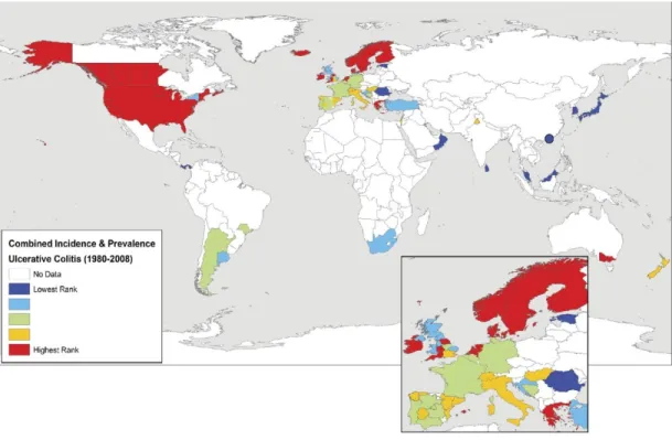
The map below, taken from the 2012 Molodecky systematic review of worldwide IBD prevalence and incidence, displays CD occurrence and prevalence rates since 1980, by region (Molodecky et al, 2012).
Figure 1: CD incidence rate by geographical area, taken from Molodecky et al., 2012
The figure shows the variation in disease incidence and prevalence among diverse regions of the world (Molodecky et al, 2012). High incidence rates are concentrated in developed countries, particularly, North America, Europe and Australia. Developing regions, such as Asia, Africa and South America have much lower rates. These results are consistent with the findings of the previous literature review, conducted by Economou et al. in 2008 (Economou et al, 2008).
It is essential to note that most IBD epidemiology studies were conducted in developed countries in Northern Europe and North America (Ng et al, 2013) (Molodecky et al, 2012). The lack of data collected from other countries might partially explain the differences in incidence.
19
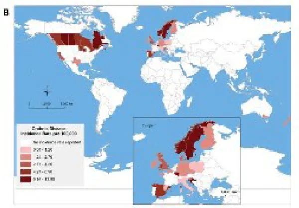
The geographic trends observed in adult CD are very similar to the paediatric CD population, as seen in the following figure from Benchimol et al, 2011.
Figure 2: Pediatric CD incidence rate by geographical area, taken from Benchimol et al., 2010 (incidence rates from 1990)
A noticeable difference has been observed in North-South CD incidence rates in Europe; the CD incidence is greater in the region North of the Alps (7/100 000) compared to the South (3.9/100 000) (Economou et al, 2008) (Castiglione et al, 2012). A similar trend has also been observed in individual countries; for instance, northern France and Scotland display higher incidence rates than their southern regions (Ng et al, 2013). In Canada, a West-East difference has been perceived, with lower rates in British Columbia and the highest rates in Quebec and Nova Scotia (Lowe et al, 2009). The origin of these gradients is still not clear. Hypotheses consist of differences between urban and rural regions, and asymmetrical immigration (Molodecky et al, 2012; Lowe et al, 2009).
Figure 3: Variation of incidence rates for CD within Canada showing an East–West disease gradient (Siew C Ng et al, 2012)
The incidence of CD remains low in Asian, Southern and Eastern European and developing countries, but it is gradually increasing. This trend is particularly apparent in Asia (Ng et al, 2013). For instance, in Japan, CD incidence has increased from 2.9/100 000 in 1986 to 13.5/100 000 in 1998. Similarly, South Korea has seen a rise in CD incidence from 7.6/100 000 in 1997 to 30.9/100 000 in 2005 (Ng et al, 2013). The rising trend in these regions is following the model of the developed countries from100 years ago; the UC incidence rate increased first, followed by the CD incidence (Ng et al, 2013).
From 2000 to 2006, the annual mean mortality rate, primarily due to CD and UC, was 75 and 39 deaths per year, respectively (Statistics Canada, 2010). Compared to deaths in Canada due to all causes, CD and UC deaths collectively accounted for 0.05% of the annual total. Although the annual mortality
21
rate for both CD and UC has been slowly increasing, the ratio of these deaths to deaths due to all causes has been constant at 0.05%. An independent meta-analysis of 13 studies found that CD patients have an age-adjusted risk of premature death that is 50% greater than the general population (standardized mortality ratio [SMR] 1.52; 95% CI 1.32 to 1.74; P<0.0001) (Canavan et al, 2007). Each year, 114 deaths in Canada are attributed to CD and UC (Fedorak et al, 2010). The economic costs of IBD are estimated to be $2.8 billion in 2012 (almost $12,000 per IBD patient). Direct medical costs exceed $1.2 billion per annum and are driven by the cost of medications ($521 million), hospitalizations ($395 million) and physician visits ($132 million). Indirect costs (societal and patient costs) total $1.6 billion and are dominated by long-term work losses of $979 million. Compared to the general population, the quality of life patients experience is low across all dimensions of health (Rocchi et al, 2012)
4. CD RISK FACTORS
CD is the result of both genetic and environmental factors (Carbonnel et al, 2009). There is recognition that the genetic risk factors do not act in isolation but in synergy with the external environment as well as the internal ‘environment’, namely the gut microbiota. The development of IBD is thought to be governed by a series of interactions between these three spheres, which simultaneously not only increase the complexity of disease pathogenesis, but also offer several avenues for intervention and improvement of patient outcomes (Ananthakrishnan, 2015).
GENETIC PREDISPOSITION
Family history is one of the strongest risk factors for CD. For instance, in a matched case-control study investigating potential risk factors for IBD, family history was found to be the most important prognosticator of the disease (OR: 4.6, 95% CI 2.6-8.3) (Baron et al, 2005). Monozygotic twins have a 50% concordance risk for Crohn’s, which is significantly higher than the concordance rate of dizygotic twins (estimated at 3%); moreover, children of parents with Crohn’s have a 33% risk of developing the disease (Baumgart et al, 2012) (Rabizadeh et al, 2013) (Frolkis et al, 2013) (Ananthakrishnan, 2013) (Brant et al, 2007).
Ethnicity has also been associated with a higher risk of CD. For example, the occurrence is especially higher in the Caucasian and the Ashkenazi Jewish population (Cohen, 2003; (Karlinger et al, 2000; Cho et al, 2011). The ethnic and familial factors indicate the existence of a genetic predisposition for CD, since gene alleles are distributed differently across populations (Cho et al, 2011). Having a single relative with confirmed IBD increases the risk of CD (OR: 3.1; 95%CI: 2.2–4.3) and UC (OR: 2.5; 95%CI: 1.9–3.5). The ‘dose effect’ was confirmed when multiple family members had IBD: CD (OR: 7.4; 95%CI: 3.4–16.1) and UC (OR: 6.8; 95%CI: 3.1–14.9) (Leong, 2010).The first CD-associated gene
23
have identified a total of 163 susceptibility loci for IBD, with 140 for CD (Jostins et al, 2012). In the past 5 years, researchers have identified several monogenic forms of severe early-onset colitis. For example, single mutations in IL10 (interleukin-10) and the genes encoding its receptor (IL10RA and IL10RB), as well as mutations in XIAP (X-linked inhibitor of apoptosis protein), have been shown to cause severe early-onset IBD (Peterson et al, 2014).
In accordance with the polygenic model for disease, a number of susceptibility loci associated with CD have been identified. The gene for NOD2/CARD15 (caspase activation recruitment domain), an important protein in innate immunity, was one of the first associated risk alleles for Crohn’s (Bonen et al, 2003). In a case-control study, the CD population attributable risk for CARD15 was estimated at 46.8% (Brant et al, 2007). There is a 20 to 40 fold increased risk of developing disease if a person has two risk alleles. The genetic loci identified in patients with Crohn’s implicate many biologically relevant immune pathways such as IL-23, IL-17 and IL-10. For the most part, though overlap exists, Crohn’s genes variations appear to be in pathways involved in microbe recognition and immune system responses such as autophagy, while in UC, genes appear to be involved in intestinal barrier integrity and function. In infants, one genetic mutation of significant interest is found in the interleukin-10 (IL-10) pathway. This rare autosomal recessive mutation leads to an infantile form of severe IBD that sometimes requires bone marrow transplantation (Rabizadeh et al, 2013).
Other genes have also been associated with CD. For instance, mutations of the ATG16L1 and IRGM genes, that play a role in the pathogen-degradation process, cause disturbance in the autophagy pathway (Spalinger et al, 2013).). The NOD2 gene has a role in peptidoglycan recognition, which are particles found on invading bacteria, and its polymorphisms significantly increase the risk of CD (Cho et al, 2011). However, these genes and other genetic loci identified thus far explain less than a third of CD cases, supporting and reinforcing the role of environmental factors and/or gene-environment interactions in the etiology of CD (Amre et al, 2003, Amre et al, 2002)
ENVIRONMENTAL FACTORS
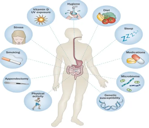
Figure 4: The interaction between genetics, immunology, environment and microbiome.
Many factors may participate in the development of IBD, such as genetic, immunological and environmental, including diet, depression, stress and the influence of free radicals. As depicted in Figure 6, IBD is thought to develop from genetic predisposition (leading to immunological abnormalities), dysbiosis of the gut microbiota and environmental influences. No single risk factor is sufficient for disease development and the complex interactions among factors lead to the development of IBD (Ananthakrishnan, 2013).
Urban living and lack of exposure to pets and vegetable gardens have been hypothesized to be associated with an increased risk of IBD. These factors all seem to be crucial for the development of IBD, each to a different degree. Studies with monozygotic twins clearly indicate the possibility of the existence
25
of environmental factors in IBD development. Another investigation on monozygotic twins revealed that, despite identical genomes, they differ in microflora. However, it was also revealed that genetic factors determine the composition of the microflora and are responsible for maintaining homeostasis in the intestine (Sobczak et al, 2014).
Dietary fibre (particularly fruits and vegetables), saturated fats, depression, impaired sleep, and low vitamin D levels have all been associated with incident IBD. Interventional studies assessing the effects of modifying these risk factors on natural history and patient outcomes are an important unmet need (Ananthakrishnan, 2015).
Oral contraceptives are known risk factors for CD. In two large prospective cohorts of women, authors observed a significant association between oral contraceptive use and risk of CD (Ananthakrishnan, 2013). Compared to women with no history of oral contraceptive use, the age-adjusted HRs for CD were 2.88 (95% CI 1.69 to 4.89) among women currently using oral contraceptives, and 1.50 (95% CI 1.13 to 1.99) among past users (Khalili et al, 2012). This effect seemingly increases with the number of years of oral contraceptives use (Ng et al, 2013). Stress, socioeconomic status (SES), diet and the use of antibiotics are among other risk factors of CD, while breastfeeding and sunlight (vitamin D) are believed to be protective (Ng et al, 2013) (Frolkis et al, 2013) (Ananthakrishnan, 2013) (Green et al, 2006). Nonetheless, research findings for these factors are still inconsistent and their association with CD is yet to be clearly established.
The role of stress in IBD also seems to be significant. Stressful conditions induce the activation of the hypothalamic–pituitary–adrenal (HPA) axis, which inhibits the immune system, impairs digestive functions and may induce a mucosal and systemic inflammatory reaction in IBD patients (Sobczak et al, 2014).
Limited physical activity (fewer than two sporting activities per week) in childhood was a risk factor for both CD and UC according to one study. A study by Persson et al. (1990) revealed that the relative risk (RR) of CD was inversely related to regular physical activity: for weekly and daily exercise, the estimated RRs were 0.6 (95% CI 0.4–0.9) and 0.5 (95% CI 0.3–0.9), respectively (Hlavaty et al, 2013).
Infrequent contact with animals in childhood (defined as less than once per week) was another independent risk factor for CD (OR 1.7). A case–control study from Canada on 581 patients with IBD and 433 controls also revealed that contact with animals, particularly cats, was protective in CD (OR 0.66, 95% CI 0.46–0.96) (Hlavaty et al, 2013).
SMOKING AND CD
ACTIVE SMOKING AND CD
The dramatic increase in the incidence of inflammatory bowel disease (IBD), especially CD, in developed countries over the second half of the 20th century highlights the important role that environmental factors play in the pathogenesis of IBD. Cigarette smoking is one of the most well documented environmental risk factors for adult-onset IBD (Jones et al, 2008). Among all the 534 studies conducted on CD risk factors, the association between smoking and IBD has gathered the most persuasive evidence (Bernstein et al, 2006). Oddly, smoking seems to have a protective effect against UC, but is a risk factor for CD (Ananthakrishnan, 2013). Based on a recent review, being a current smoker doubles one’s risk of CD (OR 2.0; 95%CI: 1.48–2.68), whereas the risk of UC is reduced (OR: 0.67; 95%CI: 0.48–0.94) (Ng et al, 2013).
27
Not all cohorts have identified an effect of smoking on IBD. In an Israeli study, smoking cessation was associated with an increased risk of UC, but not of CD. By contrast, smoking was associated with early age of onset and more frequent need for immunosuppressive therapy in CD among women, but not men (Reif et al, 2000).
PASSIVE SMOKE/ETS EXPOSURE &IBD
Environmental Tobacco Smoke (ETS)
ETS consists of two forms of smoke from the burning of tobacco products:
1. Side-stream smoke, or smoke that is emitted between the puffs of a burning cigarette, pipe, or cigar
2. Mainstream smoke, or the smoke that is exhaled by the smoker. When a cigarette is smoked, about one-half of the smoke generated is side-stream smoke. This form of smoke contains most of the same carcinogenic and toxic agents that have been identified in the mainstream smoke inhaled by the smoker, but at greater levels.
The health consequences of "passive smoking," or inhaling environmental tobacco smoke, have received considerable attention. The 1986 Surgeon General's report dealt exclusively with passive smoking and concluded that "involuntary smoking is a cause of disease, including lung cancer, in healthy non-smokers (Sandier et al, 1992).Children are more susceptible to the harmful effects of ETS. They can be exposed to tobacco smoke not only in their homes, but also in schools, restaurants, child-care settings, cars, and other public places. Home exposure is the most common type of exposure. Parental smoking in the home is known to lead to substantial maternal and fetal ETS exposure, subsequently affecting fetal and child health (Seong et al, 2008). A world-wide survey carried out in schools from WHO member states demonstrated that approximately 50% of the students were exposed to ETS in their homes (Warren et al, 2008). Not surprisingly, such a high exposure corresponds with the numerous health effects
reported. A recent comprehensive review (Cao et al, 2015) identified links between ETS exposure and upper and lower respiratory diseases, infections, cardiovascular disease, inflammation, and neurobehavioral deficits. Worldwide efforts to establish interventions to reduce ETS are ongoing.
Harries et al., (1982) were the first to suggest that a relationship might exist between passive smoke exposure and the development of IBD in adults. In a case-control study carried out in the UK, they acquired information on smoking from patients with UC (n=250), CD (n=192) and non-IBD controls (those visiting the fracture clinics of the same hospital) (n=230). Controls were matched to UC patients for age and gender. They observed that approximately 30% of UC patients belonged to smoking households, compared to 40% in CD and 50% in controls. These findings suggested that ETS exposure may be protective for UC. Although the study had a large sample size, details on ETS exposure were not specific. Furthermore, potential confounding from socio-economic status was not controlled for. A number of studies have subsequently examined this association. Persson et al., (1990) carried out a case-control study in Stockholm, Sweden, in which they identified patients with UC or CD from hospital discharge sheets maintained in a population-based registry, diagnosed between 1980-1984. Patient diagnosis was confirmed using medical records. An age-sex stratified random population-based sample of controls with age and sex distribution comparable to the IBD patients was recruited. After accounting for exclusions and refusals, 152 CD, 145 UC and 305 controls provided information on active and passive smoking (postal questionnaire and follow-up by telephone). ETS exposure was ascertained via the question: “How many people smoked regularly in your home during your childhood (0-15 years old)?”. They reported elevated risks for CD in adulthood (OR=1.5, 95% CI=1.0-2.3), but not for UC (OR=0.98, 95% CI=0.60-1.5). The study had many strengths. It was population-based and had a large sample. Diagnosis of IBD was confirmed. The response rates were sufficiently high (~80% across all 3 study groups). The major limitations were the potential for recall bias, non-specific measurement of ETS, and not controlling for of active smoking and other potential confounders such as SES. Furthermore, given
29
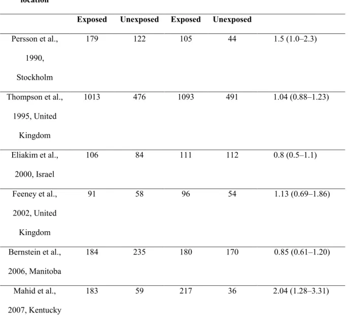
that only hospitalized patients with listed phone numbers were included, selection bias was a possibility. The findings for CD were not replicated in later studies by Thompson et al., (1995) (ETS exposure: regular smoking by either parent during childhood; OR=1.0, 95% CI=0.88-2.3), Elaikim et al., (2000) (CD=261, controls=430; ETS exposure: passive smoking in parental home; OR=0.8, 95% CI=0.5-1.1), Feeney et al., (2002) (CD=139, controls=139; ETS exposure: one or both parents smoking; OR=1.1, 95% CI=0.7-1.9), and Bernstein et al. (2006) (OR=0.8, 95% CI=0.6-1.2). However, Mahid et al. (2007) reported positive associations (CD=377, controls=384; ETS exposure: mother, father or other household member smoking; OR=2.0, 95% CI=1.3-3.3). Though all of the above studies also examined UC, none found any associations with ETS. Thus, there seems to be no association between ETS exposure and UC for adult-onset IBD; however, the findings are inconsistent for CD (Table 2).
Given the susceptibility of children to ETS exposure, they may be predisposed to the development of IBD. Some case-control studies have addressed this possibility with regards to two specific categories of passive smoke exposure: prenatal exposure due to maternal smoking during pregnancy and passive smoke exposure during childhood. As both of these exposures are relatively common in North America (Tong et al, 2009), investigating the risk of such ETS for childhood IBD is relevant. Lashner et al. (1993) carried out a hospital-based case-control study in Chicago, USA. They recruited 39 UC and 33 CD patients and 72 controls (friends of the patients). All participants were non-smokers. They defined post-natal ETS exposure as: smoking more than 5 cigarettes by a parent or sibling in the home at the time of symptom onset. They reported positive (but not statistically significant) associations with CD (OR=2.00, 95% CI=0.5-8.0) and UC (OR=1.7, 95% CI=0.7-4.3). When ETS exposure at birth (in the home) was examined, increased risks for IBD were noted (OR=3.02, 95% CI=1.28-7.1) with greater risks for CD than UC. Similarly, risks for IBD were increased with maternal smoking at birth (OR=2.09, 95% CI=1.02-4.3). Although different measures of ETS were examined, adding to the strength of the study, the results were likely to be overestimates given the small sample
sizes. Furthermore, the results were not adjusted for potential confounding of family history of IBD and SES. Moreover, it was unclear whether the associations with ETS exposure at birth were independent of those at symptom-onset and vice-versa. The effects of ETS exposure during pregnancy were also not assessed. Rigas et al. (1993) carried out a case-control study in New York, wherein they identified IBD patients from the medical records of patients diagnosed in hospitals from 1986 to 1990. Controls were patients without IBD, seen at the same pediatric gastroenterology departments at the respective hospitals. Information on maternal smoking and other potential risk factors was abstracted from the medical records. A total of 68 CD, 39 UC and 202 controls were examined. No association between maternal smoking and IBD was found. This study had some limitations. Information on maternal smoking was acquired from the medical records; information that may be incomplete or inaccurate. In addition, it was not possible to establish the timing of the ETS exposure from maternal smoking (whether at the time of IBD diagnosis, during pregnancy or during childhood). Furthermore, the study was likely not powered to detect potential associations. Another study carried out in Northern France by Baron et al. (2005) examined passive smoking by parents or caregivers along with a host of other potential risk factors. This case-control study was comprised of children diagnosed with IBD prior to age 17, identified from the EPIMAD population-based registry. Controls were randomly selected from the population using random digit dialling and matched for age, gender and area of residence. In total, 222 CD, 60 UC and 282 controls were interviewed via telephone. No association between passive smoking during pregnancy and IBD was evident (OR=0.84, 95% CI=0.55-1.3). This study was a comprehensive population-based study. The analysis accounted for potential confounding variables, including SES. Nonetheless, the measurement of ETS was likely imprecise and did not cover the appropriate susceptibility time windows. A separate analysis for CD was carried out. The latter is relevant as smoking is negatively associated with UC, and the results may be underestimated by the inclusion of these patients. A frequency distribution of the ETS variable among the subjects was not presented, precluding a clear interpretation of their negative findings.
31
The Baron et al. (2005) study was followed by a case-control study conducted in South East Scotland among children with early onset IBD, where cases of IBD diagnosed at less than 16 years of age were studied along with sex and age-matched controls attending the same general practice (Russell et al., 2005). In total, they matched 62 pairs of cases and controls, with a median age of disease onset in cases of 10.6 years. The study demonstrated that parental smoking during pregnancy and around the time of birth was more common in parents of IBD cases, 54% versus 29% in control parents (p=0.01; OR= 2.87, 95% CI= 1.23–6.66). Maternal smoking during pregnancy and at birth was also more common in IBD cases than in controls, 23% versus 6.2% (p=0.04; OR= 4.46, 95% CI= 1.16–17.1). Smoking in mothers of patients with CD was also greater, 27.8% versus 8.3% (p=0.03; OR= 4.23, 95% CI= 1.05–16.97). There was no significant effect seen when paternal smoking in pregnancy and at birth was analysed in IBD cases versus controls (p=0.27). This study was based on a very small sample, did not explore other ETS exposures and lacked control for potential confounding from SES. In a more recent study, Roberts et al. (2011) carried out a retrospective birth-cohort study in the UK. They used the Oxford record linkage study (ORLS) that comprised abstracts of records of birth registrations, maternities, day cases and inpatient admissions in the geographical region in and around Oxford. The maternity records covered a 20-year period from 1970 to 1989. These records were linked to all in-and-out patient visits until 1999. Diagnosis of IBD was based on requisite ICD codes for CD and UC. ETS exposure was “maternal smoking during pregnancy” as listed in the maternity records. Information on this exposure was available for 43 children with CD and 22 patients with UC. They observed increased risks of CD (OR=2.04, 95% CI=1.06-3.92, p=0.05) after accounting for potential confounding variables. A major strength of this study was that it was based within a well-defined geographic population, studied in a large cohort and included both inpatients and outpatients. Nonetheless, a major limitation was the unconfirmed IBD diagnosis that may have resulted in diagnostic misclassification. Furthermore, only 1 measure of ETS was assessed and the number of cases studied was small.
Table 2 - Childhood Passive Smoke Exposure and risk of adult-onset CD Author, year
of publication, location
Controls Cases OR (95% CI)
Exposed Unexposed Exposed Unexposed Persson et al., 1990, Stockholm 179 122 105 44 1.5 (1.0–2.3) Thompson et al., 1995, United Kingdom 1013 476 1093 491 1.04 (0.88–1.23) Eliakim et al., 2000, Israel 106 84 111 112 0.8 (0.5–1.1) Feeney et al., 2002, United Kingdom 91 58 96 54 1.13 (0.69–1.86) Bernstein et al., 2006, Manitoba 184 235 180 170 0.85 (0.61–1.20) Mahid et al., 2007, Kentucky 183 59 217 36 2.04 (1.28–3.31)
33
Table 3. ETS exposure in childhood and risk of childhood-onset CD Author, year
of publication, location
Controls Cases OR (95% CI)
Exposed Unexposed Exposed Unexposed Lashner et al., 1993, Chicago 26 46 17 22 2.0 (0.5–8.0) Rigas et al., 1993, New York 15 59 11 34 0.8 (0.3–2.5) Baron et al., 2005, Northern France 156 66 144 78 0.84 (0.55–1.30) Russell et al., 2005, Scotland 18 44 33 29 2.87 (1.23–6.66) Roberts et al., 2011, South East England 34229 110934 16 27 1.92 (1.03-3.56)