To cite this version : Benchikhi, Mohamed and El Ouatib, Rachida
and Guillemet-Fritsch, Sophie and Chane-Ching, Jean-Yves and
Er-Rakho, Lahcen and Durand, Bernard Sol–gel synthesis and sintering of
submicronic copper molybdate (α-CuMoO4) powders. (2014) Ceramics
International, vol. 40 (n° 4). pp. 5371-5377. ISSN 0272-8842
O
pen
A
rchive
T
OULOUSE
A
rchive
O
uverte (
OATAO
)
OATAO is an open access repository that collects the work of Toulouse researchers and
makes it freely available over the web where possible.
This is an author-deposited version published in :
http://oatao.univ-toulouse.fr/
Eprints ID : 13983
To link to this article : doi:
10.1016/j.ceramint.2013.10.118
URL :
http://dx.doi.org/10.1016/j.ceramint.2013.10.118
Any correspondance concerning this service should be sent to the repository
administrator:
[email protected]
Sol–gel synthesis and sintering of submicronic copper molybdate
(α-CuMoO
4
) powders
Mohamed Benchikhi
a,b, Rachida El Ouatib
a, Sophie Guillemet-Fritsch
b, Jean Yves Chane-Ching
b,
Lahcen Er-Rakho
a, Bernard Durand
b,naLaboratoire de Physico-Chimie des Matériaux Inorganiques, Université Hassan II, Casablanca, Morocco
bInstitut Carnot CIRIMAT, Université de Toulouse, CNRS, 118 Route de Narbonne, 31062 Toulouse Cedex 9, France
Abstract
A sol–gel method was proposed to prepare copper II molybdate α-CuMoO4powders. A gel was first obtained via the polymerizable complex
method, using citric acid as complexing and polymerizing agent, dried at 120 1C and decomposed at 300 1C. A calcination in the temperature range 400–500 1C for 2 h led to the pure phase α-CuMoO4. The different powders obtained were characterized by X ray diffraction analysis and
by transmission (TEM) and scanning (SEM) electron microscopies.
Ceramics were prepared using conventional sintering and spark plasma sintering (SPS) techniques. A maximal relative density of 94.8% was reached after conventional sintering at 520 1C for 2 h. In the case of SPS, the densification was optimized by varying the temperature, the time and the applied pressure. Higher densities, up to 98.7%, were obtained at very low temperature, i.e., 300 1C, for 5 min only under a pressure of 225 MPa.
Keywords:A. Sintering; B. Grain size; Sol–gel; Powders; Chemical preparation
1. Introduction
Copper molybdate CuMoO4exhibits two polymorphs at
atmo-spheric pressure, the α form with Mo located in a tetrahedral environment (stable form) and the g form with Mo located in an octahedral environment (metastable form). This oxide appears as a true horn of numerous note-worthy properties: it is thermochromic, trisochromic and piezochromic since it exhibits a phase transition associated with a color change from green to brown. This first order transition is accompanied by a strong change of volume and an anisotropic splitting of grains and occurs according to a domino-cascade kinetics. Moreover, the temperatures/pressures of transition are adjustable by chemical doping. So, copper molybdate CuMoO4
can find applications as catalyst, pressure and/or temperature sensor. The complexity of CuMoO4justifies the numerous studies
on this oxide[1–5].
Several methods of copper molybdate preparation have been investigated, particularly those involving solid state reactions between copper and molybdenum oxides at temperatures close to 750 1C[6,7] and those involving pyrolysis at about 500 1C of precursors precipitated at low temperature[8]or obtained by sol–gel[9].
This paper deals with the synthesis of a homogenous and finely divided powder of α-CuMoO4and the elaboration of the
corresponding dense ceramics using spark plasma sintering. The route involving the polymerizable complex, based on the Pechini method[10], was chosen. It leads to sub-micronic sized particles and also offers the advantage to increase the reactivity of reaction mixtures. The final powders synthetized this way usually present high homogeneity[11].
2. Experimental
The gels were synthesized using the following reactants: ammo-nium heptamolybdate (NH4)6Mo7O244H2O (Sigma-Aldrich
http://dx.doi.org/10.1016/j.ceramint.2013.10.118
n
Corresponding author. Tel.: þ33 56 1557751; fax: þ 33 56 1556163. E-mail address:[email protected] (B. Durand).
99%), copper salts CuX2–nH2O (X¼ NO3 "
) (Acros Organics 99.5%), Cl"
(Acros Organics 99.5%), CH3COO "
(Fluka 99.8%), citric acid (Acros 99.5%), concentrated nitric acid and ammonia.
The procedure for copper molybdate synthesis from a polymeric precursor is shown in Fig. 1. A citric acid (CA) solution was added to an equimolar solution of ammonium molybdate and copper salt in a proportion such that CA/ cation ¼ 3; the pH was close to pH1. The evaporation of the solution at 80 1C led to the formation of gels named Gi (i is the source of copper; i ¼ 1 nitrate, i ¼ 2 chloride, and i ¼ 3 acetate). The gels were dried at 120 1C for 24 h, then pre-calcined at 300 1C for 12 h.
The black powders obtained were treated under air at temperature ranging from 420 to 500 1C according to the nature of the copper source. The final calcination temperatures could be optimized after thermogravimetry analyses (SETERAM TG-DTA 92). The green powders obtained at the end of the process were characterized by X-ray diffraction (Brucker AXSD4, λCuKα¼ 1.5418 nm), by scanning (Jeol-JSM6400) and transmission (Jeol 2010) electron microscopies. Specific surface areas were determined using a BET (Micrometrics Flowsorb II 2300). The precise chemical compositions were obtained by ICP-AES (JobinYvon ULTIMA). The sintering behavior was studied by dilatometry (TMA Setsys 16/18). The ceramics were obtained using both conventional sintering in air performed in a muffle furnace (Nabertherm) and by spark plasma sintering (SPS 2080, Sumitomo).
3. Results and discussion 3.1. Powders
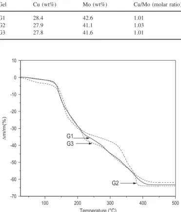
The nature of the copper precursor (nitrate, chloride or acetate) had only a very weak influence on the properties of the gels obtained. Indeed, according to the ICP analysis data (Table 1), the Cu/Mo molar ratio in different gels was close to 1, whatever the
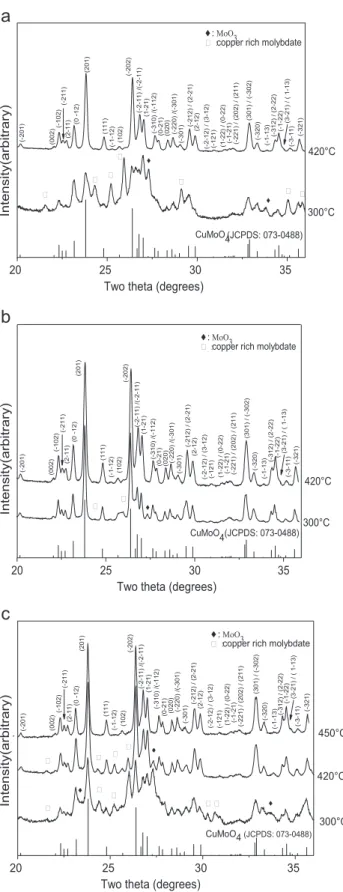
copper source. Moreover, the thermogravimetric curves of the dried gels, performed in air at a rate of 2.5 1C/min were very similar (Fig. 2) and revealed several decomposition steps (Table 2) with a total mass loss always close to 65%. The main difference was noticed for the temperature of the end of decomposition which increased following the sequence chloride (385 1C), nitrate (420 1C) and copper acetate (440 1C). The faster decomposition of the gel G2was corroborated by the X-ray diffraction analyses of
the gel powders calcined at different temperatures (Fig. 3). In the patterns of gels calcined at 300 1C for 12 h, mixtures of three phases were identified: α-CuMoO4 (JCPDS 073-0488), MoO3
Fig. 1. Flowchart of the synthesis of CuMoO4by the polymerizable complex
method.
Table 1
ICP analysis of CuMoO4powders obtained from gels Gi.
Gel Cu (wt%) Mo (wt%) Cu/Mo (molar ratio)
G1 28.4 42.6 1.01
G2 27.9 41.1 1.03
G3 27.8 41.6 1.01
Fig. 2. Thermogravimetric analysis of the xerogels Gi (G1 nitrate, G2 chloride, and G3 acetate).
Table 2
Temperature and mass loss of the different phenomena determined by TGA for the gels Gi.
Gel Temperature range (1C) Mass loss (%)
G1 25–125 2.5 G1 125–226 31.8 G1 226–420 28.7 G2 25–100 1.1 G2 100–235 32.0 G2 226–385 24.5 G3 25–120 9.5 G3 120–190 29.3 G3 231–440 24.7
(JCPDS 076-1003) and a phase that should be a mixed oxide of copper and molybdenum richer in copper than CuMoO4. The
phase α-CuMoO4was preponderant and the rays of the additional
phases were very weak only when the copper source was the chloride. For gels ex chloride or nitrate, the phase α-CuMoO4was
only identified in the powders calcined at 420 1C for 2 h. For the
Fig. 3. XRD patterns of gels Gi treated at different temperatures: (a) G1 treated at 300 and 420 1C, (b) G2 treated at 300 and 420 1C, and (c) G3 treated at 300, 420 and 450 1C.
Fig. 4. SEM micrographs of gels Gi synthesized with a CA/Cu ratio ¼ 3: (a) gel G1, (b) gel G2, and (c) gel G3.
gel ex acetate, the calcination temperature had to be raised up to 450 1C to obtain the same result.
The α-CuMoO4powders, obtained by calcination for 2 h of
gels G1 and G2 at 420 1C and G3 at 450 1C, consisted in elongated or more or less spherical elementary grains with sizes in the range 0.2–1.0 mm (Fig. 4). The crystallite sizes (calculated according to the Scherrer equation from the broad-ening of X-ray diffraction peaks), the geometrical sizes of particles (calculated from the experimental values of specific surface area assuming that powders are made of monodis-persed spherical grains) and the sizes of grains measured on TEM micrographs (Fig. 6a for G1) were of the same order of magnitude (Table 3). TEM observations could evidence some monocrystalline particles.
The influence of the CA/Cu molar ratio on the morphology of powders was examined. The ratio was varied between 3 and 12. The XRD patterns of the molybdates formed by calcination of the gels at 420 or 450 1C for 2 h showed that whatever the source of copper, beyond a molar ratio of 6, molybdenum oxide MoO3was formed beside α-CuMoO4(as shown inFig. 5for gel
G1). TEM micrographs revealed that increasing the ratio from 3 to 6 significantly raised crystallite size (Fig. 6).
3.2. Bulk ceramics
The sintering behavior was investigated on the α-CuMoO4
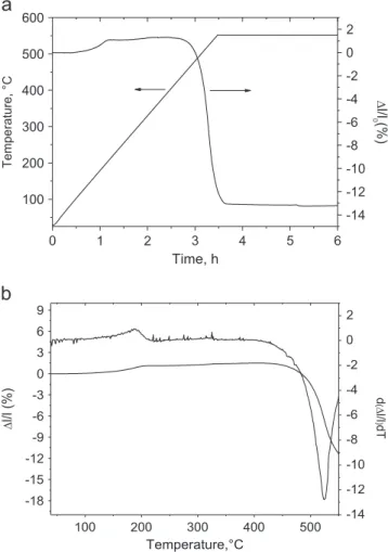
powders obtained by pyrolysis at 420 1C for 2 h of a gel prepared from copper chloride with a citrate/copper ratio of 3. For conventional sintering, discs were formed by uniaxial pressing at 25 bars at room temperature in the presence of an organic binder (Rhodoviol) so as to avoid the phenomenon of rolling. The green density reached 61%. The dilatometric curve recorded at a rate of 2.5 1C/min (Fig. 7) indicated the release of the binder at # 190 1C and the beginning of shrinkage at approximately 430 1C. The shrinking rate was maximum at 520 1C.
Conventional sintering treatments were then performed in air, in a muffle furnace at 520 1C and 570 1C, for 1, 2 and 6 h.
Table 3
Sizes estimated by different methods of particles of copper molybdate powders obtained by pyrolysis of gels Gi synthesized with a molar ratio CA/Cu ¼3.
Gel Crystallite size
DRXa(nm) SBET (m2/g) Grain size BETb(nm) Grain size TEMc(nm) G1 103 8.3 168 100–200 G2 138 6.9 202 140–250 G3 145 6.8 206 150–280
aSize of crystallites calculated from XRD peaks broadening.
bGeometrical size of particles calculated from the specific surface area. cSize of particles estimated by TEM.
Fig. 5. XRD patterns of gel nitrate (G1) treated at 420 1C for 2 h for different CA/Cu ratios.
Fig. 6. TEM micrographs of gel nitrate (G1) treated at 420 1C for 2 h for different CA/Cu ratios: (a) CA/Cu ¼ 3, and (b) CA/Cu ¼ 6.
The densification of the ceramics is shown in Table 4. The highest value, 94.8%, was reached for 2 h sintering at 520 1C, which confirmed the temperature where the shrinking rate was maximum. Prolonging the dwell time was not necessary to increase densification. Under these conditions (2 h, 520 1C), the ceramics consisted of grains sized less than 2 μm, with a relatively narrow granulometric distribution (Fig. 8a). Little residual porosity was observed. The decrease of density after the dwell time was increased to 6 h was likely due to the abnormal growth of some grains (Fig. 8b). For a higher sintering temperature (570 1C), the abnormal growth was even more pronounced (Fig. 8c).
The advantage of the spark plasma sintering technique is to allow sintering at temperatures significantly lower and for times much shorter than those required for natural sintering.
Fig. 7. Dilatometric behavior of CuMoO4: (a) Δl/l and temperature versus
time, and (b) Δl/l and derivate of Δl/l versus temperature.
Table 4
Influence of the temperature and the time on the relative density of
conventionally sintered CuMoO4ceramics.
Temperature (1C) Time (h) Relative density (%)
520 1 92.7 520 2 94.7 520 6 93.6 570 1 93.8 570 2 93.9 570 6 91.4
Fig. 8. SEM micrographs of CuMoO4 ceramics conventionally sintered: (a)
Different sintering parameters, i.e. the temperature, the dwell time and the applied pressure, were optimized in order to reach the highest densification (Fig. 9, Table 5). A sintering
temperature of only 300 1C was first arbitrarily chosen. The ceramic showed a relative density of 97.10%, which was indeed higher than the highest value obtained by natural sintering; it was reached with an applied pressure of 200 MPa and a dwell time of 2 min only. A trial at 500 1C
Fig. 9. Densification of SPS sintered CuMoO4ceramics as a function: (a) the
applied pressure, (b) the temperature, and (c) the time.
Table 5
Influence of the temperature, the time and the pressure on the relative density
of SPS sintered CuMoO4ceramics.
Temperature (1C) Time (min) Pressure (MPa) Relative density (%)
300 2 200 97.1 300 5 200 98.6 300 10 200 98.4 275 5 200 96.6 325 5 200 98.5 300 5 175 96.5 300 5 225 98.7 500 2 50 98.5
Fig. 10. SEM micrographs of CuMoO4SPS sintered ceramics: (a) at 300 1C
for 2 min with a pressure of 200 MPa, and (b) at 500 1C for 2 min with a pressure of 50 MPa.
under a lower applied pressure so as to avoid the decomposi-tion of the sample did not lead to higher densificadecomposi-tion. An increase of the level time to 5 min increased the density up to 98.6%. A further increase up to 10 min had no further effect. Taking the basic conditions of temperature and time (300 1C, 2 min), the pressure was also optimized. A maximal density of 98.7% was obtained with a pressure of 225 MPa. As the gain of density obtained when the pressure was increased from 200 to 225 MPa was only very slight, the optimal applied pressure was taken at 200 MPa, which presented less of a risk to break the sample.
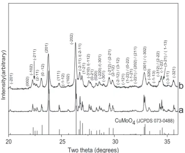
The SEM micrographs of ceramics (Fig. 10) revealed that regarding the very short time of sintering and the low temperature of densification, high densities were obtained without significant grain growth. For sintering at 300 1C for 5 min under 200 MPa and at 500 1C for 2 min under 50 MPa, the grain sizes did not exceed respectively 200 nm and 2 μm. The XRD patterns of the ceramics conventionally sintered at 520 1C for 2 h (Fig. 11a) and sintered by SPS at 300 1C for 5 min under 200 MPa (Fig. 11b) showed that, whatever the sintering procedure, the α-CuMoO4 phase is not modified by
the thermal treatment. The broadness of the peaks of the SPS sintered ceramic was comparable to that of the peaks of the non-sintered powder (Fig. 3b; 420 1C) whereas the peaks of the conventionally sintered ceramic were significantly narrower. The data of microscopy (Figs. 8 and10) are confirmed, i.e. on the contrary of conventional route, the SPS allows the limitation of the grain growth.
4. Conclusion
α-CuMoO4 submicronic powders were prepared using a
method derived from the Pechini method. Different gels, using various copper salts, i.e. nitrates, chlorides and acetates, were obtained. After calcinations at temperatures ranging from 420 1C to 490 1C, pure α-CuMoO4phases could be obtained.
The grain size was in the range 200–1000 nm.
Spark Plasma Sintering performed at temperatures as low as 300 1C for 5 min only allowed to prepare dense ceramics with the same grain size as the powders.
Acknowledgments
This work was supported by two French-Moroccan projects: Volubilis Partenariat Hubert Curien (PHC n1 MA 09 205) and Projet de Recherches sur Convention Internationale CNRS-CNRST n1 22572.
References
[1] F. Rodríguez, D. Hernández, J. Garcia-Jaca, H. Ehrenberg, H. Weitzel, Optical study of the piezochromic transition in CuMoO4 by pressure spectroscopy, Phys. Rev. B 61 (2000) 16497.
[2] M. Gaudon, P. Deniard, A. Demourgues, A.E. Thiry, C. Carbonera, A. Le Nestour, A. Largeteau, J.F. Létard, S. Jobic, Unprecedented “One-Finger-Push”-induced phase transition with a drastic color change in an inorganic material, Adv. Mater. 19 (21) (2007) 3517.
[3] A.E Thiry, M. Gaudon, C. Payen, N. Daro, J.F. Létard, S. Gorsse, P. Deniard, X. Rocquefelte, A. Demourgues, M.H. Whangbo, S. Jobic, On the cyclability of the thermochromism in CuMoO4and its tungsten derivatives CuMo1–xWxO4(x o 0.12), Chem. Mater. 20 (6) (2008) 2075.
[4] M. Gaudon, C. Carbonera, A.E. Thiry, A. Demourgues, P. Deniard, C. Payen, J.F. Létard, S. Jobic, Adaptable thermochromism in the CuMo1 " xWxO4 series (0 o x o 0.1): a behavior related to a first-order phase transition with a transition temperature depending on x, Inorg. Chem. 46 (24) (2007) 10200.
[5] H. Ehrenberg, H. Weitzel, H. Paulus, M. Wiesmann, G. Wltschek, M. Geselle, H. Fuess, Crystal structure and magnetic properties of CuMoO4at low temperature (γ-phase), J. Phys. Chem. Solids 58 (1997) 153.
[6] M. Wiesmann, H. Ehrenberg, G. Miehe, T. Peun, H. Weitzel, H. Fuess, p-TPhase diagram of CuMoO4, J. Solid State Chem. 132 (1) (1997) 88.
[7] D. Klissurski, R. Iordanova, M. Milanova, D. Radev, S. Vassilev, Mechanochemically assisted synthesis of Cu (II) molybdate, CR Acad. Bulgare Sci. 56 (8) (2003) 39.
[8] S. Mitchell, A. Gómez-Avilés, C. Gardner, W. Jones, Comparative study of the synthesis of layered transition metal molybdates, J. Solid State Chem. 183 (1) (2010) 198.
[9] J.H. Ryu, S-M Koo, J-W Yoon, C.S. Lim, K.B. Shim, Synthesis of nanocrystalline MMoO4(M ¼ Ni, Zn) phosphors via a citrate complex route assisted by microwave irradiation and their photoluminescence, Mat. Lett. 60 (13–14) (2006) 1702.
[10] M. Pechini, U.S. Patent 3,330,697, 11 July 1967.
[11] C.N.R. Rao, B. Raveau, Transition Metal Oxides, VCH, New York, 1995. Fig. 11. XRD patterns of CuMoO4: (a) conventionally sintered at 520 1C for
2 h, and (b) SPS sintered at 300 1C for 5 min with an applied pressure of 200 MPa.